pet arthritis causes

10 Common Pet Arthritis Causes & How to Help Your Stiff Pet

When your beloved furry friend begins to start slowing down as they get older, it can be very sad. As in human beings, it’s not a strange occurrence that animals experience stiff joints now and again. However, if the frequent jittery periods of stiffness become commonplace, or your pet’s lively fun suddenly turns to a refusal to jump up or go up stairs, your concerns are stirred. After all, you will eventually find yourself seeking professional guidance on pet arthritis to find out what is causing the ailment of your pet.
While the dog and cat’s body is quite resilient, the joints have to deal with years of wear and tear, and chronic inflammation is a very common ailment. You should seek the cause of your pet’s joint stiffness and ensure that your pet is in good health and comfortable. Often, chronic slowing down is just part of a larger problem and should be considered as a marker of a deeper problem in the skeletal system or daily management.
This detailed and comprehensive article will dissect the biological and environmental factors that contribute to joint degeneration, as well as medical conditions that could explain why pets struggle to move, and help you determine whether it’s something that will pass or if there’s a medical condition that you should consult your vet about.

Let's first examine the Biology of Pet Arthritis, a degenerative joint disease, and then read through the list of chronic diseases.

Osteoarthritis slowly wears away the protective cartilage that pads the ends of the bones in a joint. Protected smooth cartilage matrix senses a constant friction or repeated mechanical stress, the protective tissue deteriorates completely.
This results in the underlying bones rubbing together directly, producing tremendous friction. The joint inflames a great deal and the body tries to make the area more stable with the development of abnormal bony projections known as osteophytes (bone spurs).
When you start looking into pet arthritis, you’ll be facing anything that triggers this painful inflammatory cycle.

Main groups of pet arthritis causes and Indicators:

When your dog or cat starts to show signs of trouble with mobility, there are three main groups of pet arthritis causes and indicators: developmental skeletal conditions, lifestyle hazards, and natural biological changes. There are 10 common pet arthritis causes.

1. Hip and Elbow Dysplasia

The most prevalent cause of early pet arthritis is, without doubt, skeletal malformations, which are known as Hip and Elbow DysplasiaS. Diseases like hip dysplasia and elbow dysplasia are when a joint doesn’t develop square, which leads to the ball and socket grinding against each other. A pet that is born with such genetic structural defects will have abnormal friction within the joint capsule throughout their lives. When cartilage is gone, your pet will suddenly show signs of pain, lameness and stiffness from joint instability, rapid wear or growth spurts.

2. Excess Body Weight and Obesity

Excess body weight is another big time cause of severe pet arthritis. It is a very aggressive lifestyle factor that can have a detrimental effect on dogs’ and cats’ long-term joint health. Extra pounds on your pet’s body due to a high fat diet and/or inadequate activity put a tremendous strain on the joints that were never meant to carry such a heavy burden. This pairing leads to a significantly accelerated degradation of cartilage, and pets will not want to play or exercise due to the discomfort.

3. Cranial Cruciate Ligament (CCL) Tears

Dogs often tear the cranial cruciate ligament (CCL) which is similar to a human tearing an ACL. This injury to the structure is a huge contributor to pet arthritis since the whole knee joint is now totally unstable. If the tibia slides irregularly up and down under the femur, the meniscus cartilage which protects the femur can easily break down. If the structural instability is not corrected in a timely fashion, your dog is at a high risk of suffering from severe and rapid joint deterioration in that leg.

4. Natural Aging and Cellular Wear

When it comes to joint health, there is a common denominator: Natural Aging and Cellular WearTime. With aging, dogs and cats just naturally produce less of the cartilage matrix’s necessary building blocks, called glycosaminoglycans. The absence of natural lubrication leads to dry lining of the joint and means that it will immediately rub together or become stiff the first time your pet moves after a long nap or when it wakes early on a cold morning.

5. High Impact Jumping Injuries

Chronic micro-trauma can occur with high impact jumping injuries when your pet has been jumping for many years or off high furniture and starts to show signs of joint pain. Frequent jumping from heights to hard surfaces can create micro fractures in the bone and small tears in the cartilage. High impact household habits are one of the easier to change causes of your cat or dog slowing down because of repeated damage to the joints that it causes.

6. Slipping Kneecaps (Patellar Luxation)

Slipping Kneecaps (patellar luxation)Skeletally, the patella (kneecap) should be positioned in a deep groove at the front of the knee and glide smoothly. In some small dog breeds and some cats, however, the kneecap often completely pops out of the groove. These mechanical dislocations are important factors that alter the alignment of the leg and are important causes of progressive pet arthritis. The sliding movement needs to be properly managed and should be monitored carefully by a vet.

7. Historical Fractures and Joint Trauma

Physical injuries can have a lasting effect if they were suffered in the past, as Historical Fractures and Joint TraumaPets are naturally adventurous. Fractures of small bone fragments that occurred years ago and/or severe bouts of spraining and straining a joint often heal with subtle structural realignment changes. This results in an unbalanced transfer of weight over the smooth surface of cartilage. If the strain is not evenly distributed, the pet will have a gradual, ongoing recurrence of stiffness in the same leg that had been hurt.

8. Tick-Borne Diseases (Lyme Disease)

Another type of systemic infection that could affect a pet’s joint health is tick-borne diseases (Lyme Disease). A deer tick that is infected with Lyme disease infects the dog or cat by transferring the bacteria into the joint capsules. This can be a deep bacterial infection that breaks into the local tissues, and may lead to acute and severe joint swelling. A common overlooked cause of sudden, episodic changes in lameness in active dogs is tick-borne polyarthritis, which can resemble typical skeletal inflammation.

9. Poor Feline Muscle Mass (Sarcopenia)

If older cats have mobility problems that do not respond to treatment, they may have embedded Poor Feline Muscle Mass (Sarcopenia). Some thinning of lean body tissue can occur along the rear quarters, a normal aging process or due to metabolic changes. One of the more serious indirect causes of feline joint pain is a decrease in muscle mass, which can cause the underlying joints to bear the brunt of the shock and impact of the daily walk.

10. Immune-Mediated Polyarthritis (IMPA)

Immune-Mediated Polyarthritis (IMPA)Systemic autoimmune conditions are uncommon but devastating conditions in which the immune system attacks tissues within the body. These diseases do not begin with wear and tear, but with your pet being completely susceptible to extensive and extreme joint damage. One of the many biological reasons why pets develop arthritis can be an overactive immune system, causing normal healthy joint linings to become the source of chronic destructive pain.

Here are a few tips on helping your dog with stiffness.

There are a number of easy home care techniques you can implement while you are working with your vet to determine the exact cause of the joint issues, to keep your pet more comfortable while waiting for your veterinarian’s advice:

Provide orthopedic bedding

 Invest in a thick, high-density memory foam orthopedic bed for your pet. The soft and supportive foam distributes the weight evenly throughout their body, minimizing painful pressure points on the stiff elbows and hips.

Use non-slip flooring:

Hardwood or tile floors can be slippery, so use non-slip rubber mats or carpet runners to prevent your pets from slipping and hyperextending their joints

Use pet ramps and steps

Do not make your pet jump off high furniture or off a high vehicle door step; use specially made pet ramps in the areas where your pet sleeps or spends most of his or her time.

Gently warm the stiffest joints:

 Place a warm, safe heating pad, wrapped in a towel, against the affected area and massage for 10-15 minutes to promote blood flow and relax tight muscles, but avoid over-warming or irritating the skin.

Conclusion

It is important to carefully assess your pet’s lifestyle, home environment and physical symptoms to identify the actual pet arthritis causes. While it may be a little bit of stiffness here and there after a long day of running, if they’re having difficulty standing over and over it should not be ignored. By listening to those signs your pet is giving, your veterinarian will have all the information he needs to get your pet moving more comfortably and help pinpoint your pet’s joint discomfort.

Frequently Asked Questions (FAQs)

A1: If a pet begins to slow down but is obviously not vocalizing with pain, then there is likely no problem.

A: A pet may appear to be moving slowly with good appetite, but that doesn’t necessarily mean that they are not in a lot of pain. Chronic arthritis in animals is ingrained in them by evolution to hide it, so subtle changes in behaviour, such as animals not wanting to go up and down stairs or overall loss of energy, are a common indicator of chronic arthritis.

Q2: Do indoor cats get joint diseases like osteoarthritis?

A: Yes, cats can indeed suffer from degenerative joint diseases, even those kept indoors. Research indicates that a high percentage of older dogs have arthritis but the condition does not manifest itself in high jumps, and is easy to overlook for any cat owner who keeps her cats indoors.

Q3: What are some medical emergencies that may occur and something that should be noted if a pet is having trouble getting around?

A: If you notice a sudden complete loss of use in the rear legs, your dog doesn’t eat for over 24 hours, or if your dog is very lethargic, weeps when handled and pants heavily while resting, then a mobility problem should be given a medical priority.

cat sneezing causes

Is Your Cat Sneezing Constantly? 10 Hidden Cat Sneezing Causes Explained

10 Hidden Cat Sneezing Causes Explained

No one can deny that a little, sudden “achoo!” from your feline friend can be very cute. Just like humans, it’s not uncommon for animals to sneeze occasionally. But when an occasional tickle escalates into frequent explosive twitches, or your pet’s itchy nose and eyes are accompanied by watery eyes or nasal discharge, your concerns and concerns are aroused. Of course, you end up looking for professional advice related to cat sneezing causes in order to know what is wrong with your cat.
Cats have a very sensitive nose with a lot of specialized receptors in it, so a very strong reflex. It is important to find out what is causing your cat to sneeze so that you can help keep them healthy and comfortable. Often, chronic sneezing is just part of a larger problem and should be considered as a marker of a deeper problem in the body or environment.
This detailed and comprehensive article will dissect the biological and environmental factors that contribute to cat sneezing, as well as medical conditions that could explain why cats are sneezing, and help you determine whether it’s something that will pass or if there’s a medical condition that you should consult your vet about.

Let's first look at the Biology of the Feline Sneeze, a protective mechanism, before reviewing the list of chronic conditions.

The sneeze is an involuntary convulsion, coordinated action of the nose to expel irritants. If the thin lining of the nose passages (mucosa) senses a foreign particle, virus or allergen, the thin lining sends an electrical signal via the trigeminal nerve directly to the brainstem.
This causes a deep, quick breath to happen suddenly, and the chest muscles to tighten up. The soft palate rises, which causes the high-velocity air to rush through the nose and mouth.
If you research cat sneezing causes, you’re dealing with anything that activates this biological alarm system.

Causes for Cat Sneezing:

There are 10 common cat sneezing causes to explore when your pet begins to sneeze again and again; the cat sneezing causes generally fall into three groups, namely: infectious diseases, airborne environmental irritants, structural abnormalities within the pet’s skull.

1. Feline Herpesvirus (FHV-1)

Viral infections are certainly the most common cause of cat sneezing – feline Herpesvirus (FHV-1). Feline Herpesvirus is a very contagious disease of the respiratory system which infects a large portion of the world’s cat population. A cat that is infected will carry the virus in the nerve cells for the rest of its life. Once the virus is active again, your cat will have a sudden outbreak of thick eye discharge, squint and sneezing due to stress, illness or immune deficiency.

2. Feline Calicivirus (FCV)

Feline Calicivirus is another big time cause of viral cat sneezing. A very aggressive virus that can seriously damage oral health that affects the upper respiratory tract. When your pet sneezes because of Calicivirus, they may also develop mouth ulcers that are painful in the lining of the mouth, gums and tongue. This combination causes them to salivate quite a bit and not eat their wet food because of the discomfort.
Lorem ipsum dolor sit amet, consectetur adipiscing elit. Ut elit tellus, luctus nec ullamcorper mattis, pulvinar dapibus leo.

3. Seasonal Environmental Triggers and Pollens

Cats can also have allergies that are caused by seasonal environmental triggers and pollens. Very common spring and autumn causes of sneezing in cats are inhaling airborne particles such as pollen from trees, weed spores, grass and outdoor molds. If the windows are open or when your kitty is in an outdoor enclosure, your kitty is likely to sneeze, then an environmental allergy is likely to be the cause.

4. Household Chemical Irritants

A cat’s sense of smell is about fourteen times as powerful as a human’s. Household cleaning chemicals, synthetic perfumes, scented candles and aerosol air fresheners can be strong cat sneezing causes. The chemical odors poison the nasal lining and cause immediate coughing or a sneezing fit the first time you clean your house or use perfume close by.

5. Dusty Cat Litter

If your pet sneezes right after using the bathroom, this could be the litter box as this is happening on a regular basis. Clumping litters made of clay can generate fine dust particles of crystalline silica from the dust when stirred or when scooped. When your cat digs in the litter pan, it picks up the fine dust and inhaling it is one of the easier to correct causes of a cat sneezing.

6. Secondary Bacterial Infections

Viruses are the usual cause of the respiratory upsets, but secondary infections are likely to follow and are often caused by opportunist bacteria such as Chlamydia felis or Bordetella bronchiseptica. These secondary invaders are important factors that change watery nasal mucus to thick, yellow or green and are important causes of cat sneezing. Bacterial infections require antibiotic treatment specific to the bacteria and must be administered by a veterinarian..

7. Foreign Objects Inhaled (Blades of Grass)

Cats are inquisitive and ground-level explorers. A small piece of foreign material (like a bit of carpet fibre, a small seed or a blade of grass) may get sucked up the nares instead. This causes a great amount of localized irritation. Inhaling an object is the only one of all cat sneezing causes that causes the cat to sneeze violently and suddenly, continuously, as it attempts to remove the object.

8. Very Poor Dental Health and Tooth Root Abscesses

A cat’s upper premolars and molars have roots that lie right under the nasal cavity with only a thin layer of bone between. When a tooth is very decayed or has a deep bacterial infection, the tooth infection can penetrate that thin bone barrier into the nasal passages. Tooth root abscesses are one of the most understated causes of cat sneezing in senior cats, as it is a dental inflammation.

9. Polyps or Tumors in the Nose

If older cats have chronic, unresponsive respiratory symptoms, they could indicate the presence of structural growths. There may be slow growth of the nasal passages, either as benign nasal polyps or malignant (lymphoma or adenocarcinoma). They physically block the airflow from the nose and tire the tissue constantly; they are some of the more serious medical causes of cat sneezing.

10. Feline Immunodeficiency Virus (FIV) or FeLV

Feline Leukemia Virus (FeLV) or Feline Immunodeficiency Virus (FIV) are systemic retroviruses which weaken a cat’s immune system. These viruses do not directly cause a sneeze, but your pet is totally vulnerable to minor environmental pathogens. One of the indirect causes of cat sneezing is a weakened immune system, which can turn harmless, common germs into never-ending lung infections.

Some practical advice on how to help your cat when he is sneezing.

There are a number of easy home care techniques you can implement while you are working with your vet to determine the exact cause of the cat sneezing, to keep your cat more comfortable while waiting for your veterinarian’s advice:
Make a steamy bathroom: Take your cat in the bathroom while you take a hot, steamy shower for 10 to 15 minutes. The hot, moist breath releases the thick, irritating nasal mucus, which is a natural decongestant.
Use a low dust litter: Avoid using dusty clay litter and use a low dust paper pellet, pine wood shavings or crushed walnut shell litter instead to prevent mechanical tracking irritation.
Avoid using artificial fragrances: Avoid using plug-in air fresheners, heavy perfumes, floor cleaners, incense, or strong scented candles, in the rooms where your pet sleeps or spends most of his or her time.
Keep their face clean: Gently clean around their nostrils and eyes with a warm, moist and unmedicated washcloth to remove dried crust and secretions without causing secondary skin irritation.

Conclusion

Careful consideration of your cat’s lifestyle, home environment, and physical symptoms is necessary to pinpoint the exact cat sneezing causes. If it is just a sneezing fit every now and then from a corner of the room that needs dusting, it is not alarming but if it sneezes repeatedly, it should not be taken lightly. If you pay attention to those signs that your cat is showing, your vet will have the key information he needs to help your cat get back to a life of comfortable, clear breathing and get to the bottom of your cat’s sneezing.

Frequently Asked Questions (FAQs)

A1: If a cat is sneezing constantly but showing no other signs of illness, there is probably nothing wrong with them.

A: If your cat is active, playful and eating well, but sneezing often, he is probably reacting to some environmental irritant. Household dust bunnies, strong perfumes or a change to a dusty brand of clay cat litter are all common causes of non-medical cat sneezing.

Add Your Heading Text AVHere

A: Yes, respiratory viruses can be transmitted to indoor cats. Viruses are possible for all indoor pets, as they can be transferred to your home via your shoes, clothes or hands after encountering an infected cat in the outdoors.

Q3: What are the signs of medical emergencies to observe when a cat sneezes?

A: A sneezing should be treated as a medical priority when you see fresh blood in their nasal discharge, if they refuse feedings for more than 24 hours, or if they are very lethargic and breathing with an open mouth.

Q4: Is the dirty litter box one of the usual causes for a cat sneezing?

A: Yes. Ammonia fumes from un-scooped urine will accumulate in the litter box when it is not scooped regularly. These intense ammonia fumes affect the delicate lining of the nose and cause intense coughing and sneezing attacks.

Q5: Is dental disease a possible cause of my cat's sneezing?

A: Absolutely, dental issues are well known cat sneezing causes. Since the roots of a cat’s upper teeth are directly under the nose, if the root of a tooth becomes extremely infected, it may be able to pierce the floor of the nose and lead to chronic single-sided nasal discharge and sneezing.

Essential, Degu Care, The Complete Owners Manual

Essential Degu Care: The Complete Owners Manual

Chilean degu (Octodon degus) is a highly intelligent, diurnal, unique rodent that has become popular as a companion animal, originally native to the open matorral plains of central Chile. But good degu husbandry requires a special appreciation of their particular dietary, environmental, social and veterinary requirements.

At Grove Veterinary Hospital we know that smaller, exotic mammals deserve the same special medical and husbandic care as dogs and cats. With proper degu care, your pocket pet will have a long, enriching and healthy life. This complete reference book explains the essential elements of degu care, including creating the ideal environment and how to identify early signs of dental and metabolic diseases.

1. Understand the behavior and social dynamics of the degu.

An owner will first need to learn about the degu in the wild to have a very good base-line to care for the animal. Degus are diurnal animals, more so than many other small rodents are nocturnal. They are active during the day, and as such are very interactive and fun to have in the family. They have a complicated repertoire of up to fifteen different sounds, each with its own meaning for passing on their moods and intentions to their fellow animals and human caregivers; they use different squeaks, warbles and chitters for different purposes, depending on their mood and intent.

Understanding their strong social nature is a fundamental part of their degu care. Degus live in large communal burrows, with many excavating these themselves in their native habitat, where they forage as a group to reduce the threat of predation. They are very social animals and will not be kept in isolation. Isolation of a single degu can result in significant psychological stress, depression and behavioral abnormalities, such as bar chewing, stereotypic chewing and self mutilation.

The responsible degu care will require housing the animals in groups of two or more, which are not of opposite sexes and are compatible. Research demonstrates that female degus have an extremely high social motivation, being able to interact comfortably with familiar cagemates and other unfamiliar degus, contributing to their communal group structure. The most important factor to consider when planning degu care is having a bonded companion.

2. Dietary Management: Absolute Rule of No Sugar

There is one thing in degu care that is very important and must be kept in mind; their diet restrictions. Degus have become adapted to eat very low nutrient (high fiber) diets of grasses, leaves, seeds and roots. Their central Chilean habitat is devoid of sugars and, as a result of evolutionary pressure, the degu has an extremely divergent insulin structure and a very sensitive carbohydrate metabolism.

Thus, poor degu husbandry with the addition of sugary food items will lead to onset of diabetes mellitus, secondary cataracts and early death quickly. To avoid this, your daily degu care nutritional protocol should be:

  • Fresh Timothy hay or meadow hayis the base of degu care with unlimited access to high-quality grass hay. Hay is a good source of crude fibre for their hind-gut fermentation and is also useful for important dental wear.
  • Degu-Specific Pellets:Feed measured daily ration of commercial degu pellets or sugar free guinea pig blocks. Be sure to read the ingredient list to make sure there is no molasses, syrup or stealth sugar added.
  • Safe Treats:Leafy greens, small pieces of butternut squash and sweet potato. Neither fruits, raisins, honey sticks nor commercial hamster/gerbil mixes are suitable as a food source due to their high content of simple sugars.
  • Clean, chew proof glass water bottle– must be available at all times.

Interestingly, though, they are very similar to guinea pigs, however they are able to make their own Vitamin C for normal degu care, so they don’t need the specific vitamin in their diet.

  1. Housing and Environmental Enrichment.

Another important aspect of good degu husbandry is the provision of a safe, spacious habitat. They are extremely chewy and can climb the walls very easily; plastic cages or cages with shallow plastic bases are not suitable, as they will easily be chewed through by a determined degu in a few hours.

The ideal degu housing is a large wire or metal cage that has a solid metal base for the cage to avoid pododermatitis (bumblefoot), which can be a problem with the cage’s metal floors.

The ideal degu house is a large cage made of wire or metal, with a solid metal base for the cage to prevent their sensitive feet from getting pododermatitis (bumblefoot) which can be a problem with the metal base of a cage.

These are the structural features that should be included in your degu care enclosure checklist:

  • Space and Exercise

Degus need safe vertical and horizontal space for running, jumping and climbing. Enclosure should be equipped with solid metal shelves, heavy wooden platforms and secure ramps. They have high exercise needs and must have a large, solid metal running wheel (min. 30cm diameter) not a wire mesh as this can catch and break delicate toes or tails.

  • Bedding and Nesting Material

Good substrate choices for degu housing include dust-free wood shavings, recycled paper-based pellets and lots of hay. A deep layer of substrate and shredded unbleached paper or cardboard boxes of nesting material should be provided because degu’s will love to dig and move their nests.

  • Chinchilla Dust Baths

Just like chinchillas, degus also control the oils in their fur by bathing in fine sand. One of the most important tasks in degu care is to make sure that they have a heavy ceramic dish filled with commercial chinchilla dust, several times per week, for 10-15 minutes. It is not suggested to leave the dust bath in the cage permanently as it will get soiled with urine and feces.

4. Handling and Avoiding "Tail Slip"

You can also cause ‘tail slip’ by the way you handle, which must be avoided.

Handling is an essential skill in degu husbandry. Handle them gently from an early age and they can become highly affectionate and enjoy being handled by their human caretakers. There is one very important rule with degu care that must be followed: Do not pick up a degu by the tail.

Degus have an anti-predator defense mechanism called “tail slip” in which the skin and soft tissue of the tail will easily slip off of the underlying bone if seized or pulled. An agonizingly painful injury that is virtually always treated by removing the exposed tail bone with a general anesthetic at a veterinary clinic such as Grove Veterinary Hospital.

When handling degues, it is important to practice safe handling by corraling them and picking them up gently in two cupped hands under the main body of the animal so that it will sit safely on your palms.

  1. Veterinary Care and Common Health Problems

Information on veterinary care and common health problems.

Extensive degu care includes regular well visits and monitoring the pet’s physical condition. The average lifespan for degus is 5-8 years and as they grow older they may become prone to a variety of different medical problems.

We see the following conditions in degu patients that are very common at Grove Veterinary Hospital:

Favored areas of the plant and leaf tips die back.

Dying back of favored areas of the plant and leaf tips.

Condition

Symptoms

Prevention / Treatment

Dental Disease

Long incisors, salivation, loss of weight, selectivity, tears.

Feeds continuously growing teeth with unlimited forage of abrasive grass hay.

Diabetes Mellitus

Excessive thirst (polydipsia), frequent urination (polyuria), rapid cataract formation.

Follow a strict sugar-free diet and remove fruits, molasses and honey treats.

Pododermatitis

Reddened, swollen or ulcerated footpads; unwillingness to walk or climb.

Steer clear of wire-mesh floor materials and opt for solid metal trays with deep soft and absorbent bedding.

Tail Slip

Exposed bone on tail, after mishandling.

Ensure all people looking after the horse use the two handed scoop technique and don’t grab the tail.

As prey species, degus tend to conceal symptoms of illness or weakness until they’ve grown to extreme levels. If you observe any eating or stool changes, increased or decreased energy or cloudiness in the eyes, please reach out to Grove Veterinary Hospital at once so we can help our veterinary team intervene.

Conclusion

Learning to care for degu can be a rewarding experience and a great pleasure to have one in your home, these rodents are very active, social, and intelligent. To guarantee that your pet enjoys a full and healthy life, you must ensure that it has the right kind of same-sex companions, an adequate and enriched wire environment and that you treat it appropriately.

Keep in mind, that you’re not alone when you’re a pet owner. Grove Veterinary Hospital is always available to take care of your exotic friends, no matter what their medical issues are, with expert and compassionate care available for routine wellness exams, dietary consultations, and emergency medical issues.

ANEMIA IN DOGS, cats

ANEMIA IN DOGS AND CATS

your parent will learn about anemia in dogs and cats, and what it means when gums start looking pale and the dog or cat seems lethargic. Is your normally active dog not pulling its weight at the leash? Maybe your cat has suddenly declined to jump up on that window sill she loves so much or is sleeping more than normal? Lethargy in dogs and cats, or basic tiredness, is thought of as a regular thing when an animal ages, or a lazy daytime, but could possibly indicate a more severe, underlying issue with your pet’s circulatory system: Anemia in dogs and cats.
Anemia is not a universal condition. Rather, it is a clinical sign, which can be a serious issue of your pet’s red blood cells. They carry oxygen to all the muscles, tissues and organs of the body, and a sudden decrease in their numbers results in some form of suffocation within the body. It’s very upsetting to see your furry family member grow weaker every day. To assist them in their recovery, it is important to know how anemia occurs, how it can be detected at home, and advanced veterinary treatment to bring them back to their energy and vitality.

What is Anemia?

Anemia occurs when there is not enough red blood cells in the blood. In order to understand the effects of anemia on dogs and cats, one must examine the inside of their bone marrow, where red blood cells are continuously produced. These cells have a high concentration of a protein that contains iron, known as hemoglobin. Hemoglobin carries oxygen from the lungs to the rest of the body.
In dogs and cats, anemia is a condition in which the number of red blood cells and/or hemoglobin in the blood is abnormally low. If there are not sufficient working cells to supply oxygen, the heart has to work harder and faster to get the job done, causing this to cause exhaustion, weakness, and stress to the tissues.
There are two main types of anemia recognized by veterinarians, which are determined by the response of the bone marrow.

Regenerative Anemia: The bone marrow sees the lack and starts to make more and more of the immature red blood cells (reticulocytes) to make up for it. This is usually when there is sudden loss of blood or destruction of cells.

Non-Regenerative Anemia: The bone marrow is totally inactive, and will not generate new cells. This variant is typically a sign of chronic long-term diseases or of direct bone marrow failure.

The shortage has hidden causes.

There are many medical causes that can lead to anemia in dogs or cats. There are three primary types of causes.

Sudden or chronic blood loss (hemorrhage)

Anemia occurs rapidly when the blood flow out of the body exceeds the amount the body can replace. This may result from a more obvious trauma like being hit by a car or potentially from concealed, internal bleeding, for instance, due to a ruptured splenic tumor (hemangiosarcoma). It can also occur from severe external parasites such as fleas and ticks or from internal parasites (hookworms) which are sucking blood from the intestine over time.

Damage to red blood cells (Hemolysis)

Sometimes the body makes abundant cells which die before entering the blood stream. Many times this is related to a serious, life-threatening autoimmune condition called Immune-Mediated Hemolytic Anemia (IMHA) in which the pet’s immune system attacks its own red blood cells. Blood parasites (such as Babesia or Mycoplasma) and a number of other substances that might be accidentally ingested, such as zinc (pennies minted after 1982), onions, garlic or certain medicines for humans are all possible causes of hemolysis.

Poor production due to low levels of bone marrow.

If the bone marrow is unable to make cells, then non-regenerative anemia occurs. This is a common secondary problem of Chronic Kidney Disease. The kidneys also make an important hormone, called erythropoietin, which tells the bone marrow to make red blood cells. If the kidneys fail, the hormones begin to decrease and the cells will not produce any more. Chronic infections, cancers and viral diseases such as Feline Leukemia Virus (FeLV) or Feline Immunodeficiency Virus (FIV) directly affect the function of the bone marrow.

The symptoms that every pet parent should check are:

The signs of a declining red blood cell count may be hard to detect in the early phases of anemia in dogs and cats, so pet owners must familiarize themselves with the physical indicators.


      • Pale or White Gums: This is the most definite clinical sign. Hold your pet’s lip and check their gums. If a dog’s gums are healthy, they should be bubblegum pink. If they are pale pink or white or have a slight yellowish (icteric) tone, then your pet is probably anemic.

      • Excessive lethargy and exercise intolerance: You pet may get tired after walking a short distance, not want to play or stay in dark corners all day.

      • Rapid Breathing and Heart Rate: Tissues are oxygen deprived, so the machine must work much harder. Your pet might pant heavily when resting and/or breathe faster.

      • Loss of Appetite and Weight Loss: Gradually lessening interest in food or treats, muscle loss.

      • Jaundice (Yellow Skin or Eyes): If the anemia is due to rapid destruction of the red blood cells, a yellowish substance called bilirubin accumulates in the tissues, causing the whites of the eyes and ears to turn yellow.

    How the vet determines the severity and diagnosis.

    Once you suspect anemia in your dogs and cats, your veterinarian will immediately conduct diagnostic testing in your pet to confirm the diagnosis and determine the underlying cause.

    Complete Blood Count (CBC) and Packed Cell Volume (PCV)

    The fastest method of testing for anemia is through a PCV test. A small blood sample is taken, placed in a centrifuge and the percentage of red blood cells is compared against the volume of blood. A normal PCV for dogs is roughly 35% to 55%, and for cats, it is 30% to 45%. Pet’s PCV levels outside these ranges indicate anemia. The absolute reticulocyte count is also measured to see if the bone marrow is actively opposing the anemia.

    Blood Smear Evaluation

    A drop of blood will be examined with a microscope by a veterinary pathologist to inspect the physical structure of the cells. Abnormally-shaped cells (such as spherocytes) may confirm the diagnosis of an autoimmune disorder such as IMHA and a close examination may identify minute blood parasites attached to the cell walls.

    Advanced Treatment Options

    Treatment of anemia in dogs and cats depends solely on the extent of the anemia (the number of cells lacking), and the disease process.

    Lifesaving Blood Transfusions

    If the PCV is below the danger level (usually 15-20%), the pet may need an emergency blood transfusion to stabilize them. Dogs and cats also have blood types, and vets will do a compatibility cross match before giving donor blood. Transfusions are not a cure for the underlying condition, but do give the veterinary team valuable time to allow medications to take effect.

    Targeted Medical Protocols


        • Immunosuppressive Drugs: High doses of immunosuppressive drugs such as corticosteroids (e.g. prednisone) or more sophisticated immunomodulators (e.g. cyclosporine) may be administered to prevent the immune system from attacking its own blood cells in cases of IMHA.

        • Antibiotics/Antiparasitics: To eliminate underlying blood parasites, tick-borne diseases or bacterial infections.

        • Hormone Therapy (Erythropoietin): If your pet has chronic kidney failure, this hormone can be given in a synthetic form to stimulate your pet’s lazy bone marrow and get it producing new cells.

        • When severe flea, tick or hookworm infestations are present, they are treated aggressively to prevent active blood loss.

      Conclusion

      Anemia is a condition that can be complex and quite intimidating to the pet owner, but is one of the areas that veterinary medicine has developed thoroughly in recent years to deal with. Early detection is the key to successful recovery. Pets will have naturally varying gum colors, but intense redness, blue colors, or changes in color at any point should be cause for concern, as should be any sudden decrease in activity or rapid breathing. Routine gum checks at home, watching for sudden changes in energy or rapid breathing, and prompt veterinary action at the first sign of trouble, can mean catching the underlying disease process before it becomes life-threatening. Many pets with anemia recover and fill their blood back up and enjoy a happy, active life if the necessary diagnostic puzzle pieces are placed and the right treatment plan is followed.

      FAQ

      Q: Yes, dogs and cats have blood types that are different if used for transfusion.

      A: Yes. There are more than 12 blood group systems in dogs, known as DEA (Dog Erythrocyte Antigen). The blood typing system in cats is very strict and distinct, with only A, B, and AB blood types. The process of typing and cross-matching are important, particularly with cats, where incompatible blood transfusion reactions can be fatal.

      Q: Is there a special diet that I can give my pet to treat her anemia?

      A: Nutritional deficiencies are a very rare cause of anemia in dogs and cats, while iron-rich foods are extremely good for humans that are iron deficient. An increase in iron intake will not cure iron-deficiency anemia due to autoimmune disorders, cancer, kidney disease or rapid bleeding. Always treat the root cause, as directed by the veterinarian.

      Q: What is the recovery time for an anemic pet varies by the cause of the anemia, the severity, and the type of pet?

      A: Usually it takes the body 2 to 3 weeks to produce a normal supply of mature red blood cells and to return the PCV to normal when the bone marrow is healthy and the problem (such as flea infestation, slow bleeding etc.) has been eliminated completely.

      Allergies in dogs and cats:

      Allergies in Dogs and Cats: Symptoms, Triggers, and Relief

      Allergies in Dogs and Cats: The Ultimate Guide to Soothing Your Itchy Pet

      You aren’t alone if you’ve ever experienced a night of stress while lying awake and watching your pet shaking their head, chewing the skin, or licking their paws. Fur babies are just like us and are very sensitive to seasonal, dietary and environmental irritants. In fact, allergies in pets, both dogs and cats, are among the most common issues that dog and cat parents visit veterinary clinics around the world for.
      Humans tend to react to an allergy when their eyes get watery, their nose runs, and they sneeze. Our pets usually react in a different way, however. Pets don’t get watery eyes, a runny nose and sneezing when they have an allergy, but they react in a very different way. Their main arena of combat is the skin. Thus, allergic reactions in dogs and cats will typically appear as extreme itching, inflammation of the skin/ear and recurring ear and skin infections.
      When your furry friend is in constant pain, it’s devastating. To truly be of assistance, you have to first comprehend what is in fact causing their allergy, how to identify the subtle symptoms and how you can help bring peace and comfort back into their everyday lives.

      How Does an Allergic Reaction Work?

      Allergic reactions are essentially the body’s immune system mistaking the environment with which it is in contact as a foreign invader. Allergies in dogs and cats is a case in which the body’s immune system mistakenly targets a perfectly harmless substance from the environment (or food) and labels it as a dangerous invader. In response to this perceived threat, the immune system generates appropriate antibodies and sets off a host of inflammatory chemicals in the body, such as histamines.
      These chemicals in pets are very prone to moving towards the skin, the ears and the gastrointestinal tract. This localized inflammation produces the high temperature, redness and the desire to scratch that results in allergies in dogs and cats. The more they scratch, the more they irritate and infect, which leads to a vicious cycle of irritation and infection.

      There are three main types of allergies that are common among dogs and cats.

      There are basically three main types of causes that trigger allergies. The first major step to developing an effective treatment plan is to determine what type of variant is affecting your pet.

      1. Flea Allergy Dermatitis (FAD)

      This is most likely to be the cause of allergies in dogs and cats. The intense reaction is due to proteins found in the flea’s saliva, not the crawling flea itself. A flea’s bite puts a very small amount of saliva into the skin. A normal pet may experience itching from a bite which is only temporary. Dogs and cats with allergies caused by fleas however, will have an explosive itching reaction from a single bite by a flea, and it will happen all over their bodies. Usually, dogs will gnaw on the base of the tail and thighs and cats will get tiny, crusty scabs on their neck and back.

      2. Environmental Allergies (Atopy)

       As with people, pets can be extremely sensitive to airborne particles in the environment. Dogs and cats get environmental allergies from tiny particles, like tree, weed, and grass pollens, mold spores, house dust mites and mildews. More often, atopy begins as a seasonal disease (only in spring or fall), but can progress to a year-round condition as the pet ages.

      3. Food Allergies

      While environmental sensitivities are more common than food allergies, the latter are a big hassle to deal with. In dogs and cats, if they develop a food allergy it is to a protein that is found in their normal diet. Beef, chicken, dairy, fish and lamb are the most common allergens in both dogs and cats. These ingredients can cause chronic and severe skin problems and digestive complaints as well.

      Symptoms to be aware of:

      Dogs and cats don’t say when they are feeling lousy, so we must watch for signs and symptoms of allergies.
       Irritation: This is the most common sign. Relentless scratching, chewing and licking. Your dog or cat will be seen chewing at their paws, rubbing their face against the carpet, and scratching their underarms.
       Recurrent Ear Infection: The skin of the ear canal is a hotspot for allergies in dogs and cats. If your pet is constantly shaking its head, ears are red and swollen or there is a foul, dark discharge, there’s a strong chance your pet has some sort of allergy.
       Hair Loss and “Hot Spots”: Self-trauma creates patchy hair loss and “hot spots” are raw, weeping bacterial skin infections which occur overnight.
       Bright pink or angry red skin: Parting the pet’s fur might expose bright pink or angry red skin, especially around the belly, groin and paws.
       Food Allergy: Pets get diarrhea, vomiting, flatulence, or loose stool constantly, or they may have a lot more bowel movements than normal daily.
      Diagnostic Strategies: Finding the Root Cause
      Your veterinarian needs to investigate the cause of your dog’s or cat’s allergy symptoms slowly and systematically to determine the exact cause.
      Eliminating parasites first.
      Your veterinarian will make sure that itching isn’t due to external parasites before testing for complex sensitivities. They will do skin scrapings to see if there are mites present and will use a fine-toothed flea comb to search for hiding fleas or flea dirt.

      The Elimination Diet Trial

      Blood tests are unreliable in case a dietary trigger is suspected. Rather, the most reliable method to diagnose a dog or cat’s food allergy is by trying an elimination diet for 8–12 weeks. The pet should only be fed a prescription hydrolyzed protein diet or a novel protein source (one the pet has not eaten before – such as kangaroo or venison). It is essential to be absolute with compliance – one single kibble, one flavoured medicine, one table scraps can spoil the whole trial.

      Introduction of intradermal and serum allergy tests.

      If your pet is an environmental atopic, the veterinarian can use special blood tests (serum testing), or may refer you to a veterinary dermatologist for intradermal skin testing, which is similar to human allergy scratch tests. These diagnostics identify the specific grasses, molds or trees that are triggering the immune system to panic.

      The treatment is a multi-modal approach that relieves your pet.

      While many people think of “magic bullets” to cure their pets’ allergies, there is no single “magic cure” for allergies. Rather, it depends upon a thorough, multi-modal approach customized to your pet’s life style and sensitivities.

      Advanced Medical Therapies

      In today’s veterinary medicine there are much more targeted treatments that avoid the long-term usage of steroids, which are the traditional treatments with very high side effects:
      – Cytopoint: This is a highly targeted injection of a monoclonal antibody that works for dogs to block the specific itch-signal from the brain and can cure itching in dogs for 4-8 weeks.
      – Apoquel: Rapidly interrupts the itch and inflammation pathway in dogs that comes from the biological pathway, administered as a daily oral medication.
      – Atopica (Cyclosporine): This is an immunosuppressive drug which is safe and long-term approved for dogs and cats to help control an overactive allergic response. This is an optional item. This is an optional supplement.

      Prescription medicated shampoos that contain an ingredient like chlorhexidine, ketoconazole or a soothing oatmeal help to physically wash away the allergens in the fur and to soothe inflamed skin. Adding high quality omega-3 fatty acids (fish oil) to your pet’s food will help heal the damaged skin barrier, from the inside out, helping to lower overall inflammation.

      Allergen Immunotherapy (Allergy Shots)

      If environmental triggers are detected by testing, specific allergy shots or oral drops can be created for your pet. Over several months, the body is exposed to small, incremental doses of the allergen, and the immune system becomes accustomed to it, providing a permanent remedy for allergies in dogs and cats.

      Conclusion

      It can be an exhausting emotional roller coaster to live with a pet that has chronic allergies in dogs and cats. But by being patient, observing closely and working with your veterinarian, you should be able to break the itch-scratch cycle. If your pet requires a special food regimen, a focused flea prevention program, or even flea itch treatment with modern innovative products, it is possible to get your pet to feel better. Watch their skin and notice changes, steer clear of the things that may trigger itchy skin, and look forward to helping your companion live a happy, comfortable, itch-free life.

      Frequently asked questions (FAQs)

      Q: May I use antihistamines (such as Benadryl) on my pet human for allergies?

      A: Some antihistamines that can be bought at the pharmacy are appropriate for use in pets, but they are not as effective as they are in humans, and should be used only with strict veterinary supervision. They can be sedative, but don’t always work on intense skin itching that penetrates into the deeper layers. Always follow your vet’s instructions for the proper dose.

      Q: Yes, there are some breeds that are more susceptible to allergies than others.

      A: Yes, there is a significant genetic component. Many breeds of dogs are highly susceptible to atopy; such as French Bulldogs, Golden Retrievers, Labradors, West Highland White Terriers and Boxers. Cats with Siamese and domestic shorthair breeds often suffer from allergic skin disease.

      Q: Food allergy trial results can be seen after 1-2 weeks depending on the diet.

      A: It takes a lot of patience. It takes time for the skin to heal and dietary proteins stay in the body for weeks. The following general guidelines apply to the prescription elimination diet: It must be followed as strictly as possible for at least 8 weeks to 12 weeks before assessing the success of the diet.

      Addioson’s Disease, (Hypoadrenocorticism) in dogs:

      Addioson’s Disease (Hypoadrenocorticism) in dogs:

      Addison's Disease (Hypoadrenocorticism) in Dogs - The Parents Guide

      No pet owner wants to have an experience like this: one day your dog is normal, the next it’s shivering, weak, and lethargic. You hurried them to the clinic and the preliminary tests weren’t conclusive. After a couple of days they recover and a few weeks later, a strange illness takes hold again. The sad cycle of Addison’s disease (hypoadrenocorticism) in dogs is very common.
      Often times, veterinarians refer to this disease as “The Great Imitator” because it closely resembles 12 different diseases in dogs, ranging from common stomach viruses to serious kidney disease. Addison’s disease (hypoadrenocorticism) in dogs can be a moving target to diagnose as it can mimic other conditions. When the mystery is solved, however, it evolves from a frightening, concealed threat to a completely manageable condition. Let’s shed some light on this endocrine disorder: the way it affects your dog’s body, how you can tell if your dog is entering an emergency crisis and what it takes to give an Addisonian puppy a long, happy life.

      Addison's Disease (Hypoadrenocorticism) in Dogs: What is it?

      Understanding the issues that arise when a pet develops Addison’s disease (hypoadrenocorticism) in dogs means examining two small but powerful organs, the adrenal glands. These glands are located directly next to the kidneys and they release hormones that maintain the body’s normal functioning during stressful periods.

      The adrenal cortex secretes two important hormones:

      Glucocorticoids (Cortisol):

      The natural stress hormone in the body. It assists to control blood sugar, digest protein and fat and enables the body to deal with physical or emotional strain.

      Mineralocorticoids:

      A hormone which regulates internal thermostat for fluids and electrolytes. It regulates sodium and potassium, which are essential for blood volume, blood pressure and heart function.
      Addison’s disease (hypoadrenocorticism) in dogs is the result of the immune system attacking and destroying the outer layer of these glands. The structural damage results in a tremendous and harmful weakness in cortisol and aldosterone production. All of these hormones are essential to the ability of a dog to cope with stress and to keep their internal electrolytes in balance or balance, or they will be completely ill.

      The Triggers and Breeds at Risk

      Addison’s disease (hypoadrenocorticism) in dogs is an immune-mediated disorder in 99% of cases, in which the dog’s own immune system attacks its adrenal gland(s). It is still somewhat of a mystery why, but there is a clear trend for some breeds.
      Any dog can get this disease, but it is found more often in Standard Poodles, West Highland White Terriers, Great Danes, Bearded Collies, Portuguese Water Dogs and Nova Scotia Duck Tolling Retrievers. It is far more common in young to middle aged females, but can affect males and senior dogs.

      The Great Imitator" is a type of spotting.

      Dogs with Addison’s disease (hypoadrenocorticism) have characteristic symptoms that come and go. If a dog experiences a stressful event such as a thunderstorm, boarding, a veterinary visit, etc. and then appears “better” on their own afterward, they are likely ill because of the stress.

      Watch for the following changing indicators:

      Intermittent, chronic lethargy:

      Your dog is noticeably tired, sluggish and/or depressed for no apparent reason.

      Bad Appetite:

      Progressive pickiness with food and/or loss of appetite

      Gastrointestinal Distress: Persistent vomiting and diarrhea, frequently attributed to “dietary upset” and irritable bowel syndrome.
       Severe shaking and trembling: In severe shaking or trembling the dog shows general muscle weakness and shivering as if it was being attacked by the cold or frightened.
      High sodium and urination: The body is unable to maintain proper sodium and potassium balance, thus, the kidneys are unable to retain water.
      A painful or stiff gait: Hind limb weakness or complete refusal to go onto furniture.

      The Ultimate Danger: The Addisonian Crisis

      If the hormone deficiency is untreated, it will eventually lead to an Addisonian Crisis. It is a dangerous and chilling total collapse. The dog’s blood pressure becomes dangerously low, potassium rises to a toxic level slowing the dog’s heart rate and hypovolemic shock occurs. Addisonian crisis is a true medical emergency, in which immediate and aggressive veterinary care is required to prevent cardiac arrest.

      This is a guide to the Diagnostic Maze.

      Dogs with Addison’s disease (hypoadrenocorticism) require several special laboratory tests to confirm the diagnosis, due to the fact that symptoms can be confused with many other conditions.

      Routine blood tests and electrolyte levels.

      Typically, the first indicator is a routine blood chemistry profile. The sodium/potassium ratio will be carefully considered by vets. In Addison’s disease (hypoadrenocorticism) of dogs, potassium levels will be dangerously elevated, and sodium and chloride levels will be dangerously low. You might also have higher levels of kidney markers (BUN and creatinine), which is caused by low blood pressure.

      This test is called the ACTH Stimulation Test.

      The veterinarian may also perform an Adrenocorticotropic Hormone (ACTH) stimulation test if blood tests are abnormal and are suggestive of a problem. Addison’s disease (hypoadrenocorticism) is the only definitive diagnosis made in dogs.
      A baseline blood sample will be taken to see the level of cortisol when the animal is at rest, then an hour later, the second blood sample will be taken after the vet administers a synthetic form of the hormone, which stimulates the adrenal glands to generate cortisol. In a healthy dog, cortisol level will rise dramatically. For dogs with the Addison’s disease (hypoadrenocorticism), the low adrenal glands will not respond, thus confirming the diagnosis.
      The treatment involves stabilizing and controlling the condition. Treatment of Addison’s disease (hypoadrenocorticism) in dogs can be broken down into two stages: emergency stabilization and life long maintenance therapy.

      Emergency Crisis Management

      When a dog is rushed to the hospital with an active Addisonian crisis, the first priority is to save the dog’s life. The team of vets will insert an intravenous line to provide intense fluid replacement, increasing blood volume, blood pressure and flushing toxic build-up of potassium. Your vet will also give you some quick-release intravenous (IV) steroids to replace the missing hormone at the time.

      Lifelong Maintenance Therapy

      The long-term management of Addison’s disease (hypoadrenocorticism) in dogs starts when a dog is stable and eating. The adrenal glands cannot “recover” so, for life, the dog must be given the missing hormones. This is usually accomplished with two drugs:

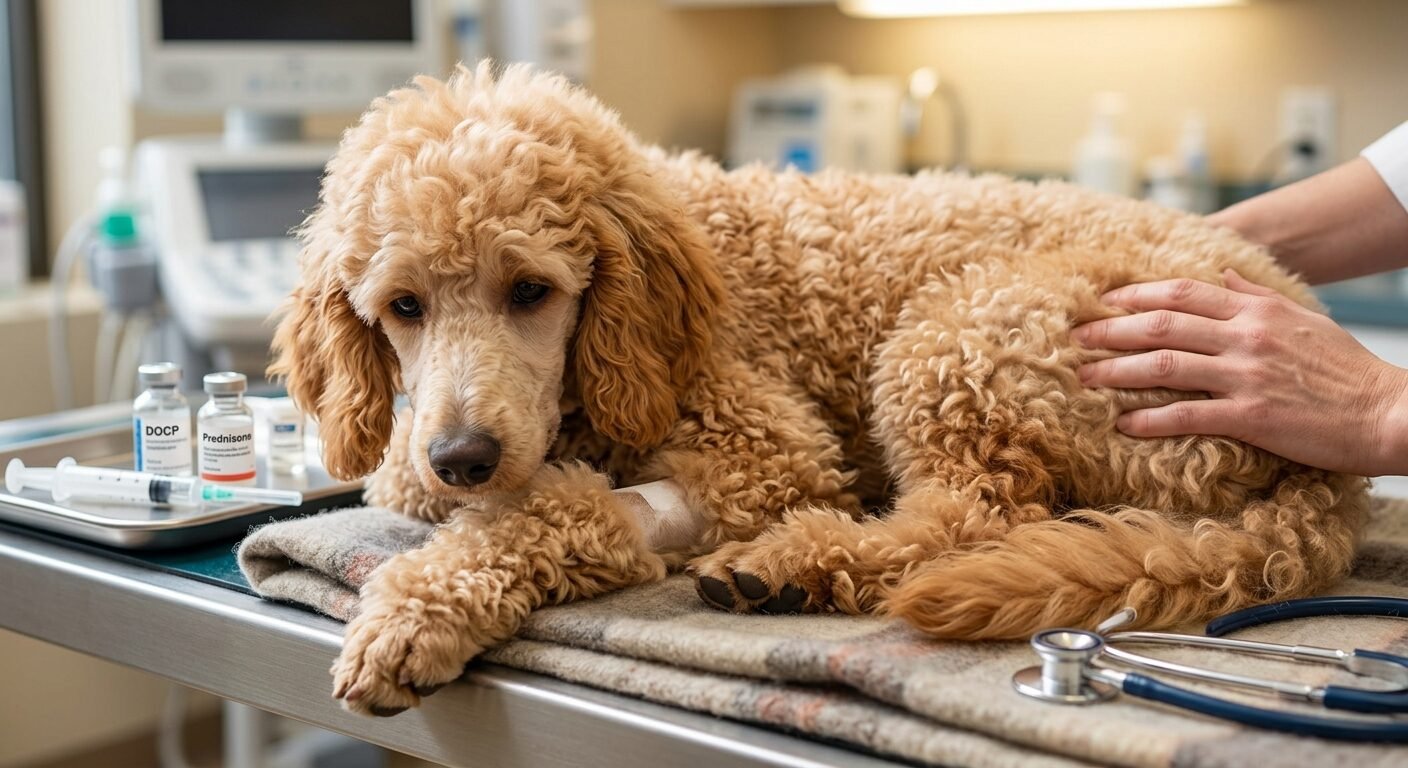
      DOCP (Percorten-V or Zycortal) Injections

       Long acting injection of a mineralocorticoid drug, administered every 25-30 days to keep sodium and potassium in balance.
       Daily Oral Corticosteroids (Prednisone): A tiny daily dose of oral prednisone to replace the missing glucocorticoids (cortisol).
       Stress Protocol: Owners should temporarily give a higher dose of oral prednisone during predictable periods of stress, like during surgery, holidays, fireworks, or when moving into a new home. The dog’s body doesn’t make additional cortisol to deal with stress, so it is necessary to give them that “stress buffer” by hand.

      Conclusion

      When your beloved is diagnosed with an endocrine disorder that is chronic and life threatening, it’s very alarming. But the prognosis is extremely optimistic for dogs with Addison’s disease (hypoadrenocorticism). After the proper dosages of the correct medications are determined, these dogs live a completely normal, very active and long life. They are able to run, play, hike, and cuddle like any healthy pup. Addison’s disease (hypoadrenocorticism) can easily be managed in dogs by learning to know the symptoms of a hormonal crisis and working closely with your veterinarian to monitor your dog on a regular basis.

      Frequently Asked Questions (FAQs)

      Q: No, Addison's disease doesn't hurt dogs.

      A: When a flare-up or crisis occurs, dogs are very uncomfortable because of the intense nausea, cramping, weakness of muscles and low blood pressure. Once stabilized and taking the daily maintenance medication, however, they are pain free and feel completely well.

      Q: In dogs, is it possible to live a normal life with Addison's disease (hypoadrenocorticism)?

      Lorem ipsum dolor sit amet, consectetur adipiscing elit. Ut elit tellus, luctus nec ullamcorper mattis, pulvinar dapibus leo.

      A: Absolutely. Dogs with this condition usually have a normal, full canine lifespan with consistent administration of medication and routine veterinary monitoring and checks for electrolyte levels.

      Q: The cost of treating Addison's disease (hypoadrenocorticism) in dogs vary depending on the severity of the condition.

      A: The first diagnosis for a crisis may be expensive and may result in crisis hospitalization. Long-term expenses are largely based on dog size since the hormone injections (DOCP) are based on weight and are administered on a monthly basis. Smaller breeds will require lower and less costly doses, whereas very large breeds will be more expensive.

      Q: If I forget to give my dog the medication, what do I do?

      A: If you miss one dose, your dog may fall into a sudden hormone deficiency, and be susceptible to illness or a life-threatening Addisonian crisis. If a dose is missed by accident, please call your vet right away for instructions on how to safely catch up.

      Distemper in Cats, A Complete Guide to Feline Panleukopenia

      Distemper in Cats: A Complete Guide to Feline Panleukopenia

      It is very upsetting to discover that your cat may have a serious disease. A veterinarian’s worst news for any pet owner is hearing about a case of distemper in cats. It is a highly contagious and potentially fatal virus that causes Feline Panleukopenia Virus (FPV) which does require urgent veterinary attention.
      Although it has the same name, it should be noted that this virus is completely different from canine distemper. Rather, it is very similar to the canine parvovirus. It causes severe damage to the gastrointestinal tract, bone marrow and developing nervous system as it targets a cat’s most rapidly dividing cells.
      Knowing how distemper works in cats, its early warning signs and what treatment protocols are available to you can mean the difference between life and death for your pet. This extensive handbook will take you through all the aspects you should know to identify, handle, and stop this hazardous condition.

      Distemper in Cats, What is it?

      In order to get a good idea of the distemper in cats, it’s important to take a moment to examine how the vitamin works. Feline panleukopenia literally means “all-white-blood-cell decrease. Once a cat becomes infected with the virus the virus invades tissues where cells are rapidly dividing.
      The virus primarily invades:

      ⦁ The Bone Marrow:

      By attacking the marrow, the virus shuts down the production of all white blood cells. This leaves the cat with no protection from secondary bacterial infections

      ⦁ Intestinal Lining:

      The virus causes damage to the lining of the intestines, causing severe ulceration and sloughing off of intestinal tissue, excessive diarrhea and deadly dehydration.

      ⦁ The Brain:

      The virus can cause problems in the developing brain of the unborn kittens of pregnant infected queens, affecting the cerebellum, the brain’s balance and coordination centre.

      The virus is very hardy and will survive in the environment for months or even years, and can not be killed by most common household disinfectants.

      Where Do Cats Get The Virus?

      Distemper spreads quickly and easily among cats. The main route of infection is via the faecal-oral route. Infected cats produce huge quantities of the virus in their vomit, saliva, urine and feces.
      There are several ways a healthy cat can become infected:

      Direct Contact: An infected cat can be contacted by sniffing, grooming or sharing the same environment.
      Contaminated Objects: Contact with contaminated food bowls, water dishes, litter boxes, bedding or toys (Fomites or Contaminated Objects).
      ⦁ Human Transmission: Transmitted by human care takers who have come in contact with an infected animal through their hands, clothing or shoes.
      ⦁ In Utero: Transmitted to the developing kittens by the infected mother across the placenta.
      Indoor cats do not have absolute protection against the pathogen because it is very stable outside of a host. A pet can pick up the virus from a walk in a part of a home where a stray cat was infected and shed the virus.

      Symptoms and Clinical Signs

      Cats do not show signs of distemper for three to ten days after exposure. The onset of clinical signs is often sudden and dramatic. A seemingly normal cat may be very sick by nightfall.
      If you think your pet may have been exposed, watch for these characteristic signs:

      1. Extreme Lethargy and Depression: The first symptom is sometimes an extreme lack of energy (extreme lethargy and depression). Your cat may get stuck in a dark corner, be unwilling to get up, and exhibit complete apathy toward cat toys or cat food.
      2. Fever and Temperature Drops: The first signs of distemper in cats are a high temperature, which can reach 104 Fahrenheit to 107 degree Fahrenheit or between 40 degree Celsius and 41.6 degree Celsius. The body temperature drops sharply as the disease advances and the cat goes into shock.
      3. Severe Gastrointestinal Distress: Signs are often severe Gastrointestinal Distress, with persistent vomiting and profuse watery diarrhea. The stool may be bloody and smell very bad from the shedding of the intestinal lining. It’s also possible you’ll find your cat leaning over the water bowl, trying to drink but can’t because of the nausea.
      4. Dehydration and Poor Coat Condition: Due to severe fluid loss from vomiting and diarrhea, the skin becomes wrinkled and dry. With gentle pinching of the skin over their shoulders it will not “pop back”. The fur soon becomes matty, unkempt and dull.
      5. Neurological Signs in Kittens (Cerebellar Hypoplasia): If a kitten is exposed to distemper in cats in the womb or during early infancy, he or she can survive and develop a condition called cerebellar hypoplasia. These kittens have an unusual “wobbly cat” walk, head tremor and poor spatial coordination. This particular problem is non-progressive and not painful, but will need to be accommodated inside where it will remain for life.

      How Veterinarians Diagnose the Condition

      These are the signs by which veterinarians diagnose the condition, so if you see any of these signs, you must go to your veterinary clinic right away. Diagnosis of distemper in cats involves the combination of clinical history, physical examination and certain blood tests:
              ⦁  Complete Blood Count (CBC): A CBC will show a severe decrease in white blood cells (leukopenia). Normal cats will have thousands of white blood cells in each microliter of blood, but infected cats can have almost none.
              ⦁ SNAP ELISA Tests: A quick fecal test, commonly used to detect canine parvovirus, is frequently used by veterinarians: SNAP ELISA Tests. These rapid tests can be used to detect the presence of the virus in a feline stool sample within minutes due to the fact that the virus is structurally very similar to the virus found in cats.
              ⦁ Polymerase Chain Reaction (PCR) Testing: If there is still any doubt about the presence of viral DNA, a laboratory can perform a PCR test on a fecal swab to confirm the presence of viral DNA.

      Treatment Options and Supportive Care

      At this time there is no antiviral drug that can cure distemper in cats. Rather, medical management is directed solely toward intensive supportive care which would be lifesaving until the cat’s immune system is able to recover and manufacture antibodies.
      The disease breaks down the barrier in the gut and reduces the number of white blood cells, leading to bacteria entering the bloodstream and causing fatal sepsis. Aggressive hospitalization is almost always required.

      ⦁ IV Fluid Therapy:

      The very backbone of therapy is IV (intravenous) fluid therapy to replace hydration, electrolyte disturbances and to support blood pressure.

      ⦁ Injectable Antibiotics:

      Antibiotics are not effective against viruses, but are essential to ward off a secondary infection by bacteria that may overpower the leukopenic patient who is already weakened by the virus.

      ⦁ Blood Transfusions or Immunotherapy:

      Blood transfusions and immunotherapy (also known as blood replacement therapy) can be used in severe cases when blood levels of protein are dangerously low, and/or anemia has set in, to give the blood proteins, proteins, and immediate immunity

      Prognosis and Survival Rates

      The prognosis of distemper and the chances of survival in cats vary significantly based on the speed of treatment. The mortality rate is more than 90% if the cat is left without veterinary treatment, particularly if it is a very young kitten under five months of age. But timely and aggressive hospitalisation and intensive supportive care can improve survival rates to 50-75%.
      If an infected animal can survive those first 3-5 crucial days of sickness, the probability of the animal making a full recovery grows rapidly. After a few weeks, once a cat has recovered from the illness, they build a strong immunity to this particular virus and don’t shed the virus into the environment again.

      Prevention: Standard Kitten Vaccination Schedule

      Prevention: Standard Kitten Vaccination Schedule
      The best way to deal with distemper in cats is to prevent it from occurring. Fortunately, we have an extremely powerful weapon: The FVRCP vaccine.
      Kittens are initially provided temporary immunity via their mums milk (maternal antibodies) which fades between 6-8 weeks of age.
      1. Initial dose: 6-8 weeks old.
      The primary vaccine gives the immune system a shot of the modified-live or inactivated virus strains.
      1. Boosters: Every 3 to 4 Weeks.
      Give booster shots every 3-4 weeks until the kitten is about 16-20 weeks.
      1. One-Year Booster: 1 Year After Series.
      A booster one year after the first kitten series to strengthen the long-term memory cells.
      1. Adult Maintenance: Every 3 Years.
      Adult healthy indoor cats will need booster shots every three years to ensure adequate protection.

      Conclusion

      As a cat owner, you are going to find it difficult to deal with distemper in cats, not only emotionally, but financially as well. The fact that feline panleukopenia is a disease that progresses so quickly demonstrates how sensitive our pets are to harmful disease-causing organisms.
      Luckily, this disease is easily preventable. You can keep your cat protected from this fatal disease by following your veterinarian’s vaccination guidelines and keeping up with good hygiene. If you ever notice your pet suddenly being lethargic, vomiting or having diarrhea, remember that every minute matters; seek emergency veterinary care right away to improve the chances that your cat will survive.

      (FAQs)

      1. Do cats transmit distemper to humans?

      No. Distemper can be caused in cats by the feline panleukopenia virus which is highly species specific. It is not communicable with humans, and does not affect dogs. Humans may however serve as mechanical vectors (carriers of the virus on their hands and clothing from an infected cat to a healthy cat).

      2. Is it possible for fully indoor cats to be contaminated?

      Distemper is a disease that can be contracted by cats in the house, yes. The virus is very hard to kill in the environment and can be easily spread indoors on your shoes, clothes or hands following exposure to an infected outdoor cat in an area where the virus is commonly found. All cats, whether indoors or outdoors, should be vaccinated.

      3. The shedding of the virus will cease after how many days in a recovering cat?

      Feces and urine may contain the active virus for up to 6 weeks after clinical recovery in a cat that has had a distemper infection. In this period the convalescent cat should be isolated from all other cats and young animals that have not been vaccinated.

      Tumors in Cats

      Feline Mammary Tumors: Symptoms, Treatment, and Survival Guide

      Any pet owner’s nightmare has to be finding a lump on their pet’s body and that is especially true for the female cat. Cats with mammary tumors are one of the most important and commonly diagnosed reproductive cancers in animals, as mammary cancer comprises about 17% of all tumors in female cats.
      Canine “mammary growths” have a 50/50 chance of being avulse, but that’s not the case with feline mammary growths. Nearly 85-95% of mammary tumours in cats are very aggressive, locally invading and are a high risk to spread to nearby lymph nodes and other organs such as the lungs. As this cancer is so quick to develop, it is paramount that the owner knows the risk factor, recognises signs of the disease early and gets their cat into a rapid veterinary treatment protocol—a treatment plan that will help to prolong their life.

      So, What causes Mammary Tumors in cats?

      Hormonal influences are a major contributing factor to why mammary tumors occur in cats and this is because they are exposed to estrogen and progesterone on a regular basis during normal heat cycles. Ovariohysterectomy (spay surgery) is the single best preventative measure against mammary tumors occurring in cats.

      Spayed before 6 months of age: decreases lifetime risk of mammary cancer by a huge 91%.
      Spayed at 7-12 months of age: decreases risk by about 86%.
      Spayed between 13 and 24 months old: Reduces risk by 11%.
      Spayed after 2 years of age: No benefit in terms of protection from mammary tumors for cats.

      Further Hormonal Risk Factors

      The use of exogenous progestins (synthetic hormones that may be prescribed by a veterinarian for certain skin problems and/or behavioral disorders) significantly increases the risk. Cats treated in this way run more than 3 times the risk of developing malignant mammary tumors in cats.

      Age and Breed Predispositions:

      ost of the cats affected are older (11-13 years median) intact females. Any domestic cat can be affected but, in Siamese cats, the tendency is quite strong, and they often get these tumors at a much younger age than other breeds.

      2. Recognizing the Signs and Symptoms

      Cats have four pairs of mammary glands from the top of the chest through to the groin area and mammary tumors can develop anywhere along these lines. When diagnosed, from 60% of felines affected will have more than one distinct mass on both chains.
      Owners should regularly palpate the abdomen at home and carefully look for these clinical signs:

      Firm, Nodular Lumps: Small, pea-sized pebbles or firm, irregular thickening under the skin, next to or under a nipple.
      Skin Ulceration and Discoloration: When a malignant growth or tumor grows, the skin is stretched until it tears apart, leaving a sore that is raw, weeping, or bleeding and giving off an unpleasant odor because of secondary bacterial infection.
      Localized Swelling and Pain: The affected mammary glands may be abnormally warm, red or swollen and the cat will be visibly uncomfortable when picked up.
      Systemic Illness or Breathing Distress: If mammary tumors in cats have been successful in metastasizing to the thoracic cavity, the cat may become lethargic, lose weight, have an uncharacteristic loss of appetite, or begin to breathe quickly and shallowly.

      3. Important Clinical Diagnosis and Staging

      When a veterinarian sees mammary tumors in a cat, she will perform a thorough staging panel to determine the extent of the disease before any surgery is done.

      Biopsy and Histopathology

      Fine-needle aspiration (FNA) may be useful to exclude skin cysts, but a biopsy or removal of the tumor and complete histopathology will be the best method to diagnose mammary tumors in cats. Pathologists analyse the cells to determine the type of carcinoma (e.g. tubular, ductal or anaplastic) and give a histological grade (Grade I, II or III), depending on how aggressively the cells are dividing.

      Advanced Imaging

      A 3-view thoracic X-ray panel is a must for a cat with mammary tumors as these can easily spread into the chest cavity and lungs. If you notice any signs of cancer spreading to the lungs (pulmonary nodules), the treatment plan changes from aggressive surgery to aggressively palliative care. An abdominal ultrasound is also commonly ordered to see if there is any spread within the abdomen to the liver, spleen or medial iliac lymph nodes.

      4. Treatment Approaches and Prognosis

      Treatment approaches and prognosis are significantly related to one basic number when it comes to mammary tumors in cats: the size of the primary tumor at the time of surgery.

      Tumor Size and Prognosis

      Tumor Size Group

      Clinical Characteristics

      Expected Median Survival Time

      Small Tumors (< 2 cm)

      Early detection, localized to the primary gland

      3+ years (with radical surgery)

      Medium Tumors (2 to 3 cm)

      Intermediate stage, potential lymph node involvement

      1 to 2 years

      Large Tumors (> 3 cm)

      Advanced local disease, high likelihood of cellular tracking

      4 to 6 months

      The Standard of Care: Radical Chain Mastectomy

      Because all the mammary glands on one side of a cat’s body are connected by shared blood vessels and lymphatic channels, conservative “lumpectomies” are highly discouraged in treating mammary tumors in cats. Surgical treatment is the gold standard, and involves either a unilateral or bilateral radical mastectomy where the whole line of mammary tissue and any regional lymph nodes are removed on the affected side to reduce the risk of aggressive local recurrence.
      When surgery is a success, an oncologist might suggest that the pet receive the same drug treatments as systemic chemotherapy, most commonly by injection with the drug carboplatin or doxorubicin. In cats with high-risk factors such as vascular invasion or high grade tumor, chemotherapy can help to postpone the progression of metastatic disease, giving cats important extra months or years.

      5. Medical Management and Palliative Care

      Medical management of the cat with an advanced, non-resectable mammary tumor and when the cancer has already spread to the lungs is chronic palliative management, where the aim is to maintain the maximum possible comfort, pain reduction and preservation of the quality of life of the cat.

      1. Offer personalized pain management:

      Use veterinary prescribed non-steroidal anti-inflammatory drugs (NSAIDs) and/or some pain modulators for the nervous system that have been developed for pets on a regular basis to ensure maximum comfort.

      2. Continue Strict Wound & Hygiene Management:

      If the tumour has broken through the skin surface, keep the area clean, wear loose protective shirts as recommended by your vet and do not let the cat over-groom or lick the raw skin.

      3. Prescribe Targeted Antibiotic Therapies:

      Make use of wide spectrum antibiotics to actively treat secondary bacterial infections in weeping, open mammary sores to help reduce inflammation and odors.

      4. Report on QoLM regularly:

      Assess daily activities of your cat within a structured QOL framework, to make sure that good days outweigh bad days for your cat as far as their appetite, hydration and general comfort are concerned.

      Frequently Asked Questions (FAQs)

      Q1. What is the survival rate for a cat diagnosed with mammary tumors?

      Tumor size at diagnosis is the almost sole determinant of survival. For cats less than 2 cm in size, a radical mastectomy may result in a few years of survival. If the tumour is bigger than 3 cm or the tumour has spread to the lungs, the median survival time decreases to less than 6 months.

      Q2. Do male cats get mammary tumors?

      Yes, but it’s unusual. Cats are less likely to develop mammary tumors if they are male, accounting for less than 1% to 5% of all mammary tumor cases. In male cats, however, once it develops, mammary cancer is as aggressive as it is for female cats and is staged and treated identically.

      Q3. Is a lump in a cat's mammary gland always cancer?

      Some mammary tumors in cats are very malignant cancers, but it is a small possibility that the mass is a benign adenoma or feline mammary hypertrophy, which is a benign fast-growing growth caused by a hormone, progesterone. They can only be distinguished safely by a professional biopsy.

      Q4. What are the causes of death of mammary tumors in cats?

      With progression of malignant mammary tumors in cats there are two ways in which complications can occur. The first is that it can cause the main external tumours to open up, become infected, bleed and cause massive systemic inflammation. Second, the cancer spreads to the lungs and, over time, becomes unmanageable and causes the cat to become uncomfortable breathing.

      Bladder Stones in Dogs. Causes, and Veterinary Treatments

      Bladder Stones in Dogs: Symptoms, Causes, and Veterinary Treatments

      The Complete Guide to Bladder Stones in Dogs: Symptoms, Causes, and Treatments

      The discovery that your cherished four-legged family member has a medical problem can be extremely stressful. You’ve heard from your veterinarian about your pet having bladder stones in dogs or you think your pet may be exhibiting symptoms of this painful condition, and you likely have a number of questions.
      Dogs are prone to developing bladder stones, which are a fairly common, but very uncomfortable, urological disorder. Scientifically called uroliths, these are rock-like structures that form inside the urinary bladder and vary in size from sand-like small particles to large and smooth stones, filling nearly the whole bladder cavity. Bladder stones in dogs can be a source of confusion, so it’s important to gain an understanding of what they are, how to identify them, and the treatment options available to help get your dog back to a happy, pain-free life. We’ll delve into the science behind bladder stones in dogs, the various types of stones, signs and symptoms, and medical and surgical treatments.

      So What Do Bladder Stones In Dogs Mean?

      It’s helpful to understand how the urinary system works in a dog to understand bladder stones in dogs. Wastes are removed from the blood by the kidneys and discharged into the bladder as urine. This urine will contain a number of the substances that are dissolved in the blood and excreted from the body.
      Under normal circumstances they are fully dissolved and excreted without causing any symptoms. These microscopic crystals of minerals can start to form if the chemical makeup of urine shifts, or if the urine becomes very concentrated. These ‘clustering’ crystals harden and increase in size with time and the crystals develop into full bladder stones in your dog.
      Note that bladder stones are different from kidney stones. Kidney stones develop in the kidneys, and are not a very common condition in dogs, however bladder stones develop in the urinary bladder, and are a much more common condition in veterinary clinics.

      The dog bladder stones are of different types.

      There are several different kinds of bladder stones in dogs.
      Dogs can have stones in their bladders that are not all of the same substance. In fact, the mineral make-up of the stone is important, the type of stone determining the exact treatment and prevention strategy. The two most prevalent are the most common veterinary cases.

      1. Struvite Bladder Stones

      Struvite stones are made up of magnesium, ammonium and phosphate. Struvite bladder stones in dogs are almost certain to occur as part of an existing urinary tract infection (UTI) in dogs. Some bacteria have an enzyme called urease which changes the pH of the urine, making it more alkaline. This alkaline environment causes struvite crystals to quickly combine to create stones. Females are more likely to develop struvite stones than males since they are more likely to get a UTI.

      2. Calcium Oxalate Bladder Stones

      Unlike struvite, calcium oxalate bladder stones in dogs form in highly acidic or neutral urine. A disease infection usually does not cause them. In reality, they are associated with genetically inherited factors, metabolic disturbances and high calcium intake. Miniature Schnauzers, Shih Tzus and Yorkshire Terriers are very susceptible to calcium oxalate bladder stones in dogs. Unfortunately, these stones can’t be dissolved with any medicine or diet and nearly always must be physically removed.

      3. Other types of stone that are less common.

      • Urate Stones: Also known as uric acid stones, they are common in Dalmatians that are genetically predisposed to a defect in their liver’s ability to break down uric acid.
      • Cystine Stones: These are a rare type of stone which is related to an inherited genetic metabolic defect and are usually only seen in undamaged male dogs.
      • Silica Stones: Very rare stones, typically associated with certain food ingredients, like foods rich in corn gluten or grain hulls.

      The symptoms of Bladder Stones in Dogs include but are not limited to:

      The symptoms are similar to that of a severe infection of the bladder in dogs, as the stones irritate the bladder’s lining, causing inflammation. If you are a conscious dog owner, you should be concerned about the following clinical signs:

      • Blood in the Urine (Hematuria): This is due to the rough surfaces of the bladder stones continually rubbing against the bladder wall, causing small tears and bleeding. Urine may appear pink, red or with blood clots.
      • Straining to Urinate (dysuria): If your dog strains to urinate for extended periods of time but only gets a few drops out, this is also a symptom to watch for. The stones are physically present, causing pain or irritation.
      • Pollakiuria (Frequent Urination): The irritation tricks the dog’s brain into thinking the bladder is always full and causes him/her to go outside more often.
      • Accidents in the House: Even a perfectly house trained dog can start to leak urine or have accidents in the house due to the inflammation that make them less able to hold their bladder.
      • Licking the Sore or Painful Areas: Dogs will often lick at sore or painful areas as a form of self-soothing.

      CRITICAL WARNING

      If the stone moves out of the bladder and blocks the narrow tube (urethra) which carries urine outside the body, it can be a complete blockage of urine. This is a life-threatening medical emergency! If your dog is straining extremely hard but no urine is produced, vomiting or is very lethargic you should go to an emergency veterinarian right away.

      Risks and root causes

      How does this condition develop in pets, and why are some never affected and never develop it? There are several interlocking factors that help cause bladder stones in dogs:

      Diet and Hydration

      If your dog does not drink sufficient amounts of water, its urine will be very concentrated. Concentrated urine allows minerals to be packed tightly together and is very easy for crystals to form in your dog’s urinary system, especially in their bladder, leading to bladder stones. In addition, too much of certain minerals (such as calcium, phosphorus, and magnesium) or protein can change the chemistry of urine, leading to the formation of stones.

      Urinary Tract Infections (UTIs)

      Like struvite stones, persistent or unaddressed UTIs completely alter the chemical makeup of the bladder thus creating an ideal environment to promote bladder stones in dogs.

      Breed and Genetics

      There are a lot of genetic factors involved. The bladder capacity of small breeds is also smaller and they tend to produce more concentrated urine, making them more susceptible to a high risk profile. There are specific bladder stones in dogs that are more common in certain breeds because of genetic anomalies, such as Dalmatians, English Bulldogs and Pugs.

      The diagnosis of the condition is made by veterinarians.

      If your dog is exhibiting urinary troubles, your veterinarian will use a number of diagnostic tools to determine if bladder stones in dogs are to blame.

      Physical Exam & Urinalysis / Diagnostic Imaging (X-rays/Ultrasound) / Stone Analysis.

      • Blood tests: The vet will take a sample of blood and test it for blood cells, signs of infection, blood chemical levels and the presence of microscopic mineral crystals.
        Lorem ipsum dolor sit amet, consectetur adipiscing elit. Ut elit tellus, luctus nec ullamcorper mattis, pulvinar dapibus leo.
      • Digital Radiographs (X-rays): Bladder stones that are radiopaque, which means they appear very bright and white on an X-ray, many types of bladder stones in dogs (like struvite and calcium oxalate) are radiopaque.
      • Urinary Ultrasound: Some stones, such as urate stones, are not detectable by the traditional X-ray. These stones can be easily identified by ultrasound, which can be used to see them in the bladder.
      • Calculus Analysis: after the stones are extracted or passed, they are taken to a special laboratory. The only way to be 100% certain of the mineral composition of the bladder stones in dogs is to have a complete X-ray analysis, so that a long-range preventive care plan can be developed.
      Treatment of Bladder Stones in Dogs

      Treatment is greatly different based on the size of the stones, the sex of your dog and the type of mineral that is involved.

      1. Surgical Removal (Cystotomy)

      If the stone is very large, will not pass on its own or if there is an immediate risk of a life-threatening blockage, a surgical procedure called a cystotomy is the gold standard. The veterinarian gives the dog general anesthetic and makes an incision into the abdomen and opens the bladder, in order to gently remove all the bladder stones in a dog and thoroughly rinse away everything. Although surgery can be frightening, it can give immediate relief from pain and clear the blockage, and also remove the risk of a sudden blockage.

      2. Dietary Dissolution

      Medical dissolution is a possibility if your veterinarian feels that your pet is not at risk of developing a urinary blockage, and you have struvite bladder stones in dogs. This includes a prescription therapeutic diet for your dog. A special food that helps to bring the pH of the urine back into the acidic range and significantly lower the consumption of mineral salts that can form stones. Dogs gradually pass away their bladder stones over a few weeks to months.

      Note:

       When dissolving the stones, your dog is not allowed to eat table scraps, regular dog food, or commercial treats, since a morsel of normal food can destroy the exact chemical balance required for dissolving the stones.

      3. Non-Invasive Procedures

      There are in modern veterinary medicine highly specialized, non-invasive methods of treating bladder stones in dogs:

      • Urohydropropulsion: If the stones are very small, the vet can sedate the dog, and use a special catheter method to flush them out from the urethra without any surgical cut.
      • Laser Lithotripsy: At specialized veterinary referral centers, a laser lithotripsy is possible, where a very small laser fiber is inserted into the urinary tract and the bladder stones are physically broken down into tiny pieces that can then be urinated out.

      Prevention: Stones not coming back.

      Unfortunately, bladder stones in dogs tend to recur very rapidly. Without modification of your dog’s lifestyle habits, there is a very high chance that stones will reappear in a year or two. A dedicated, lifelong management approach is needed for prevention.

      Increase Water Intake

      Diluting the urine is the best protection against bladder stones in dogs. Urine that is diluted prevents minerals from lumping together. To get your pet to drink more, you can mix warm water or low sodium bone broth into your dog’s food, get a circulating dog water fountain, and encourage your dog to eat more canned wet food where there is a higher moisture content.

      Prescription Preventative Diets

      If your dog has a history of calcium oxalate or urate bladder stones, your veterinarian may recommend a maintenance diet for the rest of the dog’s life. The diets are also specifically formulated to balance the body’s internal pH and reduce the raw mineral building blocks in the bladder.

      Frequent Bathroom Breaks

      Avoid holding your dog’s bladder for too long. Mineral crystals have the perfect chance to settle and adhere to each other when urine remains stagnant in the bladder for too long, resulting in complete bladder stones in dogs. The frequent urination will rinse away the microscopic crystals before they do any harm.

      Frequently Asked Questions (FAQs)

      Q1. Will bladder stones pass on their own in dogs?

      Sometimes little sand-like stones pass without any issue, especially in female dogs, who have a shorter, wider urethra than males. But, medium to large bladder stones in dogs cannot pass without the intervention. Trying to pass them on their own can be a huge risk of causing a urinary blockage that can lead to death. Don’t wait for the stones to go away, always consult a vet.

      Q2. How long does it take to dissolve bladder stones in dogs with a special diet?

      A prescription diet is usually the solution for removing bladder stones in dogs, and it takes 2 to 12 weeks to completely dissolve these stones. The amount of time it takes will vary based on the size and quantity of the stones. Continued x-rays are necessary throughout this time for monitoring.

      Q3. Can dogs use apples cider vinegar to treat bladder stones?

      No, do NOT use apple cider vinegar or other home remedies to help remove bladder stones from dogs. Changing your dog’s urine pH can be extremely dangerous if you don’t know what kind of stone is occurring. As another example, the treatment of a possible problem by making the urine more acidic may actually promote the formation of very dangerous calcium oxalate stones.
      Lorem ipsum dolor sit amet, consectetur adipiscing elit. Ut elit tellus, luctus nec ullamcorper mattis, pulvinar dapibus leo.

      Q4. On average, how long does it take for a dog to recover from bladder stone surgery?

      Dogs make a wonderful recovery after a routine cystotomy. Your dog will likely have to wear an Elizabethan Collar (the “cone of shame”) and their exercise routine limited for approximately 10-14 days until their abdominal incision has healed. Your pet’s doctor will prescribe pain medications and antibiotics to help your pet recover from the surgery in a comfortable and pain-free manner.

      oral squamous cell carcinoma in cats

      Feline Oral Squamous Cell Carcinoma: Symptoms, Treatment, and Care Guide

      This article provides a comprehensive clinical and care guide to Feline Oral Squamous Cell Carcinoma. This article offers a thorough clinical and care guide to Feline Oral Squamous Cell Carcinoma.
      The news of a beloved pet has cancer is one of the most painful things a pet owner can experience. Feline oral squamous cell carcinoma (OSCC) is one of the most common and aggressive oral cancers in cats, representing approximately 60% to 75% of all oral tumors in cats.
      This aggressive form of cancer begins in the lining of the mouth and spreads quickly and is very destructive. Oral squamous cell carcinoma in cats is a locally infiltrative disease, so prompt diagnosis and the speed with which treatment options are understood is important to ensure patient comfort and quality of life. In this guide we will look at the risk factors, common symptoms, the current treatment and importantly, the proper care practices for cats facing this disease.

      1. What is the cause of the oral squamous cell carcinoma in cats?

      Veterinary researchers have found that a combination of chronic inflammation, genetic mutations and environmental factors play a role in the development of oral squamous cell carcinoma in cats. Feline oral and head and neck cancers have different environmental predisposing risk factors than human cancers, and are not as strongly associated with alcohol or direct tobacco use.

      Environmental Risk Factors

      Add Your Heading Text Here

      Cats are known for being clean. As tobacco smoke falls on their fur they lick it off, giving the concentrated carcinogens directly to their oral tissues. This is an important action because it raises the risk of developing an oral squamous cell cancer in cats.

      Add Your Heading Text Here

       Chronic exposure to chemicals from some older flea collar models has been correlated with an increase in oral tumors

      Other Potential Risks:

      These include frequent tuna consumption (canned tuna or wet food with chemical additives), and exposure to household volatile organic compounds (VOCs), or clumping clay cat litter

      Age and Demographics

      This cancer affliction is mostly affecting older cats. In retrospective health data from a large scale, the median age of diagnosis is 13 years old. It may affect any breed, but most cases are in domestic shorthaired and longhaired cats that are not pedigree cats.

      2. Early warning of symptoms is crucial.

      In cats, one of the biggest challenges in the treatment of oral squamous cell carcinoma is that early signs are similar to that of other non-cancerous conditions such as periodontal disease or feline chronic gingivostomatitis (FCGS). As a result, few cats are diagnosed with oral squamous cell carcinoma in cats until the tumor is in an advanced stage.

      Owners should watch for these clinical warning signs in older cats:

      The cat may drop food, chew completely on one side of its mouth, or walk up to the food bowl with great enthusiasm yet walk away crying.

      Unexplained Weight Loss:

      The person will lose weight rapidly as eating is either very painful or not possible due to the main tumor

      Drooling:

      May contain a secondary bacterial infection, which blood tinges it and gives it a foul smell, or it may be necrotic (dying) tissue in the mouth.

      Add Your Heading Text Here

      An unkempt and matted coat is the result, sometimes with swelling under the eye, face and/or jawline

      3. Diagnosis and Staging

      A careful diagnostic evaluation is needed if a vet suspects a cat might have **oral squamous cell carcinoma** to confirm the diagnosis and to assess if there is any damage to the structures.

      Add Your Heading Text Here

      The only definitive diagnosis for a cat with oral squamous cell carcinoma is a tissue biopsy. Under the microscope, pathologists search for characteristic aggregations of cancerous epithelial cells, different features within the cancer cells, and rapid cell division.

      Advanced Imaging and Bone Invasion

      One amazing thing about OSCC in cats is that it tends to be osteolytic – that is, it invades its surroundings and aggressively breaks down the bone. Dental radiographs or Computed Tomography (CT) scans are used by the veterinarian to identify whether the tumour has invaded the lower jaw (mandible) or upper jaw (maxilla).

      Metastatic Assessment

      Fortunately, this tumor has a relatively low rate of regional and distant metastasis early on; it is highly destructive locally but does not spread very rapidly to other organs. Even so, when the oral squamous cell carcinoma is properly staged in cats, fine needle aspiration of the regional lymph nodes is needed as well as thoracic Xrays to confirm no lung disease.

      4. Current treatment modalities and prognosis.

      Treatment Approach

      Intended Purpose

      Expected Median Survival Time

      Palliative Care Only

      NSAIDs for pain and inflammation management

      ~44 days

      Radiation Therapy Alone

      Coarse or palliative radiotherapy

      60 to 92 days

      Accelerated Radiation + Carboplatin

      Combined radiosensitizing chemotherapy

      163 to 174 days

      5. This is an emerging research and palliative care.

      Oral squamous cell carcinoma in cats is an area of veterinary oncology that is actively being targeted for treatment breakthroughs, as the disease has been known to be resistant to traditional treatments. Due to the similarity in deep molecular and behavioural characteristics between feline oral cancers and head and neck cancers in humans, it is a valuable animal model for comparative oncology studies.
      If your cat is diagnosed with OSA, drug therapy targeted at specific molecules (tyrosine kinase inhibitors) has been used successfully and can improve survival rates – sometimes considerably – from those of the untreated cats.
      In addition, recently the oral microbiome of cats has been mapped and it has been found that cats with OSCC in cats have a dramatic reduction in microbial diversity. The tumor microenvironment is highly pro-disease and is dominated by disease-associated anaerobic bacteria, leading to a new horizon of possibilities for supportive antimicrobial treatment of secondary infections.
      Since complete resolution is uncommon, the main treatment for cats with oral squamous cell carcinoma is supportive care for comfortable, quality life.

      RWMP: Daily

      1. Establish Robust Pain Management:

      Thoroughly cooperate with your veterinarian to put a tight regimen of non-steroidal anti-inflammatory drugs (NSAIDs) or transdermal narcotics to control intense bone and soft tissue pain.

      2. Change the texture of the diet: each meal:

      Gradually switch to ultra thickened watered down purees or liquified recovery feeds. Do not feed hard kibbles or chunky foods which can mechanically rub the ulcerated tumour tissue.

      3. Impact Secondary Infections: Addressed:

      Use a specific antibiotic to control the bacterial overgrowth that usually occurs with necrotic oral tumors and minimizes the “morning breath” and inflammation in the area.

      4. Nutritional Interventions: Evaluate (Clinical Milestone):

      If the cat is very interested in living, however it is unable to swallow the food because of the size of the tumor, consult with your veterinarian about the possibility of having an esophageal or gastrostomy feeding tube placed to allow the cat to eat without stress.

      Frequently Asked Questions (FAQs)

      Q1. What's the life expectancy of an OSCA cat?

      In cats, if left untreated, the median (50th percentile) survival time is approximately 2 months after the diagnosis of oral squamous cell carcinoma. Survival times can be longer than 1-2 years, though, if the tumor is diagnosed early and placed on the bottom jaw, where radical surgery is feasible.

      Q2. Is it possible for oral squamous cell carcinoma in cats to resemble a bad tooth?

      Yes, absolutely. Oral squamous cell carcinoma in cats often presents as severe periodontal disease, loose teeth or small areas of gum swelling in its early stages. An immediate biopsy is strongly recommended if a cat’s “bad tooth” is seen in normal dental care or antibiotic treatment without improvement of the swelling to rule out oral squamous cell carcinoma in cats.

      Q3. Does the OSCC in cats hurt?

      Absolutely, cats with oral squamous cell carcinoma are very painful. It’s an invasive form of cancer that often wears down the bones of the face beneath the skin, leaves the mouth open in an ulcer and can lead to secondary bacterial infections of the affected area. Multi-modal pain management is required for these patients.

      Q4. Is oral squamous cell carcinoma contagious in cats to other organs?

      This particular cancer is very fast and very aggressive at its primary location in the mouth but it spreads (metastasizes) to other organs, such as lungs or liver, at a lower and slower rate. The most common complications of oral squamous cell carcinoma in cats are direct consequences of the primary tumor’s effects on the cat’s comfort eating and drinking.

      Q5. Should I get a feeding tube for my cat with oral cancer?

      An esophageal feeding tube can dramatically improve quality of life by taking the pain out of eating and ensuring your cat receives necessary fluids and medications. However, it should only be used if your cat’s pain is fully managed and they still display a joyful, interactive quality of life despite their diagnosis of, oral squamous cell carcinoma in cats,

      Q6. What are the earliest warning signs of oral tumors in cats?

      The earliest signs often include dropping food while eating, a sudden preference for wet food over kibble, mild drooling, and unusual bad breath. Because these signs are so subtle, early cases of **oral squamous cell carcinoma in cats  are often discovered during routine veterinary dental cleanings.