Aspergillosis in Dogs

Aspergillosis in Dogs: Symptoms, Diagnosis, and Treatment Guide

Aspergillosis in Dogs: Symptoms, Diagnosis, and Management

Being a pet parent, it is almost a distressing situation to see your dog suffering from an unexplainable respiratory problem and suffering from it for a long time. Most people immediately see a problem that can cause their dog to develop kennel cough or seasonal allergies, but there is a very serious and more sinister environmental problem that you should know about aspergillosis in dogs.
This is an opportunistic fungal infection from a common type of mold, called Aspergillus. Dogs are continually breathing in these microscopic fungal spores, which are found in soil, old compost heaps, dry hay and stable bedding. In a healthy dog the spores are easily neutralized every day, but if the immune system is compromised, the fungus can become established, multiply and continue to affect the body in a localized or systemic manner.
By understanding the development of aspergillosis, being able to recognize the early warning signs, and knowing what diagnostic procedures are likely to be used, you can help to ensure your dog’s future health. This detailed guide covers all the information needed for dealing with this complicated fungal matter.

So, what is Aspergillosis in Dogs?

It is important to familiarize yourself with the different biological types of the condition in order to be able to adequately protect your furry friend. There are two main types of aspergillosis in dogs, depending on the location of the infection in the body.

  • Nasal Aspergillosis is by far the most prevalent type of aspergillosis in dogs. The infection is confined to the dog’s nasal passages, frontal sinuses and upper respiratory tract. The fungal growth spreads to form thick “plaques” that actively destroy the fine scroll-like bony structures in the snout called “turbinates.
  • Disseminated Aspergillosis This is a much less common form but has a much greater life-threatening potential. In dogs, disseminated Aspergillus is when the fungi invade the respiratory tract completely and enter the blood stream to invade the internal organs systemically. This form usually targets the bones of the skeleton, the discs between the vertebrae of the spine, kidneys, liver and eyes.

What are the highest risk breeds?

It basically can happen to any dog exposed to high levels of spores, but there are some physical characteristics and inherited tendencies that make a dog more susceptible.

  • Long nosed dog breeds have a massive internal surface area in their nasal cavity which also serves as an uncontrolled reservoir for fungal spores inhaled into the lungs. Historically, certain breeds, such as German Shepherds, Collies, and Greyhounds are overrepresented in clinical settings.
  • Medium nosed dogs like Golden Retrievers and Labradors also experience a fair number of cases, particularly if they are working dogs outdoors and are tracking in soil or vegetation.
  • In immunocompromised Canines, dogs with immune system defects or those taking long term steroids, there are no cellular mechanisms in place to permanently suppress aspergillosis in dogs before it becomes established.
  • Specific Genetic Vulnerabilities: Interestingly, the German Shepherd breed shows a well documented genetic immunodeficiency for a specific antibody (IgA), thus making them very susceptible to both nasal and disseminated forms of aspergillosis in dogs.

Clinical Symptoms to Watch For

Dogs develop a specific progression of clinical symptoms with localized destruction that are caused by aspergillosis. Pet parents should watch for these warning signs:

  • Persistent Nasal Discharge is usually the first sign that something is wrong. Discharge generally starts in one nostril (unilateral) and may spread to both nostrils. It frequently starts with clear fluid and then becomes thick yellow-green slime or even blood (epistaxis).
  • The fungal plaques produce intense internal inflammation which is visible and sensitive. Your dog could be pawing at their face, avoiding touching their muzzle when you reach for it or whining as they eat dry kibbles.
  • Depigmentation: With the passage of time, the constant inflammation of the discharge will physically wear down the dark pigment around the outside nostrils, leaving the outer edge of the nose raw pink, or flaky white.
  • Frequent Sneezing and Reverse Sneezing – The body is constantly attempting to mechanically expel the fungal mats causing uncontrollable sneezing.
  • Dogs with the condition may lose their interest in their food bowl entirely due to the discomfort, as well as their loss of smell.

In dogs, if disseminated aspergillosis occurs, you might observe entirely different changes in structure, such as lameness, extreme pain in the back, fever, enlarged joints, and weight loss.

Diagnosis Protocols

Diagnosis is a methodical approach as the symptoms can be confused with other nasal problems such as a foreign body (such as a grass awn inhaled), or a localized cancerous tumor.
Your veterinarian will usually use a combination of specialized tools:
Advanced Imaging (CT Scan or MRI): Computed Tomography (CT) scan is highly preferred. It offers a clear insight into the internal structure of the skull, showing the precise pattern of destruction of the turbinate bones typical of aspergillosis in dogs.


Veterinarians use a small, flexible camera to examine the nose during rhinoscopy while the animal is under general anesthesia. This enables them to see the characteristic white or greyish plaques, which are caused by the fungi.

Rhinoscopy (biopsy, fungal culture): Small pieces of tissue or brushings are taken from the plaques during rhinoscopy and examined under the microscope to confirm the presence of aspergillus organisms.


Serology (Blood Tests): Blood tests can be used to identify specific antibodies to aspergillosis in dogs, but often are combined with imaging to make a definite diagnosis.

Treatment & Surgical Options

When the diagnosis is confirmed, medication (antifungal pills) is not usually effective enough when given by itself because the blood supply is poor inside the nose of a dog with the disease. Rather, the advanced localized treatment is the norm.
A specialized non-surgical technique called a topical infusion is the preferred clinical treatment for nasal aspergillosis in dogs:

  •  Airway Protection and Anesthesia: Patient Safety

The dog is under deep general anesthesia and an endotracheal tube is inserted and secured by an inflatable cuff. It is an absolute requirement to make sure that liquid drugs do not get accidentally down into the lungs.

  •                                                               Catheter Insertion and Packing :Targeted Delivery.

Carefully inserted specialized catheters are inserted into the nasal passages and frontal sinuses. Surgical sponges are placed in the back of the throat to form a tight seal around the fluid.

  •                                                               Liquid Clotrimazole Infusion :Fungal Destruction.

A strong liquid anti-fungal medication (usually 1% Clotrimazole) is sprayed into the nose. This fluid is left in for only one hour, and the patient is turned frequently throughout that hour to make sure that all the inside surfaces of the passages are covered with the medicine.

4.4.Evacuation and Flushing: Procedure Wrap-up.

After an hour, the catheters are removed, all liquid is carefully drained from the nostrils and the dog is safely awakened by flushing out the nostrils with sterile saline.
Although there is a high success rate (about 80%) with a single infusion, in some severe cases of aspergillosis in dogs, a second infusion a few weeks later will be needed to eliminate the infection. The more uncommon distribution type of aspergillosis in dogs, however, is not treated with topical flushing and must be treated for many months with systemic (oral) antifungal drugs, such as itraconazole or voriconazole.

Long-Term Prognosis and Environment Management

The prognosis for nasal aspergillosis in dogs is generally good, as long as the fungus does not have enough time to break through the thin bone wall (cribriform plate) between the nasal cavity and the brain. If this barrier is left in place and the local therapy is effective, most dogs live a normal and active life but with a structurally changed nose inside.
Unfortunately, the outlook is guarded to poor with disseminated aspergillosis because fungal infections are very difficult to cure entirely in deep skeletal tissues and organs in dogs.
The mold spores are a natural part of the outdoor environment, so it is impossible to avoid them completely. You can reduce the risks to the environment by ensuring that your dog does not go in to dusty barns, fresh excavations, or wet rotting leaf or compost heaps.

Conclusion

When faced with a dog diagnosis of aspergillosis, it can be quite a shock, particularly after dealing with advanced imaging and nasal infusion. Very aggressive fungal problem can change your dog’s upper respiratory condition if not treated.
But if you’re paying attention to your pet’s breathing, nasal discharge and comfort level, you’ll be able to take action before major structural damage occurs. With the help of your veterinarian, you can effectively fight back in dogs with aspergillosis, and help your pup live a comfortable and happy life.

Frequently Asked Questions (FAQ)

Q1. Can people catch aspergillosis directly from a dog infected with it?

No. Zoonotic disease is a disease that cannot be directly transmitted from an animal to a human. Aspergillosis in dogs is not a zoonotic disease. The infection is not transmitted between dogs and humans, but rather by breathing the microscopic spores of fungus found in the outside environment.

Q2. Would aspergillosis in dogs be the same as kennel cough?

No. Kennel cough is an upper respiratory infection caused by a particular bacteria or virus (Bordetella) and results in a dry, hacking cough which is very contagious. In dogs, aspergillosis is localized fungus infection that develops in the depths of the nasal passages and frontal sinuses and, mainly, leads to nasal discharge and erosion of the bone, rather than the primary cough.

Q3. How much does it take to apply nasal infusion treatment?

The infusion liquid part of the treatment is directly infused into the nasal passages for just one hour of nasal contact time. But don’t forget the preparation time for general anesthesia, the use of advanced imaging, the time spent on draining the area after the procedure, and the recovery period, your dog will likely spend the whole day in the veterinary hospital.

Distemper Panleukopenia in Cats

Distemper Panleukopenia in Cats: Symptoms, Causes, and Care

Distemper Panleukopenia in Cats: The Ultimate Guide to Feline Parvovirus

Being a cat owner I am keen on keeping my cat safe, active and happy. There is however a silent, extremely hardy and sometimes fatal health hazard every cat owner should be aware of, distemper panleukopenia in cats. This is a highly contagious, life threatening viral disease which attacks rapidly dividing cells in a cat’s body and is commonly known as feline distemper or Feline Panleukopenia Virus (FPV).
In the past, this infection has completely eradicated entire feline colonies and shelters. Even though it sounds like distemper, it has nothing to do with distemper itself—however, it has many of the same frightening structural and symptomatic characteristics as the canine distemper virus, canine parvovirus. With its aggressiveness, it’s essential to know how to identify the signs, be able to provide proper care, and how it can be stopped from being passed on, which can mean the difference in life and death.
In this all-encompassing guide, you’ll learn all about distemper panleukopenia in cats, how to prevent it, and how to act to navigate it.

What is DPV (Distemper Panleukopenia) in Cats?

The first step in protecting your pet is to get to know the enemy’s biology. The term “panleukopenia” literally means a multi-systemic deficit of all types of white blood cells. Your cat’s immune defense system is built from white blood cells. Once the virus attacks the body, it basically robs it of its resistance to any type of micro-organism or infection.
The cause of distemper panleukopenia in cats is a small, single-stranded DNA virus of the Parvoviridae family. The virus is active inside a host and actively targets cells that are rapidly dividing. These include the lining of the gastrointestinal (GI) tract, bone marrow and developing fetus in pregnant queens. The virus damages the bone marrow and stops it from making white blood cells, making the feline totally vulnerable to secondary bacterial infections.

How Does the Virus Spread?

Distemper panleukopenia in cats is still a problem in the cat community because cats are very susceptible to this virus. The virus is a “non-enveloped” virus because it doesn’t have a tender outer coating of lipids. It, of course, lacks such an outer layer and thus can withstand up to a year or more in an environment of freezing cold, intense heat, and the common household detergents.

  • Direct Contact: It’s transmitted mainly through the interaction of healthy cats with infected cats nasal discharge, saliva, urine or feces.
  • Fomites (Inanimate Objects): The virus is very tough and can easily transfer from food bowls, litter boxes, bedding, grooming brushes, hands or shoes of humans. These things can infect unvaccinated cats if they come into contact with them at a later time.
  • Utero Transmission: If a mother cat does contract the virus it is able to pass it directly on to her unborn kittens. This frequently leads to miscarriage, stillbirth or a major neurological birth defect.

However, this ease of infection means that an indoor pet is not 100% protected from distemper panleukopenia in cats. The microscopic virus can be transferred by a human who has been in contact with an infected animal to un-vaccinated indoor pets, by simply walking through the infected animal’s environment on clothes or shoes.

Symptoms & Clinical Signs

Cats may begin to show signs of distemper panleukopenia within 3 to 14 days after exposure, although symptoms may be very aggressive and begin to appear in a week. The clinical signs are variable and frequently involve the digestive tract and energy levels to a great degree, depending upon the cat’s age and immune status.

  • Abnormally Lethargy and Depression: The normally playful cat suddenly becomes unresponsive, hiding in dark corners and not moving.
  • Fever: The affected cats will have a very high fever, up to 107°F – 104°F but later in shock, their body temperature may drop significantly lower.
  • Severe Vomiting: Virus will actively attack the lining of the stomach and intestines. Cats will vomit clear liquid, bile or blood repeatedly and without any association with eating food.
  • Bloody Diarrhea: When the lower portion of the intestine becomes damaged, it causes diarrhea that is profuse, foul, and liquid, with blood sometimes visible.
  • Dehydration and Hunching: Due to extreme fluid loss, cats will become severely dehydrated. They may be seen sitting hunched over their water bowl, but they are unable to swallow because of the uncomfortable nausea.
  • Sudden death: In severe cases, such as in young kittens under 6 months old, the virus can be fatal within hours, before any digestive symptoms are seen.

If a pregnant queen survives a distemper panleukopenia infection in cats, or if very young newborn kittens become infected, the virus will attack rapidly multiplying cells in the cerebellum. The part of the brain that controls fine motor control, coordination and spatial awareness is the cerebellum.
This special attack results in a congenital disease called Cerebellar Hypoplasia (CH). In Utero FPV Infection leads to Destruction of Cerebellar Cells which results in Cerebellar Hypoplasia (“Wobbly Cat”).
Although this damage to the nervous system may seem frightening, it is non-progressive and non-painful. As these kittens mature, adapt to their own coordination challenges and can enjoy a long happy indoor life with a few house modifications.

Diagnostic Procedures

If you notice any symptoms from the above list, it is imperative that you seek veterinary treatment right away. A quick multi-step clinical assessment is needed to diagnose distemper panleukopenia in cats.
Your vet will use a few different tests to make a diagnosis, usually including the following:

Complete Blood Count (CBC): This is the most important diagnostic. Significant decrease in the number of white blood cells (leukopenia) strongly suggests this particular viral infection.

Fecal ELISA Snap Test: same as the rapid test for canine parvovirus. The two viruses are very structurally similar, so it is possible to detect the FPV antigen in a fresh feline stool sample in minutes.

PCR Testing: In cases of uncertainty, a veterinary lab may be able to perform a Polymerase Chain Reaction (PCR) on a fecal swab to confirm the correct genetic makeup of the virus.

Veterinary Treatment Protocols

At the present time there is no specific antiviral drug to clear distemper panleukopenia in cats. New veterinary treatment, however, is limited to intensive support measures to keep the cat alive until their immune system can recover and restore their white blood cell count.
It is usually necessary to hospitalize kittens in a special isolation room as the risk of death can be as high as 90% if no treatment is done.

                                                                ⦁ Fluid Therapy: An Urgent Treatment.

Immediate treatment of severe dehydration from repeated vomiting and diarrhea is with intravenous (IV) fluids if the dehydration is life-threatening. This helps keep the blood pressure in check and prevents vital organs from shutting down.

⦁ Broad-Spectrum Antibiotics: Combating Sepsis.

Antibiotics are helpful here although they do not fight viruses. Since the virus kills off white blood cells and weakens the barrier of the gut, normal bacteria from the gut can easily enter the bloodstream. This secondary bacterial sepsis is prevented by antibiotics.

3. Anti-Emetics and GI Support: Controlling Nausea.

Strong anti-nausea drugs are injected to prevent vomiting. This gives the damaged gastrointestinal lining a chance to rest, heal and rebuild.

4. Nutritional Restoration: Once Stabilized.

As soon as vomiting is well controlled, the feeding is started with a highly digestible recovery diet or temporary feeding tube. Nutrition supplies the energy that the cells require for repair.

Prevention: The Ultimate Shield

Prevention: The Ultimate Shield is easy and very effective versus distemper panleukopenia in cats, while treating an active infection is stressful and cost the pet owner lots of money.
The Feline Viral Rhinotracheitis, Calicivirus, Panleukopenia (FVRCP) vaccine is considered one of the core vaccines by the veterinary profession. This refers to all cats, whether indoor or outdoor, who should be given this vaccine series.
If a cat hasn’t had any vaccinations or has an incomplete vaccination history, a two shot series a couple weeks apart is usually the way to go to establish adequate immunity.

Household Disinfection Protocols

If you have unquestionably had a cat with distemper panleukopenia inside your house, just about any household cleaners or sprays that contain alcohol or bleach will not disinfected safely.
A diluted bleach solution (household bleach diluted 1:32 in water) is used to completely neutralize the viral particles on non-porous surfaces. Or, special veterinary disinfectants that are labeled to control non-enveloped viruses may be used. Items that are porous, like soft plush toys, cat trees with scratches, or soiled bedding should be tightly bagged and discarded if they can’t be thoroughly bleached.

Conclusion

When a cat is diagnosed with distemper panleukopenia, it can be a devastating shock and emotional experience for any pet owner. It’s very hard, fast and attacks the weakest feline members at incredible speed.
But armed with a knowledge of its transmission and an awareness of its early warning signs, you can act quickly and save your pet a chance. The most important is to keep your cat’s regular routine FVRCP vaccines on track which will offer a virtually impenetrable barrier against this disease. Talk with your veterinarian today and ensure your pet is properly protected.

Frequently Asked Questions (FAQ)

Q1. Can dogs catch distemper panleukopenia from infected cats?

⦁ No. Feline panleukopenia is caused by a feline parvovirus that is similar in nature to canine parvovirus, but distemper panleukopenia can be contracted by cats, but not dogs. Note that cats can sometimes be infected with certain strains of canine parvovirus, but do not contract canine distemper.

Q2. Is panleukopenia contagious to humans from their pets?

⦁ No. Distemper panleukopenia is highly species specific and caused by the parvovirus. It has absolutely no health risk to humans, children or domestic animals such as rabbits or rodents.

Q3. A cat that has been infected can infect others for how long after recovery?

⦁ If the cat survives the distemper panleukopenia outbreak, it will shed the virus in its bodily fluids and feces for up to 6 weeks after recovery. An un-vaccinated cat should be completely isolated from other cats during this period.

Q4. Should an indoor cat be given panleukenia?

⦁ Yes, absolutely. The virus is very stable in the environment and can be spread to your home in micro-particles on your clothes, shopping bags, or shoes. The only surefire way to keep an indoor cat safe is to vaccinate it.

Q5. Does a cat ever suffer from panleukopenia more than once?

⦁ Fortunately, those cats who do survive clinical infection with distemper panleukopenia become well protected against the virus for the remainder of their life.

Ovarian Remnant Syndrome in Dogs and Cats: Signs, Diagnosis, & Treatment

Ovarian Remnant Syndrome in Dogs and Cats: Signs, Diagnosis, & Treatment

Ovarian Remnant Syndrome in Dogs and Cats

When you bring your dog or cat home after the spay, you’re typically relieved. You are hoping that the days of heat cycles, behavioral changes, yowling and vocalizing are behind you. For some pet-owners, however, a strange situation arises weeks, months or even years into the future, when their dog or cat, who was completely spayed, suddenly starts to behave like a dog or cat in heat all over again.
If so, you may be experiencing some of these unusual behaviors in an unanticipated dog or cat and you may have something called ovarian remnant syndrome.
This complete guide explains everything that you would want to know about this condition, including why it occurs and how veterinarians can diagnose and treat it completely.

Add Your Heading Text Here

A spay (medically referred to as ovariohysterectomy or ovariectomy) is a procedure in which the entire animal’s uterus and both of its ovaries are completely removed. Since the production of hormone in the body is primarily from the ovaries, their removal removes the hormonal changes that cause heat cycles.
Ovarian remnant syndrome in dogs and cats is the condition that develops when the functional tissue of the ovary is removed during an ovariohysterectomy and is left inside the abdomen. This small piece of tissue is alive and continues to mature, to receive signals from the brain, and to produce hormones. This results in cyclical changes in behavior and physical characteristics of estrus (heat) even after the animal has been spayed.
The answer is as follows:
This is because:
If you’ve found out your cat or dog has ovarian remnant syndrome, it can be very frustrating. When the surgery has already been done, it’s understandable to ask how this came about. There are essentially two reasons why this syndrome may develop:

1. Surgical Complications

The most frequent occurrence is surgery leaving a small piece of the ovary in place. The ovary is held in place in the abdomen by a mass of connective tissue and blood vessels. It can be very difficult to see and completely remove tissue in deep-chested dogs, overweight pets or animals during active heat. Even a tiny fragment of the ovarian cortex can reconnect with a blood supply, and develop into a viable, hormone-producing fragment.

2. Embryological Variations

In less common cases, a pet may have “ectopic” (embryological) ovarian tissue. This resulted in a normal formation of small groups of ovarian cells in the fetal ovary outside the ovary proper. They are small pieces that are not seen with a normal spay surgery, but can develop later in life when the fragments enlarge and become active, thus causing ovarian remnant syndrome in dogs and cats.

Symptoms and Clinical Signs

The hallmark of the ovarian remnant syndrome in dogs and cats is the resumption of estrous signs and behaviors. The onset of symptoms can range dramatically—from months following the surgery to several years.
The symptoms appear slightly different when you have a dog vs. a cat.

Signs in Cats

  • Persistent, Loud Vocalization: Feline estrus is very vocal. Your cat could begin yowling or “crying” a lot, particularly at night time.
  • Lordosis Posture: The cat will usually rest the front legs on the ground, raise front legs and tail to one side.
  • Overwhelming Affection: Rubbing against the legs, furniture or other items in the house excessively.
  • Restlessness and Estrus Rolling: rolling around on the floor in a restive manner.

Signs in Dogs

  • Vulvar Swelling in Dogs: The vulva will become swollen and be enlarged.
  • Vaginal Discharge: discharge looks like a normal heat cycle and is bloody or straw colored.
  • Behavioral Shifts: Moodiness, turning tail or standing to be mounted by male dogs.
  • Male Attraction: Neighbourhood Male Dogs will be extremely attracted to her pheromones.

Long-Term Health Risks

Although it can be a big behavior problem, long-term management of an ovarian remnant syndrome in dogs and cats can have serious health implications.
Unchecked hormones over a long period of time greatly increase your pet’s risk for developing serious health issues as they get older.
The main long-term medical complications are:

  • Stump Pyometra: If a fragment of the uterine horn (the “stump”) was left after the spay procedure, the ovarian remnant secretes progesterone which can lead to the stump thickening, accumulating pus, and becoming life-threateningly infected.
  • Mammary Tumors: Abnormally high levels of estrogen can greatly raise the chance of a malignant mammary cancer occurring in both dogs and cats.
  • Ovarian Neoplasia: The remaining tissue can develop an abnormal cell change which can result in ovarian tumors or cysts.

How Veterinarians Diagnose the Condition

Diagnosing the condition is sometimes not easy for veterinarians. Tissue which is microscopic or buried in the fat of the abdomen, gastrointestinal tract or other areas is usually not detected at first by standard x-rays or ultrasounds. Rather, a combination of strategic timing and lab testing is used.
Dogs and cats with ovarian remnant syndrome can sometimes be difficult to diagnose. This tissue is usually small or located deep within the fat in the abdomen, making it difficult to detect on a routine x-ray or ultrasound scan at first. Rather, it is a combination of strategy and lab tests.

Ovarian Remnant Syndrome in Dogs and Cats.

What to Expect During and After Surgery:

An exploratory procedure and the previous incision line in the abdomen will have to be reopened. The surgeon will proceed step-by-step to examine the ovarian stumps around the kidneys. A pathologist confirms the presence of ovarian tissue after the tissue is found.

If it is found, the remaining tissue is carefully removed and submitted to a pathologist for confirmation that it is ovarian tissue.

Post-operative care is similar to that of a routine spay: a strict crate rest is recommended, activity is kept to a minimum for 10-14 days, a pain management medication is prescribed and an Elizabethan collar (e-cone) is placed on the abdomen to prevent her from licking at the incision site.


Conclusion

If you notice your dog or cat is having signs of ovarian remnant syndrome, it may make you feel uneasy, but it’s a very controllable condition and can have a very good prognosis. The most important initial step is to identify the physical and behavioral changes one will see when a spayed pet enters estrus. In conjunction with your veterinarian and with your dog in the midst of a hormonal cycle, you can have a successful chance at removing the remaining tissue with exploratory surgery and an accurate manner of hormone testing. This will overcome the unwanted behaviors and protect your pet from the potential for hazardous reproductive issues.


Frequently Asked Questions

Does a dog or cat have the ability to become pregnant with ovarian remnant syndrome?

No, if the uterus is removed entirely during the original spay, the pet with ovarian remnant syndrome will not be capable of becoming pregnant in dogs and cats. If she had only part of her uterus retained, however, she can actually have an ectopic or stump pregnancy, but this is very rare. She will still exhibit all the behaviors of a fertile animal, however.


How many days after spay does ovarian remnant syndrome occur?

There is no set period. Symptoms may be seen within a few weeks or months after the first procedure or may not be manifested for years. This depends on the amount of tissue left and how long it takes for the tissue to become sufficiently large to create a significant amount of hormone.


Dog Ear Infection Treatment

Dog Ear Infection Treatment: Veterinary Guide to Fast Relief

The Complete Veterinary Guide to Dog Ear Infection Treatment

There are few things more upsetting to a pet parent than to see a joyful dog turn into a grumpy dog with ear infection. You may find your furry friend shaking its head, rubbing its face on the carpet of the living room or whining when you scratch behind its ears, the favorite spot. If this is the case, your pet has a condition called “otitis externa,” which is the inflammation or infection of the outer ear canal.
One of the most frequent problems that dog owners have is their dog’s ears. But it’s not always easy to come up with a solution that’s effective and permanent, simply by heading to the pet store to grab an over-the-counter drop. To be able to have the right dog ear infection treatment requires an understanding of what causes ear infections, how veterinarians diagnose them under a microscope, and how to get rid of them without risking your dog’s hearing.
This article is designed to delve into all the information you’ll need to know about how to get your dog healthy and comfortable again when it comes to treating ear infections.

The Anatomy Behind the Issue: Why Canines are Prone

To understand how important professional dog ear infection treatment is, one must consider the peculiar ear shape of a dog. The ear canals of humans are straight and horizontal. When a human has water or debris in their ear, it will usually flow right out, relatively easily.
But, dogs are not so anatomically equipped in this area. The external ear canal of a dog is very deep with an S-shaped curve.
The outer ear canal extends vertically down from the outside world, with a right turn almost perpendicular, into an inner ear canal, as shown on the above normal view diagram. This horizontal pathway ends up in the thin wall of the ear drum (tympanum).
This unique configuration not only allows dogs to have their great tracking sense of hearing, but is also extremely effective (and one way) as a trap. If moisture from a bath, thick wax or outside substances falls into the vertical canal, they are then pushed down by the force of gravity into the horizontal section. The sharp bend is not conducive to the circulation of air, which results in a dark hot and very humid pocket—the best breeding ground for micro-organisms. If left untreated, they multiply quickly and can cause a lot of pain.

Signs Your Pet Needs Treatment

Dog Ear Infection Treatment

Many dogs will show signs by exhibiting physical changes and behaviors that indicate they need treatment for a dog ear infection. Does your pet exhibit any of these classic warning signs?

  • Violent Shaking of the head: Violent Shaking of the head or frequent head shaking is an initial warning sign indicating that your dog is actively struggling to get over an ear infection, and needs to get professional dog ear infection treatment. Your dog will flap or shake head back and forth repeatedly as if trying to dislodge something from the canal.
  • Pawing and Intensely Scratching: Scratching vigorously at the base of the ear with the hind legs or rubbing the face on the furniture. This can be a very aggressive scratching that can cause hair loss, bleeding and scabs in no time at all.
  • Foul Unpleasant Odor: The healthy ear of a dog doesn’t smell. If you notice that when you sit near your dog you smell a strong pungent, yeasty or sweet odor, a microbial population has taken over.
  • Color and Discharge: Healthy ears have a light pink color and are free of discharge. Check for hardened brown waxy substance, yellowish pus or black crumbly flakes in the crease of the ear flap.
  • Redness and Swelling: The ear tissue will be bright red, raw, angry and obviously swollen and hot to touch.
  • Persistent Head Tilt: Head tilted sideways, painful ear facing towards the ground. This behavior may indicate that the irritation has reached the middle and/or inner ear structures.

What's Going On? Common Triggers

There is no single “one size fits all” treatment for ear infections in dogs. This is because an ear infection is almost invariably a secondary symptom of some other underlying health issue that has damaged the defence barrier of the ear.
The most common ones can be grouped into the following categories:

Environmental and Food Allergies

Allergies are the number-one cause of chronic, recurring ear problems in dogs. It is important to note that the inflammation is in the skin of your dog, whether it is a food allergy (a chicken protein allergy or beef protein allergy) or environmental allergies (atopy to pollens, dust mites, or molds). The entire L-shaped canal is lined with skin tissue, which causes it to flare up, and compromises the natural barrier the oils in the dog’s ear put up. The result is excessive wax production, and the need for a recurring dog ear infection treatment by the owner.

Micro-organism Overgrowth

The ears have tiny, harmless amounts of yeast and bacteria that are part of a healthy ear. But when the canal’s climate changes, due to moisture or allergies, these populations go out of control.

  • Yeast Infections: Discharge is greasy, dark brown, and has a sweet corn chip smell from the fungus Malassezia pachydermatis.
  • Bacterial Infections: Moist yellow-green pus is formed by species such as Staphylococcus or Pseudomonas, and can lead to painful ulceration of the tissue walls.

Moisture and Foreign Bodies

Swimmers are often dogs that trap water in their horizontal ears, resulting in a moist environment which will allow infection to occur. In the same way, if a foreign body such as a grass seed head (foxtail) becomes stuck deep within the canal, it can cause sudden, localized swelling and severe pain which must be promptly medically cleared.

The Veterinary Diagnostic Approach

Proper dog ear infection treatment requires a professional diagnostic work-up to be performed. Randomly putting something into the ear or using old leftover ear drops from last year without knowing what they are may cause severe medical problems.
A routine examination will involve:

Otoscopic Examination

The veterinarian will use a special light instrument called an otoscope to examine the vertical and horizontal passages of the ear canal. With this step they can check for structural changes, foreign bodies or unnoticed tumors. The vet should also make sure that the ear drum is completely intact, which is a critical element for the test. If the drum of the ear is already broken, the topical liquid drugs can easily flow into the delicate inner ear cavity and result in permanent deafness or injury to the nerves.

Microscopic Ear Cytology

A sample of the discharge will be gently removed with a long cotton tip applicator by veterinary staff and smeared on a glass slide (microscopic ear cytology). The slide is stained and then looked at under the microscope.

Effective Treatment Protocols

After the type of pathogen is determined (based on the microscope findings), your veterinarian will develop a targeted dog ear infection treatment plan. The three critical phases are:

Phase 1: Clinical Cleaning and Flushing

The prescription topical medications cannot get through the thick wax and crust which will line the canal if this is fully plugged. Your veterinarian will use a safe and special earwax dissolving solution (ceruminolytic solution) to wash earwax out. If your dog is extremely itchy, painful, or aggressive because of the discomfort, a short acting sedative can be given to let the dog have a proper, deep flush without causing the painful areas to become too tense and painful.

Phase 2: Targeted Medication Delivery

Treatment of ear infections in dogs in Phase 2 involves targeted medication delivery, which is the main treatment method. They are usually very potent combination drugs with an antifungal component to clear up yeast, an antibiotic to combat bacteria, and a mild corticosteroid to rapidly decrease burning, swelling and itchiness.
If your dog is squirmy, head-shy or aggressive, consult your veterinarian about long acting extended release gels. These new drugs can be administered at the vet’s office once and the tablet disintegrates over 10-30 days and completely removes the daily hassle of home administration.

Phase 3: Treatment of the Underlying Cause

If the microscopic cytology shows that ear mites are present, a skin parasiticidal spot-on treatment will be given. If underlying food allergies are suspected to be playing a role in your pet’s symptoms, your veterinarian can help you arrange for a strict hypoallergenic elimination diet trial for 8-12 weeks to help avoid future flare-ups.

Complications: The Danger of Aural Hematomas

If you wait too long before getting your dog the correct therapy for a dog ear infection, your dog may seek relief from the intense itching and pain by licking his ears, which can result in a secondary physical injury.
From the moment it starts, a dog shaking its head violently and constantly is putting a tremendous amount of mechanical stress on the ear flap (pinna). The ear flap is made of a thin layer of cartilage enclosed between thin layers of skin and is covered with small blood vessels. If these small blood vessels get ruptured in the ear flap from aggressive head shaking, blood can leak into the ear.
When the small blood vessels get ruptured inside the ear flap due to head shaking, the blood can ooze into the ear.
Intense Ear Pain ──> Violent Head Shaking ──> Broken Blood Vessels ──> Aural Hematoma
These vessels once ruptured, fill rapidly with blood between the skin and cartilage, which results in heavy formation of a fluid-filled ‘pouch’ on the ear flap like a balloon or a pillow. This is a painful condition known as ‘aural hematoma’.
The fluid in an aural hematoma must be drained and the earflap stitched with special sutures, often a costly procedure, in order to prevent the ear from scarring closed and the ear flap from becoming permanently crinkled and distorted into a “cauliflower ear” shape. The best way to prevent this painful problem is to treat the internal ear problem early.

Conclusion

The key to a successful treatment of an ear infection in dogs is to get past the home remedies and get an accurate diagnosis from a veterinarian. The L-shaped design of the canine ear canal can easily turn a nuisance such as swimming water, too much earwax and allergens into a painful, long-lasting emergency. You can safely get rid of the infection by keeping an eye out for symptoms, such as head shaking, working with your veterinarian to get complete microscopic cytology, and completing all of the prescribed treatment. This helps to maintain your companion’s hearing and helps put him back on his feet.

Frequently Asked Questions

Are ear drops for dogs with ear infections not made from human ear drops or rubbing alcohol?

Absolutely, don’t put ear drops, rubbing alcohol or hydrogen peroxide in your dog’s ears. The inside of a dog’s ear is very sensitive and can be easily irritated. Harsh liquids will burn, cause raw ulcerations and cause extreme tissue swelling. Also, if your dog’s ear drum is hidden and has already ruptured, these liquids can enter the inner ear causing permanent neurological balance damage and irreversible deafness.

What is the usual time period for a dog ear infection to clear up?

By far the easiest, least complicated ear infections resolve in about 7-14 days with a prescription topical antibiotic. Acute or superficial bacterial infections, however, can be treated much more quickly, and may be cured entirely within a few weeks or months. After only a few days, your dog may seem to be doing fine; however, it is crucial to follow your veterinarian’s directions and finish the entire antibiotic treatment. Ceasing early gives the best bacteria the time and chance to remain alive for a subsequent antibiotic-resistant infection.

Why do my dog have recurring ear infections?

If your dog has to be given a dog ear infection treatment several times during the year then the ear infection is only a secondary symptom of the primary problem. Most common causes of chronic recurring ear inflamations are food and/or environmental allergies that lead to chronic skin inflammation. Other possible causes are deep-seated polyps, constricted ear canals and an undetectable rupture in the ear drum.

Can a dog ear infection be spread to other dogs or people?

The spread of an infection is entirely dependent on the cause of the infection. When the itching is due to an ordinary infection from bacteria or yeast, it’s 100% harmless to humans and other family pets. If ear mites are the cause, however, the mites are very contagious and will be passed between dogs and cats within the same household. Luckily, dog ear mites are unable to survive or reproduce on humans.

Can a dog get better from ear infection?

No, a dog ear infection will rarely go away on its own without a proper dog ear infection treatment. The ear canal has an L-shaped cavity and is very deep, making it hard for the body’s immune system to flush the trapped organisms away from the dark, moist environment in which yeast/bacteria populations can multiply. If left untreated, the infection will continue to spread through the head and will result in a ruptured ear drum, permanent hearing loss and extensive damage to the inner ear nerves.

Constipation and Megacolon in Dogs and Cats, Guide to Causes & Care

Constipation and Megacolon in Dogs and Cats: Guide to Causes & Care

The Complete Guide to Constipation & Megacolon in Dogs & Cats

You’re a dedicated pet owner and keep a close eye on your pet’s habits. You hear them eating it, watch them as they get active, and, of course, hear their litter box and observe their walks and outdoor habits. One or two bowel movements may be lost at any given time but when the condition becomes a recurring problem it can easily escalate into a medical crisis.
If a pet has multiple episodes of infrequent bowel movements, or experiences difficulty during bowel movements, or is unable to defecate, it indicates that there is an underlying problem with the large intestine. In dogs and cats, simple straining can progress to dangerous situations of constipation and megacolon.
In this complete reference guide, you will learn about the relationship between these diseases, learn the warning signs and discover the most recent veterinary diagnostic and treatment options for these conditions.

Understanding the spectrum from constipation to "megacolon"

You need to understand that digestive problems can be progressive to help protect your pet’s digestive health. The longer the faeces remains stagnant in the large intestine, the more serious the condition is.

1. Simple Constipation

Constipation is defined as having very few bowel movements, having trouble passing stool or not passing stool at all. The main function of the colon is to absorb the water that’s in the digested waste. If stool is not eliminated from the colon, it stays there for an extended period of time and the colon continues to extract moisture from the stool. This causes the waste to become tough, dry and rock hard, which makes it very painful and hard to pass.

2. Obstipation

If simple constipation is ignored or is not resolved by the initial treatment, it becomes obstipation. The faecal mass becomes so compacted, dry and impacted that it cannot be passed by itself. The intestine is completely obstructed and there is a severe functional blockage.

3. Megacolon

Megacolon is the last and most serious step in this process of gastrointestinal disease. Dogs and cats with constipation and megacolon are closely related as the constipation causes the large intestine smooth muscles to become over-stretched.


This extreme stretching over time damages the nerves and muscle fibres locally and eventually leads to the colon becoming permanently stretched, floppy and paralyzed. The colon changes into a large, droopy bag that can’t squeeze or propel stool through it. This condition is sometimes seen in older dogs, but is much more often seen in middle aged to older cats.

Common Causes of Constipation and Megacolon in Dogs and Cats

Constipation and megacolon in dogs and cats can be triggered by a variety of underlying lifestyle, mechanical and metabolic factors that upset the digestive tract. The most common are:

  • Idiopathic: In cats, the overwhelming majority of cases are considered “idiopathic” and the actual cause of the condition remains unknown. In this, it is a major disease of smooth muscle of the colon, and the organ simply fails to move normally.
  • Mechanical Obstructions: Anything which physically constricts the pelvic canal or prevents stool from leaving can cause back-up and stretching. Examples include poorly healed pelvic fractures from previous injuries, a very large prostate gland in male dogs, colorectal tumours or strictures.
  • Dehydration and Diet: Dry stools are very common in animals that don’t drink enough water or that are eating poor-quality diets that are not balanced for the appropriate fiber. The colon becomes hyperactive at reabsorbing water, leading to long-term constipation, because it is constantly starved of water.
  • Large Intestine Disorders: Damage to the spinal cord, pelvic nerve injuries or diseases such as Intervertebral Disc Disease (IVDD) can impair the communication of important nerves between the brain and the smooth muscles of the large intestine.
  • Metabolic and Electrolyte Imbalances: Conditions such as chronic kidney disease, hypokalemia (low potassium) and hypercalcemia (high calcium) reduce the overall muscle strength and slow down intestinal motility.
  • Ingestion of Foreign Bodies or Hair: Foreign bodies, excess hair (long haired cats) or ingestion of dirt, bones and toys can cause dense unpassable fecal mixtures.

Signs and Symptoms to Watch For

Signs and symptoms to watch for are similar to those of meningitis. A dog or cat can develop a condition known as megacolon, which is a progressive constipation caused by structural changes. The early signs of constipation and megacolon can be identified in dogs and cats and prompt veterinary treatment can prevent irreversible damage to the large bowel.
Animals affected with these diseases show any or all of the following clinical symptoms:
Frequent Straining leads to Small, Hard Stools which leads to Lethargy & Vomiting.

  • Tenesmus (Prolonged Straining): You will notice your pet straining (aggressively) outside the litter box for extended periods of time (not getting anything out) or hunched over inside the litter box for long periods of time.
  • Production of Fecaliths: Any waste will be a small, bone-dry, rock hard pellet.
  • Diarrhea of the opposite type: sometimes, owners will think they have diarrhea when they actually have constipation. As stool that is liquid comes down from the upper part of the digestive system can push around the compacted stool, which can leak out and give the impression of loose stools.
  • Systemic Illness: When the impaction progresses and the toxins are absorbed into the bloodstream, the pets lose their appetite, become lethargic, lose weight and frequently vomit.
  • Abdominal Pain: Avoid touching or lifting your dog or cat’s belly, as they might react by vocalizing, growling or moving away.

How Veterinarians Diagnose the Condition

If you think your pet may have Megacolon in dogs and cats, the veterinarians are the only people who can make an accurate diagnosis. The veterinarian will do a thorough physical exam and gently palpate the abdomen to look for a distended, stool-filled colon.
Veterinarians use objective diagnostic imaging to rule out simple constipation and differentiate from irreversible megacolon:

Abdominal Radiographs (X-rays)

This is the Gold Standard to evaluate this condition. The severity is calculated using a very precise mathematical formula that compares the colon diameter to the length of the 5th Lumbar Vertebra (L5):
Colon Diameter>1.5×Length of the 5th Lumbar Vertebra (L5)
If the diameter of the colon is greater than 1.5 times the length of the L5 vertebra, it strongly suggests that the patient has moved from normal constipation to true megacolon. The vet will also use X-rays to inspect for old pelvic fractures, arthritis or swallowed foreign bodies.

Additional Laboratory Diagnostics

Further laboratory tests may be ordered to exclude possible metabolic causes, such as bloodwork and urinalysis. These tests assess kidney function and test for serious electrolyte imbalance such as low potassium or high calcium, which can reduce the normal movement of food in the intestines.

Treatment and Management Protocols

The treatment and management of constipation and megacolon in dogs and cats is a gradual process. Treatment depends on the severity of the impaction and if the colon has any natural muscle function left.

1. Medical and Lifestyle Measures

In early or reversible constipation, medical and lifestyle measures can restore normal bowel patterns:

Treatment Type

How It Works / Protocol

Aggressive Rehydration

Fluid therapy is the first step in treatment. The pet must be hydrated with subcutaneous or intravenous fluids, which will rehydrate his body, and therefore the dry intestinal tract.

Modern Laxative Therapy

Osmotic laxatives such as Polyethylene Glycol (PEG 3350) or lactulose are very effective. These compounds don’t get absorbed into the pet but rather stay in the bowel and bring the water directly into the stool, softening the stool without any danger.

Prokinetic Medications

If the colon smooth muscle is still working, medications are used to activate the smooth muscle in the colon to move it.

Strategic Dietary Adjustments

Dietary requirements differ with patient. Others require very digestible, low-residue prescription diets which reduce the volume of faeces. Others thrive with high fibre diets using soluble fibre (such as psyllium husk) which helps to gel and retain water in the stool.

2. Clinical Deobstipation

If a pet is very affected or has extreme obstipation, the oral medications are not sufficient. Veterinarians must use only a hard mass removal to extract the hard mass.

Important Warning: Do not try to remove a hard mass at home using an over-the-counter human enema (e.g., a Fleet phosphate enema). They are highly poisonous to pets, and can cause a fatal electrolyte imbalance.
Clinical deobstipation will be carried out under general anaesthetic to prevent the pet from becoming too painful in doing it and to prevent accidental tearing of the intestinal wall. A specially formulated warm water enema is gently administered to the pet to break up and dislodge impacted stool; gentle manual manipulation is used by the veterinarian.
If that’s not an option, a temporary NG tube can be inserted and used to administer an infusion of PEG 3350 solution over 12-24 hours, which will safely break down the blockage.
3. Surgical Intervention: Subtotal Colectomy
If medical management is not effective, and the patient has chronic, irreversible megacolon, surgery is the most humane and effective treatment.
The procedure of choice is a subtotal colectomy. This is a significant abdominal operation, in which the stretched out part of the large bowel is cut away and then the surgeon sutures the healthy ends back together.
Although pets often get temporary diarrhea for several weeks after undergoing subtotal colectomy as they get used to their new body, most dogs and cats adjust very nicely. They then lead pain-free, normal lives and enjoy a good long life.

Conclusion

Managing a dog or cat’s constipation and megacolon requires attentiveness, prompt treatment by a veterinarian, and individualized treatment. If you’re having irregular bowel movements, it’s best to solve this problem early by making a few lifestyle changes and drinking more water before it gets worse. If your pet’s condition progresses into complete nonfunctional megacolon, however, do not worry—there are advanced medical treatments and surgical procedures such as a subtotal colectomy that will ultimately help your pet. With close collaboration with your veterinarian, these uncomfortable obstructions can be addressed and you can get your furry friend’s digestive system and comfort back to normal.

Frequently Asked Questions

What is the difference between constipation and megacolon in dogs and cats?

Constipation is a brief or intermittent problem of defecation, in which the colon remains structurally normal and is still capable of contracting. Megacolon is a late stage, pathological process involving a long-standing impaction which causes the smooth muscle of the colon to be so stretched that it is permanently weakened, flaccid and structurally dilated, and no longer able to expel stool.

Does a high fibre diet aggravate megacolon?

Yes, it can. Adding bulk-forming insoluble fiber to an animal with a non-functioning megacolon, due to paralysis, can be potentially harmful – fiber is used to prevent simple constipation. When the colon isn’t contracting, the extra mass is not able to be moved forward and this can rapidly exacerbate the fecal impaction. The advanced patients generally respond well on prescription low residue, high digestible diets.

Does pumpkin benefit an upset dog or cat with constipation?

Canned pumpkin (unsweetened form) is a great source of soluble fibre and water which is very beneficial in treating mild and occasional constipation. If your pet’s obstipation is deep or if it is true megacolon, however, pumpkin is not enough to provide sufficient therapeutic relief to break up a hardened fecalith, and it is necessary that you pet’s medical condition be attended to immediately.

Does megacolon hurt animals?

Yes, megacolon is extremely uncomfortable and painful. The constant pressure and stretching of the soft tissues of the intestine, combined with the intense abdominal pressure, cramping and systemic nausea and vomiting caused by the reabsorption of toxins into the pet’s bloodstream, is not a pleasant condition.

Is it normal to live with a subtotal (partial) colectomy in a pet?

Absolutely. Although the surgery is a major one (subtotal colectomy), the prognosis is good. Most pets do fine within a few weeks after the other digestive tract is able to compensate for the loss of the colon. Their bowel movements may always be softer than usual, but their pain is completely removed, and they do not need any drastic laxative treatments or manual deobstipations.

Cat Ear Infection Treatment, Veterinary Guide to Causes & Relief

Cat Ear Infection Treatment: Veterinary Guide to Causes & Relief

The Complete Guide to Cat Ear Infections

Cats are great at concealing pangs of discomfort and it is easy to overlook that they might suffer from ear infection. A dog may whimper or openly pace around when in pain while a cat will more likely seek out a quiet corner, leaving you to figure out the details of what’s bothering her. The feline ear canal is an area where pain often isn’t noticed until it reaches a more advanced stage.
If you see your pet scratching its head, shaking its ears and/or turning its face to one side, you are sharing the view of an otitis or ear inflammation/infection. Knowing what is happening in the depths of the cat ear canal is important in order to obtain an effective and safe ear infection treatment for cats.
This complete guide will take you through the different kinds of ear problems in cats, signs to look for, what you can expect from the veterinarian’s office and how you can help your cat recover.

The Anatomy of a Cat's Ear

To appreciate the importance of a correct treatment strategy, it is important to understand the anatomy of a cat’s ear. You can further see that a cat’s external ear canal is quite dramatic in shape, unlike that of humans which is a simple horizontally straight ear canal into the head.
The ear flap (pinna) directs the sound down the long vertical canal, then at a right angle into the horizontal canal and finally into the delicate ear drum (tympanic membrane).
This L-shaped configuration has a very noticeable effect on the cat’s hearing but it creates an effective natural trapping arrangement as well. Debris, moisture, thick wax and microscopic organisms easily slip down into the vertical canal and become trapped around the horizontal turn. This blockage allows a pathogen to build up in the ear, which can cause pain.

Recognizing the Warning Signs

Owners need to be aware of the Warning Signs: Our cats are adept at hiding their pain, so they need to be monitored closely for signs of changes in behavior and physical state.

  • Excessive Head Shaking: If your cat is constantly shaking or snapping its head as if something is bothering it, it’s a red flag that it’s time to talk to your vet about a cat ear infection treatment.
  • Intense Scratching or Pawing: Excessive rubbing of the ears against the floor, furniture or direct scratching with the hind claws. This frequently results in scabs around the base of the ears, with blood, and hair loss.
  • Strong, Unpleasant Odor: A healthy cat’s ear does NOT smell. A head odor, which is foul, yeasty or sweet is a classic symptom of micro-organism overgrowth.
  • Abnormal Discharge: Check for abnormal dark brown, black, yellow or pus-like (may have a foul odor) discharge in the ridges of the ear flap.
  • Head Tilt: If the head is permanently tilted to the side of the painful ear, the ear may be inflamed deeper in the middle or inner ear structures.
  • Irritability or Depressed Mood: Hissing or pulling away when trying to touch their head; general loss of appetite.

Common Root Causes of Feline Ear Infections

Any feline ear infection treatment will not be a single solution fits all type of problem. This is because ear problems are secondary symptoms that are caused by a primary health problem.

Ear Mites (Otodectes cynotis)

These tiny parasites are easily transmitted between cats and are a major contributor to irritation in the ears of young kittens and in outdoor cats. They pierce the soft lining of the ear canal and suck the earwax and tissue fluid. This causes a severe allergic response that leads to a distinctive black discharge which looks like coffee grounds and is dry and crumbly.

Microbial Overgrowth

In healthy ears, there is a small amount of bacteria and yeast organisms. But when the conditions within the canal change, such as when it gets wet or a lot of debris gets stuck in it, these organisms grow quickly.

  • Malassezia pachydermatis (yeast): forms a greasy, brown substance that has a characteristic beer or sweet odour.
  • Bacterial Infection: The bacteria (such as Pseudomonas or Staphylococcus) infection usually produces a yellow-green discharge with a lot of moisture and can cause severe ulceration and pain.

Underlying Allergies

Food allergies and environmental allergies (atopy) cause systemic inflammation of the cat’s skin. The ear canal is completely lined with skin tissue, which can flare up, overproduce wax and break the protective barrier in place, and a protective cat ear infection treatment must be applied to break the cycle.

Polyps and Foreign Bodies

Benign growths, called nasopharyngeal polyps, also can grow in the middle ear and extend into the ear canal, interfering with the process of natural air flow. Likewise, if there are foreign items in the horizontal canal, such as grass awns or dirt, the localized swelling will result in sudden infections.

How Veterinarians Diagnose the Issue

The veterinarian will need to conduct a professional evaluation before he prescribes medication. Alternatively, people may insert “cures” bought at a pharmacy, even though they do not know exactly what is wrong with their ear, and end up doing more harm than good.
Without a proper diagnosis, someone may insert an over the counter cure into a painful ear, causing them to do more damage than they could possibly imagine.
A thorough diagnostic workup generally involves:

1. Otoscopic Examination

The vet uses an otoscope to look down the vertical and horizontal pathways of the ear. This will enable them to screen for polyps, foreign bodies, and swelling. Most importantly they need to determine that the ear drum (tympanic membrane) is totally intact. When the ear drum is perforated, some common liquid medicines may seep into the middle ear which can lead to an imbalance of the nervous system or permanent deafness.

2. Ear Cytology

The vet will carefully remove the ear discharge from the ear canal and spread it out on a slide. Following staining of the slide, they analyze it using a microscope to discover the precise cause of the irritation.

Veterinary Treatment Protocols

If the cause can be identified using the microscope, your vet will tailor your cat ear infection treatment to your cat’s specific problem. This will usually involve mechanical cleaning, local treatment and systemic support.

Step 1: Professional Cleaning and Flushing

Wax and crusty discharge must be cleaned out of the canal for medicated drops to reach the tissue walls. A safe, non-irritating ceruminolytic solution (a wax dissolving solution) will be used by your vet to remove the debris. If the cat is very painful or aggressive, a sedative can be administered for a deep and thorough flushing if the animal is not causing any harm.

Step 2: Prescribed Ear Drops

The most important part of all medical treatment is the use of prescribed ear drops (step 2). The combination of these solutions often includes three potent agents: an antifungal to remove the yeast, an antibiotic that kills the bacteria causing the infection and a mild corticosteroid that helps to quickly calm burning, swelling and itchiness.
If you have a skirmishy cat, and you don’t like giving them drops every day, there are new treatments of gel that you can give them every few days. These are administered one time by the veterinarian in the clinic and gradually dissolves over 10-30 days, completely eliminating the need for worrying about home administration.

Step 3: Addressing Primary Triggers

In step 3, if ear mites are found, a specialized cat ear infection treatment will be employed that involves applying a spot-on (selamectin or fluralaner) topical treatment to the back of the neck to kill off the entire ear mite life cycle. If the veterinarian thinks that your pet may have chronic allergies, he or she will consider conducting an elimination diet trial to prevent the skin from becoming more inflamed in the future.

The Risk of Aural Hematomas

In the event that any of the cats experience excessive ear discomfort, causing them to shake their head violently, they put a lot of mechanical strain on the ear flap itself, which can lead to aural hematomas. Pinna consists of a thin layer of cartilage between layers of skin, encircled by fine blood vessels.
These small blood vessels can burst within the ear flap when the ear flap is continuously and aggressively shaken. In this case, blood accumulates quickly between the skin and cartilage and the ear flap begins to swell, much like a balloon filled with liquid or a “pillow. This is painful and is called aural hematoma.
Aggressive head shaking leads to broken blood vessels and leads to fluid-filled aural hematoma.
An aural hematoma should be treated separately from the rest of the ear surgery and will likely need a surgical incision, followed by special sutures to hold the ear flap away from the cartilage to avoid scarring it into a permanent “cauliflower ear” shape. It is the best way to prevent this painful complication by treating the primary ear problem as soon as it is present.

Conclusion

To effectively treat your cat for ear infection, you’ll need to quickly overcome home solutions and have your veterinarian make a proper diagnosis. Due to the shape of the cat’s ears, if there is an underlying cause such as ear mites, yeast infection or undiagnosed allergies, it can quickly escalate into a medical emergency. If your dog has a behaviour problem that you can identify as early signs of infection, you can proceed to regular microscopic cytology with your vet and follow the drops or long-acting clinic gels as prescribed, and the infection will be resolved without any pain. This will help maintain your pet’s sensitive ears and will help return to them their comfort of everyday life.

Frequently Asked Questions

Is it okay to use human ear drops or hydrogen peroxide for a cat ear infection treatment?

Don’t place human medication, rubbing alcohol or hydrogen peroxide in your cat’s ears. The skin in the feline ear canal is very sensitive and can cause severe chemical burns, pain and ulceration. Also, if the ear drum becomes damaged or perforates, the ear cleaning solution will get into the inner ear and cause permanent neurological damage and deafness in your cat.

What is the average duration of a treatment for cat ear infection?

Primary uncomplicated ear infections usually clear up nicely in 7 to 14 days with prescription ear drops targeted to the infection. Chronic or persistent bacterial infections might need several weeks of ongoing treatment, however. Failing to give the full course of earlots prescribed by your veterinarian no matter how clean your pet’s ears appear after a few days could prove fatal. By stopping too soon it is possible to allow the toughest bacteria to survive causing an antibiotic resistant relapse.

Do cats get ear infections?

Indeed, indoor-only cats can have ear issues. They are much less likely to pick up contagious ear mites from other cats or animals, but they can still develop yeast overgrowth, ear canal bacterial infections, underlying environmental or food allergies and inflammatory polyps in the ear canal.

What causes my cat to have constant ear infections?

If your cat suffers from cat ear infections more than once per year, the infection is only a sign of an underlying problem that has to be treated. The most frequent cause of repeated ear infections is an allergy to food or to the environment that leads to constant skin inflammation. Other causes include un-resolved polyps, the ear canal structure being narrower or a blow in the ear drum covering the middle ear cavity that was not diagnosed.

Can other animals or people contract a cat ear infection?

The contagious aspects of an infection depend on its underlying cause. It’s not contagious to humans or other pets in the household if overgrowth is caused by the routine yeast or bacteria. If ear mites are the underlying cause, however, the parasites are highly contagious and will readily be spread between a dog and cat that live together. Fortunately, feline ear mites are not parasites and will not live or reproduce on human skin.

pet first aid

Pet First Aid for Bleeding: The Essential Guide for Pet Parents

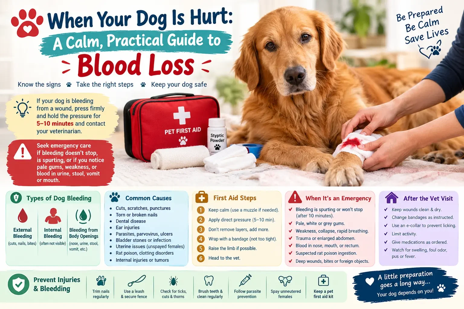
When a pets parent realizes that their animal friend is hurt it can be one of the most terrifying moments to deal with. From a broken nail in a backyard chase to a puncture wound from an unexpected encounter with a wild animal to a deep wound from stepping on broken glass, it’s essential to respond quickly, steadily, and intelligently to the loss of blood.
Pet first aid for bleeding, before you can get to a professional veterinary clinic, can literally save your pet’s life. Although it might seem a small amount or a manageable amount to us, it can be a life-threatening emergency for a small dog or cat.
This overview covers the key principles of pet first aid for bleeding, how to stay calm, how to protect yourself, how to control the bleeding, and how to safely get your animal to the closest emergency vet.

1. Safety pet First aid : Protect Yourself and Your Pet

aThe absolute first priority in an emergency situation is to secure the area and protect yourself and your pet from defensive injury.
Lorem ipsum dolor sit amet, consectetur adipiscing elit. Ut elit tellus, luctus nec ullamcorper mattis, pulvinar dapibus leo.

Recognize the Pain Response

When an animal is very physically hurt or is very afraid, its primitive survival instincts completely overtake. The most well-mannered sweet and gentle dog or cat can bite, scratch, lash out or snap at their owner. It’s not malicious or aggressive, it’s a reaction that your subconscious brain is having in response to some pain.

Restrain the Animal Safely

Before going to the wound or giving any kind of pet first aid for bleeding, you need to safely restrain your pet.

For Dogs:

If dogs do not have any of the following symptoms, they should be muzzled with a soft muzzle, or make one using a strip of soft gauze or a fabric leash that is wrapped snugly around the dog’s snout: vomiting, difficulty breathing, or a facial injury

For Cats:

In cats, gently but securely wrap the cat in a thick towel/blanket, leaving the injured area uncovered, often referred to as the “kitty burrito.” This helps to contain their legs and claws, and helps keep you and the feline safe as you try to stop the bleeding.

2. Identifying the Type of Wound

Not all wounds have the same “type”. As you apply pet first aid for bleeding, you’ll want to get it right, and the more you know about the source and severity of the fluid loss, the more context you can provide to your veterinarian when you call to get on the way to the clinic.

Arterial Bleeding

The most serious and harmful form of blood loss is called arterial bleeding. Arteries deliver blood right from the heart, and it is very oxygenated and under high pressure. If the artery has been cut, the blood will be bright red, and it will be seen to spurt or pulse with the animal’s heartbeat. This needs urgent and intense action.

Venous Bleeding

Venous blood is flowing back to the heart, and is not as pressurized. It will be darker red (almost maroon) and pour out steadily, continuously and heavily. Though less violent than an arterial injury, a venous laceration can still result in excessive and life-threatening blood loss if not contained.

Capillary Bleeding

This is common with minor abrasions, scratches and shallow cuts. The blood will slowly ooze from the skin surface. In general, capital issues will clot on their own within minutes, and can be easily cared for with simple home remedies.

3. Step-by-Step Procedure to Control Bleeding

If a wound is actively bleeding, there is a step-by-Step procedure for controlling the bleeding. This sequence ensures that you are able to treat a pet for bleeding without making it worse through pet first aid.

1.Apply Direct Pressure:Immediate Action.

Cover the wound with a clean sterile gauze pad, clean washcloth or sanitary sanitary pad. Using a flat hand apply pressure. Avoid pressing firmly to break underlying bone, as this will cause additional pain and bleeding; press firmly and steadily to promote clotting. Maintain this pressure for 3-5 minutes (no looking at the time).

2.Layer, Do Not Remove:Protect the Clot.

When the first layer of gauze is saturated, it should NOT be removed from the skin. Removing the wet gauze will cause the delicate, newly-formed blood clots to tear, thus re-initiating the bleeding. Rather, add more fresh clean gauze over the soaked and continue to apply firm pressure.

3.Use a Temporary Pressure Bandage:Securing the Area.

After a little slower flow, wrap a roll of stretch gauze or a self adhering bandage (such as VetWrap) snugly around the padding to secure it. Be sure there is enough room between the bandage and the skin for 2 fingers; if the bandage is too tight it will totally restrict blood flow resulting in serious skin damage.

4.Raise the Extremity:If Applicable.

If the injury is on a limb, paw or tail, gently lift the injured area so that it is above the animal’s heart level. A simple physics trick using gravity to lower the local blood pressure at the wound site, thus slowing the rate of fluid loss.

4. Handling Unique Body Situations

Each part of an animal’s body poses a special challenge. When it comes to dealing with bleeding injuries, treating your pet’s first aid according to the site of the injury will yield a lot more success. There are unique situations in a variety of parts of an animal’s body. When dealing with bleeding, knowing the exact location of the injury in your pet’s anatomy will lead to much better results in your pet first aid.

Torn or Broken Nails

Nails have a blood vessel and cluster of nerves in the middle area called the “quick.” A nail ripping will usually bleed a lot and look very scary, but it is not usually life threatening. Treat: Pack the nail with styptic powder, cornstarch, or baking flour, and press for a few minutes until it stops dripping.

Ear Injuries

Ear tissue is thin and packed with blood vessels. Sadly, if a dog or cat gets hurt in their ear, they will shake it vigorously! Its head shaking is similar to a centrifuge, splashing droplets across the room and continually re-opening the wound.

  • Press straight on with the aid of the gauze.
  • Whenever the ear is stopped, fold it gently over the top of pet’s head.
  • Wrap a long band aid around their head and under their chin to keep their ear flat against their head and so it doesn’t flop back open again.

Internal Bleeding

The killer is invisible: Internal Bleeding. Your pet can internal bleed without any blood visible on her coat, if it was hit by a vehicle, fell from a height or was compressed during a fight. There is no normal pet first-aid for bleeding of the internal organs, so all you can do is to be able to recognize the signs of shock.

Quick Reference Summary Matrix

This is a reference table for assessing the type of injury and selecting the proper immediate action plan.

Injury Profile

Visual Characteristics

Severity Classification

Immediate First Aid Action

Arterial Wound

Bright red fluid, pulsing or spurting out

Critical Emergency

Heavy direct pressure, instant transport to vet

Venous Wound

Dark maroon fluid, steady pouring flow

Severe Emergency

Continuous direct pressure, apply pressure bandage

Capillary Wound

Slow oozing from surface skin layer

Minor / Manageable

Clean gently, apply light pressure, monitor

Internal Damage

No bleeding to be seen but you see internal bleeding, such as pale gums and weakness.

Critical Emergency

Keep pet warm and minimize movement, rush to vet clinic

5. Recognizing and Treating Shock

When a significant amount of fluids has been lost, an animal’s blood pressure falls too low, and the body enters a “shock” state. Advanced knowledge of shock; however, essential for pet first aid – bleeding.

  • Pale or White Gums: Press your finger against above your pet’s teeth. In healthy gums, the pinkness will return in 2 seconds after pressure is released. If they will stay white, grey or light pink, your pet is in shock.
  • Hypothermia: Their paws, ears and tail will be very cold.
  • Rapid, Weak Pulse: Their heart rate will be very fast but the pulse in their inner thigh will be weak and difficult to find.
  • Lethargy: The pet will be glassy-eyed, unresponsive, weak or completely unconscious.

If your pet shows you these symptoms, apply manual pressure bandage to stop the fluid from leaving its body right away. Tie your pet securely in a warm blanket to conserve his dropping core temperature, prop up his rear a little with a small pillow to circulate blood to his brain and rush to the closest emergency room.

6. Crucial Mistakes to Avoid

When the adrenaline kicks in during a crisis it’s very easy to make well-intentioned mistakes that can actually work against your companion’s wellbeing. Avoid these common errors when performing pet first aid for bleeding:

  • Do Not Use Tourniquets: Unless a limb is completely severed, never apply a tight tourniquet. When 100% is cut off in a limb, irreversible tissue death and muscle necrosis will occur within a very short period of time, and often veterinarians will have to amputate a limb that could have been saved.
  • Do Not Use Human Medicines or Ointments: You should never pour in hydrogen peroxide, alcohol or human antiseptic liquids directly into deep open wounds. The harsh chemicals break down normal cell structure and slow the body’s ability to heal. Rinse with clean water or sterile saline only.
  • Do Not Postpone Veterinary Care: If you’ve been able to stop the surface flow of fluid with your pet first aid for bleeding, the crisis is not over. Large wounds must be closed surgically, deep punctures must be flushed internally with antibiotics to ensure that only a mild infection develops and the damage must be diagnosed to ensure that no damage has occurred inside the ball.

Conclusion

Accidents occur in a split-second time, but if you’re willing to respond to help a bleeding pet, you could become a lifesaver. You can successfully stabilize your dog or cat during the first few critical minutes if you remember to put it first: your pet’s safety comes first – secure them properly, apply steady and unyielding direct pressure without looking into the wound, cover the wound with fresh gauze over the old, and monitor for systemic signs of shock.
Make sure to have a well-equipped first aid kit in the home and in your car, have the phone number for your local veterinarian saved in your contacts and treat any time of significant blood loss with a leap straight to professional veterinary care.

Frequently Asked Questions

Q1: What is the first thing to do if my pet is bleeding profusely?

A: Your first line of defense is to safeguard yourself by properly restraining or muzzling your pet; even the gentlest animal can bite if it’s in pain. After it is safe, apply direct pressure, using clean gauze or cloth, to the wound while applying firm, continuous pressure.

Q2: May I use super glue on a dog wound which is still bleeding?

A: No, household super glue should not be used on an open wound. The superglues sold commercially can cause irritation to animal tissue, adhere the dangerous bacteria within the wound and cause damage to the tissue locally. Have a vet seal the wound properly using sterile, medical-grade adhesives or sutures.

Q3: When will I know when to measure direct pressure for a pet's wound?

A: Keep the pressure steady for 3-5 minutes and do not remove the gauze to view. If the blood clots are allowed to form “too early” the whole process of blood clots is interrupted again.

Q4: What home items can stop a torn nail from bleeding if I don't have styptic powder?

A: If you don’t have commercial styptic powder in your home kit, you can use cornstarch, baking flour, or even pack a clean bar of natural soap against the tip of the bleeding nail. Press firmly for several minutes to allow the powder to bind with the blood and seal the vessel.

Q5: How can I tell if my pet is experiencing internal bleeding after an accident?

A: Look closely for pale or white gums, rapid but shallow breathing, cold paws or ears, extreme lethargy, or weakness. If your pet has been in an accident and displays any of these symptoms, they may be losing blood internally and need to be rushed to an emergency vet clinic immediately.

This blog post is very important for pet first aid.

Signs of Heat Stroke in Dogs

Signs of Heat Stroke in Dogs: A Life-Saving Guide for Pet Parents

Discover the symptoms of a heat stroke in dogs. Learn early signs, advanced signs, immediate first aid and what you can do to prevent your dog from getting hurt.
During the summer, there are more daylight hours, outdoor activities and even more sun. But there’s an unseen and deadly threat to our furry friends with the hot weather, and that’s hyperthermia. Knowing what to look for with heat stroke in dogs is a very important skill to have, as it can become a life-threatening medical emergency in just minutes.
Dogs cannot do this as easily as humans, because they don’t have sweat glands all over their bodies to cool them off through evaporation. Their only way to dissipate excess heat is through panting, and a few sweat glands on their paw pads. These cooling mechanisms easily fail during periods of high temperature and moisture.
This complete resource will explain the earliest, later and most serious symptoms of heat stroke in dogs, and equip you with the information you need to know about this serious condition and the importance of acting quickly and decisively to save your dog’s life.

1. Early Warning Signs of Heat Stress

In the initial phase of heat exhaustion, an animal’s body goes through a transition period before it enters into heat stroke. If you can see the indicators of heat stroke in your dog at this point, you will have the highest chance of reversing the situation without any lasting effects.

Unrelenting, Heavy Panting

The dog’s main cooling system is panting. Normal panting – heat-induced however looks relaxed. A dog in its initial phases of heat stress will pant loudly, rapidly and harshly, in contrast. The tongue will be extended—extending the tongue has been found to increase the surface area available for cooling and is an instinctive response to avoid heat, so the tongue will usually be fully extended, appearing unusually wide and long, with the edges of the tongue turned up in a desperate attempt to increase the heat transfer.

Excessive Drooling and Thick Saliva

As the temperature inside the dog’s body rises, the dog will try to get rid of excess heat by producing a tremendous amount of saliva. There will be drooling, frequently leading to puddles on the floor. Once they become dehydrated, this saliva turns clear and watery to extremely thick, sticky and rope-like, clinging to the sides of their mouth.

2. Advanced Signs of Heat Stroke in Dogs

Within minutes, if the dog is not taken to a cool place and watered, his temperature will climb above 103°F (39.4°C) and enter the “danger zone”. Physiological signs are now much more evident.

Unusual Gum and Tongue Color Changes

  • Healthy Gums: PINK, moist and bubble gum.
  • Heat Stroke Gums: Will be dark, vivid red, bright purple or muddy grayish-blue.

This is an extreme blood vessel dilation in order to expel heat through the mouth. The gums can even feel very dry or sticky.

Glazed Eyes and Expressions of Discomfort

An advanced dog will show signs of panic or confusion. They might look glassy, bloodshot and wide-eyed. Often they will look unresponsive, and not react to your voice or commands as they should.

3. Critical Neurological and Systemic Failure

Once the dog’s body temperature hits or surpasses 106°F (41.1°C), cell damage is starting to happen in its internal organs. When the dog passes this point, symptoms of heat stroke become a sign of serious systemic dysfunction.

Lethargy, Weakness, and Collapse

A dog that is overheated will be losing co-ordination. They may have a wobbling gait or may stumble, or seem as if their back legs are suddenly weak. The dog will not be able to get up off the floor as it becomes very weak and depressed.

Vomiting and Diarrhea

The mucosa of the gastrointestinal tract is easily damaged by severe internal heating. Often presents with sudden onset of acute vomiting or diarrhoea. If their vomiting or feces is bright red, it means that they are experiencing dangerous internal sloughing of the intestinal lining.

Seizures and Unconsciousness

The animal will have neurological episodes as the brain begins to swell due to the extreme heat. It may involve trembling of muscles in a part of the body, convulsions of the whole body, paddling of the limbs or falling into a coma or complete unconsciousness.

Hyperthermia Symptoms Progression Matrix

Use this progression matrix of the signs of heat stroke in dogs to help you quickly assess if it’s a hot pup or a medical emergency.

Stage of Condition

Primary Behavioral Signs

Physical Cues to Watch For

Urgency Level

Normal Warmth

Seeking shade, lying on cool tile, moderate panting

Pink, moist gums; normal clear saliva

Normal Care

Early Heat Stress

Pacing, mild distress, reluctance to move

Extremely fast/loud panting, heavy watering drool

Moderate Warning (Cool down immediately)

Advanced Heat Stroke

Disorientation, dizziness, stumbling, weakness

Dark red or purple gums, thick ropey saliva, glassy eyes

Severe Emergency (Apply first aid, call vet)

Critical Organ Failure

Collapse, inability to stand, unresponsiveness

Vomiting/diarrhea (often bloody), seizures, coma

Life-Threatening (Rush to emergency clinic)

Immediate First Aid Actions for Heat Stroke

If your dog is displaying any advanced or critical signs of heat stroke in dogs, you must begin cooling them down manually before you even put them into the car to drive to the clinic.

1.Change to a Shade or AC:Environment Shift.

Immediately get your dog out of the direct sunlight. Transport them to a well-ventilated room with air conditioning or at least to a shaded outdoor area where the wind is blowing. If available, put them directly in front of an electric fan.

2.Always use Cool Water, Never Ice:Cooling Method.

Run cool or lukewarm tap water over their body, taking care to moisten their back, underarms, neck and groin. Ice cold water / ice baths should not be used. The heat stroke becomes much worse because the heat has been enclosed in the core organs and the surface blood vessels have contracted, trapping the heat.

3.Offer Water, Do Not Force It:Hydration Check.

If your dog is conscious and able to swallow, try giving him some fresh, cool water to drink. Do not force water into an unconscious dog’s throat or the dog is very lethargic, because it may end up aspirating the water into the lungs and drown or develop pneumonia.

4.Track temperature and transport:Transit Readiness.

If you have a digital rectal thermometer, check their temperature. If their temperature falls below 103°F (39.4°C), discontinue the use of the water cooling application to prevent them from becoming hypothermic. Place them in an ice-cooled truck and head directly to an emergency veterinarian.

High-Risk Factors to Keep in Mind

While any dog can succumb to high temperatures, certain factors make specific dogs drastically more susceptible to hyperthermia:

  • Brachycephalic Breeds (Flat-Faced Dogs): Pugs, Bulldogs, French Bulldogs, and Boxers have severely shortened airways. It is very hard for them to exchange air efficiently; they can get heat stroke in dogs at lower temperatures than the long-nosed ones.
  • Age and Physical Condition: Blood flow to the skin surface for dissipating heat is limited in elderly dogs, young puppies and obese dogs, which are unable to pump blood around their bodies as efficiently.
  • Thick/ Dark Coats: Heavy double coats (such as Huskies or Malamutes) retain heat like an insulation blanket and dark fur absorbs solar radiation much quicker than light colored fur.

Conclusion

It is an essential part of being a responsible dog owner to be able to recognize the signs of heat stroke in dogs. Our dogs are unconditional lovers, and will often keep playing a game of catch with a ball or keep following a hike even when they’re starting to get hot, just to please us. We have to be their voice, their protectors and their regulators.
Being hyper-aware of your pet’s panting pattern, paying attention to signs of their gum color during summertime outings, avoiding high-risk exercise in the middle of summer, and with first-aid cool water in case of trouble, you’ll keep your favorite companion safe, happy and healthy this summer.

Frequently Asked Questions

Q1: Is it possible for a dog to suffer from heatstroke in a house?

A: Yes, absolutely. Without adequate ventilation, air conditioning or airflow, the internal temperatures in a house can rapidly climb. This can be particularly harmful for at-risk dogs remaining in sun rooms, rooms with large windows which do not have blinds or shatter-resistant film, or in the upper levels of a house during a heat wave.

Q2: How fast can dog heat stroke kill a dog?

A: Heatstroke can be fatal in just 15-20 minutes. When a dog’s body temperature rises out of control, due to a failure of his or her body’s panting system, a mild heat exhaustion can lead to fatal organ failure in a matter of moments.

Q3: If a dog is suspected of being in heat stroke should I wrap them in wet towels?

A: No, don’t wrap them in wet towels. A wet towel placed over a dog’s body is beneficial at first but then serves as a trapping blanket when water boils up the dog’s body and stays there. Alternatively, drench them with water that is running, and feed them with an open fan fanning the air onto their skin.

Q4: What is the highest safe temp it is OK for dogs to walk outside at?

A: Outside temperatures above 85° F (29.4°C) pose a serious risk for heat stress, in general. Also, asphalt pavement can get much hotter than the air and can cause your dog’s paw pads to blister and burn on contact.

Q5: What is the reason that it is not safe to give ice water to a dog who has heat stroke?

A: Ice cold water will make the small blood vessels close up (vasoconstriction) close to the skin’s surface. This forms an insulating layer which keeps the superheated core blood away from the skin surface so that it never gets the chance to cool down, thus rendering their internal organs ‘cooked’ from inside out.

if Dog is in Pain

How to Tell if Dog is in Pain: The Ultimate Guide for Pet Parents

Our dogs know how to hide that they are in pain. This instinct is hardwired in us, and in the wild, it’s the first thing that goes through our minds when we see an animal that is vulnerable or injured that we are looking at. They aren’t able to communicate with words where it is hurting them, so it’s up to us to pay attention to the signs.
It’s important to be aware of any changes in your pet’s daily habits. Learn more than ever before how to recognize if dog is in pain, through this all-inclusive guide, which explains the physical, behavioral, and emotional cues that your dog reveals when they are hurt. In order to determine if dog is in pain, the first step is to analyze what he does on a daily basis. A hurting dog will tend to disrupt their routine and typically be unpredictable.

Behavioral and Routine Shifts

Familiar changes in sleep patterns

Have trouble sleeping at night and going to bed or waking up at a normal time? Or is your puppy so energetic that he doesn’t sleep at all and won’t get out of bed? Pain disrupts rest. If a dog is feeling pain in the joints or from an internal issue, it will have a really tough time laying down. These may lie down, get up right away, circle around their bed or move through it repeatedly searching for an area that will not cause pain.

Sudden decline in appetite is huge red flag and changes in eating habits

When your dog typically eats its food from the bowl and now walks away, it may be a problem of discomfort. When the pain in the mouth is caused by a broken tooth or dental conditions, eating becomes such a chore. Otherwise, food will become totally unappetizing because of nausea or internal pain throughout the body.

Physical and Postural Indicators

Physical and Postural Indicators tell a great deal about a dog’s physical health. One of the best ways to learn to look for signs of dog pain is to train your eyes to see structural and postural abnormalities.

  • The, Prayer Position: One of the classic signs of severe abdominal distress is the “prayer position. When a dog stretches out its front legs on the ground with its hindquarters off the ground. It may be as if it is a regular play bow but the dog may be unhappy, tense and not wag their tail. They’re doing this to help expand their tummy and to ease any extreme pressure that could come from situations such as pancreatitis or bloat
  • Trembling and Stiffness: Obviously limping, stiffness can be more subtle termed Trembling. If your puppy takes a long time to get up in the morning or is apprehensive about the stairs, then they might have arthritis or joint degeneration. If your dog shakes or trembles without a good reason even with warm temperatures in the room, it’s another big sign that he’s suffering from acute or chronic physical pain or discomfort.

Vocalizations and Audio Cues

Many dog owners believe that a dog in pain will whine, cry or howl. But, this is one of the biggest misconceptions in regards to dog health.
Many dogs suffer silently and stoically. Vocalization is generally only heard when the dog has an abrupt, sharp, acute pain – maybe, for instance, it has been stepped on or suffered from a sudden muscle spasm. Ongoing problems such as arthritis or progressive, long-term diseases do not usually cause ongoing whining. You may only know how your dog is feeling based on its crying, and not pick up on the signs.
Some dogs become louder, though, and whine, groan and grit their teeth more. You may observe your dog softly grunting when seated or hear some soft whining when attempting to get onto the couch. Be alert to these sounds in particular, particularly when they happen in the transition from one movement to another – they could be important indicators when learning to recognize when a dog is in pain.

Facial Expressions and Respiration

With just an attentive look at your dog’s face and an ear to the ground, you can gather a lot of information.

Heavy Panting and Shallow Breathing

It’s perfectly normal for dogs to pant after a vigorous game of fetch, or on a hot summer afternoon. If your dog is lying in a cool, air-conditioned room, and panting heavily, it is a physiological response to stress and distress. Are fast breathing, shallow breathing or panting without an environmental cause, when they are hurt.

Both “Pain Face” and Grimacing

Dogs faces are very expressive. Physical distress often causes a dog to be tight and tense. Watch for:

  • Furrowed brow, or flattened ears.
  • The “grimace,” where the lips are pulled back slightly at the corners
  • Wide eyed expression (sometimes called “whale eye” – the white parts of their eyes are more visible than normal).

Changes in Grooming Habits

Grooming shifts can be in either direction based on what the underlying problem is, so this is an interesting area of observation when deciding if dog is in pain.

Localized Grooming

When a dog is hurt in a certain localized area, they will instinctively lick and clean that spot even in the absence of an external wound that needs to be licked and cleaned. If a dog has localized joint inflammation, they will lick, bite and chew at that one area constantly, or if they have a hidden bug bite or bruise in a deep muscle, they will constantly lick, bite and chew at that one area. Lots of licking can rapidly irritate the skin and result in hot spots.

Neglecting the Coat

On the other hand, if a dog is suffering from chronic disease on a large scale, then they might even cease grooming themselves altogether! If your dog’s joints hurt a lot, then it won’t be very comfortable for your dog to turn around and brush his coat. If your usually well-groomed pup develops some matted, undressed or soiled spots, it’s time to take a closer look.

Changes in Emotion: From Aggression to Withdrawal

Pain changes the emotional threshold of a dog, and directly affects the way that a dog relates to the human family and other dogs. When a dog has a change of emotional state, it is called a behavioral shift, aggression versus withdrawal, and it will directly affect a dog’s interactions with her family and other pets.

Sudden Aggression or Guarding Behaviors

When your very sweet, very gentle dog suddenly growls, snaps or bares their teeth when you try to approach and put your hands on them, don’t conclude that they have a behavioral problem just yet. It is a reaction to the threat. They understand that touching hurts and they are learning to create a barrier around themselves through verb and dental communication.

Hiding and Isolation

Otherwise, when some dogs are not feeling well, they will withdraw from social interaction and hide. They may hide under the bed or curl up in the back of a closet or may not answer the door when you return home. This self-isolation is a protective survival mode that helps to keep them away from danger when they are feeling vulnerable.

Quick Reference Summary Matrix

To review this information in a quick manner, refer to this reference table of the typical differences between a normal dog and a dog in distress.

Observation Area

Healthy Baseline

Potential Pain Indicator

Mobility

Fluid, eager movements; easy transitions

Stiffness, limping, slow to rise, trembling

Rest

Settles quickly, sleeps peacefully

Continuous pacing, shifting positions, restlessness

Respiration

Slow, steady breathing when resting

Heavy panting or rapid, shallow breaths in a cool room

What to Do When You Suspect Discomfort

If you have carefully reviewed these signs and are now confident that your dog is hurting, your very next step must be to call your veterinarian.

Critical Safety Warning: : DO NOT EVER give your dog any human medicines such as ibuprofen, acetaminophen or aspirin. These are nonprescription medications that humans take and can be very toxic to dogs, resulting in acute kidney failure, severe gastric ulceration or fatal internal bleeding.

If you are going to the vet clinic try to safely document what you are seeing prior to arrival. Make a brief video of your dog limping, pacing or breathing abnormally. Dogs frequently come running into a veterinary clinic when they are excited or frightened, and may initially be symptom free. It’s a tremendous diagnostic tool to show your vet a video recorded at home.

Conclusion

When it comes to dog pain, knowing how to recognize it is one of the most crucial duties of being a pet owner. Our dogs can’t communicate their pain directly to us, we must learn to read the minute changes in their body language and behavior. Some subtle hints are all your dog needs to seek assistance, such as a change in sleep pattern, panting in a cool room, uncharacteristic hiding behaviour, or licking in a small area.
Seeing to your beloved companion’s every need, understanding their defensive boundaries, and working quickly with your veterinarian, you can make sure your cat receives the relief, care and comfort it deserves.

Frequently Asked Questions

Q1: If at home, how can I tell if my dog is in pain?

A: The best method of checking is by watching their posture, breathing and movement carefully, but not by touching them. If you do need to take a close look, gently run your hands over their body. Be particularly alert for flinch, blink or pant on their face. If your dog becomes stiff, growls and/or moves away, stop now and don’t get bitten.

Q2: What’s causing my dog to pant all the time when he is sleeping?

A: Panting that persists in the room, if the room is cool and your dog hasn’t just been active, is a classic indicator of stress, anxiety or physical discomfort. Their heart and breathing rate increases on pain, leading to heavy panting.

Q3: Does a dog have to whine and cry to be in pain?

A: Yes, absolutely. The majority of dogs are uncomfortable when the interaction is silent. Vocalization (whining or yelping) is generally used for sudden, sharp, acute injuries. If the dog has arthritis, or an underlying disease, it will always be covered up by him as a survival mechanism.

Q4: What do dogs do when they have joint pain?

A: If a dog has an arthritis, they will be stiff, particularly when getting out of a nap. They will walk much slower, may not want to run up and down stairs, may be reluctant to jump onto furniture, and may often lick or chew on the particular joint that hurts.

Q5: What does a doggy ‘prayer position’ refer to?

A: The prayer position is when a dog lowers its front legs and body to the ground, but leaves the back end elevated high in the air. The dog will not look as it does with a “bow,” but rather look rigid and uneasy. This position is employed to stretch out the abdomen in an attempt to alleviate high internal pressure or pain caused by such ailments as bloat or pancreatitis.

why is my cat not eating

Why Is My Cat Not Eating? 10 Reasons & When to See a Vet

As a cat owner, nothing is more alarming than when your feline friend suddenly refuses to eat. One day they are excitedly meowing for breakfast, and the next, they are completely ignoring their favorite wet food.

While a dog might skip a meal simply because they ate something in the yard, a cat refusing food is a much more serious matter. Felines are masters at hiding pain or discomfort, and a decreased appetite is often the very first sign that something is wrong.

In this comprehensive guide, our team at Grove Center Veterinary Hospital breaks down why your cat has stopped eating from a medical, environmental, and behavioral perspective—and how to know when it’s a medical emergency.

Why a Fasting Cat is a Medical Emergency

Before diving into the reasons, it is crucial to understand that cats do not tolerate starvation well. When humans or dogs don’t eat, the body safely uses stored fat for energy. However, if a cat goes without proper nutrition for even 48 to 72 hours, their liver becomes overwhelmed trying to process stored fat.

This leads to a life-threatening condition called Hepatic Lipidosis (Fatty Liver Disease). Therefore, figuring out why your cat isn’t eating is always a race against the clock.

10 Common Reasons for a Cat’s Loss of Appetite

1. Dental Pain and Mouth Soreness

It’s incredibly hard to bite down on hard kibble if you have a toothache or an ulcer in your mouth. Older cats are highly susceptible to periodontal disease, gingivitis, resorptive lesions, or fractured teeth. If your cat approaches the bowl, sniffs it, and walks away, they want to eat but it simply hurts too much to chew.

2. Gastrointestinal Obstructions or Illnesses

Cats are naturally curious and often swallow string, hairbands, yarn, or small toys. These objects can cause life-threatening blockages in the stomach or intestines. Other internal issues like Inflammatory Bowel Disease (IBD), pancreatitis, or sudden food allergies can also cause severe nausea.

3. Chronic Kidney Disease (CKD)

Kidney issues are extremely common in senior cats. As kidney function deteriorates, the body fails to filter toxins from the bloodstream. This buildup of metabolic waste leads to constant nausea, lethargy, and an unpleasant taste in the mouth, resulting in a sudden drop in food intake.

4. Respiratory Infections and Loss of Smell

Cats rely heavily on their sense of smell to stimulate their appetite; if they can’t smell it, they won’t eat it. An Upper Respiratory Infection (URI) causes nasal congestion, making it impossible for them to smell their food.

5. Stress, Anxiety, and Environmental Changes

Cats are creatures of absolute habit. Severe anxiety can be triggered by any disruption to their daily routine. Moving to a new home, bringing home a new pet, having a baby, or even just rearranging your furniture can cause a cat to go on a food strike.

6. Recent Vaccinations or Medications

If your cat recently visited the vet for routine vaccinations, a temporary loss of appetite is completely normal. Vaccines trigger an immune response that can make your cat feel slightly lethargic or feverish for about 24 hours.

7. Food Boredom or Formula Changes

Sometimes pet food manufacturers change their ingredients without changing the packaging. A cat with a sensitive stomach might reject the new formula instantly. In other cases, eating the exact same protein for years can simply lead to food boredom.

8. Whisker Fatigue

This sounds unusual, but it is a very real physical sensation. A cat’s whiskers are highly sensitive sensory organs packed with nerve endings. If their food bowl is too deep and narrow, their whiskers rub against the sides every time they try to eat, causing sensory overstimulation that makes them give up on their meal.

9. Food Temperature and Texture

As natural predators, cats are biologically wired to prefer food that is close to prey body temperature (around 100°F). Cold wet food straight from the refrigerator lacks aroma and appeal, causing picky eaters to reject it.

10. Parasites and Infections

Internal parasites like roundworms or hookworms rob your cat of essential nutrients. A heavy parasite infestation causes abdominal bloating, gas, and pain, completely wiping out their desire to eat.

Gentle Steps to Stimulate Your Cat’s Appetite at Home

While you wait for your veterinary appointment, you can try these safe methods to encourage your cat to eat:

  • Warm Up Wet Food: Microwave canned food for 5–7 seconds to bring it to room temperature. This releases the aromas and fats, making it much more enticing.
  • Use Wide, Shallow Dishes: Switch deep bowls out for a flat plate or saucer to completely eliminate whisker fatigue.
  • Add a Tasty Topper: Pour a small amount of low-sodium chicken broth (ensure it has no onion or garlic) or plain tuna juice over their food.
  • Create a Safe Dining Space: Move the food bowl away from loud appliances, busy hallways, and the litter box. If you have multiple pets, feed the sick cat in a separate, closed room to reduce competition stress.

Conclusion: When to Call the Vet

Because cats are masters at concealing illnesses, a sudden loss of appetite should never be ignored or dismissed as a “phase.” If your cat has refused food for more than 24–48 hours, or if their fasting is accompanied by lethargy, vomiting, or diarrhea, it requires urgent medical care.

If you live in the area, contact Grove Center Veterinary Hospital immediately to get your furry friend the professional care they need.

Frequently Asked Questions (FAQs)

Q1: Why is my cat not eating but acting completely normal otherwise?

A: This can happen during the early stages of dental pain, mild stress from a recent household change, or food boredom. However, because cats mask sickness very well, you should monitor them closely as an underlying medical issue may still be developing.

Q2: What is the longest a cat can survive without food?

A: Cats cannot safely go without food for long. If a cat goes 48 to 72 hours without eating, they are at an extremely high risk of developing Hepatic Lipidosis (Fatty Liver Disease), which can be fatal if left untreated.

Q3: Could a new food bowl cause my cat to stop eating?

A: Yes. Deep, narrow bowls cause “whisker fatigue” due to constant friction against the cat’s highly sensitive whiskers. Additionally, plastic bowls can scratch easily, trapping bacteria and foul chemical odors that drive cats away.

Q4: Will my cat refuse food due to stress?

A: Absolutely. Cats are highly sensitive to routine. Relocation, home construction, a new job schedule, or a new pet can induce enough anxiety to make them completely lose their appetite.

Q5: My cat used to love wet food but now refuses it. Why?

A: It could be due to a sudden recipe change by the brand, or the food is being served too cold. Wet food left out for too long can also spoil quickly, causing your cat to reject it.

Q6: What safe human food can I give to a cat that won’t eat?

A: You can offer plain, unseasoned boiled chicken breast, meat-based baby food (ensure it is 100% free of onion and garlic powder), or a spoonful of canned tuna in water.

Q7: When is a cat’s refusal to eat considered a true medical emergency?

A: If the loss of appetite is paired with vomiting, extreme lethargy, diarrhea, labored breathing, or yellowing of the eyes and gums, it is an emergency. Additionally, any cat that has gone a full 48 hours without food needs to be seen by a vet immediately, even if no other symptoms are visible.

Q8: Why is my cat not eating after a routine vet checkup?

A: If they received vaccinations, a mild loss of appetite for 24 hours is a normal immune response. The stress of the car ride and the clinic environment can also cause temporary fasting. If it persists past 48 hours, contact your veterinarian.