Grove Center

  • Home
  • About Us
    • Our Philosophy
    • Hospital Policy
    • Working Hours
    • Our Team
  • Services
  • Emergencies
  • Contacts
    • New Client
    • Pet History
  • Blog
  • Home
  • About Us
    • Our Philosophy
    • Hospital Policy
    • Working Hours
    • Our Team
  • Services
  • Emergencies
  • Contacts
    • New Client
    • Pet History
  • Blog

Grove Center

Grove Center

  • Home
  • About Us
    • Our Philosophy
    • Hospital Policy
    • Working Hours
    • Our Team
  • Services
  • Emergencies
  • Contacts
    • New Client
    • Pet History
  • Blog
dogs Tag
Home dogs Page 14

Tag: dogs

4128422
July 23, 2024
grovecenter grovecenterAllDogs

Basic Virology

This virology review is meant to help the beginner understand how viral infections work and how we survive them. Virology is a complicated subject, as is immunology, and the following is meant as a sort of kindergarten of terms and events. We think it is helpful in understanding the biology of what happens with viral infections.

What is a Virus?

Viruses are strange life forms. They have only the bare bones of biochemical equipment, barely enough to even qualify for the title of life form.

  • They have only one type of nucleic acid (either DNA or RNA, but not both.)
  • They reproduce solely from this nucleic acid, whichever type it is.
  • They do not have any metabolic enzymes.
  • They are completely dependent on their host cell.

Some Definitions:

Viral Particle

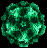
One little individual virus is called a viral particle. Viral particles are so small (some are the size of a large protein molecule) that we cannot really call them creatures; they are more like particles, hence the term. The word virus is more correctly applied to a species or genus of the virus rather than to the individual particles.

Capsid

The nucleic acids (the DNA or RNA) within the viral particles are surrounded by a protein coat and sometimes by an additional fatty envelope. The capsid consists of this protein coat and any fatty layer. The capsid proteins are crucial to the virus’ ability to attach and infect a host cell.

Viruses that have fatty envelopes generally do not last long in the environment as the fat is easily disrupted; these viruses tend to require direct contact with fresh body secretions to transmit the infection.

DNA

DNA is the double-stranded nucleic acid (deoxyribonucleic acid) that serves as the blueprint for all proteins a cell can make. It enables the cell to live and function within a body. It essentially amounts to instructions on how to make different proteins.

Messenger RNA

When it comes time to make a protein, a DNA segment unzips its double strands, allowing messenger RNA to enter and bind. The messenger RNA forms a “negative” image of the DNA segment it is “reading.”  The messenger RNA then leaves the cell nucleus and travels out into the cell’s main body, where its message is read by cell structures called ribosomes. 

Transfer RNA is another type of RNA that floats around the cell. Each piece of transfer RNA has only three base codes (as opposed to the messenger RNA which has many base codes and is a long strand). The piece of transfer RNA carries with it an amino acid that its code corresponds to.

The ribosome looks like two round pieces attached together. The two pieces grab the strand of messenger RNA and sandwich it between them. The ribosome reads the first three codes on the messenger RNA and matches them to a piece of transfer RNA. It moves to the next three codes and matches them as well. With each match, the transfer RNA leaves its amino acid for the ribosome to bind to the next amino acid, so as the ribosome moves down the strand of messenger RNA, it creates a strand of amino acids based on the matching. As we know, a strand of amino acids forms a  protein molecule. This is how our cells make proteins.

Transcription

The act of messenger RNA taking down the protein code from the original DNA segment is called transcription.

Translation

The act of producing a protein from the segment of messenger RNA using transfer RNA is called translation. It is performed by the ribosomes mentioned previously.

Viral Infections

When the match is correct, the virus binds to the cell and injects its nucleic acids inside. There are several techniques that viruses use to accomplish this, but all involve the capsid. Once the viral particle has injected its nucleic acids into the host cell, the next activity is to manufacture messenger RNA for the cell to translate into protein. The proteins that the viral nucleic acids make will shut down the cell’s normal function and convert the cell into a factory for viral particle production.

  • Some viruses simply contain ready-made messenger RNA and they just inject it into the host cell (like polio).
  • Some viruses inject a negative of the messenger RNA they need and include with it an enzyme that will trick the host into making the usable messenger RNA from the negative (like rabies).
  • Some viruses inject their DNA directly into the host cell. Messenger RNA is made from this DNA, just as it would be made from host DNA (like poxviruses).

Regardless of the tricks the virus uses to make messenger RNA, once the messenger RNA is made, the host cell is doomed. Ribosomes line up on the strand of messenger RNA, read it, and use transfer RNA to mass produce the protein coded. This will be a viral protein, and its action will be to shut down normal cell function and dedicate the cell to the production of viral capsid and viral DNA.

Soon the host cell is little more than a bag of viruses. These viral particles either bud off the surface of the host cell or the host cell simply explodes, leaving millions of new viral particles to seek new host cells. The only way to stop this process is for the immune system to recognize the infected cell early and destroy it before virus production becomes too advanced.

Infected cells express viral proteins, shapes that the immune system can recognize. Within the body are several groups of cells, all created to respond against a specific shape. For example, B lymphocytes, which live in our lymph nodes, transform into antibody-producing plasma cells once they encounter their destined antigen. They produce Y-shaped antibodies that flood the circulation and bind the viral antigen, thus preventing the virus from attaching to the host cell, clumping with other antibody-virus groupings to create a larger clump, which in turn attracts a cell called a macrophage. The macrophage is the body’s “Pac Man,” swallowing and digesting debris.

Aside from antibodies, there are patrolling T-lymphocytes that identify viral or other inappropriate shapes on the surfaces of host cells. The T-cell can thus recognize a virally infected cell or even a cancer cell, attach, and destroy it before it causes harm.

Hopefully, between T-cell and antibody attack, the viral infection is removed from the body, and the status quo resumes.

LEARN MORE
4128888
July 23, 2024
grovecenter grovecenterAllDogs

Babesia Infection in Dogs  

Most people have never heard of Babesia organisms though they have caused red blood cell destruction in their canine hosts all over the world for thousands of years. Babesia organisms are spread by ticks and are of particular significance to racing greyhounds and pit bull terriers. Humans may also become infected.

There are over 100 species of Babesia, but only a few are found in the U.S. and are transmissible to dogs. Infected red blood cells are identified and destroyed thus killing the Babesia organisms within them but, unfortunately, if many red blood cells are infected this leaves the host with anemia, a lack of red blood cells. Babesia species continue to be classified and sub-classified worldwide.

The Babesia species that infect dogs in North America are:

  • Babesia canis – a larger species of Babesia, transmitted by ticks.
  • Babesia gibsoni – a smaller Babesia species that mostly attack pit bull terriers and is transmitted by bite wound and from mother to unborn puppies. This is the most common Babesia in North America.
  • Babesia conradae – a smaller Babesia species that has only been isolated in California.
  • Babesia microti is the species of Babesia that infect humans. A “Babesia microti-like” Babesia has been found in dogs in North America.

How Infection Happens and what Happens Next

Infection occurs when a Babesia-infected tick bites a dog and releases Babesia sporozoites into the dog’s bloodstream. A tick must feed for two to three days to infect a dog with Babesia. The young Babesia organisms attach to red blood cells, eventually penetrating and making a new home within the cells for themselves. Inside the red blood cell, the Babesia organism divests its outer coating and begins to divide, becoming a new form called a merozoite that a new tick may ingest during a blood meal.

Infected pregnant dogs can spread Babesia to their unborn puppies, and dogs can transmit the organism by biting another dog as well. (In fact, for Babesia gibsoni, which is primarily a pit bull terrier infection, ticks are a minor cause of infection with maternal transmission and bite wounds as the chief routes of transmission.)

Having a parasite in your red blood cells does not go undetected by your immune system. Infected red blood cells are identified and destroyed thus killing the Babesia organisms within them but, unfortunately, if many red blood cells are infected this leaves the host with anemia, a lack of red blood cells. Often the host’s immune system will begin destroying the uninfected red cells as well, a condition called immune-mediated hemolytic anemia (IMHA). Symptoms include weakness, jaundice, fever, and red or orange-colored urine. At least 50% of patients will require blood transfusions.

Making matters worse is the fact that animals seem to get sicker than the degree of anemia would suggest so there is more to this infection than the destruction of red blood cells. The severe inflammation that is associated with this parasitism can be overwhelming and completely separate from the anemia. Platelet counts can drop, impairing normal blood clotting (especially a problem with Babesia gibsoni). An assortment of neurologic signs can occur with Babesia infection when parasites sequester inside the central nervous system and generate a more localized focus of inflammation. In severe cases, there is a lung injury similar to what people with late-stage malaria can experience. Babesia conradae seems predisposed to creating liver disease.

If the acute symptoms are relatively mild or at least non-lethal, a chronic infection can develop. This is usually without symptoms, but the dog may continue to be a source of infection by feeding ticks. Relapses can also occur with stress.

Because babesiosis is a tick-borne infection, it is not unusual for infected dogs to have other tick-borne infections such as Ehrlichiosis, Rocky Mountain Spotted Fever, and others. These infections may interact to make each other more severe.

Young dogs tend to be most severely infected, especially pit bull terriers.

Diagnosis of Babesiosis

With luck, the Babesia organisms can be seen on a blood smear. Babesia canis organisms are tear-shaped and occur in pairs. Other Babesia species have several forms in which they appear. The odds of finding the organism are improved by checking freshly drawn blood taken from a capillary source (a small cut to an ear, for example) rather than from a blood vessel. If Babesia organisms are found, the patient is definitely infected but in most cases, they are not found so an alternative method of diagnosis is needed.

Antibody testing has been problematic as infected animals may have circulating antibodies long after the organism is gone or may have no antibodies circulating while the few organisms remain hidden inside red blood cells.

The newest method of diagnosis involves testing for Babesia DNA. This type of testing is called PCR testing and is extremely sensitive, able to distinguish four different species. This is especially valuable information as the different species are sensitive to different medications.

Current recommendations are to do both antibody testing (serology) as well as DNA testing (PCR) as the information is felt to be complementary. If a patient is strongly suspected to be suffering from babesiosis, treatment should begin promptly without waiting for test results. After treatment is completed, PCR testing should be repeated starting 60 days post-treatment and again two to four weeks after that.

Babesia Treatment

Before 2004, an assortment of unpleasant drugs were used against Babesia with mixed success. What medications you will need to use to treat a Babesia infection turns out to depend on which species of Babesia the patient is infected with. There are two Babesia species that are particularly challenging: Babesia gibsoni (the one that involves primarily pit bull terriers) and Babesia conradae (the Babesia found only in Southern California).

Since 2004 a new protocol has emerged for Babesia of either the gibsoni or conradae species using atovaquone and azithromycin in combination. These two medications stop reproduction so that the host’s immune system has time to gain the upper hand and remove the organisms without their numbers increasing. Side effects are few to none, and improvement is generally obvious within the first week. Unfortunately, atovaquone is expensive, and pharmacies are reluctant to sell less than an entire bottle. It is often tempting to use the version of atovaquone that comes combined with proguanil, another anti-protozoal drug, but this version has not been evaluated thoroughly against Babesia and is famous for inducing severe nausea in the dog. Imidocarb, one of the earlier treatments, can be started while the correct formulation of atovaquone is obtained; it is best not to use azithromycin alone during that time or resistance may develop.

If atovaquone simply proves too expensive, the patient can be treated with imidocarb as we used to do in the past until such a time that it is possible to save up for the atovaquone/azithromycin protocol. Imidocarb creates a remission from the physical illness for an extended time but does not actually clear the infection.

If imidocarb is employed, a single dose is usually effective for Babesia canis but two given two weeks apart are needed for the smaller Babesia species. The injection is painful, plus it causes muscle tremors, drooling, elevated heart rate, shivering, fever, facial swelling, tearing of the eyes, and restlessness. Pre-treatment with an injection of atropine helps palliate these side effects.

Occasionally a strain of Babesia gibsoni is resistant to atovaquone. When this happens, imidocarb can be used as described above with the addition of doxycycline, clindamycin, and metronidazole for a three-month period.

As for Babesia canis, two doses of imidocarb as described above generally should completely clear the infection. The use of atovaquone appears to be unnecessary.

A vaccine is available against Babesia in France but only seems effective against certain strains. Vaccination is 89% effective in France. The best prevention is aimed at tick control.

Racing Greyhounds

Since areas where greyhounds are professionally raced tend to be areas with ticks, racing greyhounds are commonly infected with Babesia. Whether these infections become active remains to be seen as the carrier state seems to be common in infected dogs. As racing greyhounds end their careers and enter the adoption system, Babesia infection is commonly screened. An apparently healthy but positive-testing dog can still be adopted with the understanding that active infection is unlikely but possible. 

Such a dog could transmit her Babesia infection to other dogs via ticks or bite wounds and should certainly never be used as a blood donor.

Human Babesiosis

The species of Babesia that infect pets should not pose any problems to people with normal immune systems. People with compromised immune systems or people who have had their spleens removed may have some concerns. In the U.S., babesiosis usually occurs on the East Coast and along the Great Lakes and stems from tick bites. Most symptoms are mild or easily treated, but a five percent mortality rate has been reported. The usual organism is Babesia microti.

Read more about human infection from the Centers for Disease Control and Prevention.

LEARN MORE
716164
July 23, 2024
grovecenter grovecenterAllDogs

Bleeding: First Aid

Pets often suffer blood loss as a result of trauma or poisoning. Some bleeding is visible, and is a result of a cut or laceration, while internal bleeding occurs inside the body (such as in the chest or abdomen) and is not visible to the naked eye. If bleeding is severe or continuous, the pet may lose enough blood to cause shock; loss of as little as 2 teaspoons per pound of body weight may cause shock, which doctors usually detect as a high heart rate and low blood pressure. Emergencies may arise that require you to control the bleeding, even if it is just during transportation to the veterinary facility. Pet owners should know how to safely stop hemorrhage (bleeding) if their pet is injured.

Techniques to Stop External Bleeding 

The following techniques are listed in order of preference. 

Use caution; avoid injury to yourself, and take appropriate precautions (such as the use of a muzzle, wrapping your pet in a towel or blanket, and having someone assist you if possible) to avoid being bitten. For all techniques below, seek veterinary attention immediately after stopping the bleeding, or on the way to the veterinary hospital.

Direct Pressure

Gently press a compress (a pad of clean cloth, feminine sanitary pad, or gauze) over the bleeding area, so it can absorb the blood and allow it to clot. Do not disturb blood clots after they have formed. If blood soaks through, do not remove the pad; simply add additional layers of cloth and continue the direct pressure more evenly. The compress can be bound in place using loosely applied bandage material, which frees the hands of the first provider for other emergency actions. If you don’t have a compress, you can use a bare hand or finger.

Direct pressure on a wound is the most preferable way to stop bleeding

Elevation

If a severely bleeding wound is on the foot or leg, gently elevate the leg so that the wound is above the level of the heart. 

Elevation uses the force of gravity to help reduce blood pressure in the injured area, slowing the bleeding. Elevation is most effective in larger animals with longer limbs where greater distances from the wound to the heart are possible. Direct pressure with compresses should also be maintained to maximize the benefits of elevation.

Elevation of a limb combined with direct pressure is an effective way to stop bleeding.

Pressure on the Supplying Artery

If external bleeding continues after you have used direct pressure and elevation, use your finger or thumb to place pressure over the main artery to the wound. Apply pressure to the femoral artery in the groin (on the inside of the thigh) for severe bleeding of a rear leg; to the brachial artery in the inside part of the upper front leg for bleeding of a front leg; or to the caudal artery at the base of the tail if the wound is on the tail. Continue application of direct pressure.  Seek veterinary attention immediately.

Tourniquet

The use of a tourniquet is potentially dangerous and it should be used only for a severe, life-threatening hemorrhage in a limb (leg or tail) not expected to be saved. If you see blood spurting or pumping from a wound, which is a rare occurrence, consider the use of a tourniquet. Use a wide (2-inch or more) piece of cloth and wrap it around the limb twice and tie it into a knot. Then tie a short stick or similar object into the knot as well.

Twist the stick to tighten the tourniquet until the bleeding stops. Secure the stick in place with another piece of cloth and write down the time it was applied. Every 20 minutes loosen the tourniquet for 15 to 20 seconds. Remember this is potentially dangerous and can often result in disability or amputation.

A tourniquet should only be used as a last resort and a life-saving measure!

Internal Bleeding

Internal bleeding is a life-threatening condition, but it is not obvious like external bleeding. In internal bleeding, blood pools in the abdomen or chest but does not result in blood in the stool or bleeding from the rectum.

There are, however, some external signs of internal bleeding:

  • The pet is pale (check the gums- if they are white or blue, this is an emergency). 
  • The pet is cool on the legs, ears, or tail. 
  • The pet may cough up blood. 
  • The pet is unusually subdued.

If you see any of these signs, immediately transport your pet to a veterinary facility for professional help. Most cases of internal bleeding will require intensive therapy in a veterinary hospital. Remember: internal bleeding is not visible on the outside.

LEARN MORE
4127746
July 22, 2024
grovecenter grovecenterAllDogs

Bee Stings and Insect Bites: First Aid  

Any insect or spider can cause problems if they bite or sting your pet. A bite or sting can cause swelling, redness, and itching.

Some animals can have an allergic reaction to a sting or bite that may result in a range of symptoms:

  • mild to severe hives,
  • facial swelling,
  • vomiting,
  • difficulty breathing,
  • or even collapse.

Some spider bites can lead to extensive skin necrosis (skin death) which may need treatment or surgery.

Contact a veterinarian right away if your pet is showing signs of an allergic reaction. Reactions can be severe and may require emergency treatment.

What to Do:

If the stinger can be found, scrape it out with a credit card or other stiff material. Alternatively, use tweezers by grasping the stinger, which is located below the venom sac. Studies have shown that speed is of the essence in removing the stinger. If the only way is to pinch it and remove it, then do so. You’ll do more good than harm and you’ll get the stinger out faster. Don’t worry about squeezing more venom into the area – this has been disproven.    

  • Apply cool compresses to the area 3-4 times daily for 3-4 days.  
  • To help neutralize some of the acidic venom, apply a paste mixture of baking soda and water to the sting area.
  • Prevent your pet from licking at the area by using an Elizabethan collar (cone) to prevent further trauma to the area.
  • In short-faced/brachycephalic breeds like pugs, boxers, etc., facial swelling can lead to damage to the eyes. Have your veterinarian examine your pet and prevent rubbing on carpet, furniture, etc.

What NOT to Do:

Do not administer any medications without first contacting your veterinarian or a veterinary emergency hospital. The veterinarian may need to examine your pet before recommending medications.

LEARN MORE
4128950-1
July 22, 2024
grovecenter grovecenterAllDogs

Bandaging: First Aid

Home bandaging is not a substitute for proper veterinary care and improperly applied bandages can result in limb loss.

We use bandages for several reasons: to protect wounds from the environment, protect the environment from wounds, and discourage a pet from licking or irritating a wound. Bandages may be applied as support for strains or sprains or to prevent motion. Proper application is important – an improperly applied or too-tight bandage can cause decreased blood flow and potential loss of the limb.

Cleaning the Wound

The process of bandaging begins with careful cleaning of the wound. All dried blood, dirt, and debris should be washed away using mild soap and lots of water. Hair should be clipped away so that it cannot lie in the wound. If possible, the area should be patted dry.  

Materials Needed

In an ideal setting, a bandage should have a contact layer, an absorbent layer, and an outer layer.

This may include (from the innermost layer to the outer layer):

  • antibiotic ointment
  • Telfa Non-Adherent Pad
  • cotton wrap
  • gauze wrap
  • Vetrap

The Contact Layer

After cleaning the wound, apply antibiotic ointment to the contact layer and use it first. Ideally, this layer should:

  • Be sterile and inert. 
  • Stay in close contact with, but not stick to, the wound.    
  • Be very absorbent.   
  • Be free of particles or fibers that might be shed into the wound.   
  • Conform to all shapes. 
  • Allow drainage to pass to the next layer without becoming wet.  
  • Minimize pain.

A Telfa Non-Adherent Pad, available at most pharmacies, comes closest to meeting these requirements.

It is desirable to apply an antibiotic ointment, such as Neosporin, to the pad but this is not absolutely necessary. Frequent bandage changes are more important. After cleaning the wound, place a new contact layer over the wound.

The Absorbent Layer

After the contact layer is in place, apply the second (absorbent) layer to hold the contact layer snugly, but not tightly, over the wound. This layer is usually a cotton or Dacron material that comes in various widths. Generally, 1-inch rolls are used for small limbs and the tail; 2-inch rolls are for medium-sized legs; and the 3- and 4-inch rolls are for large legs and the body. It is important to use the proper size. Materials that are too narrow often cause a tourniquet effect, especially if the wound becomes swollen.

If materials are too wide, they are difficult to apply smoothly. Any wrinkles or ridges may cause the bandage to become uncomfortable for your pet. Uneven pressure may cause necrosis (tissue death) of the underlying tissues.

Begin with just enough of an absorbent layer to hold the contact layer in place. If the wound is on a leg or the tail, wrap it from the toes or the tip of the tail towards the body. If you begin at the top of the leg or the tail, the bandage is more likely to restrict blood flow and cause swelling, which may cause tissue damage. Apply several layers of absorbent material, which will soak up the fluid from the wound and increase the patient’s comfort by cushioning the wound.

Gauze wrap can be applied next to hold the cotton wrap in place and to add extra support.  This step can be skipped for small wounds or for temporary bandages.

The Outer Layer

Finally, apply the outer (third) layer, usually made up of porous adhesive tape or elastic tape (i.e., Elastikon, Vetrap). Wrapped from the toes up toward the body, this layer should also be smooth and snug. Do not stretch elastic tapes to their limits as this will interfere with circulation and result in bandage failure. It helps to unwrap the Vetrap or Elastikon first and then rewind it to remove the tension from the wrap before placing.

The tape should be in contact with the skin (hair) at the bandage margins, anchoring the bandage so it will not slip.

The outer layer of a bandage should be applied smoothly and snugly, but not tight enough to cut off blood circulation.

Bandage Changes

Bandages should be checked frequently for any signs of swelling, skin discoloration or coolness, odor, or saturation of the bandage material. The bandage should be changed whenever any of the above are noticed or any time it appears to be uncomfortable for the pet. If a bandage becomes wet, soiled or slips out of position, it should be replaced.

Be very careful when bandaging at home. Wounds that are draining heavily may require bandage changes every one or two hours.

Bandages over wounds with little or no drainage should be changed every 24-72 hours, depending on your veterinarian’s advice and evaluation. Some wounds may require up to 6 weeks of bandage changes to fully heal.

LEARN MORE
4128659
July 22, 2024
grovecenter grovecenterAllDogs

Bacterial Diarrheas in Puppies & Kittens   

Bacterial diarrheas are generally a nuisance for the adult animal but can be lethal to a small puppy, kitten, or even a human baby. Most of these problems stem from contaminated food or fecal contaminated environment. Feeding raw food diets to pets dramatically increases the exposure to infectious organisms to the humans in the family. Since these conditions are also human diseases, it is helpful to have some understanding of what they are, even if there are no young pets or immune-suppressed individuals (human or otherwise) at home.

Campylobacter

Campylobacter species are a group of bacteria capable of causing diarrhea in dogs, cats, humans, and other animals. They have a unique curved appearance under the microscope and are said to be sea gull-shaped.  They are difficult to isolate as they grow in conditions of low oxygen (making them microaerophilic as opposed to being aerobic or anaerobic bacteria). With regard to pets, Campylobacter are generally a problem for the very young. Puppies and kittens have not yet matured immunologically.

Plus, because they are small, fluid loss from diarrhea hits them much harder. Furthermore, puppies and kittens are more likely to be housed in groups where fecal cross-contamination is common so they may be more likely to become infected than adult animals. Adult animals commonly have Campylobacter organisms living in their intestines, but they do not experience any sickness due to it.

In humans, Campylobacter infection is a leading cause of gastrointestinal (GI) disease; infected dogs and cats can carry the organism and spread it even if they do not have symptoms themselves.  For this reason, pets used for therapy in assisted living communities and similar situations should be screened for Campylobacter by fecal culture before exposure to people with suppressed immunity. Humans are also infected by consumption of contaminated food, water, or raw milk; only 6% of human Campylobacter infections are attributed to dog exposure.  That said, exposure to a dog with diarrhea triples a person’s risk for developing enteritis from Campylobacter jejuni or Campylobacter coli. Studies screening pet animals for inapparent Campylobacter infections have found surprisingly high incidences of inapparent infection. In one study in the Midwest, 24% of 152 healthy cats were positive, for example.

After consuming Campylobacter organisms, they travel to the lower small intestine, attach, and multiply. They produce a toxin that destroys the lining of the intestine with the result being a bloody, mucous diarrhea (though occasionally a more watery diarrhea is described).  Sometimes a fever results, appetite becomes poor, and vomiting can occur.  Incubation is 2 to 5 days.  The organism can survive as long as a month in environmental feces.

Diagnosis is made by seeing the sea gull-shaped organisms under the microscope; however, there are so many bacterial organisms on a fecal sample that finding the culprit can be tricky. For this reason, a culture is often performed as a more accurate test. Because the organism is microaerophilic, specific culture requirements must be met; the facilities of a reference laboratory are needed.

Treatment is with appropriate antibiotics, such as erythromycin, chloramphenicol or tylosin.

Salmonella

Most people are somewhat familiar with Salmonella. They know it represents a type of food poisoning, probably know it is associated with diarrhea that can be severe, and may even know that Salmonella species are bacteria. Most human cases of Salmonella infection cause fever, diarrhea, and cramping that go away on their own, but in children, it can produce more severe disease. As with Campylobacter, the young are more susceptible to more severe illness because they are smaller and do not have mature immunity. 

The same is true with puppies and kittens; adult animals are almost never affected by Salmonella infection.

An important exception to the “Salmonella is rare in adult dogs” rule is the case of dogs fed a raw food diet. It has become popular to feed raw foods to pets with the idea that a raw food diet more closely approximates the natural diet that the feline or canine body evolved to consume, and thus, such a diet should be healthier than commercially prepared foods. In fact, cooking food is central to removing parasites, bacteria, and bacterial toxins from food. A recent study evaluating raw food diets found that 80% of food samples contained Salmonella bacteria and that 30% of the dogs in the study were shedding Salmonella bacteria in their stool. Adult dogs are often asymptomatic, but any infected animal or person will shed the organism for at least six weeks, thus acting as a source of exposure to other animals or people. Salmonella organisms are difficult to remove from the environment and easily survive three months in soil. Again, dogs used for therapy around the elderly or children should be cultured for Salmonella.

There are two syndromes associated with Salmonella: diarrhea and sepsis. Salmonella bacteria, once consumed, attach to the intestine and secrete toxins. The toxins produce diarrhea that can be severe and even life-threatening in the young. If this were not bad enough, some Salmonella can produce an even more serious “part two” (sepsis) should these bacteria invade the body through the damaged intestine, causing a more widespread and much more serious infection.

In young animals, the syndrome resulting is similar to that of canine parvovirus thus similar treatment is expected.

E. Coli

Escherichia coli may be the most common bacterial organism in the world. It lives in our intestines naturally and covers the world we live in. Unfortunately, some strains of E. coli are not so neighborly and are capable of producing diarrhea via toxin production. Like the other organisms we have discussed, this is a serious problem for the very young and more of a nuisance for adults.  There are three main types of unfriendly E. coli: Enterotoxic E. coli, enterohemorrhagic E. coli, and enteropathogenic E. coli.

Enterotoxigenic E. coli is a common cause of diarrhea in young animals as well as human infants and is responsible for the famous traveler’s diarrhea. These bacteria produce what is called an enterotoxin in the upper small intestine. This toxin, similar to the toxin of cholera, causes the intestine cells to secrete the body’s fluid into the intestine, creating spectacular watery diarrhea and what can be life-threatening dehydration for smaller living creatures.  Young pigs, cattle, and other livestock are commonly lost to this kind of dehydration. Again, the younger and smaller the patient is, the more serious this infection is.

Enteropathogenic E. coli also produce diarrhea in humans and animals. Rather than a secretory diarrhea as above, they simply destroy the intestinal cells where they attach. Diarrhea still results, but it creates more damage to the intestinal lining.

Enterohemorrhagic E. coli is similar to enteropathogenic E. coli but with more associated inflammation. This type does not seem to be a problem for small animals though they can carry it asymptomatically.

It would seem that antibiotics would be the obvious treatment for a bacterial disease, yet for E. coli it is surprisingly controversial. It seems that the use of antibiotics can enhance the synthesis of toxins by these bacteria; plus, oftentimes, antibiotic use only serves to make E. coli more resistant in the GI tract. Antibiotics are generally reserved for those animals (or people) who seem the most sick or who have evidence of bacterial invasion in the bloodstream. Basically, treatment is supportive care until the patient’s immune system regains the upper hand.

In Conclusion

Bacterial diarrheas are an especially serious consideration in the very young and in the weak or compromised, be they human or non-human. It is not unusual for apparently healthy animals to carry these organisms and shed them into the environment and feeding raw foods greatly increases the risk of this kind of latent infection. If you are considering raw food diets, especially if someone in the home is very young or has a compromised immunity, discuss prevention of these infections with your veterinarian.

LEARN MORE
4128715
July 22, 2024
grovecenter grovecenterAllDogs

Benign Sebaceous Gland Tumors

We receive a fair amount of emails related to the Papilloma article from people with older dogs with numerous “warts” wondering if their dog’s warts will go away as viral warts usually do. The problem is that in older dogs, what looks like a viral wart is probably a sebaceous gland tumor, while there is a very good chance it is benign, it will not be going away any time soon.

It is not uncommon for an elderly dog to develop scores of “warts” which are not warts at all but are sebaceous growths. Most sebaceous growths are benign but one cannot say for sure simply by looking.

Here are some reasons to remove a sebaceous growth:

  • when the growth has been bleeding.
  • when the growth is itchy or is in a location where it is bothering the pet.
  • when the growth is in a location where it interferes with normal grooming of the pet (i.e. the growth gets caught in the grooming clippers, etc.).
  • when there is a question as to whether the growth actually IS a sebaceous tumor and biopsy is needed to settle the question.
  • when you don’t want to take any chances that a sebaceous growth is malignant (as mentioned, you can’t tell by looking).

These growths are typically small (pea size or smaller) and originate from the skin’s sebaceous glands, the oil-producing glands of the skin. Because these growths are small, they are generally amenable to removal with local anesthetic. This is helpful since often patients are older and not good anesthesia candidates. It is usually not practical to remove a large number of sebaceous growths with local anesthesia at the same time but the most troublesome can be selected for removal.

Viral warts are different and occur primarily on the face of young adult and adolescent dogs. Sebaceous gland tumors occur in any location, often in large numbers, and usually in older dogs (and occasionally in older cats).

There are several types of sebaceous gland tumors:

Nodular Sebaceous Hyperplasia

About 50% of sebaceous growths are technically not tumors at all and are classified as excessive growth of the gland tissue. It is thought that growths of this group ultimately develop into actual benign sebaceous adenomas as described below. These lesions are round, cauliflower-like, and sometimes secrete material that forms a crust. Occasionally they even bleed. They are particularly common in Cocker spaniels, Beagles, Miniature Schnauzers, Poodles, and Dachshunds. This growth is technically not a tumor but is actually an area of excessive sebaceous cell division.

Sebaceous Epithelioma

Another 37% of sebaceous growths fit into this category. These look just the same as sebaceous hyperplasias to the naked eye but tend to occur in larger breeds and usually on the eyelids or head. They often pigment into a black color. They were formerly described as benign but it turns out they are able to spread in a malignant fashion if given enough time. Since it is not possible to distinguish a low-grade malignant epithelioma from hyperplasia, it is smart to remove any sebaceous growth and not take a chance.

Sebaceous Adenoma

These lesions also look the same as the others to the naked eye. These are also actual benign tumors that probably arose from areas of hyperplasia. As previously mentioned, if given enough time a sebaceous hyperplasia growth will develop into an adenoma. Both are benign.

Sebaceous Carcinoma

About 2% of sebaceous tumors are malignant and may be locally invasive but even malignant sebaceous tumors rarely spread. They have a greater tendency towards ulceration than benign growths. Cocker spaniels seem to be predisposed.

Again, in most cases, the removal of sebaceous gland tumors is straightforward and can frequently be done with a simple local anesthetic. In the event that further treatment is needed, your veterinarian will make recommendations or possibly refer you to a veterinary dermatologist.

LEARN MORE
5226265
July 22, 2024
grovecenter grovecenterAllDogs

Brucellosis from Raw Milk Consumption

Recently a case of brucellosis was diagnosed in a Texas man from drinking raw milk from a licensed raw milk dairy.  Brucellosis is a bacterial disease of cattle that was very common in Texas 50 years ago but is now rarely found due to vaccination and testing.  However, it is still in some areas and the bacteria is transmitted in the milk as well as fetal fluids at calving to other animals and people. There are also other diseases that can be carried in raw milk, which is the reason for pasteurization as this process kills the organisms.

Raw milk is not pasteurized.  Other animals commonly affected with brucellosis include sheep, goats, pigs and others.  The disease can also cause fistulous withers that is a draining tract at the withers in horses and is a cause of infection of the vertebrae in dogs. In humans, symptoms include fever, sweats, decreased appetite, headache, pain in muscles and fatigue, or basically flu-like symptoms that do not respond to normal treatment.  The condition is called undulant fever in humans because it can recur for long periods of time and can cause heart valve infection, swelling of the liver and spleen and abortion in pregnant women. 

Brucellosis is a very serious disease in animals and people and drinking raw milk should be discouraged as the risks of developing disease are not worth taking the chance.   Death rarely occurs with the disease but many people have recurring symptoms that can last a lifetime, which was common before antibiotics were discovered.  Fortunately, today the disease responds to antibiotics in most humans.  However, drinking raw milk or feeding it to your pets, just like feeding raw meat to your pets, is dangerous for you and your family and is not worth the risk. 

LEARN MORE
8202139
July 22, 2024
grovecenter grovecenterAllDogs

Black Fly Bites on Dogs

Black flies (buffalo gnats) are members of the Simullidae family. Their colors vary from black to gray to yellow to olive, and their thorax is humped over the head, giving the appearance of a bison’s hump. The female flies require a blood meal to lay eggs. Males are never blood-feeders; they feed on nectar from flowers. Black fly larvae (young) are carnivorous. There are more than 1,000 species of black flies, but only a few are considered important pests.

Black flies feed on all types of animals, including dogs, cats, cattle, horses, goats, sheep, wildlife, birds, and humans. Black flies are distributed throughout the world. The eggs are laid in fast-running water on stones or leaves just below the water. Larvae are nearly always found in swiftly flowing, well-aerated water. Some species breed in larger rivers; others live in temporary or semipermanent streams. Black flies are particularly abundant in the north temperate and subarctic zones.

Adult black flies may fly 8–11 miles (12–18 km) from swiftly flowing streams; migrating windborne swarms have been known to travel more than 155 miles (250 km). Black flies are active during the morning and evening hours, and during hot days, black flies rest near the ground on the underside of leaves.

Female black flies inflict painful bites, and animals’ ears, neck, head, and abdomen are favorite feeding sites. Local reactions such as redness, itching, and wheals (a raised, itchy spot) at the bite site are common. There can be severe effects in sensitive animals or if the animal has a large number of bites. Swarms of black flies have caused cattle to stampede and refuse to graze, and there have been cases of death in production animals. The bites may result in petechial hemorrhage (reddish spots indicating bleeding under the skin) and lesions can take weeks to heal, especially on the teats of cows and goats. Reduced milk, meat, and egg production may result from less extensive attacks.

Poultry can become anemic from a black fly attack. Death from a severe attack can result from a toxin in the fly’s saliva, which increases capillary permeability, meaning small blood vessels are compromised, permitting the fluid from the circulatory system to ooze into the body cavity and tissue spaces. Some species of black flies can transmit diseases.

Bite Prevention and Treatment

Control of black flies requires destroying breeding sites, moving animals away from the insects’ breeding grounds, and treating animals with insecticides and repellents. Black fly control is difficult because of the large number of flowing water breeding sites. Streams can be treated by using a microbial larvicide, Bacillus thuringiensisisraelensis, a product safe for mammals.

Adult black flies are small enough to pass through window screens or come indoors on or within a pet’s hair coat. The adult female flies prefer to feed outdoors and during the daylight hours. Pet owners concerned about black fly bites may use over-the-counter insect repellents. Aerosols containing pyrethrins may provide only temporary relief. Because area-wide control of black flies is difficult and expensive, people frequently use repellents daily to protect their pets and other animals.

Treatment is often unnecessary. The skin reaction usually goes away in 24 – 48 hours. But if your pet is very uncomfortable or having a more extensive reaction from the bites, your veterinarian may suggest antihistamines, steroids, anti-inflammatories, etc.

LEARN MORE
4128725
July 22, 2024
grovecenter grovecenterAllDogs

Biliary Mucocele is a Surgical Emergency in Dogs

It all starts with a sick, usually middle-aged or older dog. He is listless, not eating, and maybe has vomiting and/or diarrhea and fever. These are symptoms that could mean any number of things, but usually, when he reaches the veterinarian’s examination table, he has jaundice (yellow pigmentation visible in the whites of his eyes and possibly on his skin and gums). Hospitalization is recommended to rehydrate him and provide supportive care. Blood tests point to a liver problem. Medications are given to minimize the liver’s workload, but soon, an ultrasound is being discussed to image the liver, and costs may be rising quickly.

It all starts with a sick, usually middle-aged or older dog. He is listless, not eating, and maybe has vomiting and/or diarrhea and fever. These are symptoms that could mean any number of things, but usually, when he reaches the veterinarian’s examination table, he has jaundice (yellow pigmentation visible in the whites of his eyes and possibly on his skin and gums). Hospitalization is recommended to rehydrate him and provide supportive care. Blood tests point to a liver problem. Medications are given to minimize the liver’s workload, but soon, an ultrasound is being discussed to image the liver, and costs may be rising quickly.

Why Do We

Need an Ultrasound if We Already Know the Problem Is Liver Disease?

While blood testing can point to the liver, the fact is that numerous diseases can affect the liver. The liver can have an infection, cancer, scarring (cirrhosis), or any number of conditions. The more specific our diagnosis gets, the more specific treatment can be.

Ultrasound is a non-invasive way to evaluate the internal texture of the liver and gall bladder. By looking at the liver’s texture, it is possible to see a tumor and determine if removing it is possible or if it has invaded too far. Ultrasound can evaluate scarring and abscesses. Through ultrasound, it is possible to guide a biopsy needle to an exact area to sample tissue should this be deemed necessary. Ultrasound evaluates the gall bladder and bile ducts. 

One of the more important diseases to rule out promptly is the biliary mucocele because it is commonly a surgical emergency. As we will describe in more detail, a biliary mucocele is a wad of sludged mucus inside the gall bladder. If you wait a few days to see how the patient responds to general liver support, it may be too late for surgery. Furthermore, ultrasound can evaluate the gall bladder’s integrity; if the mucocele ruptures and spills bile into the belly, the surgeon will need to know that as additional treatment is needed. Ultrasound also evaluates concurrent abdominal conditions that might impact recovery, including pancreatitis, which can occur alone with similar symptoms or in addition to a biliary mucocele.

If the patient has a surgical disease, they will not get better until surgery is performed in the vast majority of cases. Furthermore, it may be a surgical emergency (not only will surgery be needed but it will be needed immediately). The sooner the need for surgery is identified, the better the chance of survival.

What Is the Gall Bladder and What Is the Biliary System?

The liver serves as a toxic waste processing center for the body. It filters bacterial products (as well as nutrients) entering the body from the gastrointestinal tract and it removes toxic waste products from the bloodstream. This material is bound to special biochemicals called bile acids and the body would like to get rid of it, bile acids and all.  The solution of bile acids, water, mucus, pigments, and cholesterol forms the greenish-yellow fluid we call bile.

Bile is made in the liver, and then collected into small ducts called bile ductules and bile ducts. The bile is then moved for storage into the greenish round organ called the gall bladder.

During food digestion, hormones cause the gall bladder to contract and squirt bile through the large common bile duct and into the intestine. The bile assists with digestion and carries toxins out of the body so they may be eliminated in feces. The gall bladder and its ducts represent the biliary system.

If the biliary tract becomes obstructed, the patient becomes rapidly jaundiced, painful, and sick.

What Is a Biliary Mucocele?

We mentioned that one of the components of bile is mucus. Normal bile is probably less than 3% mucus but when a mucocele develops, the bile becomes mostly mucus. Normal bile is liquid but mucocele bile is thick and goopy and will not flow easily through the common bile duct. The gall bladder distends trying to pass the mucocele bile and if it actually ruptures, the risk of serious complications and death is much higher. The gall bladder with a mucocele develops an appearance on ultrasound described as resembling the cut surface of kiwi fruit. The biliary mucocele is sometimes called a “kiwi gallbladder.”

When this is seen on ultrasound in a sick, jaundiced patient, surgery to remove the diseased gall bladder should be performed as soon as possible.

Why Does This Happen?

Mucocele development starts with delayed gall bladder contraction, which allows bile acids to accumulate. Altered fat metabolism is also associated with reduced gall bladder contraction, resulting in an accumulation of bile acid. With that accumulation, the lining of the gall bladder feels inflamed and responds by making more mucus and more mucus-producing glands. Pretty soon the bile is just a big wad of congealed mucus and it will not flow. 

Several hormone imbalances have been associated with altered fat metabolism and reduced gall bladder contraction: diabetes mellitus, hypothyroidism, and Cushing’s disease. Cushing’s disease patients have an incidence of biliary mucocele development that is 29 times the incidence of patients without Cushing’s disease. Mucocele development has been linked to pancreatitis (inflammation of the pancreas) as well.

Having Cushing’s Syndrome increases a dog’s risk of developing a biliary mucocele by 29 times.

No one knows why this might be so but it does correspond to the mainstreaming of diagnostic ultrasound into general practice.

The Shetland sheepdog, cocker spaniel, miniature schnauzer, and dachshund seem predisposed to developing biliary mucoceles.

Removing the Gall Bladder (Cholecystectomy)

Removing a dog’s gallbladder is a serious surgery that not all veterinarians are comfortable performing. Discuss with your veterinarian whether a referral to a specialist would be best for you and your pet.

The goal is to remove the gall bladder before it ruptures. If it has already ruptured, tissue damaged by the rupture must be cleansed or removed. Whether or not the gallbladder that has a mucocele is removed before or after rupture does not affect the rate of survival.

Mucocele surgery survival rate is 75-80%. However, if the gallbladder ruptures before surgery occurs, survival rate decreases significantly to 25-40%, depending on the study.

The gall bladder’s function is mainly one of bile storage. Without the gall bladder, bile simply dribbles into the intestine constantly rather than in controlled squirts. You might think this would be a problem, but it turns out not to be. Some patients require long-term medication for liver support but generally speaking, if the dog recovers from surgery, the prognosis is excellent, and the patient can return to normal life.

What if Surgery is Not an Option, or What if the Dog Isn’t that Sick?

There is no question that surgery is the best treatment choice, but there is more to the story. Biliary mucoceles can be an incidental finding in dogs who are not sick or who are sick from something else and are having an ultrasound of the belly for some other reason. In a patient that is not experiencing problems with a mucocele, medication may be able to stave off illness in some cases.

As for skipping surgery on a dog that is sick from its mucocele, this is a very risky move.

It is possible to attempt treatment with general liver support medications, a low-fat diet, and choleretics (medications to help liquefy bile, such as ursodiol), but the problem is that the gall bladder is obstructed with a big wad of goop. This goop is unlikely to liquefy in a timely fashion, if at all, no matter what we do. If medical management is attempted, it is important to regularly recheck the gall bladder by ultrasound to watch for any sign of progression that would indicate that surgery should no longer be postponed.

LEARN MORE
  • 1
  • …
  • 12
  • 13
  • 14
  • 15
  • 16
CATEGORIES
  • All
  • Cats
  • Dogs
  • Small Mammals

Recent Posts

  • Fur Mowing in Cats  
  • Plasma Cell Stomatitis in Cats 
  • Mealtime Enrichment for Cats
  • Mammary Tumors in Cats
  • Notoedric Mange in Cats (Feline Scabies)  
TAGS
Antibiotics Aural Hematoma Bloat Cannabis Toxicity cat and dogs cats cats & dogs cats and dogs Cellular Delinquent Clinical Signs Corticosteroids Cyclosporine Dental Disease Diagnosis Diarrhea dog dog & cats dogs dogs & cats Dogs and Cats Fluids gastrointestinal Glomerulonephritis Guinea Pigs Hepatic Necrosis Hyperplasia Keratoconjunctivitis Sicca mammals Milbemycin Oxime Moxidectin muzzle Nutrition pets Prevention Prognosis rabbits rats Small Mammals Symptoms Trilostane veterinarian veterinarians Vomiting and diarrhea Whipworm Infection Workups for Itchy

Copyright © 2021 Pawscare by WebGeniusLab. All Rights Reserved