 grovercentervet
Dog Bleeding: Causes, First Aid & When to See a Vet
When Your Dog Is Hurt: A Calm, Practical Guide to Blood Loss
There’s nothing more terrifying to a pet owner than spotting blood on their pet. Dogs can bleed from a tear in his nails leaving red paw prints, a cut from a walk, or blood in the urine or feces, from a minor scrape to a life-threatening emergency. Deciding what to do in the first few minutes can save your dog’s life and knowing when to stop treating at home and go to your veterinarian can save a life.
Answer:
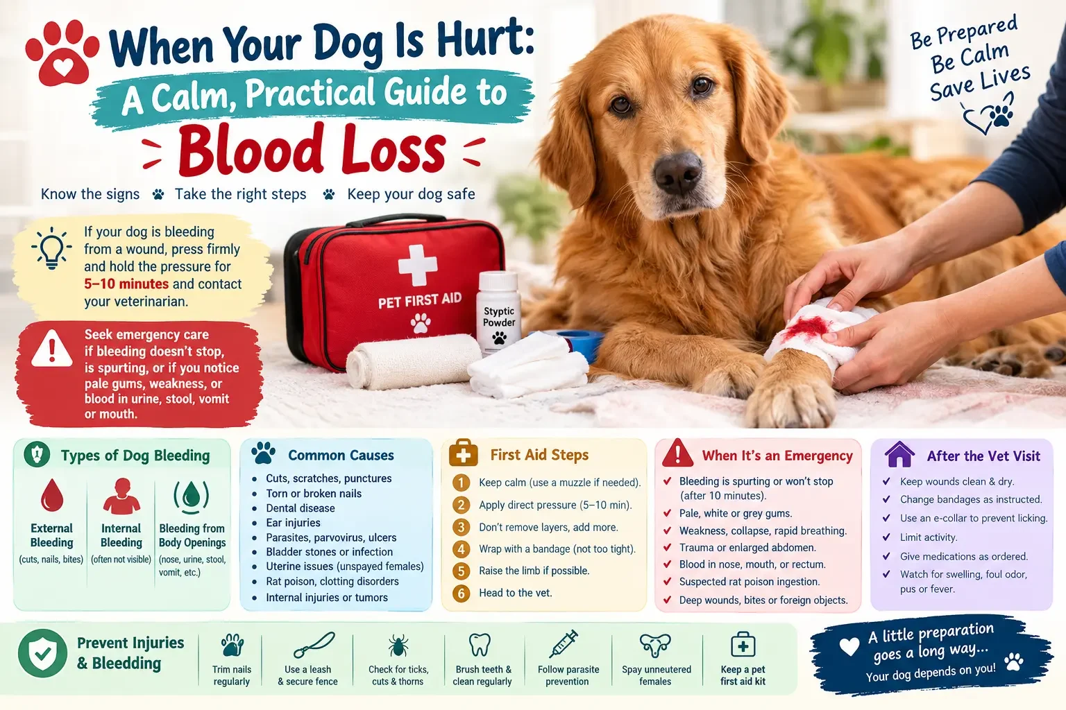
If your dog is bleeding from a wound, press it firmly and hold the pressure for 5-10 minutes and contact your veterinarian. If bleeding does not stop, if it is spurting, if your dog’s gums are pale or weak, or if blood is seen in urine, stool, vomit or mouth, visit an emergency clinic immediately.
Types of Dog Bleeding
Bleeding can come in many forms and each form gives clues to the severity of the bleeding.
- External Bleeding: Blood is flowing out of the body through the skin. It contains cuts, bite wounds, torn nails and scapes. Oozing blood is less serious than blood that spurts or pours, indicating damage to an artery.
- Internal Bleeding: Internal bleeding is bleeding inside the body and usually there will be no blood evident. This can happen after a car accident, after a fall, after a ruptured tumour or after poisoning, and it is always an emergency.
- Bleeding from Body Openings: Bleeding from body openings refers to nosebleeds, bloody vomit, blood in the stool, blood in the urine and vaginal discharge. These can be a sign of illness, infection, clotting disorders or injury and should be treated by a vet.
Common Causes
There are numerous causes of bleeding in dogs. Most frequently come across are:
- Fights, sharp objects or rough play causing cuts, scratches or punctures
- Bleeding nails are also torn, broken nails that have great blood supply in the nail.
- Blood from gums due to dental disease or chewed object
- Bleeding of ear flaps due to head shaking, biting or ear hematoma.
- Parasites, parvovirus, ulcers and diet problems can cause blood in the stool or vomit.
- Infections, bladder stones or bladder inflammation can cause blood in the urine.
- Uterine infection or tumors in unspayed females, or bleeding from a heat cycle.
- Ingestion of rat poison that prevents blood clotting
- Clotting disorders and low platelets – such as those associated with tick-borne disease or immune disorders
- Internal bleeding, including ruptured splenic mass
When to Seek Emergency Care
In the event of an emergency, this guide should be consulted to determine which veterinary services are needed. If you see any of the following, consider dog bleeding an emergency:
- Sputtering, pouring or oozing blood through a bandage in minutes.
- If bleeding does not stop after 10 minutes of firm pressure,
- Gums that are pale, white or grey.
- Weakness, collapse, rapid breathing, rapid heart rate
- Trauma-induced hard, enlarged abdomen
- Blood in the nose, mouth or rectum from an unknown cause
- If you suspect that your pet has eaten rat poison or experienced bleeding after eating some, please contact your veterinarian.
- A wound that is deep or has been bitten or a foreign object in the body.
Note: Internal bleeding may not be visible. If your dog is quiet, cold or “off” after an accident, visit the same day, even if there is no wound apparent.
First Aid for Dog Bleeding
The first aid kit for dogs with bleeding. Should your dog be bleeding from a wound, follow these steps until you can get him to the vet:
- Assure your dog and yourself that you are both calm. Pain and fear can make even gentle dogs bite. Use a soft muzzle if necessary, but not if a dog is vomiting, is having trouble breathing or is unconscious.
- Apply direct pressure. Use clean gauze, towel or clothing. You should press down and hold for 5-10 minutes without lifting your hand to look; this can disrupt the clot that is forming.
- Do not delete layers, add layers. Add more material and continue pressing if it seepes through.
- Wrap it. After bleeding subsides, cover with a bandage or self adhesive wrap. Tie snugly, but not too tight, and be sure to get a finger under it. To determine if the wrap is too tight, look for swelling or coolness on the toes of a leg.
- Raise up, if possible. Elevating a bleeding limb above the heart can help to reduce the flow of blood.
- Head to the vet. Deep wounds, bites and punctures often require cleaning, stitching, or antibiotics, even when the bleeding has stopped.
What Not to Do
A tourniquet should only be applied for life-threatening bleeding from the limbs as a last resort because it can cause serious damage to the affected tissue. Never remove objects from the body. Avoid using hydrogen peroxide or alcohol on wounds or using human medications like ibuprofen or paracetamol (acetaminophen) as these can be harmful to dogs.
Handling Common Situations
- Bleeding toenail: Apply a Styptic powder to the tip of the nail. If you don’t have one, cornstarch or flour pressed hard on the nail will suffice. With dog still, hold for several minutes.
- Nosebleed: Try to calm your dog down and apply a cold compress to the bridge of his nose. Avoid tilting the head back. Nosebleeds may be a sign of serious illness or poisoning so contact your vet.
- Minor scrape: Thoroughly flush with clean water or sterile saline, dry and maintain clean. Keep away from licking, and monitor for redness, swelling and discharge.
- Presence of blood in urine or faeces: If possible take a new sample, and say how long this has been occurring and make a vet call as soon as possible. If your dog is drowsy, vomits or the blood is thick, go in right away.
Diagnosis and Treatment at the Vet
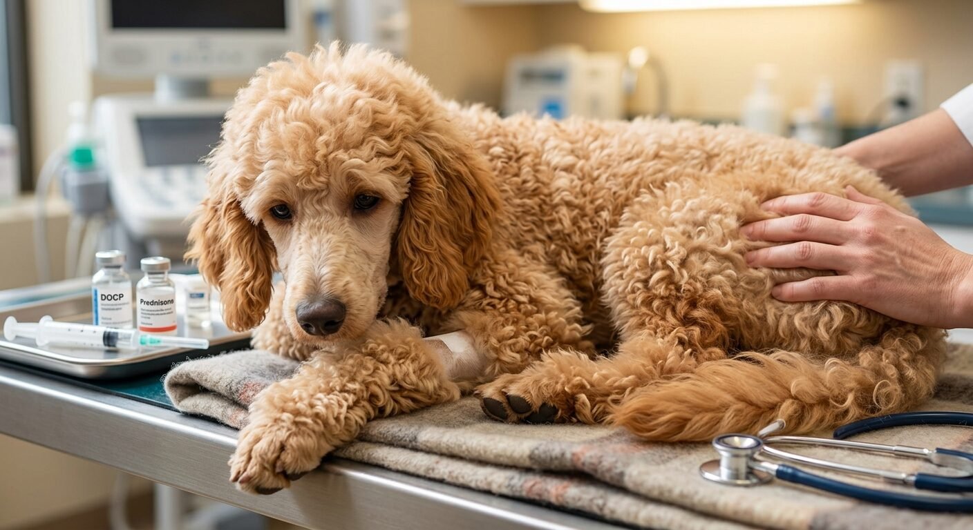
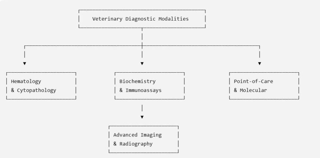
The diagnosis and treatment of bleeding in Vets. The first priority is to calm your dog down. Your veterinarian may insert an IV catheter into your goat or provide intravenous fluids, pain medication, and medication to help stop the bleeding. Diagnostic tests then pinpoint the cause and may include:
- Blood tests: examination of red cells, platelets and organ function
- Blood sugar levels: indicate blood sugar status and if the blood glucose is becoming elevated.
- X-ray and ultrasound: check for internal injuries, tumors or abdominal fluid.
- Tests of urine and stool: detect infection, parasites or crystals.
The treatment will be directed by the cause. Cleaning and suturing of wounds may be necessary. The treatment for rat poison is to give it vitamin K and sometimes a transfusion. If blood loss is severe, a blood transfusion will be needed, and if there’s a ruptured tumour or internal injury, emergency surgery might be necessary. Promptly intervening can mean the difference between full recovery and a critical outcome in many cases.
Home Care After Treatment
If a dog was seen by a veterinarian for bleeding, it is important to follow the veterinarian’s instructions carefully:
- Wounds must remain clean and dry, bandages changed as instructed and an e-collar used to prevent licking and chewing of wounds.
- Limit activity to allow wounds to heal and administer all medications as ordered.
- If you notice any fresh blood, swelling, foul odor, pus or fever, call your vet.
How to Prevent Injuries and Bleeding
How to stop injuries and bleeding. While not every accident will be avoided, there are some habits that will help reduce the risk of dog bleeding:
- Store rodent baits and poisons securely and do not apply rodenticides in areas where pets are likely to be.
- Regularly cut nails to prevent snags and tears
- Always wear a leash in areas with lots of activity, and keep a secure fence at home.
- Inspect paws and coat after walks for cuts, thorns and ticks.
- Follow your veterinarian’s recommendations for tick and flea prevention.
- Brush teeth and get dental cleaning.
- To prevent some tumors and uterine infections, spay unneutered females.
- Have a pet first aid kit (with a salvinus, self adhesive wrap, styptic powder, saline and your veterinarian’s number).
Conclusion
A calm response makes all the difference when it comes to dog bleeding. Apply firm pressure, cover wound and contact a veterinarian promptly if the bleeding is profuse, does not stop, or is accompanied by pale gums, weakness or collapse. Have a first aid kit at home, know the emergency symptoms and have the number of your vet and the closest emergency clinic saved in your mobile phone. If you have any questions, speak with a vet. Better to be checked than not.
Frequently Asked Questions
What is the maximum time to apply pressure to get bleeding to stop?
Apply sustained, firm pressure for 5-10 minutes (no checking). If it doesn’t slow down by that point, place additional layers on top, and visit a vet right away.
When is bleeding in dogs an emergency?
If the bleeding is spurting, if it doesn’t stop after 10 minutes of pressure, if the gums are pale, if your dog is weak or collapsing, or if it is followed by trauma or poison, it is an emergency.
Can I use human antiseptics or painkillers on my dog?
Don’t use hydrogen peroxide or alcohol on wounds because they can be harmful. Do not administer human painkillers such as ibuprofen, naproxen or paracetamol (commonly used for human pain) as these can be toxic to dogs. Ask your vet first.
What do I do if my dog’s nail is bleeding?
Styptic powder or cornstarch can be applied to the tip of the nail and pressure applied firmly. The majority of nail bleeds will fade within a couple of minutes. Do not wait, if it does not stop, see a vet if it is badly broken.
Why is my dog’s nose bleeding?
It is caused by injury, foreign objects, infections, tumors, clotting disorders and poisoning. As it may be a serious issue, reach out to a vet on the day of the occurrence.
What are some of the symptoms of internal bleeding?
Check for pale gums, weakness, cold limbs, rapid breathing, a swollen abdomen and collapse. Dogs can have these symptoms without any blood apparent.
Should my dog be taken to the vet if the blood is in its stool?
It can be. This may be caused by a stomach upset, parasites, parvovirus or ulcers. Black, tarry stool or blood is heavy with vomiting or lethargy requires immediate veterinary attention.
What to do at home in case of emergencies?
Create an emergency first aid kit for pets, make a list of your veterinarian’s and emergency clinic’s phone numbers and know how to apply pressure bandage. Practicing before helps keep you cool.