Cooperative care training is the art of teaching animals how to take part in care routines rather than forcing them to comply. In veterinary medicine, the goal is to help pets feel safe during care and enjoy care routines; therefore, pets become more treatable in the future, not less. The practice of cooperative care also teaches veterinary staff, caregivers, and animal trainers how to communicate effectively with and support the emotional well-being of the individual pet based on that pet’s specific needs and progress. These concepts are core values of Low Stress Handling®.
Pets Can Participate in Their Own Care Routines
Teaching pets to take part in care routines should be prioritized over modifying the pet’s behavior for other reasons, such as obedience training.
Modern zoos care for animals, which are often massive, without resorting to physical restraint. Imagine a giraffe voluntarily stepping onto a giant platform to get weighed. Picture a tiger presenting his tail through a small window of an enclosure to have a blood sample taken. For so many reasons, it is not effective to repeatedly dart and fully anesthetize these cherished animals every time healthcare is needed. Veterinary teams can implement these sophisticated but user–friendly techniques by prioritizing Low Stress Handling®.
This is an exciting time in veterinary medicine. Practitioners certified in Low Stress Handling® are joining forces with clients, board-certified veterinary behaviorists, and trainers who use positive reinforcement methods. The collaborative goal is to strengthen the human-animal bond by implementing these powerful cooperative care techniques for the benefit of clients and patients.
When Pets Say “No” To Care Routines
Cats and dogs sometimes growl or hiss when the veterinarian walks into the room, cower during examinations, or try to flee at the site of a nail trimmer. Fear can escalate to a point where the pet refuses to allow veterinary personnel to touch them. Pets can become so fearful, resistant, and defensively aggressive when visiting the veterinarian that the staff runs out of options to provide care to these patients. These pets may then need to be referred to board-certified veterinary behaviorists.
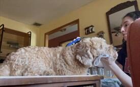
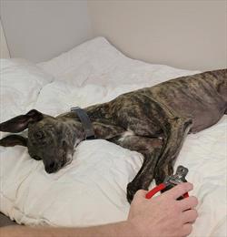
It is also common for pets to become frightened during home care routines. One painful or scary experience, such as exposure to a loud set of clippers, a cleaning of a painful, infected ear, or an accidental clip of a toenail to the quick can result in a pet developing lasting memories of that event. Each animal experiences handling, restraint, and care in different ways. Sensitivity levels vary among pets, with some being more prone to stress and anxiety than others. These pets that show avoidance or resistance during care routines at home, in grooming salons, or at animal hospitals are exhibiting signs of anxiety, fear, or both.
Low Stress Handling® veterinary practitioners will do everything they can to provide a pleasurable experience during veterinary care. Your pet’s veterinarian may prescribe short-term, anti-anxiety medications before scheduled appointments. They may also make environmental modifications, such as bringing your pet into the exam room by skipping the busy lobby. The veterinary team will treat pets in the exam room (rather than “the back”) whenever possible. Low Stress Handling® practitioners will make sure clients are included in the decisions being made about the pet’s care. When caregivers understand their pet’s emotional state, it helps everyone make better decisions together. It is imperative to slow down and realize that continuing a procedure with a fearful pet makes the patient less treatable in the future. Realizing when pets are anxious or frightened is half the battle.
Cooperative Care Teaches Pets to Say “Yes” to Care Routines
Core Concepts in Cooperative Care:
Emotional health status and medication use. Before teaching cooperative care routines, a veterinarian should evaluate a pet’s physical and emotional health. Some patients are simply too anxious to learn. Medications can be helpful in creating a comfortable starting point by relieving the pet’s anxiety enough to allow them to participate and receive food for counterconditioning.
Veterinarians may prescribe medications to reduce overall anxiety or situational medications intended for use before stressful events, and sometimes both. Using multiple medications can help to avoid the sedating effects that may come with a larger dose of a single medication. Additionally, many pets may require short-term anesthesia for veterinary care or grooming until they can be successfully retrained through cooperative care routines.
Body Language. Each species of animal communicates with their own unique body language. Interpreting what pets are communicating with their body language does not come naturally to most people. Your Low Stress Handling® Certified veterinary practitioner can guide you to the appropriate professional who can help you interpret your pet’s body language. These professionals include board-certified veterinary behaviorists (DACVB), board-certified veterinary technician specialists in behavior (VTS), and progressive positive reinforcement trainers who have a close association with the veterinary behavior community. Trainers who use any form of punishment or recommend prong or shock collars, choke chains, etc., should be avoided. These techniques break down the human-animal bond and potentially intensify fear and fear aggression.
Prevention. Resilience conditioning is defined as supporting an individual’s ability to recover from stress. If young pets display sensitivity to being touched, during routine handling, it is time for intervention and changing the approach to improve their ability to recuperate from stress. Support and agency are core concepts in both resilience conditioning and cooperative care.
Low Stress Handling® veterinary practitioners have a crucial responsibility to support each patient’s resilience for future care events. This is achieved by providing predictability, social support, and agency during veterinary care. For example, kittens might lick baby food off a lick mat during procedures, puppies can be trained to stand on a platform for treats while undergoing a gentle examination, and any non-essential treatments should be postponed if a patient shows signs of being overwhelmed. Each time cooperative care routines are practiced with puppies and kittens, they start to remember that veterinary visits are safe and predictable.
Veterinarians and veterinary technicians are integral in preserving the human-animal bond (HAB). This bond can deteriorate when clients struggle to care for pets at home or when veterinary professionals cannot effectively treat patients who have become fearful or dangerous. A simple yet effective preventive measure is to continuously feed a puppy or kitten throughout their veterinary experience. This approach helps these young pets develop resilience and reduces fear in care contexts.
By gradually introducing pets of all ages to home and veterinary care routines and by pairing those routines with high-value food and other pleasant experiences, pets find pleasure in care procedures. When pets remember these experiences as enjoyable, they want to do those activities again.
Important Tip: The frequency of treats during cooperative care is continuous at first, 10 to 15 per minute. The treats should be pea-sized and delicious.
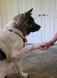
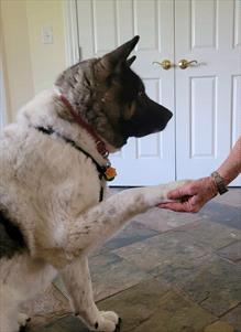
Targeting and Stationing. Targeting and stationing are valuable techniques for training animals to participate willingly in their care. Targeting involves training an animal to direct a specific body part, or their entire body, toward a designated point or object. This method can be applied to various animals and for different purposes. For instance, a parrot may be trained to target their wing into a person’s palm for feather inspection or a dog can be taught to touch their nose against a wall and stand still for a short veterinary examination.
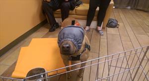
The practice of targeting behaviors paired with positive reinforcement (e.g., food) for each repetition helps to make targeting and the target object safe and predictable for the animal. For example, if a dog is taught to target the inside of a basket muzzle with their nose, the muzzle becomes a familiar and non-threatening object. This principle can be extended to a variety of grooming and veterinary tools.
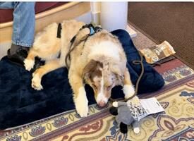
Stationing is a technique closely related to targeting and is particularly useful in teaching animals to stay at a specific location for an extended period. This concept is highly effective in various settings and for different species. For instance, a horse can learn to station themselves in a particular spot for voluntary grooming and veterinary care, eliminating the need for restraints like cross ties. Similarly, a dog can be taught to stand on a yoga mat for home care routines, and this mat can then be brought to the veterinary clinic to provide a familiar and comforting environment. Cats can be trained to jump onto a pedestal to receive oral medication, making the process smoother and less stressful for the cat and the caregiver.
Choice, control, and consent. In traditional pet care or treatment methods, animals often have no say in the process, which can be confusing and frightening for them as they do not understand the intentions behind these actions. However, modern techniques in animal care are changing this dynamic. These techniques involve communicating intentions to the animals and teaching them to express “yes” or “no” to their care.
This approach of allowing pets, zoo animals, wildlife, and livestock to consent to or deny their own care has shown surprising results. Contrary to the assumption that when given a choice, pets would say “no” all the time, the opposite is often true. The more we acknowledge their “no” the more they begin to say “yes”. They begin to engage in their care routine.
Start Buttons. Start buttons are an innovative concept in animal husbandry and a key element in cooperative care routines, emphasizing the importance of animal consent and autonomy. By observing and reinforcing animals with food as they display natural and comfortable behaviors, these behaviors can be encouraged and, therefore, more frequently offered. The concept is to teach the animal to present these behaviors as a signal to initiate a care routine, effectively giving them a “start button” communicating that they are ready to start their care routine.
This method is being applied across various species. For instance, horses can be taught to nod their heads to communicate you can start brushing them. Dogs can be trained to rest their chin on a towel to indicate readiness for home care and to lift their chin as a signal of discomfort or the need to stop.
When a pet stops offering their start button behavior, they are withdrawing their active participation in the care routine, communicating a “no” or “I need a break.” The more a caregiver acknowledges when a pet says “no”, the more comfortable and less fearful or defensively aggressive the animal becomes in future care situations.
By empowering pets with the control to start and stop their own care routines at will, caregivers are providing their pets with autonomy. This practice not only improves the animal’s experience during care routines but also strengthens the trust and communication between the pet and their caregiver, enhancing the overall quality of care and the human-animal bond.