Tag: cats

4128976

Cholangitis and Cholangiohepatitis in Cats

Diseases associated with the liver, gallbladder, and/or bile duct system can be confusing for pet owners because these diseases can have really vague symptoms. It’s not always clear why they happen, and the terminology used to describe them tends to sound like a foreign language. Cholangitis/cholangiohepatitis, the second most common type of liver disease seen in cats, is a prime example. This disease is sometimes referred to as cholangitis, sometimes cholangiohepatitis, and sometimes cholangitis/cholangiohepatitis syndrome. In addition to these names, several different forms of the disease exist, which all have their own names. To add to the confusion, sometimes other diseases can occur at the same time as cholangitis/cholangiohepatitis, which makes figuring out the cause for the symptoms and diagnosing the problem pretty difficult. 

Helpful Definitions

Cholangitis means inflammation of the biliary tree (also called the biliary tract or bile duct system), which connects the gallbladder and liver and helps these organs make, process, store, and secrete bile. Bile is used to break down fat from food in the intestines. Cholangiohepatitis means inflammation of the biliary tree as well as the surrounding liver cells. Cholangitis/cholangiohepatitis can be caused by bacterial infection, in which case the disease may be called neutrophilic cholangitis. It can also be caused by a parasite known as a liver fluke, which causes inflammation after it invades the liver. Another type is called lymphocytic cholangitis. It is associated with lymphocytes, a type of white blood cell involved with the immune system, and it likely occurs because of an overreaction by the immune system.

Pancreatitis, or inflammation of the pancreas, and inflammatory bowel disease, an immune-mediated condition associated with inflammation, poor digestion, and poor absorption of nutrients within the gastrointestinal tract are common conditions that occur in cats that develop cholangitis/cholangiohepatitis. Veterinary scientists don’t know exactly why these diseases happen together, but over 50% of cats have one or both of these diseases in addition to cholangitis/cholangiohepatitis. 

Clinical Signs and Symptoms

Depending on what type of cholangitis/cholangiohepatitis a cat has, the symptoms may occur slowly, over a long period of time (chronic) or happen suddenly (acute). Common signs include poor appetite, throwing up, diarrhea, acting tired, drinking and urinating a lot, fever, weight loss, belly pain, swollen belly, and a yellow tinge (jaundice) to the skin, gums, and eyes may be seen.

Diagnosing Cholangitis/Cholangiohepatitis

Lab tests will be run on the cat’s blood to see how the red blood cells, white blood cells, and platelets are handling the disease. This type of test is called a complete blood count or CBC. A chemistry lab test will also be run, which will show the disease’s effects on the liver as well as other organs. Findings that point towards cholangitis/cholangiohepatitis include high liver enzymes. Increased levels of bilirubin, which is a product of the breakdown of red blood cells by the liver, can also be seen. At high levels, bilirubin can cause jaundice (yellow-tinged skin, eyes, and gums). Other tests include abdominal x-rays and ultrasound to examine the liver, gallbladder, and biliary tree. Ultrasound can also be used to sample the bile to check for and culture bacteria. Finally, to officially diagnose cholangitis/cholangiohepatitis, biopsies of the liver tissue are needed. These can occasionally be done with abdominal ultrasound, but often surgery is needed to get large enough pieces of diseased tissues for a pathologist to properly examine. 

Treatment

Treatment may depend on the cause or reason for the cholangitis/cholangiohepatitis (e.g. immune-mediated vs. bacterial infection). If the cat has pancreatitis or inflammatory bowel disease, those will require treatment as well. Antibiotics are commonly used although they may be switched to different kinds depending on the bile culture results. Antibiotics are usually needed for at least 1-2 months and should be continued even once the pet starts feeling better. Steroids to suppress the immune system may be needed. Supplements or medications to help support the liver and biliary tree may also be prescribed, including ursodiol, vitamin E, and SAMe. Do not start any supplements without consulting your vet.

In addition to medications, very sick cats may need hospitalization, IV fluids, a feeding tube, and/or surgery to remove the gallbladder. Surgery is usually only needed if an obstruction or blockage occurs within the biliary tree/bile ducts. Prognosis, or chance for improvement, varies depending on the cause. Cats with acute neutrophilic cholangitis, which is a sudden onset bacterial infection of the biliary tree, usually recover. Those with chronic or long-term forms, or lymphocytic forms of cholangitis, tend to respond poorly to medications and the disease can worsen over time to the point of liver failure. 

If you suspect your cat has a form of cholangitis/cholangiohepatitis, or you have questions about the disease, call your veterinarian for further information. 

4127784

Cerebellar Hypoplasia Cats Are Clumsy from Birth

A cat with cerebellar hypoplasia is abnormal from infancy, though it may not be clear that something is wrong until the kittens are toddling around with some coordination. The cat with this disease clearly has balance issues: he walks with his legs widely placed for fear of falling and places his feet clumsily. He often leans against walls for support. Sitting still, he may look like a fairly normal cat but once focused on a toy, food bowl, or friendly hand, tremors begin and get more severe as concentration becomes more intense. Still, despite the obvious impairment, cats with this condition are happy, able to feed themselves and use a litter box, and are felt to have good life quality.

Your Cerebellum and You

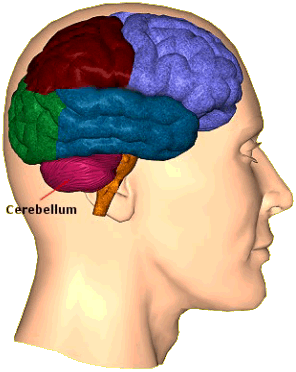
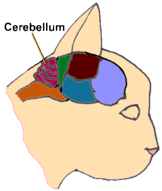
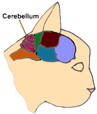
The cerebellum is a part of the brain nestled inside the base of the skull, as shown in Image 1. The cerebellum is involved in the involuntary aspects of moving our bodies, whether we are walking around, reaching for something, or just turning our heads. These all seem like voluntary activities so it might be difficult to consider that there is something involuntary going on as well.

Consider walking across an uneven, perhaps rocky, terrain while carrying on a conversation with another person.

Certainly, the decision to move from point A to point B is voluntary but the chances are you can walk across pretty uneven terrain without falling down or even thinking about where to put your feet. You can thank your cerebellum for that.

Your cerebellum receives information from your feet as well as from the vestibular apparatus in your middle ear. This information tells you where up and down are and where your body is in relation to up and down. Your cerebellum integrates all this information so that you can not only walk or run across uneven terrain without devoting any conscious thought to how to do so, you can turn your head, maybe even drink from a bottle of water, and look at another person while you are walking without (much) chance of falling down.

Your Cat’s Cerebellum and the Feline Distemper Virus

The cerebellum is obviously a complex piece of equipment. During fetal development, the cerebellum is maturing up until the last few days before birth and finishes its development in the first couple of weeks after birth.  Having a long period of development leaves the cerebellum especially vulnerable to an assortment of toxins for a long period of time. In the case of cerebellar hypoplasia, the culprit is the feline distemper virus (also called the panleukopenia virus). Following infection in the pregnant mother, growing nerves in the unborn kitten’s cerebellum are destroyed, ultimately yielding a tiny improperly developed cerebellum and a disability for the kitten.

The mother cat may not be actually infected during pregnancy to create this syndrome; if while pregnant she has been vaccinated with a modified live distemper vaccine, the most basic of all cat vaccines, it can have the same detrimental effects.

.Kittens infected with distemper under age 2 weeks may also experience enough cerebellar damage to suffer cerebellar hypoplasia. This means that mother cats due for vaccines should not be vaccinated during pregnancy or in the first 2 weeks following kitten delivery.

Diagnosis

While imaging such as CT scanning or MRI can be used to confirm the underdeveloped tiny cerebellum, the diagnosis of this condition is usually obvious because of the following features:

  • The condition is non-progressive (it doesn’t get worse).
  • The cat is not weak, he’s just not coordinated.
  • Intention tremors are evident when the cat is focused but the cat looks largely normal if still.
  • Walking is uncoordinated with a wide stance, often using the wall for support.

Treatment

No treatment can resolve the damage in a kitten with cerebellar hypoplasia. Some kittens are more severely affected than others. Owning a cat with cerebellar hypoplasia emphasizes managing the condition. The cat should be indoors only and non-spill food dishes should be obtained. Most cats have good life quality and make-up in personality what they lack in motor skills.

Prevention

This syndrome is easily prevented by vaccinating female cats for feline distemper when they are neither pregnant nor nursing kittens under age 2 weeks. This means that regular vaccination will prevent cerebellar hypoplasia in kittens. If a cat is pregnant, vaccination should be withheld during pregnancy and for a good three to four weeks after the kittens are born. 

5186769

Body Language of Cats

Cats use body language to communicate a variety of information and emotions to others. The primary body parts they use to convey their state of mind are their ears, tail, eyes, body posture, and vocal cords. Types of emotions and behaviors that can be demonstrated are fear or anxiety, frustration, pain, play, mating, contentedness or relaxation, and curiosity. Often body language can be used to display multiple emotions. Thus, when trying to figure out your cat’s body language, try to understand the context in which he is communicating.

Fear/Anxiety Avoidance

These types of behaviors are an effort to maintain safety from a perceived threat by avoiding the threat.

  1. freezing (in an effort to be less noticeable), especially if confined or unable to escape; such cats are often quiet and do not vocalize or make noise. Note that a “frozen” cat can switch quickly to “repulsion,” becoming aggressive towards the cause of fear
  2. freezing results in stopping other normal behaviors like eating or using the litterbox; this is why cats wait until quiet hours to eat/drink when a lot of commotion is happening at home
  3. moving away slowly, i.e., crawling or slinking away
  4. fleeing, especially if startled
  5. hiding
  6. crouched body with feet flat on the ground (often underneath the cat) to allow for quick escape and protect important organs
  7. trembling
  8. tensed muscles
  9. dilated pupils (enlarged black part of the eyes)
  10. ears turned back or flattened with brow furrowed
  11. sleepy appearance
  12. walking with back flat, tail down, and head down
  13. excessive and sudden grooming in one area
  14. standing tall with hair standing on end (an effort to appear larger)
  15. tail held tightly to the body
  16. averting the eyes

Fear/Anxiety Repulsion

These types of behaviors are an effort to maintain safety from a perceived threat by getting rid of it.

  1. hissing, growling
  2. swiping
  3. spitting
  4. scratching
  5. biting
  6. directly and intensely staring at the source of anxiety
  7. using the body to physically block another being (eg, blocking another being from the food bowl if worried about that resource)
  8. ears flattened and out to the side
  9. standing tall with hair standing on end (an effort to appear larger)

Fear/Anxiety/Curiosity

These information-gathering behaviors are an effort to learn about an object, person, environment, and/or if perceived as a threat, to determine the severity.

  1. hypervigilance and scanning the surroundings frequently
  2. fixation on the object of concern
  3. ears erect and directed at the source of concern
  4. sniffing
  5. Flehmen response, which is raising or curling the lips to detect information such as pheromones
  6. may startle easy if intensely focused
  7. tail swishing
  8. straight legs and upright posture
  9. watching object of interest casually/calmly (as opposed to the intense staring of repulsion)

Frustration

These types of behaviors are reactions to something that is not meeting the cat’s expectations or their inability to control something (e.g., hunger, not wanting to be held). These behaviors are an effort to achieve what they desire.

  1. vocalization- meowing continuously
  2. circling or pacing
  3. rubbing body or face on people, animals, or objects
  4. scratching on objects
  5. cats may switch behaviors quickly (unlike with fear/anxiety, where in some cases behaviors are prolonged)
  6. very active
  7. flattening or rotating ears
  8. thrashing or firm swishing of tail
  9. gazing upward
  10. half-blinking
  11. showing the tongue
  12. Flehman response
  13. nose licking or nose wrinkling
  14. rippling and twitching skin
  15. hair standing up if very frustrated
  16. grooming specific areas (eg, shoulder or base of tail) quickly and in an intense manner

Pain

It is extremely difficult to notice pain in cats as they tend to hide pain well, but they do demonstrate some noticeable behaviors.

  1. hiding
  2. limping
  3. more withdrawn or quiet
  4. ears back
  5. pupils dilated
  6. change in normal behavior
  7. squinted eyes
  8. hunched back

Play

These behaviors are used during play and to initiate it. Hunting behaviors are similar to those of play.

  1. ears forward
  2. tail pointed up (an indicator that the cat is game for a friendly interaction)
  3. whiskers forward
  4. tail twitching
  5. predatory/hunting behavior such as stalking, pouncing, swiping, or biting
  6. immature cat rolling towards older cat (an indication of appeasing behavior)

Relaxed/Content

These show the cat is content and often demonstrates trust in their environment.

  1. eyes half-closed
  2. sitting or lying down with minimal muscle tension
  3. minimal tail movement
  4. purring
  5. kneading (“making biscuits”)
  6. rubbing on people or things
  7. belly exposed (demonstrates trust because cats will often protect their belly in fearful situations)
  8. grooming the body rhythmically, in a relaxed manner

Mating

These are an effort to demonstrate receptiveness to mating, such as showing signs of going into heat or estrus.

  1. female rolling around a male
  2. purring
  3. stretching
  4. rubbing on objects, people, or other animals
  5. extra affection
  6. raising hindquarters, treading with back legs
  7. frequent and loud vocalizations
  8. backing up to furniture or horizontal object, twitching tail, often urinating in this position (marking behavior).

Behavior changes in cats can be quite subtle and difficult to notice unless you are really paying attention.

4128586

Bartonella and Cat Scratch Fever

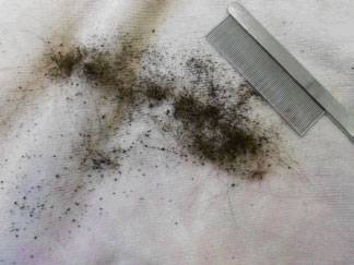
Most people know very little about this infection other than it involves a fever spread by cat scratches. In fact, it involves infection by bacteria of the genus Bartonella. There are 24 Bartonella species, 14 of which can infect humans and five of which are harbored by cats. The five Bartonella species harbored by cats are spread by fleas. The most well-studied and most common Bartonella organism is Bartonella henselae. Classically, cats transmit the organism when they are parasitized by fleas, scratch themselves, get infected flea dirt (digested host’s blood excreted by fleas) in their claws, and scratch a person or another cat with their dirty claws.

The Human Disease

Infection with Bartonella henselae in an immunocompetent person (i.e., a normal, healthy person) person leads to cat scratch disease. The inoculation site, a scratch from a claw containing bits of flea dirt, develops a small red bump called a papule. About 2 to 3 weeks following contact with the infected cat, the lymph node in the area will swell and become painful, and a fever will develop. These signs generally resolve on their own, and the condition is minor, though lymph node enlargement can persist for several months.

If the patient does not have a competent immune system (in other words, they are very old, very young, debilitated from another condition, etc.), one of several far more serious syndromes can result. The infection goes deeper into the body and causes spleen enlargement, and potentially encephalitis, heart valve infection, and other conditions. These syndromes are rare, but they are potentially quite serious.

How Likely Is It for a Cat To Be Infected?

Since fleas carry the bacteria, cats with insufficient flea control are at the highest risk. This means cats living in climates that are warm and humid (conditions where fleas thrive best) are most likely to be infected. If conditions are right, up to 40% of cats in an area may be infected. If a person is diagnosed with cat scratch disease, there is a 90% chance that the cats they own will be found infected as well.

This sounds somewhat concerning for the cat-owners in a flea area but it is important to realize that an infected cat cannot transmit the infection without a claw full of flea dirt. If the fleas are removed from the infected cat, there will be no flea dirt in the coat and no risk of disease transmission. Experimental studies have not been able to demonstrate cat-to-cat transmission in cats that mutually groom each other, fight with each other, mate with each other, or share food or litter with each other, nor has transmission been demonstrated between infected mother cats and their kittens. Short of an actual blood transfusion from an infected cat, fleas or ticks are needed to spread the infection.

Do Infected Cats Get Sick?

This is a highly controversial question. It was only relatively recently discovered (1992) that cats themselves were more than simple carriers of Bartonella henselae and that they could actually become actively infected themselves.

Several illnesses seem to have been associated with Bartonella infection (fever, deep eye inflammation, lymph node enlargement, muscle pain, reproductive failure, and bacterial heart valve deposits called endocarditis), but these seem to be isolated cases for the most part. The Bartonella organisms are highly adapted to live in the feline body without causing disease, and it is only in rare situations that cats actually experience issues from this infection.

It has been suggested that Bartonella infection may be at the root of numerous chronic inflammatory conditions of the cat. With such high numbers of infected cats occurring regionally (up to 40%), it is going to be difficult to prove one way or the other whether there is a real association or just coincidence.

Can Dogs Get Infected?

The short answer is yes, though the species they get is Bartonella vinsonii rather than Bartonella henslae. Fleas may carry the infection as they do for cats, plus it appears that ticks may also be carriers. Since there are numerous infectious agents spread by ticks and it is not unusual for a dog to have multiple tick-borne infections, it is difficult to determine which infection is causing which signs.

Is My Cat Infected?

Testing is not recommended for the average cat, even if the cat is sick. Testing would be a good idea for any cat that may become a blood donor, for the rare sick cat where Bartonella-related disease is actually being considered if a human in the home has been diagnosed with a Bartonella-related disease, or if there is an immune-compromised person in the home, in which case testing is actually important.

There are five tests available to detect Bartonella henselae: ELISA, IFA, PCR, Culture, and Western Blot. All the tests have pros and cons, and no method seems to shine above the others.

The ELISA, IFA, and Western Blot tests are tests for antibody detection, the idea being that if antibodies against Bartonella are there, then Bartonella must be there as well. For most diseases where antibody levels are used to establish a diagnosis, a minimum titer or antibody amount is considered necessary to say “Yes, this patient is infected.”  A problem with this is that we know that up to 11 percent of cats with Bartonella organisms happily circulating in their bloodstreams will not make antibodies and will thus test negative. At least this means that when the test is negative, there is an 89 percent or greater chance that the cat is truly negative. For screening, this kind of antibody testing is frequently paired with PCR testing.

PCR is a very sensitive DNA test for Bartonella, but because the organism only intermittently circulates, false negatives can result if not very much Bartonella DNA is circulating. Results from PCR testing can rapidly distinguish different species of Bartonella. 

The most reliable test is the blood culture; however, several consecutive cultures are needed because the organism tends to circulate only intermittently. A positive culture is proof of infection, though a negative culture may simply not have been taken at the time when the organism is circulating. 

In humans, a delayed hypersensitivity skin test is used as part of the diagnostic criteria for cat scratch disease but this test has not been useful in cats. In this test, similar to the tuberculosis test most of us are familiar with, a scratch on the skin is made, and a reaction to the introduced antigens may occur either right away or in approximately 48 hours (delayed hypersensitivity reaction). Cats are poor delayed hypersensitivity responders. 

All in all, it is hard to rule out a cat as a carrier of a Bartonella organism. If the results of testing are suggestive of infection, treatment can be considered if the cat is truly believed to have an illness from the infection or if there is realistic concern about exposure to an immune-compromised person.

Treatment for Cats

An assortment of antibiotics have been used against Bartonella henselae in cats: clavulanic acid-amoxicillin combination, azithromycin, doxycycline, and quinolone class medications (enrofloxacin, marbofloxacin, etc). Efficacy has been mixed, and Bartonella henselae rapidly becomes resistant to therapy. Treatment is currently recommended for cats showing symptoms of disease or for cats living with immune-compromised individuals.  Treatment lasts a minimum of three weeks.

Preventing Human Infection of Bartonella: Guidelines from the Centers for Disease Control and Prevention

Guidelines for Prevention of Human Infection

The following guidelines have been published by both the CDC and the European Advisory Board on Cat Diseases:

  • Immune-suppressed people should adopt only apparently healthy cats less than 1 year of age.
  • Immune-suppressed people should not adopt from the shelter or from multi-cat homes.
  • Cats belonging to immune-compromised people should be kept indoors only and should not have contact with cats that go outdoors.
  • Claws should be kept trimmed.
  • Cat scratches should be promptly washed. (This goes for everyone, not just immune-compromised people.)
  • Flea control should be strict. 

Preventing Disease

No data support chemoprophylaxis (i.e., preventive drug treatment) for Bartonella-associated disease. In simpler terms, if a suspicious cat scratch has occurred, there is no point in using medication to prevent infection.

4128401

Blocked Cats are an Emergency

When something happens to stem the flow of a cat’s urine, trouble ensues – and fast.

Urine has lots of good things in it. In many cases, they are substances that cats or people can’t live without, such as potassium, sodium, and water. The kidneys sense and adjust the composition of bodily fluids and drop the excess into the urine. If a person eats a large order of fries, covered with salt, the kidneys dump the unwanted excess of sodium into the urine.

 The same is true with many other substances, like water, that need to be regulated. Urine is (usually) sterile, so unless there is a urinary tract infection, urine is pure. It’s not the terrible stuff that many third graders make it out to be. True, it does have the waste products of metabolism in it, which a body needs to remove.

And that’s where some of the problems begin. If the flow of urine stops, those waste products build up and negatively impact the way the body works. One of the most common ways that happens is when a cat’s urethra (the tube that carries urine from the bladder to the litter box) gets blocked. Known in veterinary parlance as a ‘blocked cat’ or ‘blocked tom,’ this poorly understood disorder is seen with alarming frequency in veterinary hospitals and ERs.

Many ERs see about two to three cats per week who cannot urinate. Cats can be in all stages of the disease, from the early onset ones who just seem a little painful and have a big, hard bladder to the nearly dead ones that are in many cases beyond saving.

The actual plug that stops the flow can be made of bladder stones (often erroneously called kidney stones), tumors or a gooey mix of mucus and protein known as ‘matrix’ that has the consistency of toothpaste. How and why matrix forms, no one knows, despite a few decades of investigation. Adding to the confusion, the name of the disorder has changed no less than four times in the past 20 years from feline lower urinary tract disorder (FLUTD) to feline urologic disorder (FUS) to feline interstitial cystitis (FIC) to the most recent iteration of Pandora Syndrome, which hasn’t really caught on yet.

The causes go beyond a mucousy plug, as well. A host of other factors, such as stress, lack of access to water, diet, infectious agents, indoor lifestyle, and many other causes have been implicated as being responsible for the lead-up to getting blocked. Those little plugs don’t form in a vacuum: something causes them to form, and we don’t know with any certainty what factors contribute to it.

Cats that are blocked often show the following signs:

  1. Straining repeatedly in the litter box (often mistaken for constipation)
  2. Crying or howling
  3. Licking at the genitals/below the base of the tail
  4. Hiding

If you notice your cat showing any of the above signs, get right in to see your veterinarian or go to your nearest ER as soon as possible. Don’t delay as a few hours can make a big difference. The longer those toxins circulate unchecked, the more pain the cat experiences, the more work the veterinarian has to do, and the bigger the final bill will be.

In advanced cases, where the urine flow has been stopped for more than 24 hours, cats can become systemically ill from retained toxins and start vomiting, or become very weak and lethargic. Death usually happens within 48 hours, and it’s not a pleasant way to go. The pain with this disease is immense, and some cat owners understandably choose euthanasia over trying to reestablish the flow of urine.

The course after unblocking these cats is just as unpredictable and mysterious as the factors leading up to the obstruction; some cats are released from the hospital never to suffer another episode, while others will have repeated occurrences days, weeks or years later. This is an inhumane disease.

Managing these cases medically can go way beyond relieving the obstruction in some cases. First priority is fixing the plumbing problem: getting urine to flow. This is usually done with anesthesia and a catheter to remove the obstruction. Managing the havoc wreaked by the toxins is next. This can necessitate some medical dancing as veterinarians try to put things back in place. Disorders of deadly potassium, elevated renal values, and severe dehydration can mean days in the hospital, even long after the urine is flowing again. It can get complex, expensive, and can wear down even the most committed of owners for the really medically complex (and expensive) ones.

However, getting the urine to flow and taking the cat home is the easy part. After this episode, lifestyle changes are necessary: medication tweaks, medical rechecks, and diet changes that try to extend the initial complexity of this disease across months or years.

Compared to 20 years ago, cats with this disease do go home and get better, even some of the tough cases.  Someday, science will provide an answer and veterinarians will have some means to prevent this disease in the first place, or some surefire way to treat it. Until then, rush your cat in to be seen if you see the signs.

6665143

Bacterial Conjunctivitis in Cats

Conjunctivitis is inflammation of the conjunctiva, a tissue that makes up part of the eye and eyelid. In people, the conjunctiva is the white part. Conjunctivitis is common in cats and can be a result of infection or injury.

The primary cause of most conjunctival infections in cats is usually feline herpes virus. The treatment for herpes-based conjunctivitis involves a combination of antibiotics and antivirals, both in eye drops and pills. Even cats who have had the herpes vaccine can get conjunctivitis because the vaccination does not prevent herpes, but allows a milder case.  

However, some conjunctival infections that are primarily viral also have a bacterial infection at the same time.

The two bacteria species best known to cause conjunctivitis in cats are Chlamydia felis and Mycoplasma spp.

An infection caused by Chlamydia may cause redness, discharge, and excessive tears. Bacterial conjunctivitis caused by Chlamydia is treated with antibiotics. All cats in a household may need to be treated, as some cats who have it do not show any symptoms.

Mycoplasma is a natural inhabitant of every cat’s eye, so infection happens when there is an overgrowth of that bacteria. Treatment for the Mycoplasma infection is similar to that of Chlamydia: antibioticsThe goal in treating Mycoplasma conjunctivitis is to reduce the overgrown number of bacteria back down to a normal level because it is impossible to eliminate them entirely.

Who gets bacterial conjunctivitis?

Both cats and dogs can get conjunctivitis caused by bacteria or viruses. Conjunctivitis is the most common cat eye disease, and most cats will develop the infection once in their life. All breeds can develop infection, and juvenile and young adults are more likely to get it.

Clinical Signs of Either Form

  1. Red eyes
  2. Eyelid redness
  3. Eyelid swelling
  4. Excessive blinking
  5. Eye discharge
  6. Eye crusting
  7. Tearing
  8. Squinting
  9. Scratching the face to relieve irritation

 Diagnosis

A physical exam of the eye by your veterinarian is the first step, as this will rule out other causes of redness, such as trauma.  The easiest diagnosis is a cytology test, which means looking at scrapings of the irritated tissue under a microscope. If a lot of bacterial and inflammatory cells are seen, it is likely that the eye is infected. Checking scrapings is a quick test for infection that can be done at the clinic during your appointment. PCR is another test that can be used to detect the specific organism causing the infection, but typically needs to be sent out to a lab.

Treatment

Antibiotics are used to treat bacterial conjunctivitis, mainly doxycycline.  Mycoplasma and Chlamydia are the most common bacteria involved in eye infections, and doxycycline is effective against both.

Prognosis

Treatment should clear the signs and infection. However, if the eye still looks infected or just doesn’t look normal, contact your veterinarian. The ongoing bacterial infection may also be caused by an unknown factor that needs to be treated before the eye infection can be cured.

Prevention

There is a vaccine for Chlamydophila felis, but it is not required and is usually only suggested if your cat is at a higher risk for an infection or in a multi-cat household. There is no vaccine for a Mycoplasma spp. infection because it is normally in the eye anyway.

The key to preventing infection is separating sick cats away from the unaffected ones and bringing them to your veterinarian at the first sign of infection.

4118703 (1)

Asthma in Cats

What is Asthma?

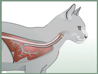
Asthma is a recurring respiratory compromise featuring the constriction (narrowing) of the lung’s airways.

There are three features that define asthma:

• Airway inflammation
• Airway hyper-responsiveness
• Reduced airflow (which is at least partly reversible)

More simply, this means the airways of the lungs are inflamed, plus they are extra reactive to mental stress or respiratory irritants, and the flow of air through the lungs is reduced (either sometimes or all the time). When all three features are present, we can be comfortable diagnosing asthma.

What Exactly is Happening in the Lungs with Asthma? 

First, excess mucus forms in the airways. After that, the airway walls swell with inflammation and can actually ulcerate. Finally, the airway muscles go into spasms leading to constriction. Airway constriction leads to an inability to draw a deep breath, intolerance to exercise, coughing, and musical sighing sounds called “wheezes,” though not all these symptoms may be apparent at the same time.

Sometimes a low-grade chronic cough is the only evident sign, but it should be remembered that a sudden asthmatic crisis can arise at any time and can represent a life-threatening event. Asthmatic airway constriction can happen spontaneously, in reaction to stress or anxiety, or as a type of allergic reaction. Relieving and preventing airway constriction is the main focus of treatment.

Is Feline Asthma the Same as Human Asthma?

The feline condition was named asthma because of the clinical features shared with the human disease. As of this time, it appears that all the diagnostic criteria needed to diagnose asthma in people are shared by cats, yet we are still working out the mechanics of this syndrome in cats and have a great deal to learn. For example, in humans, we know that while actual symptoms of asthma occur in episodes, the airways of the lung are diseased all the time. We do not know yet if feline airways are also diseased all the time or if the actual airway structural changes occur only when there are clinical signs of distress.

How is the Diagnosis Made?

Because of the constricted airways, the actual volume of air this patient can move in and out of the lungs with each breath is reduced. There is often a great deal of effort seen in the cat’s breathing. The abdomen appears to be working to push air out and the breaths are shallow and rapid. The cat may even be breathing with its mouth open in an effort to move the largest possible amount of air.

The next step toward making a diagnosis of feline asthma is the chest radiograph, assuming the cat is not in too much distress to hold still for this procedure. Classically, this radiograph will show what is called air-trapping, meaning that the small airways have constricted so that inhaled air cannot be exhaled. The lungs are larger in appearance than normal as they are over-inflated. The diaphragm may seem flattened due to this over-inflation.

It is possible to view the tiny airways in much higher detail on radiographs. This is partly because they are made more prominent because of the mucus and inflammatory fluid inside them (the doughnuts and tramlines, as mentioned below) and partly because they are surrounded by more air than usual (air-trapping).

Inflammation and mucus build up within the airways, causing their walls to appear thickened in the radiograph. The terms used for such airway appearance are doughnuts (when viewing the airway end-on) or tramlines (when viewing the airway from the side). You may hear your veterinarian use these terms, and they are classical findings in airway disease. 

Some Asthmatic Cats Have Normal Radiographs

Since visible changes are not always evident on radiographs, which can lead to diagnosing asthma when it is not there. Indeed, diagnosing asthma can be complicated, and it is famously underdiagnosed because up to 23% of asthmatic cats can have completely normal radiographs.

The coughing cat with normal chest radiographs poses a diagnostic dilemma. It is hard enough to sort out potential issues when there actually are radiographic changes, as many conditions can mimic others. Because of the nature of feline heart disease, where the heart muscle can thicken without the overall external shape of the heart changing, heart disease should be considered. In heartworm-endemic areas, feline heartworm disease should be considered. If the patient is thought not to have too high of an anesthetic risk, a tracheal wash and/or bronchoscopy can be considered but often have indefinite results. CT (cat scan) imaging is emerging as a helpful diagnostic tool, but it is not yet available in many areas.

Response to Therapy as a Diagnostic Test

One important asthma feature is that the airway constriction it engenders is reversible. In an emergency situation, a small dose of epinephrine (adrenalin) or terbutaline (which expands airways) can reverse an asthmatic crisis in as little as 15 minutes. Response to an injectable corticosteroid (such as dexamethasone) generally yields a positive response within 30 minutes.

Sometimes, diagnostic tests still leave room for questions, and you have to simply go with medical treatment for asthma and regard response to therapy as evidence that the diagnosis is correct. See below for a list of medications commonly used in the long-term management of this problem.

Treatment Options

Oral and Injectable Steroids 

It is crucial to realize that the underlying problem in the airway is inflammation. Inflammation is what is responsible for constriction. To resolve inflammation, corticosteroid medications have been the cornerstone of therapy. These can be given in pills, by injection, or, more recently, through a metered dose inhaler. Usually, treatment is started with either an oral corticosteroid such as prednisolone or a long-acting injection such as Depo-Medrol. These medications are relatively inexpensive, and a good response to them helps confirm the diagnosis, as mentioned. A better strategy might be to use oral or injectable steroids to ensure that they work and if they do, change to a metered dose inhaler (see below), as there is much less potential for corticosteroid side effects with an inhaler.

If the response to oral corticosteroids is good, they may be continued long-term, supplemented with some of the other medications mentioned below, or used only during flare-ups. Long-term corticosteroid use has some potential for side effects, though feline patients tend to be resistant to these problems. If giving pills is too difficult, especially in a cat that is stressed and having some trouble breathing, a long-acting corticosteroid injection can be used periodically to control asthma symptoms. Cats are more sensitive to potential side effects from these injections as they are much stronger than oral corticosteroids. This sensitivity means that injections can only be given periodically without the risk of developing diabetes mellitus. If the cat seems to require an injection more frequently than every other month, a metered dose inhaler should be considered (see below for details).

When using oral corticosteroids, it is important to taper the dose over time so as to find the minimum dose needed to control symptoms. If higher doses are used indefinitely, asthma symptoms may become resistant to steroids. This resistance is more of a problem with injectable steroids and manifests as a shorter and shorter interval between the need for the injection. Again, if this is seen, consider changing to an inhaled form of medication.

Metered Dose Inhalers

Human asthmatics have enjoyed the benefits of portable inhalers for years. These handy devices deliver medication locally to the airways, thus minimizing drug side effects to the rest of the body while maximizing desired response.

There were two problems with using these devices in cats:

  • Cats cannot be told when to inhale. 
  • Cats tend to object strongly to the spray puff delivered by the device.

Interestingly these same problems apply to human infants with asthma and a device called a pediatric spacer was invented for them. This tubular chamber is attached to the inhaler on one end and a face mask on the other. The inhaler is actuated into the spacer and the infant simply breathes in the spacer’s contents.


The discovery that the pediatric spacer could also be used for cats has solved the feline inhaler problem as well. The setup is the same as described above for young children and the cat takes 7 to 10 breaths from the face mask to be properly dosed.

Corticosteroid inhalers, Flovent® being the most popular, are typically used twice daily long term while airway dilator inhalers such as albuterol-containing Proventil® or Ventolin® are used for flare-ups.

Cats are generally started on a combination of oral prednisone and the metered dose inhaler and gradually maintained on only the metered dose inhaler.

Pediatric equipment is generally available from most human pharmacies, however, a device created specifically for feline use can be ordered. It comes with a spacer and an appropriately sized face mask. Your veterinarian will need to prescribe the metered dose inhaler usually from a regular human pharmacy.

See the Aerokat device.

Are there other Treatment Options?

Other medications that might be helpful include:

Allergen-Specific Immunotherapy 

Since allergy is generally a part of the asthma picture, it makes sense that “allergy shots” might be helpful. In fact, they are helpful to many cats. It is important to realize that this form of therapy takes months before benefits are obvious so it is not a method for fast relief but more for the “bigger picture.” The owner must be able to give injections to the patient at home regularly. A veterinary dermatologist is generally needed to oversee this type of therapy.

Airway Dilators

Terbutaline (Brethine®) and theophylline are airway dilators commonly used in the management of asthma. It makes sense that if constriction is an important feature of this disease that eliminating constriction would be therapeutically helpful. Terbutaline is available orally or as an injectable. Some veterinarians encourage owners to keep a bottle of injectable terbutaline at home in case of a crisis and show them how to give it. If you are interested in this, let your veterinarian know. Theophylline is an oral medication usually given once a day at bedtime.

Cyclosporine

The use of cyclosporine in asthma is relatively new. Cyclosporine is an immunomodulator often used in organ transplant patients. It has been used occasionally in cats where adequate suppression of inflammation has not been possible with combinations of the other medications listed or when the cat is unable to take corticosteroids for other reasons (concurrent diabetes mellitus, infection, history of calcium oxalate bladder stones, etc.). Cyclosporine should not be prohibitively expensive since cats, being small, require relatively small doses but blood levels of cyclosporine may be periodically recommended which increases the expense of this treatment significantly.

Don’t Forget…

Minimizing irritants in the air is always helpful to an asthmatic cat.

  1. Do not allow cigarette smoke in the cat’s environment.
  2. Use dustless cat litter.
  3. Consider non-topical insecticides. No sprays, either.
  4. Regularly replace air filters at home.

It is important to realize that asthma can culminate in a respiratory crisis that can become life-threatening if ignored. If your cat begins to breathe with an open mouth or if you see excessive abdominal movement during breathing and the cat is not purring, you may have an emergency situation. Contact your veterinarian immediately.

For more details, including a video on the use of the inhaled system, we recommend visiting Fritz the Brave, a website set up by one family devoted to their asthmatic cat. It has grown into a detailed instructional site for both pet owners and veterinarians interested in the details of inhalers for feline asthma treatment.
7644496

What To Do When Your Dog’s Toenail Trims Don’t Go as Planned

When your dog’s nails get too long, it can affect their health and overall comfort. Overgrown nails are more prone to nail breakage and injury. Over time, too-long nails can keep your dog from placing their feet correctly as they walk or run, and broken nails can get snagged in carpet. 

You’ve watched the staff at your veterinarian’s office trim your dog’s (or cat’s) nails many times. Your well-trained dog has no problem with nail clippers and is always cooperative with the technicians, so how hard can this be?

If you aren’t planning to visit the veterinarian for any other reason, you think, why go in just for a quick nail trim?

So, you have corralled the needed tools for the job, which in your mind are:

     1.  your dog; and
     2.  the nail clippers.

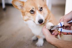
As expected, your always-calm dog sniffs the clippers and holds their paw up for you to begin. Clearly, this is not a big deal. You grasp the paw in your hand and gently expose the first toenail by squeezing a bit. You put the trimmer around the nail and squeeze the handles. With a satisfying click, you see the nail trimming pop away, and at the very same moment, your sweet dog lets out a sharp yelp, frightening you both. And there is blood.

Blood? Why is there blood? That never happens when the veterinarian technician does the nail trimming. 

Your very good dog is still sitting there, looking at you with what you think is betrayed reproach. What now? Panic? No.

Nail Trim Essentials

Let’s back up. 

Let’s return to the last time the veterinary technician trimmed your dog’s nails. This time, really watch the process and ask questions, such as, how short should you trim the nails?

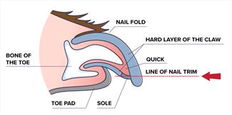
If you aren’t familiar with the term “quick”, it refers to the blood and nerve-filled nail bed. If nails are clipped too short, the quick is nicked, and the nail will bleed. If your dog has fairly clear nails, you can see the quick. If there is an assortment of clear and dark nails, you can judge where the quick might be on the dark ones by looking at a clear nail.

There is a theory that when nails are trimmed or broken, the quick recedes further up the nail. Therefore the“quick line” may not be the same on every nail. If your dog or cat has a nail that seems ragged or shorter than the rest or has a bit of dried blood on it, this nail may have been torn previously. Keep in mind that this nail may not need as much trimming as the longer ones. However, it’s a good idea to clean it up to avoid further tearing.

So, you feel more confident trimming your dog or cat’s nails, right?

Handling “Quicked” Nails:

But now you know that things can happen, and you have revised your list of tools. You gather: 

  1. your dog (or cat);
  2. nail clippers;
  3. clean towel;
  4. a paper plate or small dish; and
  5. a coagulant to help stop any bleeding.

Over-the-counter coagulants to stop nail bleeding are readily available. Styptic powder is the most common substance used for this type of first aid. Made from natural ingredients, including potassium aluminum sulfate (alum) and diatomaceous earth, styptic powder can be used on minor cuts and toenails. It’s safe for use on pets.

You can find styptic powder at any pet supply store. Your veterinarian probably carries it if the clinic sells over-the-counter products. Corn starch will also stop bleeding effectively, and baking powder or flour can be used as a second choice. Keep styptic powder or one of these products on hand with your nail-clipping supplies.

If a toenail does start to bleed, press the clean towel against the nail and hold it firmly for a few moments. Remove the towel and press the bleeding nail into some coagulant powder you’ve already prepared by pouring it onto the paper towel or dish. Use your fingers to pinch some powder to pack into the nail. Continue to hold the towel firmly around the toe.

You could also use a cotton-tipped applicator with styptic powder on the end and hold that firmly to the toenail.

A few minutes of this will likely stop the bleeding, but if it doesn’t, continue the pressure with the towel and reapply the styptic powder.

If your pet isn’t exactly the quiet dog described here, have someone help you by holding your dog or cat. A trusted pet holder can help calm an upset animal and keep the affected foot still, helping to avoid a mess if your pet flings their paw around. Make sure you have trained your pet prior to beginning nail trims. Food distractions may also be helpful.

While uncomfortable for your pet, a toenail trimmed too short and bleeding is not necessarily an emergency. You will become more confident trimming your pet’s nails and less likely to nip the quick the more times you do it. If the bleeding continues for longer than 10 minutes or is very heavy, call your veterinarian.

On rare occasions, your dog may need to be seen by a veterinarian to assess why a nail is bleeding excessively, and sometimes an injured or broken toenail must be removed in order to heal properly.

4536889

Wound Healing in Dogs and Cats

One thing is certain about life: we can all expect to experience some wounds. The good news is that we are fundamentally designed to heal.

While the statement above has philosophical implications, we will stick to the physical ones in this discussion. In particular, we will be considering the skin and the wounds experienced in the skin and underlying tissues.

The skin and its associated tissues exist in layers: the epidermis on the outside (layered in and of itself), the dermis below, the subcutis below that, and fat and muscle below that. When we are injured, these areas may be cleanly cut, punctured, scraped, ulcerated, or burned. 

These wounds can be sterile, unclean (relatively clean but not sterile), or even heavily contaminated. The body is designed to address all of these situations, and often, as caregivers, we can help.

The Healing Process Starts as Soon as the Wound is Inflicted

There are four phases of wound healing: Inflammation, debridement, repair, and maturation.

Inflammation (Starts Immediately)

This is the first phase of healing and is all about controlling bleeding and activating the immune system. Without going into too much biochemical detail, blood clots are forming, and blood vessels are constricting to limit blood loss in the area. This process also calls in “clean up” cells of the immune system to address contaminating bacteria and any dead tissue.

Debridement (Starts in a Few Hours)

Wound fluid, dead tissue, and immunologic cells form pus, which is designed to flow as a liquid from the wound and carry debris with it. The cells that were called to the wound in the inflammation phase are now actively working on consuming dead tissue and cleansing the area.

Repair (Starts in a Couple Of Days)

Collagen begins to fill in the wound to bind the torn tissues, a process that will take several weeks to complete. New blood vessels begin to grow into the area from the uninjured blood vessels nearby. The wound edge begins to produce granulation tissue, the moist pink tissue that will ultimately fill in the wound. The wound will shrink in a process called wound contraction so that new skin can form and cover it.

Maturation (Starts in 2-3 Weeks and can Take Months or Even Years)

Once plenty of collagen has been deposited, the final phase of scarring can form. The scar becomes stronger and stronger over time as new blood vessels and nerves grow in, and the tissue reorganizes. The final result will never be as strong as un-injured tissue but should ultimately achieve approximately 80% of the original strength

Spay Incision

A spay incision is an example of wound healing by primary intention.

Primary Intention

When the wound is a surgical incision with sutures in place, there is no area for the body to fill with granulation tissue. Instead, the wound margins are already held together and the two margins simply need to bond. New skin begins to form across the margin within two days. The four stages of healing continue as above but go much faster (10-14 days total) because there is no gap in the tissue to fill in.

Healing occurs across the wound margin, not down its length, which means long incisions heal just as fast as short ones.

Secondary Intention

If the wound cannot be closed with sutures (it is too big, there is too much tension on the wound margins pulling them apart, the wound is too infected, etc.), then a process called second intention comes into play. This is the part of wound healing where granulation tissue must form to fill in the gap. Once the wound is filling with granulation tissue, contraction soon follows, which means the wound will be getting smaller and smaller. Eventually, it can be allowed to simply close on its own or when it is small enough, the margins can be trimmed and the wound surgically closed with primary intention of a smaller scar and better fur coverage. In the right circumstances, skin grafts can be applied, but only if there is a healthy granulation bed.

We Love Granulation Tissue

It looks like it would be sore. Many people incorrectly feel granulation tissue is not supposed to be there when, in fact, it is a sign of a healthy healing wound. When a wound is cleansed of debris, scabs, or crusts (and often when a bandage is removed), granulation tissue is evident. Many people, especially those not familiar with wound management, find granulation to be disturbing: it is red or bright pink, moist, bleeds easily, and is often confused with underlying muscle.

Granulation tissue:

  1. Should be moist so as to allow better blood flow and a proper debridement phase.
  2. Bleeds easily as it is rich in blood vessels.
  3. Generally is not painful as nerves grow into granulation tissue late in the healing process.

How Can We Help?

The body is actually pretty good at healing, but there are some things that can go wrong, as well as ways that we can facilitate the healing process.

  • Deeper pus pockets must be drained. Eventually, these will probably burst out on their own. Depending on the size of the pus pocket (abscess), a large amount of tissue may slough off when the abscess bursts, so, if possible, the pocket is lanced, flushed, and drained before it gets to that stage. Sometimes, actual latex strips are sewn in place to facilitate pus drainage.
  • The wound must be kept moist. This can be accomplished with bandages and/or ointments. A moist wound has better blood flow and can heal more effectively.
  • Gross contamination should be cleaned up. Dirt, hair, pus, and other bacteria-rich substances should be flushed from the wound. Antibiotics may be needed either orally, topically, or both to address infection.
  • Dead tissue should be trimmed. If there is non-viable tissue in the wound, it should be removed so the body will not have to liquefy it. This can be done through surgery, through certain types of bandages, or by topical applications, depending on the type of wound.
  • Wound-enhancing topical products can be applied. There are a number of topicals touted to reduce infection and/or enhance the formation of granulation tissue. It often seems like new products are available annually, so we won’t review them, but your veterinarian may select one to potentially assist the body’s wound healing efforts.

First Aid Tip: If your pet’s wound is fresh and your pet will allow it, try to wash out large debris particles with tap water (saline flush as used for eyes is even better as it is balanced for tissue exposure). Cover the wound with clean, dry bandage material, if possible. See your veterinarian for professional wound care.

Non-healing Wounds

If a wound seems to be ongoing, either healing and then getting worse again or simply not showing signs of healing, be sure to bring your pet to the veterinarian. Unhealing wounds may have tumor involvement or may simply be infected. Do not try to do it yourself and end up with an advanced and possibly untreatable problem.

7327458

When You Need Antibiotics for Your Pets, and When You Don’t

Antibiotics can be literal life savers for your pet when they have the kind of infection that responds to the medication. However, when given for something where they are not appropriate — such as a virus — antibiotics can create a long-lasting problem of antibiotic resistance, lessening the future ability of antibiotics to cure.

Using antibiotics for viral illnesses such as colds simply doesn’t do anything to get rid of the cold, but it may create drug-resistant bacteria that are much harder to kill because they no longer respond to typical doses of regular antibiotics. Using large doses of less commonly used antibiotics may not treat the problem either or may simply cause more resistance.

Both bacteria and viruses are germs and can make dogs or cats (or people) sick. The two types of germs can cause diseases with similar symptoms, but each one has a different way of multiplying and spreading disease. Diarrhea, for example, can have several different causes, some of which are bacterial, and some are viral.

The difference between the two types of germs is that bacteria are living organisms and viruses are not. Bacteria are only single-cell organisms, but they are still considered alive. Viruses are essentially a collection of molecules that work together to replicate, so they only grow and reproduce after they’ve invaded living cells. Like all other species, their main goal is to replicate. Antibiotics fight living organisms – the bacteria – by killing them or stopping their growth and reproduction, but they can’t do that for viruses.

Your own immune system can successfully fight some viruses, but sometimes, such as after catching a common cold, viruses must simply run their course and there is little anyone can do to shorten the time frame. You can treat symptoms such as coughing and headache, but not the disease itself. Your pets are the same way.

Many diseases veterinarians see are only viral in nature and that’s the point at which you do not want to give your pet antibiotics. Such viruses include:

  1. Distemper
  2. Upper respiratory infection (rhinitis or bronchitis, such as kennel cough)
  3. Viral infections in the eye (keratitis or conjunctivitis)
  4. Parvovirus
  5. Influenza
  6. Rabies
  7. Feline immunodeficiency virus (FIV)

Some bacterial diseases develop after a virus has made it easier for bacteria to infect. Examples include:

  1. Upper respiratory infections
  2. Eye infections
  3. Pneumonia (after, for example, distemper)
  4. Bacterial septicemia (bacteria in the blood) after parvovirus

Many organs can be infected with bacteria. Bacterial infections include:

  1. Ear infections, usually combined with yeast infections
  2. Leptospirosis
  3. Tick-borne infections such as Ehrlichia, Lyme’s disease or bartonellosis
  4. Clostridial diarrhea
  5. Tetanus
  6. Gingivitis or stomatitis
  7. Pyothorax (infection in the chest cavity)
  8. Respiratory infection, including pneumonia
  9. Urinary tract infections in the bladder (cystitis)
  10. Pyelonephritis (kidney infection)
  11. Enteritis or Colitis
  12. Bacterial conjunctivitis
  13. Pyoderma (infection of the skin)
  14. Deep wound infections

Taking antibiotics too often or for the wrong reasons can change bacteria so much that antibiotics don’t work against them. Giving antibiotics when they are not indicated can have two effects that can profoundly impact the health of your patient.

The first is killing healthy bacteria. Not all bacteria are bad, in fact most are probably beneficial. Beneficial bacteria protect from disease-causing bacteria, promote a healthy immune system, and help the body with metabolism. Bacteria in the gut are particularly important, however, bacteria are crucial to the health of many organs.

The second effect of antibiotic use is the potential for antibiotic resistance. This means that when a pet has a bacterial infection that could have been treated by antibiotics, the drugs may not be able to help end the infection; your pet will still be sick despite treatment. This can be true if the pet already has received antibiotics, even a different antibiotic.  One example is skin infections.  In some cases, pets can end up with methicillin-resistant Staphylococcus pseudintermedius (MRSP or MRSA). MRSA and MRSP are resistant to numerous drugs and adapt quickly to the immune system, so they are a big concern in the veterinary community. Another common organism that rapidly develops resistance is Escherichia coli, (usually called E. coli), which usually infects the urinary bladder.

Your veterinarian understands these risks and may suggest that antibiotics not be used if alternative treatments might be better. Examples include: 

  1. Mild skin infections (bathing).
  2. Ear infections (cleaning the ears).
  3. Feline bite wound abscesses (opening the wound).
  4. Gingivitis (cleaning the teeth).
  5. Asymptomatic bacteriuria (bacteria in the bladder but your pet does not act like they have an infection).
  6. Most diarrheas (Kaopectate in dogs or cats or bismuth subsalicylate [Pepto-Bismol] in dogs might be better).

Thankfully, you can help your pet avoid antibiotic resistance in several ways:

  1. Let milder illnesses (especially those caused by viruses) run their course.
  2. Take antibiotics for the entire time as prescribed by the veterinarian. Otherwise, the infection may come back stronger than it was, and it will be harder to get rid of than it was in the first place.
  3. Don’t give your pet antibiotics longer than prescribed.
  4. Do not use leftover antibiotics or save extra antibiotics for next time. The reason bacteria are cultured is to find out which antibiotic has the best chance of fighting the specific bacteria.
  5. Don’t give your pet antibiotics that were prescribed for another pet or person.
  6. Use the same caution about antibiotics for pets for everyone in your family. Antibiotic resistance affects people too.