Tag: cats

7613596

Hyperesthesia Syndrome in Cats  

Feline hyperesthesia syndrome (FHS) is known by several names including “rolling skin disease”, “neurodermatitis”, neuritis, psychomotor epilepsy, and pruritic dermatitis. Hyperesthesia means “too much feeling.” It can be difficult to determine whether the cause is behavioral or medical without additional diagnostics, such as an MRI or EEG.

Clinical signs of feline hyperesthesia syndrome (FSH) can range from mild to self-mutilating.

Common behaviors seen include:

  1. rippling or rolling of skin and muscles over the back, ears, head, or tail;
  2. excessive staring at their tail, then attacking their tail or sides;
  3. biting at the base of their tail, front legs, and paws;
  4. running around while meowing in a distressed manner;
  5. aggression towards people or other cats. Some cats may show an increase in affectionate behavior;
  6. big, dark, round (dilated) pupils.

To diagnose FHS, other conditions related to the skin and nervous system must be ruled out first. These include:

  1. dermatologic causes: flea, food, environmental, or seasonal allergies; skin infections or parasites, auto-immune diseases, anal sac disease;
  2. neurological conditions such as seizures or neuromas (painful nerve bundle) which may form in cats after declaws or tail injuries;
  3. pain: frostbite and orthopedic conditions such as arthritis, injury to the tail, hips, or back;
  4. compulsive disorders: may be a primary behavior problem starting from one of the above conditions, then worsening over time;
  5. toxins: Pyrethrin/pyrethroid, Organophosphate/carbamate toxicosis;
  6. gastrointestinal diseases that are accompanied by changes to appetite, vomiting or diarrhea, flatulence or constipation;
  7. urinary conditions, particularly in male cats with a urinary blockage.

To rule out underlying medical causes, expect your veterinarian to do a full physical, orthopedic, and neurological exam to determine the cause. Diagnostics may include radiographs, blood work or urinalysis, skin swabs or skin scrapes.

Treatment for FHS depends on the cause. Your veterinarian may recommend medications, supplements, weight loss or rehabilitation. Treatment of skin conditions may require antibiotics, medications for itch and inflammation or diet change.

If a physical cause cannot be found, antidepressant and anti-anxiety medications may be prescribed.

In addition to medications, there are some treatments that apply to all cats with clinical signs of FHS such as:

  1. avoid known situations that cause the behavior in your cat, such as petting;
  2. avoid punishing the behavior verbally or physically. This causes conflict and is not likely to stop the behavior;
  3. provide a calm, predictable environment for your cat;
  4. provide different types of enrichment;
    •    vertical spaces and areas to hide;
    •    offer toys to chase;
    •    encourage hunting with food dispensing and puzzle toys;
    •    train with positive reinforcement techniques.
5107512

Haws Syndrome in Cats

Haws syndrome is an elevation of the third eyelid in both eyes. The third eyelid, or nictitating membrane, is a transparent eyelid some animals have that moistens and covers the eye for protection. In Latin, nictare is to blink. Haw is what the third eyelid in horses are called, although in cats it’s a nictitating membrane. Haws syndrome is not associated with diseases inside or outside of the eyeball, nor the socket. It is also called bilateral third eyelid protrusion, bilateral prolapse of the third eyelid, and bilateral prolapse of the nictitating membrane.

The condition has not been reported in dogs.

The cause is not known. However, the condition is frequently associated with gastrointestinal (GI) inflammatory disorders. The elevation of the third eyelid may be due to a change in the way nerves are supplied to the third eyelid.

A cat’s vision is normal with this condition, but may be functionally decreased simply because the elevated third eyelids are blocking the cat’s vision. Affected cats may have watery diarrhea that began before the onset of elevation. 

Diagnosis

Diagnosis is primarily made on finding classic clinical signs and eliminating other diseases with a thorough ophthalmic and physical examination. The main sign is a protrusion of both third eyelids. Thankfully, the eyes are not painful.

Your veterinarian may do a phenylephrine challenge. Phenylephrine eye drops are applied to the eyeballs to see if the third eyelids return to their normal position within 20 minutes; If they do, then Haws can be confirmed. 

Treatment

This condition is usually self-limiting, so no specific treatment is necessary.

Any diarrhea a cat has should be treated as that may be a contributing factor.

Monitoring and Prognosis

The prognosis is good because the condition is often self-limiting. In one report about cats, the average time for the third eyelids to return to normal was 47 days; however, the number of cases evaluated was limited. In a separate report, 17 out of 45 cats had a third eyelid protrusion that persisted for more than 4 weeks. If the condition persists, your veterinarian may suggest further diagnostic tests, especially if your cat has GI signs.

5458065

Tail Pull Injuries in Cats

First, an Anatomy Lesson

Tails are wonderful, expressive body parts used by cats for communication purposes as well as for balance. The tail consists of a varying number of vertebrae (called caudal vertebrae; caudal means near or at the tail) and voluntary muscles with ligaments and tendons holding it all together. The tail attaches to the body at an area called the tail head. The first caudal vertebra attaches to a backbone called the sacrum, which connects the tail and lower back (lumbar) vertebrae.

The “Horse’s Tail”

The spinal cord does not extend down to the tail (it typically ends at the level of the fifth lumbar vertebra), which means that tail injuries do not damage the spinal cord. Unfortunately, injuries to the tail can still cause serious nerve damage.

Cats get their tails pulled or broken through an assortment of traumas: A child might pull a tail or it might get caught in a closing door. A tail can get bitten during a cat fight and, of course, automobile accidents can easily lead to dislocated or broken tails. One might think a tail break would involve an obvious external wound but usually this is not the case; instead, signs of nerve damage from stretching of the cauda equina nerves are often the only tip off.

In many cases, the tail pull injury is eclipsed by bigger injuries. In one study presented at the World Small Animal Veterinary Association Congress in 2016, 46% of tail pull injuries were accompanied by pelvic fractures or sacroiliac dislocations, 18% were accompanied by broken rear legs, and 11% had additional spinal fractures. Only 21% had the tail pull as the only injury.

Because the spinal cord ends so high up, nerves exiting the cord to provide control and sensation to the tail, hind legs, urinary bladder, large intestine, and anus must extend long branches to reach their destinations.  When you look down at these nerves (an aerial view if you will), these nerves are said to look like a horse’s tail, and thus anatomists call this area the cauda equina (which literally means horse’s tail).

What Might be Noticeable at Home After a Tail Pull Injury

Cats with a tail pull injury might show any of these symptoms:

  1. A tail that drags or is never held high.
  2. Involuntary dribbling of urine.
  3. A dilated, flaccid anal sphincter with or without diarrhea or fecal incontinence.
  4. Incoordination of the rear legs.

Any of these symptoms might lead the cat into the vet’s office for an evaluation. Some additional findings the veterinarian might notice include:

  1. A distended bladder that is relatively easy to express manually (in other words, the bladder is full but can be emptied with careful squeezing.)
  2. Bloody urine (if the tail trauma is recent).
  3. Painful tail head.
  4. Loss of sensation in the tail.

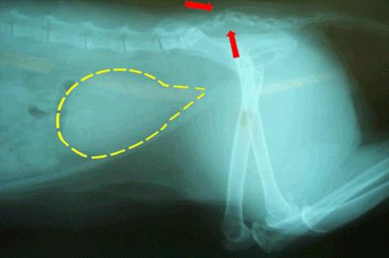
A radiograph will often show a break in the tail or a dislocation, although in some cases the tail bones are intact. How permanent these signs are largely depends on whether the cauda equina nerves have been over-stretched or actually torn. 

Classification of the Injury

In a 1985 study by Smeak and Olmstead, sacrocaudal fractures in cats were divided into five groups.

Group One: Cats With Painful Tail Heads as their Only Sign
These individuals are minimally affected and, while they may or may not have a chronically sensitive tail head, they are expected to otherwise have a full recovery.

Group Two: Cats With Lack of Tail Mobility and Sensation as Their Only Sign
These cats are expected to recover tail function completely and have an excellent prognosis.

Group Three: Cats With Lack of Tail Mobility and Sensation Plus Urine Retention as Their Only Signs
Most cats will have a complete recovery.

Group Four: Cats With Lack of Tail Mobility and Sensation and Diminished Anal Tone
This group of cats has about a 75% recovery rate, meaning 75% of the cats that fit this description should recover.

Group Five: Cats With Lack of Tail Mobility and Sensation Plus a Dilated/Flaccid Anus (i.e., Zero Anal Tone)
This group of cats has about a 50% recovery rate, meaning about half of the cats with this description will eventually recover.

If it is not clear whether a cat has partial anal sphincter tone or not, it may be possible to have a test called an electromyogram. The muscles of the anal sphincter and tail can be tested to see if they are receiving any nerve input at all. If they are, there is reasonable potential for recovery. The EMG test is highly specialized and not readily available except at practices with a neurology specialty. Referral will probably be necessary.

In a more recent study (Nov 2009, Journal of Small Animal Practice) researchers found that an excellent predictor of bladder function return is pain detection at the tail head. In the 21 cats with sacrocaudal fracture/dislocation, all 11 cats that had pain sensation at the tail head on the first day after the injury had regained bladder function within 3 days. Absence of pain sensation in this area on the first day did not necessarily mean that bladder control would not return; 60% of cats without tail head sensation on the first day had recovered bladder control by 30 days after the injury.

Should the Tail be Amputated?

If the tail is not expected to recover mobility or sensation, there are some reasons to consider amputation. If the cat cannot lift his tail, he may soil it regularly, creating an infection issue or simply an odor issue. Further, the weight of the tail dragging may further stretch the cauda equina nerves. Whether or not tail amputation is helpful or speeds recovery is controversial but certainly amputation could solve a soiling problem.

Bladder Management

It is important not to allow the bladder to over-stretch. The fine muscles of the bladder can become so stretched out that even if the nerves recover, the bladder may still remain unable to empty fully. This means the cat’s bladder must be gently squeezed three or four times daily to keep it from over-distending. Medications can also be used to assist the bladder’s own ability to contract (bethanechol chloride) or to relax the urinary sphincter (prazosin or phenoxybenzamine).  Since urine retention tends to promote infection, patients will periodically require some sort of monitoring urine tests. If constipation is a problem, periodic enemas or stool softening medications can be given regularly.

Recovery Time

Nerves heal notoriously slowly. It has been said that a good six months must pass before one can say the maximum recovery has occurred and no more positive progress can be expected. Most cats who are going to recover function do so in a one-week period, and most cats who do not recover urinary control after a month probably will remain incontinent. Tail function and sensation tends to take longer.

4128626

Tooth Resorption in Cats

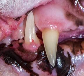
A common feline oral malady is tooth resorption (TR). Greater than half of all cats older than three years old will have at least one tooth affected by resorption; it affects dogs less frequently.   These tooth defects have been called cavities, neck lesions, external or internal root resorptions, feline odontoclastic resorption lesions (FORLs), and cervical line erosions.

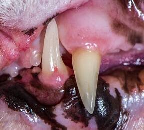
Tooth resorptions are usually found on the outside of the tooth where the gingiva (gum) meets the dental hard tissue. The lower jaw premolars are mostly affected, however tooth resorption can be found on any tooth. 

The cause is unknown, but theories supporting an autoimmune response, calicivirus, and metabolic imbalances relating to calcium regulation have been proposed.  The resorption generally starts at the cemento enamel junction (gum line) and progresses inward, eroding sensitive dentin. Once the resorption is exposed to the oral cavity, bacteria invade causing pain and jaw spasms. Increased salivation, oral bleeding, or difficulty eating are other signs. Unfortunately, most times there are no outward signs. It is up to the veterinarian or astute owner to diagnose tooth resorption.

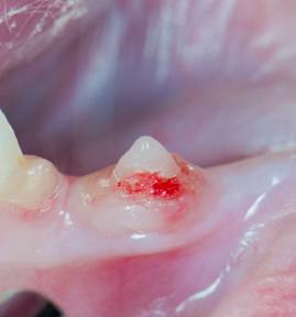
There are five recognized stages and three types of tooth resorption. Initially in stage 1, only an enamel defect is noted. The lesion is usually minimally sensitive because it has not entered the dentin. In stage 2, the lesion penetrates enamel and dentin. When resorption progresses into the pulp chamber (nerve) stage 3 has occurred.  In stage 4, large amounts of the tooth’s hard structure have been destroyed. By the time stage 5 has occurred, most of the tooth has been resorbed, leaving only a bump covered by gum tissue.

Intraoral radiographs (X-rays) are essential to evaluate all the teeth to determine the best course of therapy. Depending on what is seen on the visual patient exam and the intraoral X-rays (tooth resorption type 1, 2, 3), treatment involves following the resorption until it is exposed to the oral cavity, extraction of the entire tooth and roots, or a partial tooth extraction.  In cats affected by stage 5 without inflammation, treatment is not necessary.

3096810

Tritrichomoniasis in Cats  

Tritrichomoniasis is a major cause of diarrhea in cats and is caused by Tritrichomonas blagburni. The disease is seen most commonly in young cats and kittens.

T. blagburni is a type of protozoa, which are microscopic, single-celled parasites. They are not related to bacteria or viruses. T. blagburni is transmitted to cats when they come into contact with the organism directly or accidentally eat the infected feces of another cat. Infections are common in crowded environments such as catteries or animal shelters. Thankully, T. blagburni is not contagious to humans or other types of animals, so only cats get it.

Symptoms

Chronic (i.e. long term), voluminous, watery diarrhea is the most common symptom. Diarrhea can come and go so that the cat seems fine sometimes, which can help delay a diagnosis. Cats can also experience excess gas, going more often, foul odor to the stool (we mean more than usual!), and/or trouble defecating.

In general, cats infected with T. blagburni are otherwise normal, have a healthy weight and appetite, and are well hydrated. Sometimes, cats can become dehydrated or develop a poor appetite if the diarrhea is severe or excessive.

What to Expect at a Veterinary Visit

If you suspect your cat has tritrichomoniasis, bring some fresh feces to the veterinary appointment with you. Sandwich-sized plastic zippered bags are great for carrying the diarrhea sample.

Your veterinarian will start with a thorough physical examination, and then check for signs of dehydration and tummy pain. There will likely be an examination of your cat’s feces for intestinal parasites or increased levels of bacteria by looking at the feces under a microscope. Some cats are infected with multiple types of parasites at one time, so it is important that all issues be diagnosed and treated properly.

Unfortunately, T. blagburni is difficult to identify just by looking at the feces. Laboratory tests may be needed and/or blood tests to get an accurate diagnosis. Occasionally, a biopsy may be needed, but this is not common.

Treatment

Medications are available to treat tritrichomoniasis, but they are not always effective.  In some cats, especially in those less than one year of age, infections will improve without specific treatment. However, this can take several months.

Supportive care is important to keep your cat healthy during the infection. Feline diets high in fiber can help firm up the stools. Probiotics may also be useful to improve and maintain normal gut/GI flora. Any additional intestinal parasitic diseases need to be treated appropriately as well.

Control and Prevention

Keep your cat’s environment clean and free of feces. Minimize stress where possible by providing plenty of exercise and individual resting spaces. Scoop litterboxes daily and disinfect them regularly. All cats in the household should be tested for tritrichomoniasis because some cats may be infected without showing symptoms. Uninfected cats can be separated from the infected ones to prevent the disease from spreading, although that’s difficult in a home setting.

8226812

Hairballs (Trichobezoars) In Cats

“He’s throwing up hairballs all the time. I’m tired of stepping on slimy hair.”

To the client, the problem may be simple.

But to the veterinarian, it’s much more complicated.

When cats groom themselves and their feline friends, hair gets caught on the barbs of the tongue. That hair then is swallowed. In a “healthy” cat, that hair will go through the stomach and intestines (gastrointestinal tract) and come out in the feces.

So, if a cat is throwing up or regurgitating hairballs, there is more going on than most owners realize.

Grooming

Grooming allows cats to keep themselves clean. They remove old hair, surface dirt, and foreign bodies when they groom themselves and other cats.

But if a cat is long-haired, he has more hair to remove. (Mother Nature designed felines with short hair. Life is easier for wild cats when they have short hair.) So, long-haired cats who have to do all the grooming themselves — who aren’t being groomed (brushed) by their owners — are ingesting more hair than Mother Nature planned. The elimination of the extra hair may be too much for a healthy long-haired cat’s digestive system.

Also, if grooming becomes excessive, then more hair is entering the digestive tract, whether the cat is a short-haired or a long-haired cat.

Over-grooming can occur when a cat is stressed. Anxiety, emotional stress, etc., can cause a cat to groom himself more often and for longer times. It’s a coping mechanism, like the same way many children suck their thumbs to seek solace.

Over-grooming can also occur in cats experiencing pain. They may lick and groom the painful area to try to make it “feel better” and/or to reduce the stress. Urinary tract pain, musculoskeletal pain, and abdominal pain may cause over-grooming by the unhappy cat.

But hairballs also occur in animals who aren’t “putting too much hair” into their digestive tracts.

What Are Other Potential Causes?

  1. Intestinal motility problems
  2. Stress and stereotypic behaviors
  3. Skin diseases
  4. Abdominal problems
  5. Urinary tract problems
  6. Musculoskeletal problems

If the stomach and small intestine don’t allow the ingested hair to move through normally, the cat will produce hairballs.

If the cat has inflammatory bowel disease, hair – and food – won’t pass through it at a normal speed, or at all. The stomach and intestinal tissue is damaged and doesn’t provide normal motility. It also doesn’t produce the normal digestive enzymes or lubricants that allow the intestinal contents to move along.

The same is true of gastrointestinal cancer. The contents of the tract (hair and food) don’t move along the way they should.

To find the more specific causes of hairballs, your veterinarian may need to explore various body systems and consider conditions that may lead to hairballs. This may include megaesophagus, diaphragmatic hernia, gastrointestinal neoplasia, ileus, intestinal parasites, inflammatory bowel disease, pancreatitis, gallbladder disease, cystitis, bladder stones, kidney stones, arthritis, boredom, frustration, skin parasites, fungal skin diseases, and more.

This is not quite as simple as the problem the client discussed.

Diagnostic Tests

Your veterinarian may recommend tests to look for causes of hairballs including a complete blood count, blood chemical analysis, fecal examination, urinalysis, ultrasonography, radiography, contrast radiography, skin cytology, histopathology, behavior evaluation, etc.

Finding and treating the primary problem is always preferable to just treating the symptom (hairballs). The cat and the client will be happier if everything can be put back to normal. In addition to treating the inciting problem, the veterinarian may recommend diet changes, more brushing by the owner, more play time with the owner, etc.

With diagnostic skills and a little luck, the veterinarian can make the owner happier and the cat healthier and more content.

7486367

Rat Poison’s Effect on Dogs and Cats

The EPA has been at odds with Reckitt Benckiser (the makers of d-CON) since 1999 when they began seriously scrutinizing the risks of rat poison products to pets, children, and wildlife. At last, Reckitt Benckiser has aggreed to comply with the EPA directives. This means that second-generation rat poisons (which kill rats in one feeding) will be banned from residential use and that rat poisons for residential use must be secured within bait stations (not in open trays). The traditional d-CON products have not been manufactured since December 2014 and have not been distributed since March 15, 2015. Bromethalin-based rat poisons have since captured the residential market. Commercial exterminators may still use the anticoagulant rodenticides discussed here.

Mankind and the rat have been in conflict for thousands of years. Rats spread disease, eat our crops, leave droppings and make nests in our storage areas, and infest our homes. Rodent removal services are an important part of pest extermination even in this century. Rat poison can be obtained in most hardware stores, grocery stores, and even for free from city agencies in some areas. While you may want to get rid of rats, you certainly do not want to pose a hazard to the children or pets of the family.

Research continues to create a product that fits this bill but in the meantime be aware of the signs of rat poisoning, particularly if your pet travels with you to places outside the home where bait may be left out.

There are several types of rodenticides available. The traditional products are called anticoagulant rodenticides and are discussed here. If you intend to use a rodenticide, we encourage you to choose this type over others as there is a readily available antidote for the anticoagulant rodenticides. Other rodenticides are more toxic and no antidote is available. You should know what product, if any, you use at home. Common anticoagulant rodenticides are: brodifacoum, dopaquinone, warfarin, bromadiolone, and others.

Most of these products include green dyes for a characteristic appearance; however, dogs and cats have poor color vision and to them these pellets may look like kibbled pet food.

Anticoagulant rodenticides do not produce signs of poisoning for several days after the toxic dose has been consumed. Anticoagulant rodenticides cause internal bleeding. A poisoning victim will show weakness and pallor but bleeding will likely not be obvious externally.

Symptoms

Most of the time external bleeding is not obvious and you only notice the pet is weak and/or cold. If you look at the gums, they are pale. Sometimes bloody urine or stool is evident or nose bleeds may be seen. Signs of bleeding in more than one body location are a good hint that there is a problem with blood coagulation and appropriate testing and treatment can be started.

How Does Rat Poison Work?

You would expect the signs of a poisoning to be evident quickly but anticoagulant rat poisoning signs require at least 5 days to show up. Furthermore, unlike most poisons where treatment involves managing symptoms until the poison is out of the system, there is a true antidote for anticoagulant rat poison and it is actually a vitamin. How can this be? To understand what these poisons do, it is necessary to have some understanding of how blood clots.

A blood vessel is sort of like a pipe carrying rapidly flowing blood along its path. The pipe is lined by smooth flat cells called endothelial cells that facilitate the smooth flow of blood. If the pipe breaks, the structure of the pipe below the lining is exposed to the flowing blood inside.

From there the sequence of events is as follows:

  1. The blood vessel automatically constricts and spasms. This restricts the blood flowing to the damaged area and helps minimize blood loss.
  2. The exposed pipe attracts circulating platelets, cloud-like cells that circulate ready to assist in clotting should the need arise. Platelets clump together over the tear in the blood vessel, forming a plug within the first five minutes of the injury. This is all a good thing but the platelets will not stay in place unless a substance called fibrin can be made to bind them.
  3. Generating fibrin is complicated and beyond the scope of this article, but a cascade of activating proteins is needed to make the tiny protein threads (fibrin) that bind the platelets and makes a permanent platelet plug on the wound. Four of the proteins involved are called serine proteases, and these are the factors relevant to anticoagulant rat poisoning. These four factors must be able to work or there will be no fibrin, the platelets will not clump properly, and bleeding will continue without clotting.

About those Four Clotting Factors

The four clotting factor proteins are also called the K-factors because they depend on vitamin K for activation. After the clotting situation is under control, the used Vitamin K is recycled in the liver so it will be ready for the next time bleeding needs to be stopped. Anticoagulant rat poisons interfere with Vitamin K recycling. It takes several days to deplete the Vitamin K but after that is used up, there is no more and bleeding cannot be stopped. That is why it takes some 5 days for symptoms to show up and how it is possible to reverse this poison by giving more Vitamin K.

As long as there is plenty of vitamin K, the serine proteases can be activated and clotting can proceed normally. The anticoagulant rodenticides abolish vitamin K recycling. This means that as soon as the body’s active vitamin K reserves are depleted there can be no meaningful blood clotting.

In cases of poisoning you would expect symptoms to be nearly immediate but in the case of anticoagulant rodenticide poisoning, it takes several days to deplete vitamin K. After that, even the smallest of jostles and traumas can lead to life-threatening bleeds.

Testing

Testing can be done using clotting tests called a PTT (partial thromboplastin time) and PT (prothrombin time).  These tests evaluate the activity of the four K-factors. If both these tests are abnormal, there is an excellent chance that anticoagulant rat poison is in the patient’s system. The PT test in particular bears special mention as it tests the least stable of the K-factors called Factor VII. This means that the PT test becomes abnormal before the PTT test becomes abnormal. When it’s time to monitor a patient for recovery, the PT test must be normal in order to declare the poisoning resolved.

Another test called the PIVKA (Proteins Induced by Vitamin K Antagonism) test is more specific. The PIVKA test detects inactive serine proteases. An unusually high amount of inactive K-factors circulating indicates something is wrong with Vitamin K recycling.

Therapy

If the patient has only just ingested the poison, he or she may be made to vomit it up. Cathartics and adsorbents can be used to prevent the poison from entering the patient’s system. Still, it is best to use the antidote anyway. Certainly, if there is evidence that the patient is bleeding, the antidote obviously is required.

The antidote is simply vitamin K.

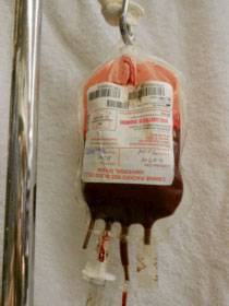
Vitamin K is generally started as an injection, and when the patient is stable, tablets are prescribed. The human formulation, available as a prescription drug at most drug stores, is a 5 mg tablet. The veterinary strength is a 25 mg tablet. Blood transfusions may be needed to stabilize a patient who has suffered significant blood loss.

There are different classes of anticoagulant rodenticides, and they remain in the body for several weeks. It is hard to know when to discontinue therapy, especially if the particular rodenticide is not known. After a couple of weeks of therapy, the medication is discontinued. Forty-eight hours later, a PT test is run. If there is still rodenticide in the patient’s system, the PT will be abnormal, but the patient will not yet have started to bleed. The results of the PT test will tell the veterinarian whether or not another couple of weeks of vitamin K are needed.

It is important to return for the recheck PT test on schedule. Waiting an extra day or two will allow internal bleeding to recur. There is no point in doing the PT test while the patient is still taking vitamin K. The test must be done 48 hours after discontinuing the medication.

When the PT test has returned to normal, it is safe to discontinue therapy.

Vitamin K1 Vs. Vitamin K2 Vs. Vitamin K3

There are three forms of vitamin K but only vitamin K1 is used therapeutically. Vitamin K1 is a natural form of vitamin K that is found in plants and absorbed nutritionally. Its more technical name is phylloquinone. Vitamin K2 (menaquinone) is also natural and is produced by the body’s intestinal bacteria but apparently not in amounts adequate for rescue from the anticoagulant rodenticides. Vitamin K3 (menadione) is a synthetic version that may be injected or taken orally. You may even see it available as a vitamin supplement tablet.

Within the body vitamin K1 and vitamin K3 are converted to vitamin K2. Vitamin K3 might seem like an inexpensive way to treat a pet with rat poisoning but unfortunately, K3 is sometimes toxic and can actually lead to red blood cell destruction. Inexpensive vitamin K3 pills on the drugstore shelf for over-the-counter sale are not acceptable antidotes. Vitamin K1 is used because it is absorbed early in the GI tract and concentrates directly in the liver, which is where the K-factors are activated.

It is only vitamin K1 that should be considered to be the antidote for anticoagulant rodenticide poisoning.

Other Rodenticides

While anticoagulant rodenticide poisoning is a life-threatening event, at least there is an antidote readily available. Other rodenticides are not as readily reversed. These rodenticides include:

  • Quintox, Rat-B-Gone,  Bromethalin (see an additional article)
  • Strychnine, gopher bait
  • Zinc phosphide, gopher bait 

Pets Who Eat a Poisoned Rodent

We are commonly asked about the risk to a cat or dog who eats a rodent that has been poisoned with an anticoagulant rodenticide. The rodent might have already died or simply not have died yet given that several days are required to feel the effect of these poisons. The fact is that when you’re talking about the newer generation anticoagulant rat poisons, such as diphacinone, the risk is real. A greedy rat can eat enough poison to kill 20 rats before he starts to feel sick, and if this was a second generation rodenticide it will accumulate in the rat’s liver ready to poison the cat that eats the rat’s liver. Fortunately, second-generation rodenticides have been banned for residential use. First-generation rodenticides are no longer in the rat’s body after several hours, making pet poisoning less of a concern. Furthermore, most rats do not overindulge in poison. The usual patient for secondary poisoning is a pet or predator that depends heavily on rats for food (a barn cat, for example). There is some controversy over how often this actually happens, as most pets do not consume numerous rats.

4128299

Neuropathic Pain in Dogs and Cats

Neuropathic pain is an abnormal painful response. It is caused by injury to or disease of the nerves, spinal cord or parts of the brain that are involved in receiving, conducting and interpreting painful stimuli. 

Both physical and chemical changes within the spinal nerves that convey pain contribute to neuropathic pain.

Such changes include: an increase in the chemicals and receptors that transmit signals between nerves; an imbalance between pain signals coming into the brain and the brain suppressing those signals; and a change that makes nerves that don’t normally relay pain signals into ones that do.

There are different types of neuropathic pain.  An exaggerated response to a painful stimulus, such as a pin prick, is called hyperalgesia.  Pain from a normal stimulus that shouldn’t be painful at all, such as a light touch, is called allodynia. Hyperpathia is increasing pain after repetitive stimulation, which includes continued pain after the stimulus is gone, and pain that radiates to adjacent areas.

People who experience  neuropathic pain describe it as “a pins and needles sensation,” tingling, burning, itching, numbness or cold, and sometimes feeling as if they had received a small electric shock.  Grades of neuropathic pain that have been established in human medicine are definite, probable and possible.

Pets cannot describe their pain to us but we believe it’s likely to be like what people have.  Neuroma formation in horses is known to cause definite neuropathic pain.  A neuroma consists of an abnormal sprouting of nerve endings from a severed nerve, and affected horses develop lameness or an abnormal gait. 

An example of probable neuropathic pain in dogs is the abnormal scratching behavior that is seen in King Charles Cavalier Spaniels with a spinal cord abnormality called syringomyelia. Cats with hyperesthesia syndrome incessantly lick and attack their back, and become extremely painful when petted. Neuropathic pain may be one of the causes for hyperesthesia syndrome.

Diagnosis

Diagnosing neuropathic pain in animals involves ruling out other painful conditions, such as osteoarthritis and bone cancer, as well a thorough neurological workup. A neurological examination evaluates the different parts of the nervous system (brain, spinal cord, nerves); if those parts are not normal, further diagnostic tests are indicated.  Laboratory tests, radiographs, CT or MRI imaging, and in some cases electrophysiologic testing may be recommended to look for diseases that are known to cause neurological injury and secondary pain.  Examples of such diseases include: diabetes mellitus, intervertebral disk herniation, spinal cord or nerve sheath tumors, cervical malformation, and syringomyelia.

Treatment

In some cases treatment is started early in conditions that are normally expected to be painful to try to prevent neuropathic pain from developing.  In other cases, neuropathic pain may be the first sign.

Combining drugs that reduce pain in different ways has proven to work better than using a single drug alone.  Depending upon the species, one or more drugs may be prescribed by a veterinarian.

Gabapentin, pregabalin, dextromethorphan, and amantadine are drugs that act on the relay centers for pain within the spinal cord. Amitriptyline acts both on the brain and within the spinal cord. The antibiotic minocycline has shown potential for modifying neuropathic pain.

These agents may be combined with one or more of the traditional analgesic drugs: tramadol and codeine, narcotic drugs that modulate the brain’s perception of pain; non-steroidal antiinflammatory drugs (NSAIDs) such as carprofen, deracoxib, and firocoxib; local anesthetics such as lidocaine and alpha-2 agonists; drugs such as medetomidine that modify pain, heart rate and blood pressure.

Some veterinarians have found success using combinations of herbs, diet, exercise, and acupuncture.

It is important to work closely with your veterinarian to manage neuropathic pain. Balancing the benefit of multiple medications against side effects can be challenging. Discussing the perceived effects of medications with your veterinarian is essential for the best outcome for your pet.

info-box-img-6

Ovarian Remnant Syndrome in Dogs and Cats

Whether the patient is a dog or a cat, ovarian remnant syndrome is suspected when a spayed female pet appears to be coming into heat, something that should be impossible after spaying. A spayed female has had both her ovaries removed and should not cycle and should not display behaviors typical of heat. If she is displaying signs of an active heat cycle, a search for an estrogen source is required. In particular, we need to rule out the presence of a piece of ovarian tissue left inside. If a piece of ovary (or similarly active tissue) is indeed inside, the only treatment is surgically removing it, and since this is an invasive solution, we want to be as certain as possible that there is no other likely explanation.

The Girl Dog in Heat

A female dog comes into heat every 6 to 8 months on average. During this time, the most prominent feature is the vaginal discharge which begins bloody and switches to a more golden straw color. The girl dog in heat will attract males and will most likely flirt playfully with them. This period of hormonal activity lasts about 3 weeks and culminates in a false pregnancy (or potentially a real one if she has been bred). The false pregnancy period lasts roughly a couple of months.

Drawing of a dog

The Girl Cat in Heat

A female cat in heat does not have an apparent vaginal discharge. For her, the signs of heat are more behavioral. She becomes restless and loudly vocal. She may urinate in the house in an attempt to notify local tomcats of her state. She is often unusually affectionate and has a tendency to stick her rear in the air and even tread her feet when she is petted over her back.

A female cat cycles seasonally, when the days are longer, and tends to stop cycling when the days begin to get shorter. She is in heat for five days or so and then out of heat for a couple of weeks, then back in, then back out over and over until she is either bred or the seasons change. If she is bred, she will ovulate and go into a false pregnancy or a real one if the breeding was successful. In any case, it is the behavior that is the clue that her hormones are active, especially since the occasional female cat will not follow the usual rules about the seasons.

Signs of heat (also referred to as estrus) imply estrogen in the pet’s system. The first step is to confirm that there genuinely are estrogens in her system.

Testing for Hormones

Vaginal Smear

You might expect a blood test to be necessary to look for estrogen or progesterone, but in fact, the best test is a vaginal smear test, at least for dogs. Using a cotton swab, the veterinarian harvests some cells from the vaginal wall and looks at them under the microscope. If the female is in heat at the time of the test, the cells will have a characteristic appearance called cornification. This means there are estrogens in her body.

Blood Testing

Testing for estradiol, one of the estrogen hormones, can be done but is not as accurate as the vaginal smear test. If the spayed female has high estrogen levels during the time she appears to be in heat, this would be considered a positive test. The problem is that low levels can still be present in the bloodstream even if there is an ovarian remnant, so low levels are hard to interpret.

Another approach is to give her a shot of GnRH (gonadotropin releasing hormone) while she appears NOT to be in heat. If she responds to the injection by producing progesterone two weeks later (detectable with a blood test), this would be consistent with an ovarian remnant.

The vaginal smear costs a fraction of the amount of blood testing and can generally be done in a matter of minutes in the doctor’s office, so these types of blood tests are rarely performed.

Witness LH and SpayChek® Testing

There is an in-house test kit for luteinizing hormone (LH) that can determine if a female cat or dog has been spayed or not, provided she is not experiencing a heat at the time of the test. This test could be performed to assist in diagnosis but would have to be done during a time without symptoms of heat. A low LH level is compatible with the presence of estrogen and presumably ovarian tissue.

The newer SpayChek® test for anti-mullerian hormone is reportedly more reliable but requires that at least two to three months have passed since the original spay surgery for accuracy. It is best performed in conjunction with a progesterone level for the best accuracy.

Your veterinarian may suggest more than one type of test to be sure there is little room for ambiguity. After it is clear that estrogens are definitely present, the next question to address is, “Where did they come from?”

Where Did they Come from?

Estrogen Creams for Human Use

Topical estrogen-containing creams have numerous human uses. These creams invariably end up on the user’s hands or arms, where a loving pet can lick them. A female pet exposed to a hormone crème in this way can easily manifest signs of heat but will not show a hormone cycle in any predictable way. Generally, people are aware that hormone crèmes are in use at home and can be more careful about avoiding exposure to pets. Creams may be applied using gloves that can be discarded away from the pet, hands can be washed carefully, etc.

Did Something go Wrong with the Original Surgery?

One would think that if there is a remnant of ovarian tissue in the spayed pet’s abdomen, the surgeon must have left it behind. This is not necessarily so. It appears that some females possess accessory ovarian tissue separate from the main ovary, and this tissue becomes active only after the main ovaries are removed. Some cats actually grow invisible ovarian tissue down the length of their ovarian ligament, which is cut during the spay surgery but not fully removed. Furthermore, if an ovary contacts the abdominal wall, cells appear able to adhere and vascularize effectively, creating a new, albeit small, ovary. In many cases, the

Gloved hands removing feline ovary

original spay surgery was performed months or even years before. These secondary bits of ovary are growing in this time and do not show themselves until they have achieved sufficient hormone-producing power. Once it has been determined that a pet has an ovarian remnant, surgery should be performed to locate and remove it.

It is worth mentioning that adrenal tumors can produce hormones. These can be difficult, if not impossible, to remove, but exploratory surgery should at least identify them over an ovarian remnant. In the situation of this type of tumor, signs of heat are constant; they do not cycle as with ovarian tissue.

Treatment

The ovarian tissue will be most prominent or visible just following the active heat period. This is the time after ovulation when the ovary is normally ripe with large progesterone-secreting structures called corpora lutea, and this is the best time for surgical exploration. If errant ovarian tissue is found, it can be removed generally without difficulty. It is not unheard of, however, for the ovarian remnant or remnants to be impossible to find. If that is the case with your pet, Discuss with your veterinarian whether referral to a surgical specialist would be best for you and your pet; however, a surgery specialist may be necessary to find the remnants. Biopsy of the removed tissue should settle the question as to whether the hormone source was removed intact or not.

Why Do We Need to do Anything at all?

The problem with all this is not limited to the inconvenience of hormone-related behaviors and discharges. The actual problem is that hormones can cause harm. Long-term exposure to female hormones can promote mammary cancer. If a small piece of uterus persists after spaying, the influence of female hormones can promote chronic infection (“stump pyometra”). While neither of these outcomes is definite, their repercussions are serious, and it is best to take these hormones out of play.

What if it isn’t Heat?

Female dogs can have a bloody vaginal discharge for reasons other than heat. It is possible the owner is seeing bloody urine, for example, and the medical workup will need to shift to address the different causes of this problem. Alternatively, a transmissible venereal tumor, urinary tract infection, or vaginal injury might lead to bleeding.

As for a female cat, she may simply have behavioral issues to address.


4128901

Giardia in Pets

What are Giardia?

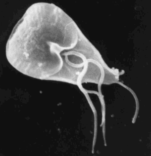
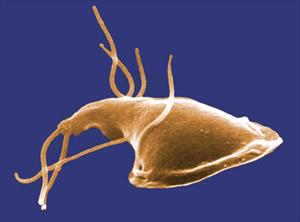
Giardia are single-celled organisms, infectious to many types of animals (including humans) all over the world. As you can see in the above image, Giardia organisms have little whip-like tentacles called flagella that classify them as flagellates. They use their flagella to move around from place to place, but when they find a spot where they wish to stay (like a cozy nook in the host’s intestine), they use a suction cup-like structure (visible in the image) to attach.

Their presence in the host intestine can cause diarrhea, though some hosts are symptom-free carriers. Different types of Giardia infect different types of animals; it is rare for Giardia from a pet to transmit to a human; furthermore, dog and cat Giardia species are separate and are unlikely to cross from dog to cat or vice versa.

Giardia have two forms: the trophozoite and the cyst. The trophozoite is the form that lives within the host, swimming around and attaching with its suction cup. The cyst, however, is the form that lives out in the environment. Trophozoites round up to cysts as they approach the colon and then are passed in feces. Trophozoites that don’t round up into cysts and form a shell prior to passing into the cold, cruel world cannot withstand the temperature/moisture variability of the outside world. Cysts are the contagious stage. Trophozoites are in the parasitic stage.

Life as a Giardia Organism

As mentioned, trophozoites and cysts may be passed in fresh feces, but only the hard-shelled little cysts can withstand the conditions of the outside world. The cysts live in the environment (outside the host’s body) potentially for months until they are consumed by a host. Inside the host, the cyst’s shell is digested away, releasing two trophozoites into the intestine, and the cycle begins again. Contaminated water is the classical source of a Giardia infection.

When a fecal sample is analyzed, the appearance of the Giardia organism depends on whether the sample is freshly obtained or if it has been outside of the host’s body for a while. Giardia organisms begin to round up into cysts in a matter of hours. The active trophozoites rather look like funny faces with the two nuclei forming the eyes and median bodies forming the mouth. Cysts look a bit more generic.

In the environment, cysts survive in water and soil as long as it is relatively cool and wet. A host animal will accidentally swallow a cyst when drinking from a puddle, or toilet, or when licking fur. After the cyst has been swallowed, the cyst’s shell is digested away, freeing the two trophozoites that go forth and attach to the intestinal lining.

As mentioned, the trophozoite will swim to a spot using its flagella and attach with its suction cup (more correctly called its “ventral disc”). Trophozoites tend to live in different intestinal areas in different host species but will move to other areas depending on the diet the host is eating. The trophozoite may round itself up and form a cyst while still inside the host’s body. If the host has diarrhea, both trophozoites and cysts may be shed in diarrhea; either form can be found in fresh stool.

After infection, it takes 5 to 12 days in dogs or 5 to 16 days in cats for Giardia to be found in the host’s stool. Diarrhea can precede the shedding of the Giardia. Infection is more common in kennel situations where animals are housed in groups.

How Does Giardia Cause Diarrhea?

No one is completely sure, but infection seems to cause problems with normal intestinal absorption of vitamins and other nutrients. Diarrhea is generally not bloody with a Giardia infection. Immune-suppressive medications, such as corticosteroids, can re-activate an old Giardia infection. We do not know why some infected hosts get diarrhea while others never do.

Diagnosis

In the past, diagnosis was difficult. The stool sample being examined needed to be fresh, plus Giardia rarely show up on the usual fecal testing methods used to detect other parasites. Several tricks have been developed to make Giardia easier to find (special stains, using special processing solutions, etc.), but what has made the biggest difference in the diagnosis of Giardia is the ELISA test kit, which is similar in format to a home pregnancy test. This method has dramatically improved the ability to detect Giardia infections and the test can be completed in just a few minutes while you wait.

Giardia shed organisms intermittently and may be difficult to detect. Sometimes pets must be retested in order to find an infection, and asymptomatic carrier animals are common. It should also be mentioned that the ELISA test can remain positive for some time after the infection has been eradicated so if re-testing is desired after a positive test, another test format may be more helpful than the ELISA test kit.

Treatment

A broad-spectrum dewormer called fenbendazole (Panacur®) seems to be the most reliable treatment at this time. Metronidazole (Flagyl®) has been a classical treatment for Giardia but studies show it to only be effective in 67% of cases. For some resistant cases, both medications are used concurrently. Febantel is also commonly used for Giardia as it is converted to fenbendazole in the body.

Because cysts can stick to the fur of the infected patient and be a source for re-infection, the positive animal should receive a bath at least once in the course of treatment. 

At the least, the patient should have a bath at the end of treatment, plus it is especially important to promptly remove infected fecal matter to minimize environmental contamination.

Can Humans Be Infected?

The short answer is only rarely, so the concern is pretty low in general. However, maintain good hygiene practices such as regular hand-washing and removing fresh pet fecal matter promptly, as mentioned.

That said, here is a more detailed answer: Giardia duodenalis is classified into several subcategories called assemblages and designated A through G. Some assemblages are specific as to which host animals they can infect, and other assemblages are not so picky. Assemblage F, for example, only infects cats, and assemblages C and D only infect dogs but assemblage A will infect dogs, cats, people, rodents, wild mammals, and cattle. Common testing methods do not indicate what assemblage has been detected, so there is always a possibility of human transmission as long as the assemblage is unknown. 

Environmental Decontamination

Giardia cysts are killed in the environment by freezing temperatures and by direct sunlight. If neither of these is practical for the area to be disinfected, a chemical disinfectant will be needed. Organic matter such as dirt or stool is protective of the cyst, so on a concrete surface, do basic cleaning before disinfecting. Quaternary ammonia compounds can be used to kill Giardia cysts.

Animals should be thoroughly bathed before being reintroduced into a clean area. A properly chlorinated swimming pool should not be able to become contaminated. As for areas with lawns or plants, decontamination will not be possible without killing the plants and allowing the area to dry out in direct sunlight.

A Footnote on Vaccination

A vaccine against Giardia was previously available, not to prevent infection in a vaccinated pet but to reduce the shedding of cysts by the vaccinated patient. In other words, the vaccine was designed to reduce the contamination of a kennel where Giardia was expected to be a problem. This would be helpful during an outbreak in a shelter or rescue situation but is not particularly helpful to the average dog whose owner wants to simply prevent infection. Because of the limited usefulness of the vaccine, manufacturing was discontinued in 2009.

In Summary:

  1. Giardia is a parasite that sticks to its host’s small intestine to feed.
  2. It has two forms: one that lives in the environment (cyst) and one that lives in the host (trophozoite).
  3. Cysts are the contagious stage. Trophozoites are in the parasitic stage.
  4. Transmission is by the fecal-oral route from infected stool or contaminated water.
  5. After infection, it takes 5 to 12 days in dogs or 5 to 16 days in cats to be found in the host’s stool.
  6. Infection is more common in kennels or shelters where animals are housed in groups.
  7. The parasite can cause diarrhea ranging from minor to severe.
  8. We do not know why some infected hosts get diarrhea while others never do.
  9. Giardia rarely transmits from a pet to a human or vice versa.
  10. Diagnosis uses a fresh fecal sample. Several tests may be needed as false negatives can occur.
  11. Treatment is dewormers and metronidazole (antibiotic).
  12. A low-residue, highly digestible diet may be beneficial until stools are firm.
  13. Because infective cysts can stick to fur, patients should receive a bath during and at the end of treatment.
  14. The environment has to be cleaned after a case of giardiasis because it is so contagious. Giardia can be killed with common disinfectants. Quaternary ammonia compounds are the most effective.
  15. Giardia cysts are killed in the environment by freezing temperatures and by direct sunlight.
  16. To kill Giardia on a concrete surface, do basic cleaning before disinfecting.
  17. Lawns and plants cannot be decontaminated without killing the plants and allowing them to dry out in direct sunlight.
  18. No vaccine is available.