Adrenal Hormones
The adrenal gland is so named because it is located just forward of the kidney (renal means kidney). The center of the gland is called the medulla and the outer area is the cortex. While both areas produce hormones, Addison’s disease concerns the hormones produced by the cortex; these hormones are called corticosteroids.
Corticosteroids are the hormones that enable us to adapt physiologically to stress. The glucocorticoids (such as cortisol and related synthetics, prednisone, dexamethasone, and numerous others) act on the mechanics of sugar, fat, and protein metabolism.
They gear the metabolism towards the preparation of burning, rather than storing, fuels so as to be ready for a fight or flight situation.
The mineralocorticoids (such as aldosterone and related synthetics, fludrocortisone acetate, and desoxycorticosterone pivalate) influence the electrolytes sodium and potassium.
You may wonder what electrolytes have to do with fight or flight but it is important to remember that where there is sodium (salt), water soon follows. Conserving sodium will pull water from other tissues and bring it into circulation ready to support blood pressure should a life-threatening bleed occur. The mineralocorticoid hormones instruct the kidney to retain sodium so that the circulatory changes of stress can be handled smoothly. As sodium is retained, potassium is lost in the exchange.
Corticosteroid hormones are needed to adapt to stressful situations and without these hormones, even small stresses could lead to physiologic disaster.
Hypoadrenocorticism (Addison’s Disease) is a Deficiency in Corticosteroid Hormones
In animals with Addison’s disease, there is a deficiency of corticosteroid hormones. There are several reasons why this might happen: the adrenal gland might be damaged or drugs may be involved. We usually never find out what caused an individual patient’s Addison’s disease but the good news is that we don’t need to know why it happened. Treatment is straightforward: we simply provide the hormones that the body is not making on its own.
Clinical Signs
Patients are usually young (age four to five years) but any age dog can be affected. (This disease can occur in cats but is very rare.) There is a genetic predisposition for Addison’s disease in the standard poodle and bearded collie, Nova Scotia Duck Tolling Retriever, and Portuguese water dog. Female dogs are affected twice as often as males.
At first, signs may be vague: listlessness, possibly some vomiting, or diarrhea. The dog just does not seem to feel right but not in an obvious way and may seem more or less normal most of the time as symptoms wax and wane with stress. This vague waxing and waning go on and on with the dog never really getting fully sick but never staying well either. Eventually, if the disease is not diagnosed, it can come to a head in a phenomenon known as an Addisonian crisis. The animal collapses in shock due to an inability to adapt to the caloric and circulatory requirements of stress. Blood sugar may drop dangerously low. Potassium levels can soar and disrupt the heart. Heart rate slows and arrhythmias result. The patient may not survive this episode.
About 30% of dogs with Addison’s disease are diagnosed at the time of an Addisonian crisis. Approximately 90% of the adrenal cortex must be non-functional before clinical signs are observed.
Making the Diagnosis
Because of the numerous symptoms Addison’s disease can present with, Addison’s disease has earned the medical nickname “The Great Imitator.” One would think that one could simply look for an increase in potassium and/or a drop in sodium on a basic laboratory blood panel, but it turns out spot checks of electrolyte values like this are not reliable enough for a diagnosis of Addison’s disease.
Shock
Veterinarians can be presented with a young animal in shock. There is usually no history of trauma or toxic exposure so general treatment for shock is initiated. This consists of rapid administration of fluids (usually Lactated Ringers solution which has little potassium and a moderate amount of sodium) plus some glucocorticoids. By coincidence, this also happens to be similar to the specific treatment for Addison’s disease so often the patient simply recovers without the veterinarian really knowing why.
Imitating Kidney Disease
The blood panel often comes back showing elevations in the renal parameters (BUN and creatinine), and, thus the elevated potassium is suggestive of acute renal failure, a condition with an extremely poor prognosis. The veterinarian may become suspicious of another diagnosis as the patient will respond well to fluid administration and most renal failure patients do not respond as well.
Low Blood Sugar
Addison’s disease may present in more unusual ways. The inability to maintain normal sugar levels (ultimately manifesting as a seizure disorder) may be strongly suggestive of an insulin-secreting pancreatic tumor but before major abdominal surgery is planned, it is important to test for Addison’s disease.
Megaesophagus or Recurring GI Disease
In a similarly unexpected way, regurgitation of undigested food due to abnormal nerve function in the esophagus (a condition called megaesophagus) can be caused ultimately by Addison’s disease. Megaesophagus has many causes but it is important not to forget Addison’s disease. Of course, Addison’s disease can also manifest with chronic waxing/waning diarrhea and/or poor appetite which would suggest a gastrointestinal problem such as inflammatory bowel disease. Before investing in the expense and potential anesthetic risk for endoscopy and intestinal biopsy, a screening test to rule out Addison’s disease is prudent.
Sorting it out with the ACTH Stimulation Test
Your veterinarian may use a resting cortisol test to rule out Addison’s but the only definitive test for Addison’s disease is the ACTH stimulation test. The patient receives a dose of ACTH, the pituitary hormone responsible for the release of corticosteroids in times of stress. A normal animal will show an elevation in cortisol in response to ACTH while an Addisonian has no corticosteroids to respond with. This lack of response is diagnostic for Addison’s disease; however, a false positive may be obtained if corticosteroids have been used in the treatment of the crisis prior to the test. Of all the commonly used corticosteroids, only dexamethasone does not interfere with the assay for cortisol; if any other steroid has been used, the test will not be valid for at least 24 hours.
Treatment
The most important aspect of treatment for hypoadrenocorticism is replacing the missing mineralocorticoid hormones. One way to do this is with oral fludrocortisone (brand name Florinef®). Fludrocortisone is given twice a day at a dose determined by the patient’s sodium and potassium blood tests. At first, these electrolytes are monitored weekly. When levels seem stable, these blood tests are repeated two to four times per year. Often with time, it will be found that the fludrocortisone dose needed will increase. This increase is unfortunate as the medication is relatively expensive. Since fludrocortisone has glucocorticoid activity as well as mineralocorticoid activity, only 50% of patients need to receive additional glucocorticoids. Using a compounding pharmacy or looking into the cost of generic fludrocortisone may be helpful in managing the costs of this particular medication, especially in a larger dog.
Another way to treat this condition is with an injectable medication called DOCP desoxycorticosterone pivalate, (brand names Percorten-V or Zycortal®). This treatment is given approximately every 25 days. Electrolytes are measured prior to injections at first but testing can usually eventually be tapered to once or twice a year. There is some feeling among experts that DOCP produces better regulation of electrolytes than oral Florinef. Dogs on DOCP, however, do require glucocorticoid supplementation (such as a low dose of prednisone).
What is Atypical Addison’s Disease?
Most dogs become Addisonian when they lose the ability to produce both mineralocorticoids and glucocorticoids. They need both types of hormones replaced. It turns out that there is a subset of Addisonian dogs that are able to control their sodium/potassium imbalance and only the glucocorticoids need to be supplemented. These patients have what is called atypical Addison’s disease. They can have all of the symptoms of typical Addison’s as described above (chronic relapsing diarrhea/appetite loss, low blood sugar crisis etc.) but not the electrolyte abnormalities. Treatment is supplementing with glucocorticoid hormones, such as prednisone.
Atypical Addison’s patients can progress to the typical form, which means they will eventually need more aggressive treatment with fludrocortisone tablets or DOCP injections. The current recommendation is to monitor sodium and potassium levels every couple of months in atypical patients for up to one year, to watch for the switch to the typical (and more serious) form of Addison’s disease. Becoming atypical Addison’s can still occur after one year. If an atypical Addisonian patient becomes ill, sodium and potassium should be checked to see if they have progressed to the typical form.
What is Secondary Addison’s Disease?
Addison’s disease is most often caused by the destruction of the adrenal glands. In some cases, the problem is a failure of the pituitary gland. ACTH is a hormone made by the pituitary that tells the adrenal glands to produce and release more glucocorticoids. Without ACTH, the adrenal glands atrophy and stop producing glucocorticoids. Measuring the hormone ACTH in the blood can distinguish adrenal and pituitary failure. This is not simply of academic interest. A patient with secondary Addison’s disease (i.e., pituitary failure) will never progress to typical Addison’s disease and does not require long-term, periodic monitoring of electrolytes.
What is Pacific Rimism?
Dog breeds originating in the Pacific Rim, such as the Akita and Shiba Inu, commonly have elevated potassium levels on blood tests. This can be confusing when a patient has symptoms that suggest Addison’s. These patients will have normal ACTH stimulation test results if they do not have Addison’s disease.
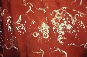
Whipworm Infection?
Whipworm infection has been known to create a syndrome nearly identical to Addisonian crisis, complete with abnormal sodium and potassium values. These patients will have normal ACTH stimulation tests but because whipworms only periodically shed eggs, fecal testing may not detect whipworm infection. If there is any question about whipworm infection, treatment should be instituted.