Biliary Mucocele is a Surgical Emergency in Dogs
It all starts with a sick, usually middle-aged or older dog. He is listless, not eating, and maybe has vomiting and/or diarrhea and fever. These are symptoms that could mean any number of things, but usually, when he reaches the veterinarian’s examination table, he has jaundice (yellow pigmentation visible in the whites of his eyes and possibly on his skin and gums). Hospitalization is recommended to rehydrate him and provide supportive care. Blood tests point to a liver problem. Medications are given to minimize the liver’s workload, but soon, an ultrasound is being discussed to image the liver, and costs may be rising quickly.
It all starts with a sick, usually middle-aged or older dog. He is listless, not eating, and maybe has vomiting and/or diarrhea and fever. These are symptoms that could mean any number of things, but usually, when he reaches the veterinarian’s examination table, he has jaundice (yellow pigmentation visible in the whites of his eyes and possibly on his skin and gums). Hospitalization is recommended to rehydrate him and provide supportive care. Blood tests point to a liver problem. Medications are given to minimize the liver’s workload, but soon, an ultrasound is being discussed to image the liver, and costs may be rising quickly.

Why Do We
Need an Ultrasound if We Already Know the Problem Is Liver Disease?
While blood testing can point to the liver, the fact is that numerous diseases can affect the liver. The liver can have an infection, cancer, scarring (cirrhosis), or any number of conditions. The more specific our diagnosis gets, the more specific treatment can be.
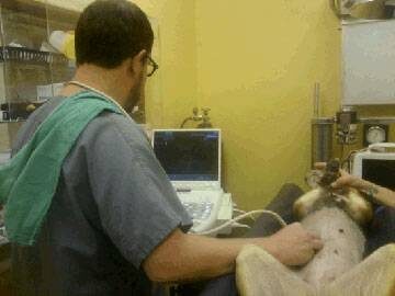
Ultrasound is a non-invasive way to evaluate the internal texture of the liver and gall bladder. By looking at the liver’s texture, it is possible to see a tumor and determine if removing it is possible or if it has invaded too far. Ultrasound can evaluate scarring and abscesses. Through ultrasound, it is possible to guide a biopsy needle to an exact area to sample tissue should this be deemed necessary. Ultrasound evaluates the gall bladder and bile ducts.

One of the more important diseases to rule out promptly is the biliary mucocele because it is commonly a surgical emergency. As we will describe in more detail, a biliary mucocele is a wad of sludged mucus inside the gall bladder. If you wait a few days to see how the patient responds to general liver support, it may be too late for surgery. Furthermore, ultrasound can evaluate the gall bladder’s integrity; if the mucocele ruptures and spills bile into the belly, the surgeon will need to know that as additional treatment is needed. Ultrasound also evaluates concurrent abdominal conditions that might impact recovery, including pancreatitis, which can occur alone with similar symptoms or in addition to a biliary mucocele.
If the patient has a surgical disease, they will not get better until surgery is performed in the vast majority of cases. Furthermore, it may be a surgical emergency (not only will surgery be needed but it will be needed immediately). The sooner the need for surgery is identified, the better the chance of survival.
What Is the Gall Bladder and What Is the Biliary System?
The liver serves as a toxic waste processing center for the body. It filters bacterial products (as well as nutrients) entering the body from the gastrointestinal tract and it removes toxic waste products from the bloodstream. This material is bound to special biochemicals called bile acids and the body would like to get rid of it, bile acids and all. The solution of bile acids, water, mucus, pigments, and cholesterol forms the greenish-yellow fluid we call bile.
Bile is made in the liver, and then collected into small ducts called bile ductules and bile ducts. The bile is then moved for storage into the greenish round organ called the gall bladder.
During food digestion, hormones cause the gall bladder to contract and squirt bile through the large common bile duct and into the intestine. The bile assists with digestion and carries toxins out of the body so they may be eliminated in feces. The gall bladder and its ducts represent the biliary system.
If the biliary tract becomes obstructed, the patient becomes rapidly jaundiced, painful, and sick.
What Is a Biliary Mucocele?
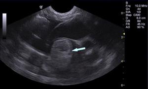
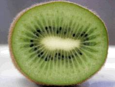
We mentioned that one of the components of bile is mucus. Normal bile is probably less than 3% mucus but when a mucocele develops, the bile becomes mostly mucus. Normal bile is liquid but mucocele bile is thick and goopy and will not flow easily through the common bile duct. The gall bladder distends trying to pass the mucocele bile and if it actually ruptures, the risk of serious complications and death is much higher. The gall bladder with a mucocele develops an appearance on ultrasound described as resembling the cut surface of kiwi fruit. The biliary mucocele is sometimes called a “kiwi gallbladder.”
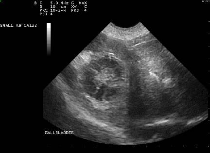
When this is seen on ultrasound in a sick, jaundiced patient, surgery to remove the diseased gall bladder should be performed as soon as possible.
Why Does This Happen?
Mucocele development starts with delayed gall bladder contraction, which allows bile acids to accumulate. Altered fat metabolism is also associated with reduced gall bladder contraction, resulting in an accumulation of bile acid. With that accumulation, the lining of the gall bladder feels inflamed and responds by making more mucus and more mucus-producing glands. Pretty soon the bile is just a big wad of congealed mucus and it will not flow.
Several hormone imbalances have been associated with altered fat metabolism and reduced gall bladder contraction: diabetes mellitus, hypothyroidism, and Cushing’s disease. Cushing’s disease patients have an incidence of biliary mucocele development that is 29 times the incidence of patients without Cushing’s disease. Mucocele development has been linked to pancreatitis (inflammation of the pancreas) as well.
Having Cushing’s Syndrome increases a dog’s risk of developing a biliary mucocele by 29 times.
No one knows why this might be so but it does correspond to the mainstreaming of diagnostic ultrasound into general practice.
The Shetland sheepdog, cocker spaniel, miniature schnauzer, and dachshund seem predisposed to developing biliary mucoceles.
Removing the Gall Bladder (Cholecystectomy)
Removing a dog’s gallbladder is a serious surgery that not all veterinarians are comfortable performing. Discuss with your veterinarian whether a referral to a specialist would be best for you and your pet.
The goal is to remove the gall bladder before it ruptures. If it has already ruptured, tissue damaged by the rupture must be cleansed or removed. Whether or not the gallbladder that has a mucocele is removed before or after rupture does not affect the rate of survival.
Mucocele surgery survival rate is 75-80%. However, if the gallbladder ruptures before surgery occurs, survival rate decreases significantly to 25-40%, depending on the study.
The gall bladder’s function is mainly one of bile storage. Without the gall bladder, bile simply dribbles into the intestine constantly rather than in controlled squirts. You might think this would be a problem, but it turns out not to be. Some patients require long-term medication for liver support but generally speaking, if the dog recovers from surgery, the prognosis is excellent, and the patient can return to normal life.
What if Surgery is Not an Option, or What if the Dog Isn’t that Sick?
There is no question that surgery is the best treatment choice, but there is more to the story. Biliary mucoceles can be an incidental finding in dogs who are not sick or who are sick from something else and are having an ultrasound of the belly for some other reason. In a patient that is not experiencing problems with a mucocele, medication may be able to stave off illness in some cases.
As for skipping surgery on a dog that is sick from its mucocele, this is a very risky move.
It is possible to attempt treatment with general liver support medications, a low-fat diet, and choleretics (medications to help liquefy bile, such as ursodiol), but the problem is that the gall bladder is obstructed with a big wad of goop. This goop is unlikely to liquefy in a timely fashion, if at all, no matter what we do. If medical management is attempted, it is important to regularly recheck the gall bladder by ultrasound to watch for any sign of progression that would indicate that surgery should no longer be postponed.