Tag: Prognosis

4128273

Cognitive Dysfunction Syndrome in Dogs  

What is Cognitive Dysfunction Syndrome?

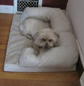
Cognitive dysfunction syndrome (CDS) is essentially the dog equivalent of Alzheimer’s disease. With CDS, a dog’s brain gradually degenerates, leading to abnormal and senile behaviors that reflect declining cognitive function. CDS is common in older dogs, generally occurring after 9 years of age.

CDS is caused by age-related changes to the brain. In dogs with CDS, a substance toxic to the brain called “beta-amyloid protein” accumulates. Other changes in the brain include reduced blood flow and dysfunctional neurons. Neurons are the cells that carry information throughout the brain and body. When neurons don’t function correctly, the brain’s ability to remember, process information and tell the body what to do is impaired.
What are the Signs?

The acronym “DISHAAL” can be used to describe the signs of CDS. It stands for: Disorientation, Abnormal Interactions, Sleep/wake cycle disturbances, House soiling, Activity changes, Anxiety and Learning/memory changes.

Signs of CDS an owner may recognize include:

  • Wandering
  • Anxiety
  • Confusion
  • Urinating/defecating in the house
  • Pacing, often at night
  • Less interaction with owners
  • Not recognizing familiar people, animals or commands
  • Less interest in eating, playing, walking and socializing
  • Restlessness
  • Waking up in the night; increased daytime sleeping
  • Inactivity
  • Increased vocalization, often at night
  • Going to unusual places
  • Can’t locate food dropped on the floor
  • Getting lost in familiar environment

How is it Diagnosed?

To diagnose CDS, a veterinarian will rely on information given by the owner, the dog’s symptoms and physical exam findings. To rule out other causes of the dog’s symptoms, the veterinarian may use additional tools such as blood and urine tests. An MRI may be done to look for abnormalities in the dog’s brain.

How is CDS Treated?

There is no cure for canine CDS. However, there are a number of treatments that may slow progression of the disease and relieve some of the dog’s symptoms.

Treatments for CDS include:

Dietary changes: Your dog may be put on a specific therapeutic diet designed to help. These diets contain ingredients such as antioxidants, fats and fatty acids that may protect and promote healthy brain cells.

Dietary supplements: Your veterinarian may recommend dietary supplements such as Senilife®, which is rich in antioxidants, or oils rich in a type of fat called “medium-chain triglycerides.” Medium-chain triglycerides provide energy to the dog’s brain, which is helpful because the brain is less able to use glucose for energy in CDS.

Drugs: Your veterinarian may recommend medications that could improve your dog’s cognitive function. These include MAO inhibitors such as Anipryl, which may help neurons communicate with each other and protect the brain from damage. Drugs such as propentofylline, which is licensed for use in some countries in Europe, increase blood flow in the brain and may help dogs with CDS.

Cognitive enrichment: Cognitive enrichment may improve your dog’s brain function. Cognitive enrichment consists of exercise, social interactions, providing new toys and teaching new commands to your dog.

Some veterinarians may suggest trying herbal therapies and acupuncture. These methods have the potential to help affected dogs, but they have not been well-studied in dogs with CDS.

What is the Prognosis for CDS?

There is no cure for canine CDS, so the disease will progress. However, if CDS is caught early and treated effectively, the dog could live a full, quality lifespan. Unfortunately, dogs with severe cases of CDS generally have a worse outcome, often being euthanized about 2 years after signs of CDS appear.

If you notice signs of CDS in your dog, it’s best not to just attribute them to old age; see your veterinarian.

36322

Cleft Palate or Lip in Puppies and Kittens

Congenital clefts in the palate or lip are birth defects that puppies and kittens can get in the tissues connecting parts of the mouth. Human babies also get them. These cleft defects happen when the roof of the mouth does not close correctly before the baby is born.

Cleft lip is a defect of the lips, underlying bone, and/or front area of the roof of the mouth, also known as a hard palate. Oftentimes, part of the lip will be missing or misshapen. Sometimes, one or both nostrils appear to connect directly to the mouth. Cleft lip may also be called a harelip because the face appears similar to that of a hare’s. 

Cleft palate is a malformation or disconnection of the tissues that form the roof of the mouth. The soft palate towards the back of the mouth can be involved. A hole may be in the palate because the tissues never fully connected to each other while the fetus was developing. Severe palate defects result in holes from the palate that connect directly to the nasal passages. 

The big problem with a cleft lip or palate is that it is difficult, if not impossible, for the affected puppy or kitten to latch on properly to a nipple and therefore they cannot eat properly. Depending on the severity of the cleft, the infant may not be able to get sufficient nourishment at an age where lack of nutrition can completely compromise the infant’s health and ability to grow.

Causes

Many conditions and issues can cause cleft defects. Congenital clefts can be an inherited birth defect; genes may be passed from the mother or father to the developing fetus. Because of this, pets with a cleft lip or cleft palate should not be bred as they have a high likelihood of passing on the cleft genes to their offspring.

Occasionally there are other causes, such as trauma during fetal development. During pregnancy, too little folic acid or too much vitamin A given to the mother can result in cleft defects, although that is rare. Many medications, if given to a pregnant cat or dog, may also cause cleft birth defects. These include steroids such as prednisone, aspirin, anti-seizure drugs, and griseofulvin. Certain viral infections or illnesses can lead to congenital clefts in puppies and kittens if the mother is sick while pregnant. Luckily, most of these issues are rare.

Signs

Signs are not always obvious, especially if the defect is minor. An abnormal appearance to the lip may be visible, or you may not even see any problems because they are so far back in the mouth. Animals with severe defects where the mouth connects to the nose may have nasal discharge. Milk, saliva, and/or food may come out of the nose during or after feeding. If food material gets lodged in the nasal passage way, nasal discharge may be green or mucus-like.

In addition to signs of cleft, signs of complications can be seen at home. If the cleft connects with the nasal cavity, the puppy or kitten may cough when trying to eat or drink. Pneumonia can occur due to cleft palates or cleft lips: trouble chewing and properly swallowing food can lead to food or milk in the lower airways and lungs instead of the stomach. Pneumonia will also result in coughing as well as trouble breathing.

For those who appear to eat well enough on their own, poor growth, decreased activity level, and slow development may result from not getting enough nutrition.

Diagnosis

Your veterinarian will do a thorough physical examination. Sedation is sometimes needed for a good look in the mouth. If the pet has symptoms of pneumonia, X-rays and lab work may be needed. If surgery is planned, advanced imaging, such as computed tomography (CT) or magnetic resonance imaging (MRI) scans, may be necessary but that is rare. 

Treatment

Cleft lips that do not connect the mouth directly to the nose can be left alone if they do not cause any symptoms. However, for those with symptoms, surgery is usually needed. Without surgery, food can get stuck in the malformed areas, leading to serious infections. In severe cases, pets with cleft palates can’t eat or swallow properly, and will become ill from lack of nutrition. 

Unfortunately, surgery is costly, and multiple attempts may be needed to close the defect.

Most surgeons recommend waiting until the puppy or kitten is older and strong enough to handle anesthesia, if the pet can wait that long. If surgery is delayed, tube feeding (either through a tube inserted into the back of the mouth, through the nose, or directly into the stomach) may be necessary to help keep up with the growing baby’s nutritional needs. Treatment for associated respiratory diseases may require hospitalization where the infant can get IV fluids and IV or oral antibiotics.

Prognosis

If a pet has no signs, it may survive and live on to be a healthy adult.

Improvement from surgery is not guaranteed, and chances of survival are decreased if the pet is sick or if the symptoms are severe.

If you are a breeder, call your veterinarian for a checkup if you notice your puppies or kittens have a facial deformity, have trouble drinking milk, cough or spit up frequently, or do not thrive like the rest of the litter. Also call your veterinarian if you see similar signs in your new puppy or kitten. Early intervention can prevent some of the issues associated with clefts such as infections and may help improve your pet’s chances for a healthy outcome.

4127904

Chronic Bronchitis in Dogs is Neither Infectious nor Contagious

Definition and Causes

Chronic bronchitis is usually a non-infectious, inflammatory condition affecting the lining (mucosa) of the large airways (the trachea and bronchi) that results in a cough on most days for more than two months. In most cases, the specific cause of chronic bronchitis in dogs is not identified.  Chronic bronchitis is neither infectious nor contagious to other dogs; dogs will not pass it on to other dogs as they can in the acute form (see below).

Acute bronchitis, on the other hand, is most often caused by infectious agents, such as viruses (e.g. parainfluenza virus, adenovirus, canine influenza virus, distemper virus, or bacterial agents Bordetella bronchiseptica is the most common).  It is commonly termed kennel cough or canine infectious tracheobronchitis. Inflammation can also be caused by noxious irritants such as second hand smoke, foreign bodies, allergens (similar to hay fever in humans), foods allergens, and either migrating or primary lung parasites. In many of these cases, this acute bronchitis, occurring at the time of the infection or insult, will resolve with appropriate therapy from your veterinarian.  However, if the bronchitis persists long enough – generally, more than a few months – it is labeled chronic, and the cough itself can begin to cause and perpetuate airway inflammation.

Clinical Signs

Dogs with chronic bronchitis generally have a persistent hacking cough.  Some people describe it as sounding like a goose honking.  However, any trachea-bronchial inflammation/irritation can produce a similar sounding cough.  Often, the coughing occurs during the night or when the dog first starts to move around upon waking.  It also commonly occurs with excitement or exercise.

Diagnosis

Chronic bronchitis is a diagnosis of exclusion.  Other diseases can cause clinical signs similar to chronic bronchitis.  These include tracheal collapse, laryngeal paralysis, lung diseases, lung cancer, heart failure, heartworm disease and infectious causes of acute bronchitis (listed above).  Your veterinarian will likely attempt to rule out many of these causes with a history, physical examination, and additional testing.

Diagnostic tests that are used to establish a diagnosis of chronic bronchitis include chest x-rays (thoracic radiographs), airway endoscopy (passing a small camera into the larynx, trachea and bronchi), collecting samples from the lower airways for culture and analysis of the cells in the airways (broncho-alveolar lavage or a transtracheal washing), and sometimes even echocardiography (examining the heart using ultrasound).  A fecal analysis might be needed to rule out parasites.

Treatment

Once a diagnosis of chronic bronchitis is made, your veterinarian will generally treat this condition with a variety of medications.

There are several things to remember about treating chronic bronchitis:

  • The cough in chronic bronchitis is unlikely to completely disappear.  The aim is to reduce the coughing by more than 70-80%, and to reduce the severity of the coughing fits.
  • Coughing begets coughing – the more a dog coughs, the more they irritate their airways, which stimulates more coughing.  Therefore, the aim is to initially break the coughing cycle.

Treatment of chronic bronchitis generally relies on using a combination of medications:

  • Corticosteroids. These drugs are the main therapy for chronic bronchitis. They reduce and suppress the inflammation in the airways.  They can be given orally (pills) or with an inhaler.  Specific canine inhalers are required to properly administer steroids.
  • Certain antibiotics, such as doxycycline, minocycline, or azithromycin may be given to rule out common infectious agents of the airways.  These particular antibiotics also have an anti-inflammatory effect, and may decrease coughing even if there aren’t any infectious agents.
  • Cough suppressants.  These are usually narcotic-based preparations that require a prescription. They sometimes need to be given to the point of sedating the dog, especially early in treatment.
  • Bronchodilators.  These may help by relaxing the airways, allowing air to move more easily into and out of the lungs. They can be given orally, by nebulizer, or by inhalers.

Your veterinarian might prescribe a combination of these drugs, and alter the doses as the disease is controlled to minimize side effects while still maintaining good control of the coughing.

An important factor in successful therapy of chronic bronchitis is weight loss.  If your pet is overweight, a loss of even 5-10% of the body weight can dramatically improve the coughing.  When the dog is overweight, improving your dog’s condition is completely in your hands; therefore, your veterinarian might prescribe a weight-loss program for your pet in addition to the medications.

Your veterinarian might also ask you to keep a “coughing log” to keep track of the severity and frequency of coughing, as this can help determine how successful the treatment is.

Prognosis

The prognosis for treating chronic bronchitis is good.  However, because this condition generally occurs in older dogs, other causes of chronic coughing (listed above) might complicate the management.  If coughing persists or recurs despite appropriate therapy, your veterinarian might be required to pursue other causes, or refer you to a specialist for additional diagnostic tests.

7736303

Cellulitis in Dogs and Cats

Cellulitis is a painful inflammation of the tissue under the skin. In animals, it is generally caused by a bacterial infection that has entered the skin from a cut, open wound, or broken skin from an animal bite.

The most common bacteria causing cellulitis are Staphylococcus and Streptococcus. These bacteria are found in your pets’ environment (in the soil, water, and air), on humans and other animals, and are known opportunists. An open wound is a perfect environment for bacteria to thrive.

You may have heard infections caused by these bacteria referred to as “staph” or “strep.”

It is extremely important to seek your veterinarian’s help as quickly as possible if you suspect a wound has become infected. If left untreated, cellulitis can quickly become very serious, sometimes resulting in the tissues becoming necrotic (tissue death), and can be fatal to your pet in severe cases.

Symptoms

Cellulitis can occur on any part of your dog’s or cat’s body where there is a break in the skin, allowing bacteria to enter. For this reason, cellulitis is often the secondary health issue, the first being the wound that breaks the skin. A wound does not always need to be large for bacteria to enter and may go unnoticed until the symptoms of cellulitis appear.

In the affected area, your pet may have:

  • sensitivity and pain;
  • skin that feels hot to the touch, redness;
  • an abscess;
  • accumulated pus or green discharge;
  • swelling;
  • obsessive licking of the area.

The infection may cause your pet to have a fever, refuse to eat, act lethargic, or appear depressed. You may notice an unpleasant odor if there is an abscess or discharge. Often, this odor is the first clue that something is going on with your pet if you are unaware that they have been injured.

Treatment

Your veterinarian will take your pet’s history and examine them for wounds or abscesses. Blood tests and cultures may be needed to determine the type of bacteria in your pet’s body and their overall health.

If an abscess is found and hasn’t already burst, it will be opened, drained, and cleaned, usually under anesthesia or sedation. A piece of sterile tubing, cloth, or other material is often placed into the abscess to allow continued drainage. Stitches may be used to keep this drain in place, and drains are often covered with a bandage.

A lab test may be performed on the fluid from the abscess to identify further the culprit or culprits causing the infection. This is important to ensure that the correct antibiotics are prescribed. Culture results can take several days to return, and a change in antibiotics may be needed. Your pet may have other issues stemming from the original injury, which will be treated as needed.

Your veterinarian will send you home with antibiotics to tackle the infection, possibly anti-inflammatories, and pain medication as needed. Based on culture results, antibiotic therapy can be tailored to your pet’s specific needs, and multiple antibiotics are sometimes needed. Additionally, topical antibiotic ointments are available. You will likely be given a list of aftercare instructions, which may suggest the continued use of warm compresses on the abscess site to encourage drainage. Use a protective e-collar if your pet won’t leave the treated area alone. These cones are available at your veterinary clinic or a pet or feed store.

Be sure to finish the prescription medications as directed.

Sometimes, the drain is not enough, and the abscess fills up again despite your best efforts. Contact your veterinarian if this happens.

Should You Be Concerned About Catching This from Your Pet?

Always wash your hands well before and after treating your pet, and wear gloves if possible. If your immune system is compromised, let your veterinarian know, and pay special attention to hygiene when dealing with your pet’s wound.

Prognosis

Your veterinarian is your best source for information and treatment and for receiving a positive outcome when dealing with cellulitis.   

In minor cases and with proper and timely care, your pet should recover without problems.

4127238

Blastomycosis is a Systemic Fungal infection Affecting Dogs and Cats

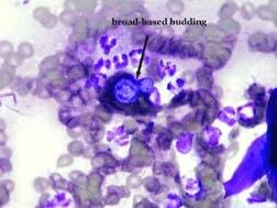
Blastomycosis, caused by Blastomyces dermatitidis, is a systemic fungal infection that affects dogs and cats. Blastomycosis is most common in certain geographic areas in North America, most often the Mississippi, Ohio, Missouri, Tennessee, and St. Lawrence River valleys, and in three provinces of Canada (Quebec, Ontario, Manitoba). It has also been reported in Africa, India, Europe, and Central America. (Fungal growth is supported by wet, sandy, acidic soils rich in organic matter, which is why it is found in valleys and is seen most frequently near water.)

Pathophysiology

Infection with Blastomyces occurs when a cat or dog inhales the fungal spores into the lungs. The incubation period is from 5 to 12 weeks. Some animals don’t show clinical signs for a long time after being infected, but those animals are not contagious to other animals and people. Blastomycosis organisms have a predilection for the respiratory tract, and pulmonary disease is the most common sign (88-94% of canine cases). Once the lung disease develops, yeast forms of the organism spread throughout the body. Organs typically affected include eyes, bones, skin, lymph nodes, subcutaneous tissues, brain, and testes. It can also be found in the nose, prostate, liver, mammary glands, and heart, but those locations are less common.

Dogs appear to be much more susceptible to infection than other species. Although the disease does occur in both people and cats, the incidence is much lower than in dogs. Dogs are ten times more likely to contract the disease than are people, and 100 times more likely than are cats. The incubation period in dogs is also shorter than in people. The reason dogs are more susceptible is unknown, but immune-deficiency may play a role. Annual prevalence in dogs in endemic areas is estimated at 1-2%. Many infected animals live within 0.25 mile of water. An increased number of cases can occur after periods of unusually heavy rainfall. Historically, young (i.e. 1-5 years), male, large-breed (e.g. hounds, pointers) dogs have had the highest risk of infection. (This is probably because these animals would tend to have more contact, due to hunting activities, with the organism’s geographical area.) However, any age, breed, or sex of dog can be infected.

Clinical Signs

The signs of illness will depend on what organs are infected. Some dogs will have eye problems, and some will have neurological signs (seizures, head tilt, etc.). Lameness may occur, if the infection is in the bones. Owners of dogs in the specific geographic areas should watch for coughing, difficulty breathing, eye inflammation, enlarged testicles, fever, swollen lymph nodes, ulcerated/draining skin lesions, bloody urine, difficulty urinating, nasal cavity signs (including bloody nasal discharge), and lack of appetite. (Weight loss occurs because of the decreased appetite.) Infections in the nasal passages may result in skull damage, and lead to infection of the brain. Large skin abscesses and neurologic signs are more common in cats than in dogs, while bone lesions are more common in dogs than in cats. Hypercalcemia can occur in dogs, although it’s rare in cats and non-domestic feline species.

Diagnosis

Diagnosis involves physical exam, blood tests, imaging (radiography, ultrasonography, CT, etc.), urinalysis to look for Blastomyces yeast, cultures, serology, and biopsies of affected organs. Fungal serology, to look for antibodies, is not always accurate and has been known to produce false negatives. The enzyme immunoassay (EIA) for B. dermatitidis galactomannan antigen appears to have a high sensitivity in urine (93.5%) and serum (87%). The EIA assay is commercially available; cross reactions with Histoplasma capsulatumCoccidioides immitis, and Penicillium marneffei can occur with this assay. The definitive diagnosis is finding the fungus in the tissues.

Treatment

Treatment involves various antifungal medications, including itraconazole, ketoconazole, fluconazole, amphotericin B, and some combination therapies. These medications usually need to be taken for a long period of time; how long depends on the specific case. Treatment usually can be done at home, unless the disease is severe. Pets with respiratory involvement should be on restricted activity. In severely ill dogs, intravenous fluids, oxygen, antibiotics for secondary infections, and pain medication may be necessary. Skin lesions may require wound cleaning and debridement.

Eyes that are severely affected may not respond well to the treatment because the medication does not penetrate eyes very well. Ocular blastomycosis cases may need systemic and topical corticosteroid therapy, topical anti-glaucoma medications, etc. Significantly affected eyes may require enucleation (removal of the eyeball).

Pets with severe lung disease may get worse at the beginning of treatment because the fungal organisms are dying; the mass death of organisms can cause severe respiratory problems.

Patients may not appear to improve for one to two weeks after the start of treatment. Close monitoring should be done for the first two weeks, and then rechecks are usually scheduled on a monthly basis. Rechecks may involve blood tests, biochemistry profiles, and imaging, if appropriate.

Prognosis

The prognosis for many pets is good, especially if owners can afford to treat the pet for long enough. Approximately 50% to 75% of affected dogs recover when treated with itraconazole, fluconazole, or an amphotericin-ketoconazole combination. Prognosis is poor for dogs with severely affected lungs, but if the dog survives the first 7 to 10 days of therapy, the prognosis improves. However, mortality rates in dogs with Blastomyces dermatitidis can be as high as 41%.

The prognosis for the retention of vision, in animals that have eye disease, is variable.

Dogs with brain involvement often die. Seizures are common upon death of the organism and may be uncontrollable.

Prognosis is also poor when at least 3 body systems are involved.

Relapses are most likely to occur in dogs that had a severe case at onset, or dogs that were not treated long enough. Relapses are most common within the first 6 months after treatment. Dogs that recover from the disease are probably not immune to getting it again.

After discontinuing therapy, animals may be rechecked at 1, 3, and 6 months for evidence of relapse. In one study, relapse rates for itraconazole and fluconazole were 18% and 22%, respectively. Relapses are treated like a new infection.

There is no way to prevent your pets’ exposure to Blastomyces other than by keeping them away from affected geographic areas.

Zoonotic Potential

Blastomycosis is not considered to be a zoonotic disease. It is acquired by humans via inhalation or direct contact with infective conidia/spores. Risk of infection is higher for excavation workers, and for people working or playing in wooded areas with waterways. Blastomycosis cannot be spread between dogs and other animals, or between dogs and people. However, immunocompromised people should limit their contact with infected pets and should wear gloves when cleaning and treating draining lesions.

4128649

Bad Breath in Dogs and Cats

What Is Halitosis?

Halitosis, also called bad breath, is as an offensive odor emanating from the oral cavity. Bad breath is a common presenting pet odor complaint. Common causes may be related to the mouth or, rarely, related to other health problems.

What Causes Halitosis?

The most common cause of halitosis is periodontal disease caused by plaque (bacteria). Bacteria is attracted to the tooth surface within hours of teeth cleaning. Within days, the plaque becomes mineralized producing calculus. As plaque ages and gingivitis develops into periodontitis (bone loss), bacteria changes from somewhat irritating strains to bone destroying types that produce hydrogen sulfide causing halitosis.

Other causes include eating malodorous food; metabolic disease (diabetes, uremia); respiratory disease (rhinitis, sinusitis, neoplasia); gastrointestinal (megaesophagus, neoplasia, foreign body); dermatologic (lip fold pyoderma); dietary (fetid foodstuffs, eating stool); non-periodontal oral disease (orthodontic, pharyngitis, tonsilitis, neoplasia); foreign bodies; trauma including electric cord injury; open fractures; caustic agents; infectious agents including bacteria, fungi, and viruses; autoimmune diseases; and eosinophilic granuloma complex.

What Are The Signs Of Halitosis?

Periodontal disease is painful. Some dogs and cats will have problems chewing hard food, others will paw at their mouths. Unfortunately most will not show any signs.

How Is Halitosis Diagnosed?

Halitosis is easily diagnosed by smelling your dog or cats breath. If there is a disagreeable odor, halitosis is present. A veterinary examination is necessary to diagnose the specific cause of bad breath. If the diagnosis is not obvious after oral examination, blood tests will be taken to check for internal disease.

How Is Halitosis Treated?

Halitosis treatment depends on the cause. There are four recognized stages of periodontal disease. The first two (early gingivitis and advanced gingivitis) are treated by professional teeth cleaning. As the disease advances bone loss occurs causing periodontitis, which may require surgery or tooth extraction.

Odor neutralization of hydrogen sulfide occurs with the use of zinc citrate.

What Is The Prognosis For Halitosis?

Once the underlying disease has been treated, halitosis will disappear. If due to periodontal disease, daily tooth brushing will help maintain good oral health and sweet breath.

canine_Brachial Plexus_brachial_plexus_closeup_2

Brachial Plexus Avulsion in Dogs and Cats 

Brachial Plexus avulsion or brachial plexus injury can occur when an animal suffers an injury involving the front limbs or shoulders.

Caused by trauma such as an automobile accident, a bite or gunshot wound, a surgical complication, or a fall, brachial plexus avulsion or injury can happen to any breed or age of dog or cat.

Like people, dogs and cats have a network of nerves that arise from the cervical spinal cord in the region of the neck, cross each other in the armpit of the dog, and extend to supply stimulation to the muscles of the neck, shoulders, and front limbs. These nerves convey sensation from this region to the brain via the spinal cord. The brachial plexus is the term for the bundle of nerves in the armpit.

In the case of injury, damaged nerves can affect the function of a front leg. Usually, the damage is on only one side. 

Brachial plexus injuries usually involve more than one nerve. Nerve roots which arise from the spinal cord and contribute fibers to one or more nerves are more apt to be damaged than the nerve itself because the roots are less elastic.

When the nerve roots in the neck region are torn away from the spinal cord, the injury is called a brachial plexus avulsion. When the nerves located in the armpit are stretched or torn, the injury is called a brachial plexus injury.

Depending on the severity of the injury and which nerves are involved, your dog may be unable to move the injured shoulder or leg, may not be able to flex the elbow or toes, or may not put weight on the injured leg. They may lose feeling in the leg or paw. The seriousness of this type of injury depends on what kind of nerve damage your pet has.

How is it Diagnosed?

After hearing your pet’s history, a neurological examination will help your veterinarian determine if your pet has suffered a brachial plexus avulsion or brachial plexus injury and, if so, which type and how severe it is.

canine_Brachial Plexus_brachial_plexus_closeup_2

X-rays, ultrasound, and advanced imaging tests such as computed tomography (CT) and MRI may be recommended.

Treatment

Your veterinarian will treat any life-threatening injuries resulting from the accident or trauma first. If one or more nerves have been stretched rather than torn away from the spinal cord, recovery may occur given time. Treatment involves mainly supportive care. This care should be started early. Your veterinarian may prescribe analgesic drugs for pain as needed. Range-of-motion exercises to do when your pet is ready may be discussed and demonstrated for you. 

Monitoring

Watch your pet closely after this type of injury. An abrasion to the front of the paw may occur if the pet drags the limb. You may find a wound that was not visible at first. Your pet may fuss at or lick a wound or the affected leg. E-collars or other protective measures can be used if your pet starts to self-mutilate.

Your veterinarian will continue to check your pet’s progress and evaluate pain perception and the use of the injured leg.

Prognosis

How well your pet does will depend on the type and location of the injury and how severe it was. A mild injury may show signs of improvement within 1-2 weeks after the trauma happened. In severe cases, the affected leg may not improve even after several months. If there has been avulsion of multiple nerve roots, loss of sensation to the limb, or no progress is seen after several months, and lack of normal limb function is resulting in poor quality of life for the pet, amputation may be recommended.

Prevention is best: do not let your pets have free access to traffic or other hazards, and don’t let your dog ride in open truck beds.

5770367

Alcohol Poisoning

Dogs and cats can get more than just drunk when they drink ethanol, isopropanol, or methanol — they can get a trip to the emergency room. Pets can die from ingesting alcohol. 

What are ethanol, isopropanol, and methanol?

Ethanol (ethyl alcohol) is most commonly formed from the fermentation of sugars. Ethanol is found in alcoholic beverages, some liquid medications and mouthwash bases, rotting/fermenting fruits, and rising (raw) yeast-containing bread dough. The “proof” of ethanol is twice the percentage of alcohol concentration (i.e. 80 proof = 40% ethanol). Although ethanol is also in some household inks, cleaners, and solvents, the levels are low enough to generally be of no clinical significance if these products are ingested by pets.

Isopropanol (isopropyl alcohol) is in rubbing alcohol (70%), some anti-freezes, some detergents, glass/window cleaners, perfumes/colognes, and alcohol-based topical sprays, including some pet flea-control and grooming products. Isopropyl alcohol is more than twice as potent as ethanol or methanol.

Methanol (methyl alcohol, wood alcohol) is most commonly found in automotive windshield washer fluids (20-80%) but is also in some gasoline additives, “canned heat” fuels (e.g., Sterno®), and some household solvents (e.g., paint removers).

Although the strength of these three alcohols varies, the clinical signs associated with their ingestion by dogs and cats are quite similar.

All poisoning problems boil down to the amount of alcohol ingested compared to weight, just like a dosage of medicine. Thus, when pets drink an alcoholic beverage that was left within reach or that was given to them intentionally by someone, it can cause a significant toxicity problem. In addition, significant absorption can occur through the skin or by inhalation.

Dogs are highly susceptible to the effects of alcohol. Signs of mild inebriation may occur with even minor exposure. Within 15 to 30 minutes after the pet has drunk the alcohol on an empty stomach (or within 1 to 2 hours on a full stomach), central nervous system (CNS) signs, such as staggering, excitement, or decreased reflexes, can begin. Behavioral changes can be seen, as can an increased need to urinate. As the problem gets worse, the pet may become depressed, have a slow respiratory rate, or go into cardiac arrest. Puppies and kittens are at particular risk because of their small size and immature organ systems. 

Alcohols are depressants, so many of the clinical signs associated with them are due to their effect on the CNS. Alcohols irritate the gastrointestinal tract. They also act as diuretics, and the alcohol and its metabolites are eliminated by the kidneys. Liver damage may occur after exposure, although it is much more common in animals that have repeated or chronic exposure. 

Clinical Signs

Clinical signs of intoxication can occur within 15-30 minutes of ingestion of alcohol. Signs include nausea, vomiting, thirst, urination, dehydration, lethargy, incoordination, disorientation, becoming cold (hypothermia), low blood pressure, and alcoholic bad breath. In severe cases, blindness, tremors, tetraplegia, respiratory depression, coma, or seizures may develop. Death is uncommon but may occur, especially if the pet has severe respiratory and cardiovascular depression, low blood pressure, and/or hypoglycemia. 

Diagnosis

Diagnosis is based on a history of exposure, combined with appropriate clinical signs and laboratory tests.  

Treatment/Management/Prevention

The treatments your veterinarian may use can include assisted ventilation, intravenous fluids (to improve elimination of the alcohol/metabolites, to provide cardiovascular support, and to correct electrolyte abnormalities), seizure control, and other supportive therapies. In addition, if the alcohol toxicosis is due to the ingestion of bread dough, your veterinarian may need to wash out your pet’s stomach with cold water to inhibit further alcohol production and to break up the dough mass for removal. In addition, your pet will be confined to prevent accidental self-injury.

Prognosis

Most dogs with alcohol intoxication can recover with adequate symptomatic and supportive care. Recovery may take 8-12 hours and tends to take longer if it was a severe intoxication or if isopropanol was involved.

Preventing Alcohol Toxicosis

All alcoholic beverages and alcohol-containing fluids should be kept out of reach of your dogs and cats. Consult your veterinarian before giving any ethanol-containing liquid medications.

Do not feed raw bread dough to your pets or leave bread dough out to rise in areas that can be reached by them. Always dispose of discarded bread dough carefully. 

The ASPCA National Animal Poison Control is available 24 hours a day at 888-426-4435. Expect an initial consultation fee of around $100.00 and additional follow-up is at no charge. You will be assigned a case number your veterinarian can use to communicate with a toxicology specialist before beginning treatment.