Tag: Prognosis

4127255

Oral Squamous Cell Carcinoma in Dogs and Cats

When squamous cell carcinoma (SCC) occurs in the mouth and throat, it’s called oral squamous cell carcinoma. In these oral cases, the lesion is usually located on the gums or tonsils. Squamous cell carcinoma is the most common oral cancer in cats. In dogs, SCC is the second most common oral tumor.

The most common location of oral SCC in cats is on the base of the tongue on the underside. SCC may also come from the gingiva (gums), particularly along the maxillary (upper) teeth.

SCC on the gingiva expand and progress locally and are usually associated with destruction of the bone. SCC of the maxilla (upper jaw) often appears as a depressed, ulcerated area. The tumors on the tongue, mandible (lower jaw), and pharynx (throat) tend to be proliferative and raised.

SCC metastasizes (spreads) to the regional lymph nodes less than 10% of the time in dogs and 31% of the time in cats. Metastasis to the lungs occurs 3-36% of the time in dogs and 10% of the time in cats. SCC are typically very invasive and can become quite large, especially in cats.

SCC affects middle-aged to older cats (range 7-20 years). There is no increased likelihood based on breed or gender.

Signs

Signs can include drooling (with or without blood), difficulty eating, and halitosis (very bad breath). Depending on the tumor’s location, the pet can have trouble swallowing or may cough. If the mouth is too uncomfortable for the pet to eat normally, they will lose weight. As is true with many cancers, affected dogs and cats tend to be older animals.

Diagnostic Tests

Diagnostics include radiographs (X-rays) of the local site, radiographs of the lungs to see if it has spread (metastasized) to other locations, CT scans, biomarker assessment (laboratory tests), and biopsies. Sometimes a fine needle aspirate will provide enough sample tissue for diagnosis. In a study of oral SCCs, biomarkers (such as proliferating cell nuclear antigen and Ki-67) were associated with higher-grade tumors and increased likelihood of spread. 

Treatment

Treatment may involve surgery, radiation therapy, chemotherapy, electromagnetic thermoablation, supportive therapy, or a combination of these depending on location, the amount of tissue involved, etc. Your primary veterinarian or veterinary oncologist will recommend treatment options specifically for your pet’s condition.

Surgery

If the tumor hasn’t spread, surgery is the preferred treatment. The entire tumor, including the extensions into underlying tissue and bone, will be removed. Often, part of the jawbone has to be removed. Surgery can provide a cure if the pet has clean margins (the tumor was completely removed). Dogs do quite well with partial jaws. It doesn’t typically alter the dog’s appearance as much as people might expect. Even if surgery isn’t curative, surgery can extend survival.

As for cats, following radical mandibulectomy (removal of the lower jaw) most can eat independently, but a few may require hand feeding. In cats, surgical excision, with or without partial removal of the tongue, may be considered for SCC of the tongue. When dealing with SCC of the pharynx (throat) or tonsils, the specialist may recommend removing as much of the mass as possible to make the cat more comfortable. This is palliative, but it’s not curative.

Radiation Therapy

Radiation therapy can be used if surgery isn’t an option, or if surgery can’t completely remove the tumor.

In cats, radiation therapy is not very effective as the sole therapy for SCC because median survival times (MST) range from only a few weeks to months. In a report of combined radiotherapy and chemotherapy, the overall MST was 163 days, and cats with tumors of the tonsil or cheek had MST of 724 days. Radiation therapy has been used as palliative care for cats with nonresectable SCC.

Chemotherapy

Chemotherapy may be added to therapy, depending on the circumstances. Chemotherapy has some drawbacks, so your veterinarian or oncology specialist will have to determine if this treatment would be useful.

Electromagnetic Thermoablation 

Electromagnetic thermoablation (hyperthermia, in which body tissue is exposed to high temperatures) may be used. This technique applies a high-frequency alternating electromagnetic field to heat alloy needles. The needles are placed into and surrounding the tumor to destroy the malignant tissue.

Supportive Therapy

Supportive therapy includes pain medications, acupuncture, feeding tubes to provide nutritional support, antibiotics for secondary infections, etc. A few cats with oral SCC have been treated with the medication zoledronate in an attempt to reduce bony destruction and pain. Pamidronate is also a medication option for cats. Your veterinarian will work with you to determine what therapies apply to your pet.

Monitoring

Frequent examinations are needed to watch for recurrence or progression. Periodic monitoring to watch for evidence of spreading disease, such as feeling the lymph nodes, lymph node aspiration, and chest X-rays are also indicated.

Prognosis

Prognosis For Dogs 
The median survival time for dogs that have mandibular (lower jaw) SCC treated with surgery alone varies from 19-43 months, with a 1-year survival of 88-100%, a 2-year survival of 79%, and a 3-year survival of 58%. 

The median survival time for maxillary (upper jaw) SCC that was treated with maxillectomy varies from 10-39 months. The local recurrence rate after mandibulectomy or maxillectomy is less than 10%. 

Tumor-associated inflammation and invasion of the lymphatic system are indicators of a poorer prognosis. Overall survival times are lower with SCC of the tonsils. In one study, the median survival time was only 243 days, with a 1-year survival rate of 40% and a 2-year survival rate of 20%. The longest survival times occurred when surgery and chemotherapy were used together.

Prognosis For Cats

Overall prognosis is poor. One study of 21 cats treated with mandibulectomy reported a median survival time (MST) of 217 days, with 1 and 2-year survival of 43%. The local recurrence rate was 38%. Cats with rostral (nose/mouth) tumors had a longer MST of 911 days. When SCCs of all oral tissues are included, MST with no therapy is only 30-45 days. MST was 106 days following stereotactic radiotherapy for oral SCC of any location, with 38.5% of the cats showing a positive response. Most cats with this tumor type die of uncontrolled local disease. Another factor adversely affecting survival is that many cats with advanced SCC have metastatic disease (lymph node 31%, lungs 10%) at the time of diagnosis. 

4128293

Otitis Media (Middle Ear Infection) in Dogs and Cats

The word otitis means ear infection, but that one word doesn’t indicate which part of the ear is infected. Otitis media is inflammation and/or infection of the middle ear, whereas otitis externa begins in the external ear canal. Some nerves are closely associated with the middle ear, so infection there can damage them, causing the neurologic signs often seen with this infection: a head tilt, lack of balance, nystagmus (back-and-forth eye movements). These symptoms are called vestibular signs. Middle ear infections can also cause paralysis of the facial nerve, leading to a slack-jawed appearance on that side of the face.

Otitis media most often develops after an existing otitis externa travels from the external ear canal through the tympanic membrane and into the middle ear. The infection in the external ear canal leads to inflammation and damage to the ear canal and tympanic membrane, allowing the infection to enter the middle ear. 

Otitis media has been found in 16% of dogs that have acute (rapid onset) otitis externa and in 52% of dogs that have chronic (long-term) otitis externa. In contrast, 63% of cats that had otitis media/interna did not have a previous history of ear infection. This is why keeping your pet’s ears clean and watching for infection are important to your pet’s health.

Other causes of otitis media can include infections in the nose and throat, trauma, foreign bodies, fungal infections, inflammatory polyps, cancer, etc. In addition, developmental abnormalities of the external ear canal and pharynx can lead to otitis media.

Otitis media does not affect any particular age or animal, and males and females are affected equally.

Dogs that have long, pendulous ears, such as beagles and basset hounds, are more likely to develop otitis externa, which can lead to otitis media.

The Cavalier King Charles Spaniel breed is known to have a condition called primary secretory otitis media (PSOM). Dogs with PSOM typically have mild to moderate pain in the head/neck, neurologic signs (e.g. ataxia, facial paralysis, head tilt), and itchy ears, but without otitis externa. Owners of this breed will want to keep this disease in mind. 

Diagnosis

Diagnosis includes physical examination, ear examination with an otoscope, bacterial/fungal culture, radiography, and possibly advanced imaging.

Physical Examination

During the physical examination, your veterinarian will be looking for some of these signs: head shaking, pawing at the ear, pain when opening the mouth, signs of otitis externa (ear odor, redness, etc.), head tilt, facial nerve signs, Horner syndrome, ataxia, etc. Not every affected pet will exhibit all of these signs. In addition, cats are more likely to have neurologic signs than dogs are.  

Ear Examination (Otoscopy)


In order to do a complete examination of the ear, a deep ear cleaning may be necessary. To keep your pet comfortable, your veterinarian may decide to sedate or anesthetize your pet for the procedure. Then the infection can be more easily seen with an otoscope, which can see the eardrum.

Culture and Sensitivity

Your veterinarian will usually take samples of the material in the ear and culture those samples to determine what medications will work best.

Radiographs

Your veterinarian may take radiographs, also called X-rays. Radiographs can help show changes (thickening, destruction) to the bony structures of the ear. However, even if the radiographs appear normal, that does not mean that your pet does not have otitis media.

Advanced Imaging

In some cases, your veterinarian may suggest using computed tomography (CT) and/or magnetic resonance imaging (MRI) to further evaluate the tympanic bulla. CT is usually better than MRI at detecting bony changes, while MRI is better at detecting soft tissue abnormalities.

Treatment

Cleaning both the external and middle ear canals is an important step in treating otitis media. Debris can inactivate some topical medications and prevent medications from contacting infected tissues. Anesthesia will be necessary in order to do the deep ear cleaning and ear flushing that will remove the debris.

Your veterinarian may need to infuse (fill) the ear with medication in order to deliver a high concentration of antimicrobial products or corticosteroids directly to the affected area.

Topical medications may need to be used to control an otitis externa.

Systemic therapy with antibiotics, antifungals, and corticosteroids may also be used, depending on the infection.

If severe, irreversible changes to the external ear canal are found, if there are masses in the inner ear, or if medical therapy has failed to resolve the otitis media, your veterinarian may advise surgery, such as an ear canal ablation. This surgery is not undertaken lightly, but it may be necessary to control the infection. An ear canal ablation involves removing the vertical and horizontal portions of the ear canal, along with the bones of the middle ear and eardrum.

Monitoring (Rechecks)


During treatment, your veterinarian will usually want to recheck your pet’s external ear canal and tympanic membrane every 10-14 days. (A ruptured tympanic membrane will generally begin to heal within 21-35 days.) Topical medications will be given until the external ear canal and the tympanic membrane are normal, and until cytology of the ear canal reveals no infection. Systemic therapy is typically given for at least 4-6 weeks.

Prognosis

In general, the prognosis for dogs is good if appropriate therapy is started in time. The prognosis is not as good if the infection is resistant to the medications; if the concurrent otitis externa is not managed adequately; if there is significant bone infection; or if there is no response to surgery. In addition, some neurologic signs (e.g., facial nerve problems, Horner syndrome) may be permanent.

Reminder

Keeping your pet’s ears clean is critical to preventing ear infections of any kind. However, It is not always possible to prevent every infection in some pets. 

6665143

Bacterial Conjunctivitis in Cats

Conjunctivitis is inflammation of the conjunctiva, a tissue that makes up part of the eye and eyelid. In people, the conjunctiva is the white part. Conjunctivitis is common in cats and can be a result of infection or injury.

The primary cause of most conjunctival infections in cats is usually feline herpes virus. The treatment for herpes-based conjunctivitis involves a combination of antibiotics and antivirals, both in eye drops and pills. Even cats who have had the herpes vaccine can get conjunctivitis because the vaccination does not prevent herpes, but allows a milder case.  

However, some conjunctival infections that are primarily viral also have a bacterial infection at the same time.

The two bacteria species best known to cause conjunctivitis in cats are Chlamydia felis and Mycoplasma spp.

An infection caused by Chlamydia may cause redness, discharge, and excessive tears. Bacterial conjunctivitis caused by Chlamydia is treated with antibiotics. All cats in a household may need to be treated, as some cats who have it do not show any symptoms.

Mycoplasma is a natural inhabitant of every cat’s eye, so infection happens when there is an overgrowth of that bacteria. Treatment for the Mycoplasma infection is similar to that of Chlamydia: antibioticsThe goal in treating Mycoplasma conjunctivitis is to reduce the overgrown number of bacteria back down to a normal level because it is impossible to eliminate them entirely.

Who gets bacterial conjunctivitis?

Both cats and dogs can get conjunctivitis caused by bacteria or viruses. Conjunctivitis is the most common cat eye disease, and most cats will develop the infection once in their life. All breeds can develop infection, and juvenile and young adults are more likely to get it.

Clinical Signs of Either Form

  1. Red eyes
  2. Eyelid redness
  3. Eyelid swelling
  4. Excessive blinking
  5. Eye discharge
  6. Eye crusting
  7. Tearing
  8. Squinting
  9. Scratching the face to relieve irritation

 Diagnosis

A physical exam of the eye by your veterinarian is the first step, as this will rule out other causes of redness, such as trauma.  The easiest diagnosis is a cytology test, which means looking at scrapings of the irritated tissue under a microscope. If a lot of bacterial and inflammatory cells are seen, it is likely that the eye is infected. Checking scrapings is a quick test for infection that can be done at the clinic during your appointment. PCR is another test that can be used to detect the specific organism causing the infection, but typically needs to be sent out to a lab.

Treatment

Antibiotics are used to treat bacterial conjunctivitis, mainly doxycycline.  Mycoplasma and Chlamydia are the most common bacteria involved in eye infections, and doxycycline is effective against both.

Prognosis

Treatment should clear the signs and infection. However, if the eye still looks infected or just doesn’t look normal, contact your veterinarian. The ongoing bacterial infection may also be caused by an unknown factor that needs to be treated before the eye infection can be cured.

Prevention

There is a vaccine for Chlamydophila felis, but it is not required and is usually only suggested if your cat is at a higher risk for an infection or in a multi-cat household. There is no vaccine for a Mycoplasma spp. infection because it is normally in the eye anyway.

The key to preventing infection is separating sick cats away from the unaffected ones and bringing them to your veterinarian at the first sign of infection.

7941098

Arthritis in Dogs and Cats: What Can be Done?

Arthritis, also known as osteoarthritis or degenerative joint disease (DJD), is a degenerative, progressive, and irreversible condition of the joints. It is characterized by the progressive loss of joint cartilage, bony spurs/growths, and the thickening and scarring of connective tissue around the joint, usually as a result of injury.

Approximately 25 percent of dogs are diagnosed with arthritis in their life time, and as many as 60 percent of dogs have radiographic evidence of it.

Degenerative joint disorders are probably as common in cats as in dogs but are less likely to be associated with obvious clinical signs, such as lameness. In one study, 90 percent of cats over 12 years of age had radiographic signs of osteoarthritis.

Arthritis is classified as primary or secondary. Primary arthritis is associated with aging, due to years of wear and tear on the joints. Secondary arthritis is the result of an external event or force (e.g., trauma, poor joint alignment, etc.) that once damaged the joint cartilage.

Arthritis can affect any age, sex, and breed of dog and cat. Most predispositions to it relate to underlying causes, such as elbow dysplasia, hip dysplasia, osteochondrosis, and so forth.

Diagnosis

Diagnosis is based on what is found in the physical examination, X-rays and other diagnostic imaging, joint taps, cytology, force plate gain analysis, therapeutic drug trials, and other tests.

Physical Examination Findings in Dogs

Lameness is the most common sign. It may happen once in a while (episodic) progressive (gets worse over time), or be persistent. Stiffness is common after periods of rest. Stiffness and lameness may decrease when the dog warms up a bit with some activity. Lameness often gets worse after periods of overexertion. Pain, swelling, and decreased range of motion may be seen. Thickened joints, excess fluid in the joint space, and muscle weakening are likely to occur.

Physical Examination Findings in Cats

As opposed to the visible lameness seen commonly in dogs, many cats simply become less active, may hide, or develop behavioral changes, such as irritability, decreased grooming, or difficulty getting into position in the litterbox. Cats also may have joint swelling/thickening, too much fluid in the joint space, and decreased range of motion. There may or may not be pain when the cat’s affected joint is moved by you or your veterinarian.

Diagnostic Imaging

Radiographs (X-rays) and CT scans may show the excess fluid in the joints; the bony spurs; signs of an underlying disorder, such as elbow dysplasia, osteochondritis dissecans, hip dysplasia, or cruciate ligament rupture; and so forth.

Kinetic Tests

Force plate gait analysis in dogs can help find where the lameness is within the joint. It can also show the severity of lameness, both before and after therapy. A force plate is mounted to the floor, and the dog walks over it. Measurements are taken to see which areas use the least force (more affected) and vice versa. The gait analysis hasn’t been used as often in cats.

Therapeutic Trial

In some cases, a therapeutic trial of nonsteroidal anti-inflammatory drugs (NSAIDs) may help confirm the diagnosis if the lameness is less noticeable after taking them.

Treatment, Management, and Prevention

It is not possible to cure arthritis.

The goals are to alleviate your pet’s discomfort, to minimize further degenerative changes to the joint, and to restore the joint’s functionality. Multiple types of treatment are usually necessary to relieve pain, stiffness, and discomfort.

Managing your pet’s weight is important. Excess weight increases stress on the joints and muscles. If your pet is obese, your veterinarian will want your pet to lose weight. Daily, low-impact activities, such as walking and swimming, will not only help your pet with losing some pounds but can also improve joint mobility, muscle mass, and exercise tolerance.

Joint supplements known as chondroprotective agents will help support the cartilage and will have some anti-inflammatory effects. These agents will slow the breakdown of cartilage and/or provide the building blocks that can help build it. Some agents also increase joint fluid secretion and thus decrease inflammation.

The main components of chondroprotective agents are polysulfated glycosaminoglycan (PSGAG), glucosamine, and chondroitin sulfate. Oftentimes there is no improvement using chondroprotective therapies. The failure may be due to too little cartilage left in the affected joint as there’s bone on bone; unresponsive joint inflammation; variable bioavailability of the drug between different products; and the lack of analgesia.

Although the injectable PSGAGs are more expensive, they provide a faster and longer-lasting response than the oral forms. If there is no response after 4-6 weeks, your veterinarian may discontinue use of PSGAGs.

Diets containing omega-3 fatty acids may help reduce inflammation. Some studies have found that dietary supplementation with fish oil omega-3 fatty acids can improve the clinical signs of arthritis in dogs, and may allow the NSAID dose to be reduced.

NSAIDS are an important component of arthritis therapy in dogs. Side effects that you should be aware of include stomach upset, elevated liver enzymes, and potential worsening of chronic kidney disease. Few NSAIDs are licensed for use in cats; your veterinarian will advise you about what NSAID options are available for your cat.

Analgesics such as tramadol, gabapentin, and amantadine may provide pain relief in dogs.

Alternative therapies such as acupuncture, stem cell therapy, platelet-rich or conditioned plasma, physical therapy, rehabilitation therapy (e.g. radial shock wave therapy, pulsed signal therapy), green-lipped mussel supplements, vitamin E, and zoledronic acid may be beneficial in some canine patients. Studies to support their use are still being done.

Surgery 

Your veterinarian may consider surgical options if your pet’s response to medical treatment is low. In addition, your veterinarian may suggest surgery for certain underlying causes of arthritis, such as cranial cruciate ligament rupture, elbow dysplasia, osteochondritis dissecans, joint incongruity, intra-articular fractures, and joint instability. Reconstructive procedures can eliminate joint instability and correct the anatomic defects. If your pet has severe hip dysplasia, your veterinarian may suggest a total hip replacement and femoral head/neck ostectomy. If the arthritis is in the wrist or ankle (then joint fusion may be considered; this surgery is usually well tolerated and can result in reasonable functionality.

Monitoring and Prognosis

Your veterinarian may need to do periodic physical examinations every 1-4 months to monitor your pet’s response to therapy and the progression of the disease. In addition, if your pet is on an NSAID, blood tests including complete blood counts and biochemistry profiles, should be done every few months to ensure there are no side effects impacting the liver or kidneys.

With therapy and careful monitoring, arthritis can be managed in many dogs and cats, resulting in a good quality of life that you and your pet will appreciate. 

6512549

Yeast Dough Dangers in Pets

If you bake yeast bread from scratch, it is essential that you keep all raw bread dough away from your pets. Ingesting it can cause serious problems and be life threatening for your pet.

Doughs that do not contain yeast are less of a concern, but can still cause gastrointestinal problems.

Why is it so dangerous to eat?

Yeast is a microscopic fungus that, under the right conditions, makes carbon dioxide gas and ethanol (alcohol) from sugar. This effect is great for making bread because gas makes the dough rise and ethanol flavors the bread before burning off in the oven. However, this process is dangerous when it occurs in your animal’s stomach.

Being moist and warm, the stomach is essentially the perfect environment for yeast to grow and pump out carbon dioxide and ethanol. The buildup of gas and rising dough in the stomach is painful and can lead to a condition called bloat: “the mother of all emergencies.” Also known as gastric dilatation and volvulus, or GDV, bloat is where the expanding stomach puts pressure on surrounding areas and decreases blood flow to important organs. Even more dangerous, the stomach may eventually twist, cutting off blood flow even more. The twisted stomach can reduce blood flow throughout the whole body, leading to a serious condition called shock.

Several steps are needed to save a bloated dog’s life. Part of the problem is that the main three steps – decompress the stomach, give IV fluids to fight shock, and stabilize the heart rhythm – should all be done at the same time and as quickly as possible.

Although GDV can be life threatening, death from yeast dough in pets is usually due to the effects of ethanol. Ethanol is alcohol, like that found in beer, wine and spirits,  so the ethanol produced by yeast can cause alcohol poisoning. This poisoning can lead to low blood sugar, low blood pressure, decreased body temperature, seizures and trouble breathing.

Signs

Early signs of yeast dough ingestion are generally those associated with bloat. Your pet may try to vomit or retch but not produce anything or maybe just a little white foam. Additional signs include an enlarged abdomen due to the buildup of gas and dough, in addition to weakness, increased heart rate, and collapse.

Signs of alcohol poisoning generally occur two or more hours after dough is ingested. You might be able to smell alcohol on your pet’s breath. Your pet may seem drunk, becoming uncoordinated and disoriented. They may also feel cold to the touch. Seizures and trouble breathing are two other serious signs of alcohol poisoning.

What should I Do?

Get to a veterinarian as soon as possible, as this is potentially a life-threatening emergency.

If your pet just recently ate the dough and isn’t showing any symptoms yet, your veterinarian may try to induce vomiting to remove it from the stomach. As you can imagine, bread dough is so gummy and sticky that it is hard to vomit up. So, other methods of removal will likely be necessary.

Yeast love the warmth in a stomach. If you make the stomach cold, you can slow the yeast’s growth and the production of alcohol and gas. This slow down can be accomplished by feeding ice chips or flushing the stomach with cold water. These treatments can also break up the dough and make it easier to pass through the rest of the digestive system. In rare cases, the dough may need to be surgically removed.

Treatment also involves dealing with your pet’s specific clinical signs and may entail giving intravenous fluids to correct any blood abnormalities and to speed up removing alcohol from the bloodstream. If your pet is cold, heating blankets, warm intravenous fluids and other techniques may be used to warm them up. Depending on your pet’s symptoms, medications may be given to reduce pain, treat seizures, correct breathing abnormalities, manage vomiting, etc.

The veterinarian will need to monitor your pet for at least 12 hours until they recover.

Prognosis

Pets generally do well if the yeast dough is removed quickly and they are treated soon after ingestion. Unfortunately, pets suffering from severe alcohol poisoning, bloat, or GDV have a more guarded prognosis.

To prevent this life-threatening situation altogether, avoid letting your pets have access to raw yeast dough by letting it rise in areas your pets can’t reach, such as elevated places in the kitchen, or in a closed oven or microwave.

5171558

Head Tilt in Pet Rabbits

Head tilt in rabbits is seen with some frequency and can be caused by a variety of diseases.  Another common name for head tilt is wry neck.  The correct medical term is vestibular disease, which can include other signs besides a head tilt. Another term that is often used is torticollis. Rabbits with vestibular disease can have a head position that ranges from a few degrees to 180 degrees off the normal position.  They can fall over, circle, have difficulties standing and develop eye injuries because the prominent eye globe (especially of the “down” eye, the one facing the ground when the head is tilted) is prone to trauma.  The cardinal signs of true vestibular disease in the rabbit are a persistent head tilt and a loss of balance. 

Anatomy

First let’s look at the anatomy and function of some vital areas in order to understand what does and does not contribute to head tilt in the rabbit.

External Ear

Disease in this area alone can cause head shaking, drooping ear and pain but does not cause a persistent head tilt or loss of balance. 

Middle Ear

Disease of the middle ear can cause head shaking, drooping ear and pain as well as deafness but does not cause a persistent head tilt. 

Inner Ear

The inner ear controls balance and hearing.  Signs of disease of the inner ear include deafness, head tilt, loss of balance and horizontal or rotary nystagmus. Proprioception (the ability to sense where the feet and legs are) and postural reactions (the ability to try to return to a normal standing position) are normal.  The rabbit will not act weak in other parts of the body and will continue to try to maintain normal body position even if it is difficult and the head is tilted. 

Brain

A specific area of the brain stem contains the vestibular nuclei, the origin of the vestibular nerve in the inner ear. The vestibular nuclei serve as the body’s central balance control. Signs of disease to this tiny area of the brain stem include head tilt, loss of balance, circling toward the affected side, rolling, vertical nystagmus, positional nystagmus, delayed or absent proprioception and loss of postural reactions.

Spinal cord

Head tilt is not a sign of primary spinal cord disease. 

Diseases Resulting in Head Tilt

A major differentiation that has to be made when diagnosing the cause of head tilts is whether it is peripheral (affecting areas other than the brain) or central (involving the brain and most specifically the vestibular nuclei).

Otitis Interna (Inflammation of the inner ear)

Causes can include the following: infectious disease, foreign bodies, trauma, neoplasia, and toxins. Signs of otitis interna include persistent head tilt toward the affected side, circling, nystagmus, ataxia (inability to walk normally), and deafness. The most accurate way to diagnose otitis interna is with a CT scan or MRI. A negative finding on an x-ray may not rule out otitis interna. 

Treatment for otitis interna depends on the primary cause, but since the majority of head tilts in rabbits are likely caused by bacterial otitis interna, it is advantageous to use a long-term course of antibiotics (3 to 6 weeks up to several months).  It is currently recommended to avoid the use of corticosteroids in rabbits. Rabbits may be more sensitive than other animals to developing immunosuppression when taking corticosteroids, either topically, orally or parenterally.

Otitis Media (Inflammation of the middle ear)

This is also a common disease of rabbits and may occur along with or even be the cause of otitis interna.  However, disease in this area alone does not cause a persistent head tilt.  Signs of otitis media include periodic head tilting and shaking. Diagnosis and treatment are generally the same as for otitis interna. 

Brain Stem Disease

Disease at the brain stem, specifically the vestibular nuclei, can cause similar signs as seen with inner ear disease.  Because the vestibular nuclei are deep in the brain, it is likely that disease affecting this area will also affect surrounding brain tissue.  Therefore, additional neurologic signs may be seen such as loss of appetite, mental dullness, paralysis and sudden death.  If the disease is also affecting the cerebrum, additional signs such as seizures can be seen. Bacteria, fungi and viruses can affect the brain stem.

Encephalitozoon cuniculi is a one-celled organism called a microsporidium that can infect rabbits. There is an ongoing controversy over the prevalence of E. cuniculi as a cause of primary head tilt in the rabbit.  It has been extremely difficult to demonstrate a definitive correlation between head tilt and active E. cuniculi infection.  Serological testing for E. cuniculi has some value but is not definitive and, if not interpreted appropriately may be misleading.  The only way to diagnose E. cuniculi as the definitive cause of a head tilt is to take brain tissue samples from the rabbit and find the organism and its damage in the microscopic samples.  No one has yet proven this correlation because a brain biopsy is dangerous for the rabbit and the E. cuniculi organism can be difficult to find in brain tissue. There are few, if any, case reports or studies definitely proving E. cuniculi is a significant pathogen in the rabbit nervous system. If a rabbit shows signs compatible with central vestibular disease, has a positive test for E. cuniculi, and all other diseases have been ruled out, some veterinarians will choose to treat for E. cuniculi empirically.  Proper and effective treatment for E. cuniculi is controversial. Some of the medications that have had been used to treat infection with E. cuniculi include albendazole, fenbendazole and oxibendazole.

The most common parasite associated with head tilt in a rabbit is the raccoon roundworm Baylisascaris procyonis.  Signs observed in rabbits with Baylisascaris may include head tilt, tremors, weakness, blindness, seizures or sudden death. Prevention of exposure to the parasite eggs is clearly the best way to counteract this disease.

Other causes of head tilt in rabbits may include cerebrovascular accident (stroke), cancer, trauma, toxins, metabolic disease, and heat stroke.

Diagnostic Approach to Head Tilt

History

A detailed history is of vital importance to determine the cause of disease. 

  1. History of any prior illness
  2. History any prior bouts with head tilt, weakness or incontinence
  3. Possibility of exposure to environmental toxins or parasites
  4. Exposure to other rabbits that are or were ill (particularly with neurological disease)
  5. Possibility of trauma
  6. Possible contact with human with active herpes viral infection

Physical Exam

A thorough physical exam and a thorough neurological exam is essential to diagnosing the cause of head tilt.

  1. Mental attitude: Is the rabbit still alert and active, or dull and depressed?
  2. Head tilt:  Look at persistence, side to which rabbit tilts, is circling involved?
  3. Balance:  Does the rabbit try to right itself if given support?
  4. Gait:  Any abnormalities in gait?
  5. Nystagmus: If present, is it spontaneous or positional, is it horizontal or vertical?
  6. Ear exam:  Are there signs of external or tympanic membrane disease?  Are there signs of fluid, blood or pus beyond the tympanic membrane?
  7. Respiratory:  Are there signs of respiratory disease?
  8. Systemic: Any other neurological signs, weakness (particularly hind limb), paralysis, incontinence, behavioral changes, external signs of trauma, particularly around the head and neck?   

Blood Tests

  • Complete Blood Cell Count:  This test may be helpful to determine if there is anemia or infection.  
  • Serum biochemistries:  These tests are helpful to rule in or out a number of metabolic diseases
  • Serology for E. cuniculi – These tests are of limited use in definitively diagnosing active disease.
  • Blood testing for heavy metals – These tests are particularly important if heavy metal intoxication is suspected.

Bacterial Cultures

Unfortunately, it is frequently not possible to safely or easily collect a sample to culture from a rabbit with vestibular disease.

Endoscopy

Endoscopy of the ear canal may be useful if middle ear infection is present, or possibly used to obtain cultures through the tympanic membrane with a surgical technique called myringotomy. 

Radiographs (X-rays)

Radiographs are useful to detect any heavy metal in the GI tract and for diagnosing head trauma.  Radiographs are also helpful in screening for disease of the tympanic bulla where the middle ear is housed

CSF (cerebrospinal fluid) Analysis

This may be useful if central disease such as encephalitis is suspected.

Biopsy

If it is possible to obtain a sample of the affected tissue, then a microscopic analysis can be extremely helpful in making a diagnosis.

CT scan or MRI

These imaging techniques are the most accurate and safest means of diagnosing disease of the inner and middle ear.

Treatment and Nursing Care

  1. If a definitive diagnosis of the cause cannot be made (very common situation), but peripheral disease is the suspected due to physical exam, history and whatever diagnostics could be performed, the rabbit should be put on a course of broad spectrum antibiotics for an extended period of time ranging from 3 weeks to several months.
  2. Generally, corticosteroids (cortisone-like drugs) should be avoided if possible, because rabbits may be especially sensitive to the immunosuppressive qualities of these drugs, and their use may cause further complications. 
  3. If a diagnosis of E. cuniculi infection is strongly suspected based on multiple signs of central disease, serology, and ruling out other disease,  a short-term use of oxibendazole,  fenbendazole, or albendazole, can be considered. 
  4. Non-steroidal anti-inflammatory drugs should be considered to reduce inflammation and control pain that may be present.  These drugs may be needed only at the beginning of therapy.
  5. The use of anti-nausea drugs is controversial as there is no clear evidence that rabbits experience feelings of nausea.  There is no substantiated evidence that the use of anti-nausea drugs helps improve the condition of rabbits with head tilt.  Some veterinarians feel anti-nausea drugs, like diphenhydramine or meclizine, are useful in a rolling rabbit or one who is not eating.
  6. Eye lubrication is useful, particularly in those animals that have a severe head tilt.  The down eye is prone to injury due to the protruding nature of rabbit eyes.  Rabbits do not blink often and this eye may become dry, abraded or infected.  Daily attention is necessary.
  7. Fluid therapy and nutritional therapy (assisted feeding) may be necessary in any rabbit with vestibular disease.
  8. Rabbits with vestibular disease from any cause often cannot access their cecotropes.  These nutrient-rich droppings can be collected while still moist and placed in a rabbit’s food bowl along with the pelleted food. 
  9. It is essential to modify the environment of a rabbit with severe vestibular disease. This involves providing an enclosed padded or smooth-sided cage or enclosure. 
  10. Aside from occasional anecdotal reports or testimonials, there is no evidence that any kind of physiotherapy or acupuncture will reduce the length of time a head tilt persists or will resolve a residual head tilt.
  11. It is important to remember that the course of vestibular disease, even with the best prognosis, can take many weeks to months of committed care before improvement is seen. 
  12. If a rabbit shows a continual decline or continued mental depression, loss of appetite or other weakness over a 2 to 3 week period, then the prognostic outlook is fairly grim and euthanasia should be a consideration. 

Prognosis

The prognosis for recovery from vestibular disease is variable, depending on the cause.  For most rabbits with peripheral disease, the prognosis is good to guarded. Some rabbits will have a lifelong residual head tilt even if the inner ear disease is cured. For rabbits with central vestibular disease the prognosis becomes guarded to poor for recovery to a sustainable state.

Key Points

  1. Persistent head tilts accompanied by nystagmus and loss of balance are either peripheral (inner ear) or central (vestibular nuclei of brain).  
  2. Peripheral vestibular disease is probably the most common cause of head tilt and is usually confined to head tilt, spontaneous nystagmus, circling and loss of balance.  The majority of cases are still mentally alert, maintain an appetite and do not exhibit other signs of weakness, gait abnormalities or seizures.
  3. Peripheral vestibular disease is most commonly caused by inflammatory disease of the inner ear with bacterial disease being the most common.  There is no current evidence that E. cuniculi causes disease of the inner ear.
  4. Peripheral vestibular disease carries a good to guarded prognosis for clinical recovery.  There is often a residual head tilt, but the rabbit can learn to re-establish balance and live a relatively normal life. 
  5. Central vestibular disease is less common, and also includes head tilt, positional nystagmus, circling and loss of balance. 
  6. Rabbits with central vestibular disease may also have histories of other signs compatible with central disease, potential exposure to toxins, parasites, or trauma.
  7. Central vestibular disease may be caused by a variety of conditions including bacterial infections, E. cuniculi, parasites and trauma, and carries a guarded to poor prognosis for recovery. 
  8. Radiographs are necessary to rule out trauma and may detect middle ear disease.  However, in many cases there will be no radiographic change even in middle or inner ear disease. Therefore, a negative x-ray is not proof that this disease does not exist.
  9. CT scan or MRI is the most accurate and safe means of detecting inner ear disease as well as some types of central disease.
  10. It is probably best at the minimum to treat rabbits with strictly peripheral signs that are confined exclusively to head tilt, nystagmus, circling and loss of balance with appropriate antibiotics because bacterial disease of the inner ear is common.
  11. Non-steroidal anti-inflammatory drugs should also be considered in many head tilt cases to reduce inflammation (since inflammatory disease is so common in both peripheral and central disease) and reduce any pain. 
  12. Appropriate nursing care for a rabbit with vestibular disease is crucial and requires a long-term commitment to both environmental and patient management.
  13. The sooner you get veterinary care for a rabbit with vestibular disease, the greater the chances for successful resolution with a relatively short recuperative period.
6313300

Distichiasis Requires Permanent Eyelash Removal in Dogs

Distichiasis is quite common in dogs. Distichiasis is a condition in which extra hairs grow out of the eyelash area. It happens when there are two or more hairs growing out of a Meibomian gland opening. (Meibomian glands are located along the margin of the eyelid.) These hairs are not supposed to be there. In some cases, these extra hairs can be long and stiff and irritate the eye, resulting in a corneal ulcer. The severity of the problem depends on how stiff the hairs are, how long they are, where they’re located, and how many extra hairs there are.

Distichiasis is different from entropion. Distichiasis involves extra eyelashes; entropion is an inward roll of the eyelid that causes eye irritation from normal eyelashes or hair.

Untreated distichiasis can cause corneal ulcers, chronic eye and eyelid pain, and excessive tearing. It is quite uncomfortable and/or painful for the animal, depending on the amount irritation. If the excessive hair causes any clinical signs at all, the hair should be permanently removed.

Signs include increased blinking, lots of extra tears that often look like a tear streak, and squinting. Dogs don’t typically paw at the eye. The more severe cases are the most easily diagnosed, as the milder cases often involve small softer hairs that can easily be missed.

It’s seen most often in puppies or young adults and is typically diagnosed before a dog is three years old. Any dog can have it, but it’s considered to be one of the most commonly inherited diseases in dogs, and considered by some to be the most common congenital eye problem.

Breeds that seem to be predisposed to distichiasis include:

  • Cocker spaniel
  • Cocker Spaniel, American
  • English bulldog
  • Flat-coated Retriever
  • Golden retriever
  • Lhasa apso
  • Miniature Longhaired Dachshund
  • Miniature poodle
  • Pekingese
  • Poodle
  • Pug
  • Shetland sheepdog
  • Shih tzu
  • Toy poodle
  • Yorkshire terrier

Treatment

There are several treatment options and your veterinarian’s choice will generally depend on how many extra hairs are involved and what equipment the veterinary facility has. General anesthesia is usually needed. Occasionally a very cooperative dog might allow treatment with only a local anesthetic, but that would be extremely rare.

  1. Cryosurgery freezes the lid margin at the places where there are extra hairs.
  2. Surgery will remove the hairs permanently.
  3. Electrolysis will remove the hairs permanently.

These procedures destroy the hair follicles, preventing hair regrowth. However, no procedure can be guaranteed, so retreatment may be necessary in some cases. Several follow-up visits will be needed, to make sure that the hairs are not going to regrow.

Plucked hairs will just grow back, so plucking is not a permanent solution. Lid splitting and thermocautery can destroy the normal lid margins, leading to severe permanent scarring and entropion, so they are not good treatment options.

The eyelids will have some post-operative inflammation, which your veterinarian will also treat.

Prognosis

Once the hairs have been removed permanently, the prognosis is good. However, dogs with distichiasis should not be used for breeding, because of the hereditary aspects.

4128595

Demodectic Mange in Dogs

The Culprit – Demodex Canis

Demodectic mange, also called demodicosis, is caused by one of the microscopic mites of the Demodex genus. Three species of Demodex mites have been identified in dogs: Demodex canis, Demodex cornei, and Demodex injai. The most common mite of demodectic mange is Demodex canis. All dogs raised normally by their mothers possess this mite as mites are transferred from mother to pup via cuddling during the first few days of life.

Graphic by MarVistaVet

Most dogs live in harmony with their mites, never suffering any consequences from being parasitized. If, however, conditions change to upset the natural equilibrium (such as some kind of suppression of the dog’s immune system), the Demodex mites may gain the upper hand. The mites proliferate and can cause serious skin diseases.

Is Demodicosis Contagious?

Unlike sarcoptic mange, demodectic mange is not considered a contagious disease, and isolation of affected dogs is generally not considered necessary. That said, there are some circumstances under which the mites could spread from one dog to another. Classically Demodex mites have been felt to only be transferable from mother to newborn pup. After the pup is a week or so old, it has developed enough immunity so that infection is no longer possible. In other words, after age one week or so, a dog will no longer accept new mites on its body. Recently this idea has been challenged as occasionally multiple unrelated dogs break with demodicosis in the same household. It is not clear if some species of Demodex are more contagious than others or if some contagion is possible under certain circumstances. Current thinking is that mites actually can be transferred from one dog to another but as long as the dog is healthy, the mites simply add into the dog’s natural mite population, and no skin disease results. In rare circumstances, the spread of disease is possible if severe infection is involved. While there are still assorted theories about dog-to-dog transmission of Demodex mites, there is no question that mites cannot be transmitted to humans or to cats.

  • Mites live inside hair follicles — a difficult place for miticides (chemicals that kill mites) to reach.
  • Mites are normal residents of dog skin; it is only in some individual dogs that mites cause problems.

Demodicosis Has Three Forms

Form #1: Localized

Localized demodicosis occurs as isolated, scaly bald patches, usually on the dog’s face, creating a polka-dot appearance. Localized demodicosis is considered a common puppyhood ailment, and approximately 90% of cases resolve with no treatment of any kind. This is quite a contrast to generalized demodicosis, as described below, so it is important to be able to distinguish localized from generalized disease. It seems like this would be a simple task since localized demodicosis classically involves several round facial bald spots, and generalized demodicosis involves a bald scaly entire dog; still, reality does not always fit into neat categories in this way. Some guidelines used to distinguish localized demodicosis include:

  • Localized disease does not involve more than two body regions. (One spot or two on the face and one spot or two on a leg would still qualify as localized even though the spots are not close together.)
  • Localized disease involves no more than four spots total on the dog.

Treatment is not necessary nor recommended for localized demodicosis but there are so many regular flea products that kill Demodex mites that most puppies will clear simply by starting their normal prescription flea control regimen.

Not all flea products will kill Demodex; one needs a product of the isoxazoline class, and the puppy must be old enough for such a product. 

If there is a reason why the puppy cannot take an isoxazoline flea product, there are alternatives. Goodwinol ointment, an insecticide ointment, may be used daily to control localized demodicosis. Antibacterial gels are also used against localized demodicosis and associated skin infections. It is important to note that rubbing a creme or ointment on a demodicosis lesion can cause reddening of the lesion, making it appear to get worse. It is also possible for rubbing the medication on the area to break off the weaker hairs at the margin of the lesion, causing the lesion to appear to get bigger. Neither of these situations truly represents an exacerbation of the disease.

Without treatment, the resolution of a localized demodicosis lesion should be at least partially apparent after one month though total resolution can take up to three months.

Approximately 10% of localized demodicosis cases will progress to generalized demodicosis. Enlarged lymph nodes are a bad sign — often foretelling generalized mange.

Can the Pup be Bred Later?

Sometimes the puppy with localized demodicosis was obtained for breeding purposes. The current recommendation is not to treat these puppies so that we can determine if the condition will stay localized and resolve or if it will generalize. If it stays localized and eventually resolves without treatment, the animal is still a candidate for breeding. If the condition generalizes to cover the entire body, the animal should be sterilized. If the condition receives treatment and resolves, we will never know how the disease would have gone in its natural state and will not know whether the pup is carrying the genetic predisposition for generalized demodectic mange. In this case, it is best to be conservative and not take the chance of passing on genetic predisposition for this disease.

Localized demodicosis is almost exclusively a puppyhood disease. When a puppy develops localized demodicosis the chance of the condition resolving is 90% unless there is a family history of demodicosis in related dogs. In this case, the chance of spontaneous resolution drops to 50%.

Occasionally an adult dog develops localized demodicosis. We currently do not have a good understanding of the prognosis or significance of this condition in an adult dog.

It is important that dogs with a history of generalized demodectic mange not be bred, as there is a hereditary component to the disease.

Form #2: Generalized

Generalized demodicosis entails much more extensive involvement of the skin. Large patches of skin are affected and, if allowed to progress untreated, the entire surface of the dog may be impacted. Sometimes a polka dot appearance results but if there are more than four spots present, the patient is no longer classified as having the “localized” form, and treatment rules for generalized disease may be applied. 

Photo by MarVistaVet

The secondary bacterial infections make this a very itchy and often smelly skin disease. These infections may require antibiotics but it is important to realize that infection will probably not resolve fully until the mites are gone.

The goal of treatment is the total eradication of the mite population on the dog’s body.

Juvenile Onset (The Most Common Form)

Young dogs that develop demodicosis are believed to have a genetic immunological defect that allows the mite to gain the upper hand. As the puppy grows up and his or her immune system matures, the immune system tends to naturally regain control of their mite infestation; in fact, 30-50% of dogs under age 1 year recover spontaneously from generalized demodicosis without any form of treatment. Usually, treatment is recommended, though, to facilitate recovery.

Adult Onset

Most demodicosis occurs in young dogs under age one and a half. An older dog should not get demodicosis unless there is an underlying problem with the immune system. In such cases, demodicosis is considered an indication to seek a more serious hidden condition such as cancer, liver or kidney disease, or an immune-suppressive hormone imbalance. A more extensive medical work-up will be required. In the case of shelter dogs, sometimes the stress of abandonment, street living, starvation or other poor husbandry can compound into the immune suppression needed to generate a case of adult-onset demodectic mange.

Years ago, generalized demodicosis was nearly untreatable, and many dogs were euthanized for uncontrollable disease. Today, treatment for most dogs is surprisingly simple.

Treatment

The biggest change in treating this condition came, as mentioned, with the release of the isoxazoline flea products (sarolaner/Simparica®, fluralaner/Bravecto®, afoxalaner/Nexgard®, lotilaner/Credelio®). The isoxazoline products are oral medications labeled to kill fleas and ticks in dogs. It turns out they have substantial activity against Demodex mites and are successful enough that they have become the treatment of choice. These products are typically given orally every two to four weeks, depending on the protocol. Most dogs are simply cured by this protocol.

Occasionally, a dog fails this treatment and is relegated to the more traditional methods such as high-dose oral ivermectin, a protocol that is not safe for many herding breeds; topical moxidectin (Advantage multi®); daily oral milbemycin (Interceptor®); or even labor-intensive dipping with amitraz (Mitaban®).

The common anti-itch medication oclacitinib (Apoquel®) is an immunosuppressant and thus decreases the body’s ability to fight the Demodex infection. Understand before you use it to treat the itch of the Demodex infection that it can make the mites worse. Other forms of itch relief should be sought if necessary. Oclatacinib use can actually lead to a recurrence of demodicosis years after recovery, so it should not be used in any dog that has ever suffered from demodicosis previously.

Prognosis

The younger the dog, the better the chance of cure. Most dogs under one-and-a-half years of age recover completely from generalized demodicosis. In many cases of adult-onset demodicosis, the disease is controlled with treatment, but a cure is not always possible. Some cases can never be controlled.

No matter which option you choose, treatment should be accompanied by skin scrapes every two to four weeks. In this way, the effectiveness of treatment is assessed, and dosing can be changed. There are several protocols describing how to determine if treatment can be stopped. The idea is to eradicate every single mite from the dog’s body so that the condition cannot recur. This typically entails either continuing treatment for a significant time after the patient appears recovered and/or rechecking skin scrapes a significant time after treatment is finished.

Relapse?

When relapse occurs, it is often because the dog appeared to be normal, and treatment was discontinued before all Demodex mites were killed off. Relapse is always a possibility with generalized demodicosis as there is no easy way to confirm that every mite has been killed, but most dogs that relapse do so within a six to 12-month period from the time they appear to have been cured.

Sarcoptic mange is a completely different disease.

We Wish it Weren’t Necessary to Add This

Decades ago, dipping dogs with demodectic mange in motor oil was a popular home remedy. However, skin exposure to motor oil can cause rashes and skin destruction in severe cases. The hydrocarbons can be absorbed through the skin and cause a dangerous drop in blood pressure. If motor oil is licked off the coat, resultant vomiting can lead to aspiration of motor oil into the lungs and pneumonia. Kidney and liver damage can result from motor oil dipping.

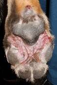
Form #3: Demodectic Pododermatitis

This condition represents demodectic mange confined to the paws. Bacterial infections usually accompany this condition. Often as generalized demodicosis is treated, the foot is the last stronghold of the mite. Old English Sheepdogs and Shar-pei tend to get severe forms of this condition. The infection can be so deep that a biopsy is needed to find the mites and make the diagnosis.

It is one of the most resistant forms of demodicosis, and deep infections and proliferative tissue that result can take months and great expense to resolve.

In the past, special organophosphate foot soaks, along with daily medications, were needed to resolve this condition, and months of treatment were common. As with the other forms of demodicosis, the isoxazoline flea products (sarolaner/Simparica®, fluralaner/Bravecto®, afoxalaner/Nexgard®, lotilaner/Credelio®) have changed everything. In most cases, the mites can be quickly killed, and the deep secondary infections can be resolved in a matter of weeks (instead of months) once the mites are gone. 

4128893

Cryptorchidism (Retained Testicles) in Dogs and Cats

Cryptorchidism is a condition in which a male’s testicles have not descended (dropped) into the scrotum. At birth, a male puppy’s or kitten’s testicles are located near the inguinal ring. 

By 8 weeks of age, testes are palpable in the puppy’s scrotum. However, scrotal and suprascrotal testes can be difficult to palpate if the testes are small or the puppy is obese. Testes may also freely move between the scrotum and inguinal area in young puppies.

By 8 to 16 weeks, they’re palpable in the kitten’s scrotum. Some retained testicles can be palpated. However, testicles retained in the abdomen are typically not palpable on physical exam.

In the fetus, a structure called the gubernaculum connects the testicle (located next to the kidney during development) to the scrotum. If this structure fails to develop properly, the testicle will not end up in the scrotum but will end up in the abdomen, the inguinal canal, etc.

Cryptorchidism can be unilateral (only occurring on one side) or bilateral (occurring on both sides). Unilateral cryptorchidism usually involves the right testicle. Bilaterally cryptorchid animals are usually sterile because the higher body temperature inside the abdomen is enough to prevent sperm production. (The animals will, however, still exhibit male behaviors.)

Cryptorchidism is a fairly common defect in dogs. Dog breeds most likely to be affected include Yorkshire terrier, Pomeranian, French poodle, Siberian husky, miniature schnauzer, Shetland sheepdog, Chihuahua, German shepherd, dachshund, and brachycephalic breeds. Still, it can happen in any breed or mixed breed.

Since cryptorchidism is considered to have a genetic basis, animals with this condition should not be used for breeding.

Dogs with cryptorchid testicles are prone to testicular torsion (twisting) and testicular cancer, so these dogs should be neutered to prevent problems later.

Cryptorchidism in cats is uncommon. The most common breed associated with cryptorchidism is the Persian. Congenital abnormalities that have been known to occur simultaneously with cryptorchidism are patellar luxation, shortened tail, kinked tail, tetralogy of Fallot, tarsal deformity, microphthalmia, and upper eyelid agenesis. Unlike dogs, it may be possible to visually differentiate between a castrated cat and one with retained testicles, because cryptorchid cats have barbs on the penis.

Laboratory Diagnosis

A testosterone assay can be used to differentiate between a castrated male and a cryptorchid male.

Treatment

Surgical removal is the only treatment for cryptorchidism. Even if the animal is a unilateral cryptorchid, both testicles should still be surgically removed. (The cryptorchid testicle should be removed to prevent testicular torsion and testicular cancer, and the normal testicle should be removed to prevent cryptorchid offspring.) This surgery is more complicated than the usual neuter surgery because the cryptorchid testicle can be difficult to locate. Depending on the case, some pets will be able to go home on the day of the surgery, and some may have to stay in the hospital overnight. A 2-week recuperation (reduced activity) is advised because this surgery usually involves opening the abdomen, and the surgical site has to have time to heal before the pet resumes normal activities.

Many males will need to wear a protective Elizabethan collar during recuperation to prevent them from licking or chewing at the incision. Owners should check the incision regularly for redness and swelling, which could indicate a post-operative infection or self-trauma. If non-dissolvable skin sutures are used, they will need to be removed by your veterinarian about 10 to 14 days after surgery.

Prognosis

Cryptorchid animals that have had both testicles removed, and have no other defects, will generally live a normal lifespan for the breed.

5379732

Congestive Heart Failure in Dogs and Cats

What is Congestive Heart Failure?

Congestive heart failure (CHF) is the clinical syndrome of fluid retention due to severe heart disease. In dogs and cats, the fluid is retained in specific parts of the body, depending on the side of the heart that is diseased.   

  • If the left side of the heart is diseased (e.g. mitral valve disease, most dilated cardiomyopathies, hypertrophic cardiomyopathy, and most common congenital cardiac defects), fluid is mostly retained in the lungs or within the pleural cavity (the chest space around the lungs).
  • If the right side of the heart is diseased, fluid is retained mostly in the belly or within the pleural cavity.

Why Does CHF Occur?

CHF occurs because the pressure in the veins and capillaries draining into the diseased side of the heart increases to the point that fluid leaks out of the veins and capillaries. The capillaries and veins that drain into the left side of the heart are those coming from the lungs, so fluid leaks into the lungs. The capillaries and veins that drain into the right side of the heart are those coming from the body, so fluid leaks into the belly (ascites) or creates swelling in the legs, the skin under the belly, etc.; swelling of the legs or the skin is uncommon in dogs and cats with right-sided CHF, but much more common in humans and horses.

It can be useful to think of this like a garden soaker hose. If water is flowing through the hose under low pressure, only a little bit of water leaks through the soaker hose. However, if we increase the pressure inside the hose, water starts to emerge from the soaker hose at a much greater rate and floods your garden. The same happens with the capillaries – under low pressure, only a little fluid leaks out of them (and is resorbed). But if the pressure is too high, a large volume of fluid leaks out into the surrounding tissue, and overwhelms the resorption mechanisms.

What Causes CHF?

CHF requires severe heart disease that increases the resting (diastolic) cardiac pressure. For example, diseases that cause leakage of valves (mitral or tricuspid valve disease) can result in CHF if the leak is big enough. Cardiomyopathies also cause the resting cardiac pressure to increase and can result in CHF. Pericardial diseases can prevent the heart from relaxing and stretching properly, which can result in right-sided CHF. Heartworm disease can cause right-sided CHF. Many congenital defects that shunt blood can result in CHF (e.g. patent ductus arteriosus, ventricular or atrial septal defects).

Importantly, congenital diseases that obstruct outflow from the heart, such as pulmonic stenosis or aortic stenosis, do not normally cause CHF.

What Are the Clinical Signs of CHF?

This depends on the side of the heart that is diseased. Let’s start with left-sided CHF, which causes pulmonary edema and, sometimes, pleural effusion. As fluid starts to deposit fluid into animals’ lungs, the amount of oxygen in the blood decreases because it can’t be absorbed from the lungs. Human patients describe this as shortness of breath, initially during exertion, such as climbing stairs, and eventually even when stationary. However, animals cannot tell us if they are short of breath. Sometimes, owners will notice a decrease in exercise ability, such as not going as far on walks and getting winded on walks more quickly. However, many things can cause exercise intolerance. Therefore, we tend to look for an increase in breathing (respiratory) rate, especially when sleeping.

Coughing has been described as a feature of CHF in dogs, but there is some doubt as to whether CHF really causes coughing or not. Again, many dogs and cats cough for reasons other than heart disease. As the fluid continues to build up, dogs and cats will also show difficulty in breathing as well as a faster respiratory rate. This is especially true of cats, who can hide their disease until it becomes really advanced. Consequently, many cats see veterinarians with labored, open-mouth breathing that is almost gulping for air, which appears to have started all of a sudden. In dogs, the clinical signs can be more gradual and subtle.

With right-sided CHF, the most common presentation is a swelling of the abdomen (ascites), making the dog appear pregnant. The discomfort from a belly full of fluid results in difficulty getting comfortable or breathing comfortably when lying down. These dogs will even resort to sleeping in a sitting position at times! Appetite often decreases slightly because of the abdominal pressure.
If there is a buildup of fluid in the chest cavity, the animal might show difficulty in breathing.

How Do Veterinarians Diagnose CHF?

The diagnosis of CHF relies on pairing the clinical signs of increased respiratory rate and difficulty in breathing with the severe heart disease that is responsible for these clinical signs. Many times, a murmur can be heard for the first time. The pet’s heart rate will be elevated, their respiratory rate will be elevated. Some cats come in with a low body temperature because they are somewhat shocky from inadequate oxygen.

If veterinarians suspect CHF, they will generally take chest X-rays to see if there is evidence of (A) severe heart disease that appears as an enlarged heart; and (B) opacity (areas through which light does not pass) in the lungs consistent with pulmonary edema, or fluid in the chest cavity consistent with pleural effusion. If they are unsure or require additional information, they might perform or recommend a cardiac ultrasound. Those are often performed by specialists, typically veterinary cardiologists or radiologists, who have the necessary equipment.

How Do we Treat CHF?

Treatment is directed at both the underlying heart disease and the accumulation of fluid. If possible, the cause should be corrected. For example, closing a congenital shunt, such as a patent ductus arteriosus, will immediately correct the problem permanently. Repairing a leaking mitral valve will also correct the problem almost immediately, although this procedure is currently very expensive and performed by a limited number of surgeons.

The mainstay of medical treatment of left-sided CHF is the use of diuretics called loop diuretics, which include furosemide or torsemide. Diuretics reduce blood volume and consequently reduce the pressure in the veins, forcing the fluid out into the lungs or the abdomen.

Other drugs that are commonly used to treat CHF include angiotensin-converting enzyme inhibitors, pimobendan, thiazide diuretics, and spironolactone. These are much less effective than loop diuretics and should never be given as the only drugs for managing CHF. Indeed, if a dog or cat does not require a diuretic, they most likely do not have CHF.

The fluid buildup in right-sided CHF often requires repeated manual removal, using large catheters to make the patient feel better. In many dogs, this can be done as frequently as every week or two. Ideally, when coupled with medical treatment, the frequency of belly taps can be reduced somewhat.

How Do we Monitor CHF?

The most important thing to monitor with left-sided CHF is respiratory rate. Provided the respiratory rate when sleeping is in the normal range, we can be reasonably confident that we have good control of the CHF. When the sleeping respiratory rate starts to increase, adjustments in treatment might be necessary; a veterinarian will evaluate the patient to determine exactly what to do in such instances. (See monitoring videos below.)

Many veterinarians will monitor bloodwork to make sure that the medications being given are not causing problems with kidneys or elsewhere.

If a pet appears to destabilize after a period of control, additional x-rays might help better evaluate the situation.

What is the Prognosis?

This depends somewhat on the underlying disease. With the more common diseases, such as mitral valve disease in dogs or hypertrophic cardiomyopathy in cats, once CHF is diagnosed and treatment instituted, survival is generally less than two years. With mitral valve disease, approximately 50 percent of dogs will succumb to their disease within 8 to 10 months, and only 20 percent live for 18 to 24 months. With hypertrophic cardiomyopathy survival data are less clear, but some older studies suggest that 50 percent of cats will succumb to the disease within 7 to 10 months. As is always the case, a few individuals will live longer than expected.