Tag: pets

7963100

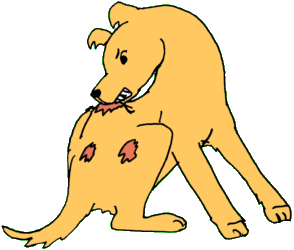
Hypertrophic Osteodystrophy (HOD) in Dogs

Hypertrophic osteodystrophy is a developmental, auto-inflammatory disease of the bones that is usually first seen between 7 weeks and 8 months of age.  In general, most cases will appear for the first time when the puppy is between 3 – 5 months of age. Relapses may occur until about 20 months of age.

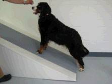
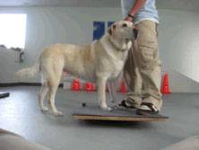
HOD is most often seen in fast-growing puppies of large and giant breeds.

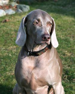
Some medium-sized breeds, such as the Australian cattle dog and pit bull may also develop HOD. The Weimaraner, in particular, appears to be predisposed to HOD.

Predisposed breeds include:

  • Australian kelpie
  • Boxer
  • Bullmastiff
  • Doberman pinscher
  • German shepherd dog
  • Giant breed dogs
  • Great Dane
  • Irish setter
  • Irish wolfhound
  • Kuvasz
  • Labrador retriever
  • Large breed dogs
  • Rottweiler
  • Saint Bernard
  • Standard poodle
  • Weimaraner

In HOD, blood flow decreases to a part of the bone next to the joint. This interrupts bone formation. The interruption means that bones don’t harden appropriately, nor do they grow as strong as those of a healthy puppy.

HOD can be very painful. Sometimes HOD is straightforward and responds quickly to treatment, but it can also be tricky to diagnose and treat.

HOD is somewhat similar to panosteitis (pano) in that it affects the growing leg bones of large- or giant-breed puppies. However, pano usually affects only one leg at a time, and is thought of as “growing pains.” HOD can affect more than one leg at the same time and is more painful than panosteitis. In addition, unlike pano, HOD can permanently damage the growth plates.

HOD doesn’t appear to be inherited, and at this time no one knows how puppies get it, although auto-immune diseases and dietary excesses can trigger it. However, because it is more common in some breeds if the puppy is being considered for breeding purposes, your veterinarian may recommend a thorough review of the pup’s relatives, to see how many of them have had HOD. If the puppy comes from a bloodline that has few cases, or the cases were mild and self-limiting, that may be better for breeding purposes than bloodlines that have had many cases, or have severe cases. (Every pup, of every breed, being purchased for breeding purposes should always have an extensive review of the relatives, to avoid known breed-related issues.)

Clinical Signs

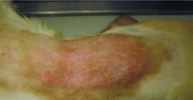
Clinical signs depend on how mild or severe the HOD is. For mild cases, the puppy usually has a slight limp and appears to have pain in the affected bone. Puppies with more severe cases may have a decreased appetite and subsequent weight loss, fever, and depression. Unfortunately, these non-specific signs can happen with many diseases.

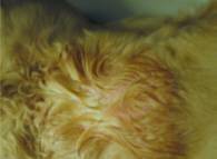
More specific signs of HOD include swollen, warm, and painful leg bones. The puppy may refuse to bear weight. If multiple limbs are affected, the puppy may prefer to remain lying down, be reluctant to get up, and be unwilling to walk.

And not just the legs may be involved. Some cases have swollen muzzles, excessive salivation, and pain when the dog tries to open his mouth; these cases may involve the jaw. Also, radiographs may show changes in the jaw, spine, ribs, shoulder, and eye socket bones.

Systemic signs can include:

  1. Fever ranging up to 106⁰F (a dog’s normal body temperature is 101-102.5⁰F)
  2. Lack of appetite
  3. Discharge from the eyes or nose
  4. Bumps on the skin, some with pus
  5. Diarrhea that can be bloody
  6. Inflamed vulva or vagina
  7. Increased respiratory sounds

At the very least, HOD dogs will have a fever, lameness, and typical HOD lesions showing on the radiographs. The more signs the puppy has, the more severe the case. (In severe cases, with multiple systems and organs affected, more aggressive treatment to suppress the immune system will be necessary.)

A complicating factor to remember is that, for the most part, HOD patients are very young puppies that do not have bodily reserves and can decline rapidly with fever and lack of appetite.

HOD can be a painful condition and typical treatment to control even significant pain may not work.

HOD generally comes in episodes that last a few weeks. Often the first episode lasts a week and it can be followed by a full and spontaneous remission.

Diagnosis

Diagnosis is made via physical exam (seeing obviously swollen soft tissue over the leg bones), radiographs, blood tests, etc.

Treatment

Your veterinarian will want to control the fever, lethargy, and bone pain. Nonsteroidal anti-inflammatory drugs (NSAIDs) are often used for this. However, if the puppy develops gastrointestinal signs, the NSAIDs may have to be discontinued.

Some puppies may need immunosuppressive doses of corticosteroids, usually prednisone. Some dogs may need to take them for a year, in which case the drug must be tapered off slowly, rather than simply stopped. Your veterinarian will recommend tapering only once the dog is free of pain and walks normally.

In some cases, narcotics may be necessary to control severe pain. These may have to be given by intravenous injection.

Some breed-specific studies have shown better responses to corticosteroids than to NSAIDs. In one study of 53 Weimaraners, 50 percent did not respond to NSAIDs. In another study of six related Weimaraners, NSAIDs had little effect and the dogs had to be switched to corticosteroids. A similar experience has been reported in affected Irish setters and Australian kelpies.

Your veterinarian may need to prescribe antibiotics for pneumonia or to prevent secondary infections resulting from immune systems that have been depressed by the corticosteroids. Antacids, such as famotidine, may be given while the dog is taking the corticosteroids. Some veterinarians may recommend probiotics when the GI tract is involved. Pain medications may be needed.

Other care may include fluid therapy, analgesics (e.g. tramadol, opiates), and rest or restricted activity. Even when the puppy starts feeling better, the veterinarian may still want exercise to be restricted until the bones have structurally recovered.

Vaccines should be avoided during an active HOD episode.

Monitoring and Prognosis

Mild cases of HOD may resolve on their own or with only supportive care. Watch for a possible return of any clinical signs once the dog has stopped/tapered the NSAIDs or prednisone because recurrences are common until the leg bone has finished growing. Dogs with affected litter mates are more likely to relapse. Complete recovery is expected once the leg is finished growing, but relapses (episodes of fever and malaise) have been reported in adult dogs. Most relapses in adult dogs respond to NSAIDs or corticosteroids.

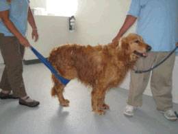
Permanent limb deformities are rare. However, if they occur, surgery may help. Your veterinarian will use radiographs to determine if surgery is indicated.

Prevention

While it’s difficult to prevent a disease when you don’t know what causes it, some common sense can boost the chances that a puppy won’t get it. Large- or giant-breed puppies should not be given any mineral or vitamin supplements that aim to increase their growth rate and adult size. Slow and consistent growth is ideal for the puppy’s health because rapid growth can cause skeletal abnormalities. Puppies need a completely balanced diet. Puppies should be discouraged from doing jumping exercises until their growth plates have closed, which is at about a year of age. (Check with your veterinarian for an appropriate estimate of growth plate closure based on your puppy’s breed and growth.) Avoid housing puppies on hard surfaces, such as concrete. If floors are not carpeted, rugs may help so the puppies don’t slip as much.  

Prognosis

Most cases of HOD are resolved with or without medical care. Unfortunately, sometimes HOD is so severe, painful, and uncontrolled by treatment that owners are forced to discuss the pet’s quality of life issues, resulting in euthanasia.

4128244

Hot Spots (Pyotraumatic Dermatitis) in Dogs and Cats

What are Hot Spots?

When the weather gets warm, we start seeing more dogs with hot spots. These wounds are weepy, wet, red, and sometimes bloody when they are fresh, and they are dry and scabby when they are resolving. They can cover large expanses of the pet’s skin or they can appear as smaller solitary lesions (sometimes in multiple locations). Hot spots generally have very clear margins defining them and separating them from normal skin.

They are caused by over-zealous self-licking, chewing, scratching, and rubbing (the pet creates them him/herself), and they can arise especially quickly (10 minutes of chewing can create a big one). The good news is that they almost always look worse than they are. The infection is quite superficial and often will resolve with topical treatment alone. The bad news is that about 30 percent of the pets who come to my hospital for hot spots actually have other kinds of skin diseases, such as deeper skin infections, bite wounds or other trauma, or even immune-mediated ulceration. If you aren’t entirely sure about what a hot spot looks like, it is probably best to have the veterinarian look at it.

The dog causes the hot spot by self-licking, chewing, scratching, and rubbing. What causes the pet to self-chew and lick is another story.

The pet may have an allergy, may have come in contact with an irritating substance, may have irritation from a grooming clipper, or may have some pain in the area from the underlying tissues. In many cases, the pet simply has fleas and is allergic to their bites. Anything that makes the dog itch will make the dog lick and chew, and if the licking and chewing is obsessive enough, a hot spot will soon follow.

First Aid

Treating a hot spot may or may not be a do-it-yourself project. Smaller hot spots can be treated at home with topical products made for this use. The important thing is to be aware that these areas are tender and so the pet may bite if you use something on the area that stings. Also, be careful about using human topical products as these may be toxic to pets when licked. Zinc oxide, for example, can be toxic when licked and is common in many human skin ointments.

Initial treatment usually involves removing the surrounding hair so that the hot spot can be disinfected. Once the superficial infection is properly cleansed, topical products can be used to relieve the associated inflammation. The lesion dries and scabs while it is healing. If the pet is really itchy or there are multiple hot spots, pills or injectable medication become necessary. Hot spots just under the ear/on the facial cheek, for example, are notorious for covering up a deeper skin infection below and often require more extensive treatment, especially in Golden Retrievers. More extensive treatments might include oral corticosteroids and/or oral antibiotics in addition to topical antiseptics and anti-inflammatories.

Good flea control is important for any itchy pet and is the foundation of itch prevention for most dogs and cats. Always be sure you have flea control secured in the approach to managing itchy skin in pets.

6039086

Hepatitis in Dogs

What is Hepatitis?

Hepatitis is inflammation of the liver. It is a syndrome – a group of clinical signs or symptoms – rather than a specific disease, and it has many causes. Your dog can have immediate inflammation in their liver called acute hepatitis or long-term inflammation called chronic hepatitis. Chronic hepatitis (CH), also called chronic active hepatitis, can lead to scar tissue formation and cirrhosis in the liver. Cirrhosis is extensive, end-stage scar tissue. Some dogs with acute hepatitis progress to the chronic form.

Infectious Canine Hepatitis

One disease that causes chronic hepatitis is infectious canine hepatitis (ICH). ICH is caused by a virus but not the same as the Hepatitis A, B or C viruses that cause disease in humans; you and your family won’t get sick if your dog is infected. Similarly, The hepatitis viruses that cause human hepatitis do not affect dogs. ICH is spread through animal feces, urine, saliva and objects. Since most dogs are vaccinated as puppies against ICH, it is considered a rare infection. If you have a young puppy or an unvaccinated dog in your household, they can get ill with this virus. However, outbreaks sometimes occur, mostly in dogs less than 1-year-old or unvaccinated dogs. An infected dog may show signs of depression, fever, vomiting, diarrhea, and abdominal pain. Treatment involves supportive care such as fluids and medications to boost liver function; the latter may include SAMe, milk thistle, ursodiol, and vitamin E.

Causes 

Aside from ICH, there are many other causes of hepatitis. Causes of acute hepatitis include toxins, drugs, viruses, bacteria (particularly leptospirosis), and fungi. On the other hand, chronic hepatitis is caused by bacteria, viruses, excess copper in the liver, drugs, and immune system issues. If a cause cannot be determined, this is referred to as idiopathic hepatitis and is the most common cause. Idiopathic means no cause can be found.

Age and Breed Predispositions

Any dog can have an acute episode of hepatitis. Chronic hepatitis, on the other hand, is most common in middle-aged to older dogs; most dogs diagnosed with chronic hepatitis are 4-10 years old.

Some breeds are suspected to have a genetic predisposition to CH, making them more likely to develop it. These include the Bedlington Terrier, Doberman pinscher, West Highland white terrier, Dalmatian, English springer spaniel, American and English cocker spaniels, Jack Russell terrier, and standard poodle, Great Dane, Labrador retriever, and others. CH is also more common in females than males.

Signs

Dogs with acute hepatitis often have more severe signs than those with CH. Symptoms may include anorexia (lack of appetite), vomiting, diarrhea, dehydration, depression, fever, and jaundice (yellow discoloration of the eyes, gums, and skin), enlarged abdomen and weight loss. How serious your dog’s symptoms depend on how much liver damage they have and this may vary from mild to severe. In severe cases, there are signs of bleeding and bruising. Bleeding can occur inside the body or through any opening of the body, such as the nose and mouth.

Dogs with chronic hepatitis often appear normal early on. It is often surprising how normal the dog appears, even with significant progressing disease. This is why it is a good idea to pay attention to elevations in the liver enzymes (particularly ALT), as there is a better chance of slowing down the disease the sooner it is detected and appropriate management begins. As the disease gets worse, common signs are poor appetite, vomiting, diarrhea, depression, increased thirst and urination, weakness, jaundice, and weight loss. A small number of dogs have severe signs such as bleeding, incoordination, and behavior changes. Similar to acute hepatitis, bleeding can occur through any opening in the body.

Diagnosis

Similar to many illnesses, a thorough history and physical examination from your veterinarian are crucial to making a diagnosis. Drug history and potential exposure to toxins are particularly important as these can cause significant liver damage. Some dogs with hepatitis have increased levels of liver enzymes that can be seen on lab work. These increases do not have to be large to be important; they may indicate liver damage but can also be caused by many different diseases.

Elevated liver values are a common abnormality, but they are often non-specific and can be confusing to pet owners. Not all cases of elevated liver enzymes indicate specific liver disease. The liver performs many functions and, in some cases, the liver enzymes may elevate due to problems elsewhere in the body, or simply due to age. It is a good idea to look into these sooner rather than later, especially if your dog is one of the breeds predisposed to hepatitis. They may be the only abnormality a dog with chronic hepatitis has at that time.

To add to the confusion, some dogs with hepatitis can have normal liver enzymes. This normalcy does not rule out chronic hepatitis: CH can be so severe in some dogs that there is little functioning liver tissue left to release enzymes, making test results look ‘normal.’ If hepatitis is suspected, your veterinarian may recommend a serum bile acids test specifically to evaluate liver function. Another diagnostic test is an abdominal ultrasound. Abnormalities on these tests may strongly suggest hepatitis, but often do not pinpoint a cause. A liver biopsy is the most accurate way to make a diagnosis.

Treatment

Treating a dog with acute hepatitis involves supportive therapy such as fluids, medications and antioxidants.

Treating chronic hepatitis has several goals: treat the underlying cause, reduce inflammation and scarring/cirrhosis, provide supportive care, and treat complications. This support may involve changing current medications, use of antibiotics, and medication meant to reduce excess copper. Steroids, and/or other immunosuppressive drugs, may be used to reduce inflammation if infection is not suspected. Supportive care works to boost liver function, while treating complications depends on the specific problems.

Your dog may benefit from specific dietary changes depending on their individual condition and the cause of their chronic hepatitis. Dogs whose chronic hepatitis is associated with excess copper in the liver should be fed diets reduced in copper. They can also have a zinc gluconate supplement added to their food which acts to reduce copper levels as well. Regardless of cause, dogs with chronic hepatitis may benefit from diets with restricted protein levels. Restricted protein diets are not always necessary however and are only considered if your dog has evidence of protein intolerance. Your veterinarian will work with you to determine if your dog needs to be on a protein restricted diet. Another important dietary factor to consider for a dog with CH is how tasty and nutritious the food is. Dogs with CH often do not want to or are reluctant to eat. It’s important your pet’s food is appetizing to them and has high levels of carbohydrates and moderate levels of fat to provide them with their necessary calories. There is evidence that fiber may benefit dogs with liver disease and is another factor to consider increasing in your dog’s diet.

Prognosis

Your pet’s prognosis depends on the underlying cause of their hepatitis. In general, acute hepatitis has a better prognosis than the chronic form. If the liver is not too damaged, most dogs recover. Some, however, progress to chronic hepatitis.

According to one study, the average survival time with chronic hepatitis was two to three years, although individual results depend on the condition and response to treatment. Identification of the liver disease (via biopsies) is the best way to ensure the proper treatment. The prognosis is much worse if scarring and/or cirrhosis is extensive or blood clotting test results are abnormal. To ensure the health of your pet, it is important to not only treat hepatitis as soon as it is diagnosed, but also to look into abnormal test results such as elevated liver enzymes; these may be early signs of liver issues.

715962

Hepatic Encephalopathy in Dogs and Cats

Hepatic encephalopathy is a neurological condition that can occur in pets, more commonly in dogs, that already have liver disease. Neurological conditions affect the nervous system, which includes the brain, nerves, and spinal cord. The condition is potentially life threatening.

The liver normally filters out certain substances that are toxic to the body’s nervous system, such as ammonia. When the liver isn’t working properly, it can lead to a buildup of these substances in the blood stream. The most common liver disease that causes hepatic encephalopathy is a portosystemic shunt, a condition in which certain blood vessels bypass the liver’s filtration system. Hepatic lipidosis, a build-up of fat within liver cells, is another common cause of hepatic encephalopathy, especially among cats.

Signs

Signs of hepatic encephalopathy include unusual behavior, trouble or wobbliness when walking, seizures, drooling, vocalizing (i.e. whimpering, whining, crying, and other unusual noises), blindness, weakness, and/or coma. Signs of liver disease may also be noted, which include poor appetite, weight loss, yellow skin, gums, and eyes, enlarged belly, drinking and urinating often, throwing up, and/or loose stool. Any or all of these signs may be worse after eating. That is because the gastrointestinal (GI) tract is one of the main organs from which ammonia is filtered, so eating potentially causes an influx of this toxin into the blood stream.

Diagnosis

To diagnose the condition, your veterinarian will give the pet a physical examination looking for signs of neurologic or liver disease. Bloodwork will assess the body’s immune system and check for evidence of inflammation or infection (e.g. complete blood count/CBC) and determine how well the major organ systems are working (e.g. serum biochemistry profile). Common findings with liver disease include anemia, low red blood cell percentage; elevated liver enzymes e.g. ALT, alkaline phosphatase, and bilirubin; and decreased blood glucose. Sometimes with liver disease, pets are at increased risk for bleeding. Coagulation tests, which can determine how well the blood is clotting, may be run if bleeding tendencies are suspected.

Bile acid tests and ammonia measurements, also known as ammonia tolerance tests, can help confirm liver disease and hepatic encephalopathy, especially when combined with signs and laboratory findings. Occasionally, such tests do not provide a full diagnosis. Additional tests may be needed to figure out what caused the liver disease, such as X-rays and an abdominal ultrasound. Treatment may be started before all tests are finished if most signs point to liver disease and hepatic encephalopathy. This speed allows veterinarians to help the patient as quickly as possible and prevent the disease from getting worse.

Treatment

Hepatic encephalopathy can be life-threatening, so treating symptoms quickly is important. Hospitalization may be required. In some cases, brain swelling can occur, which is treated with intravenous (IV) medications. Patients with brain swelling need to be monitored very closely. Many such patients are admitted to veterinary ER hospitals for round-the-clock care. Seizures are treated with anti-epileptic medications such as diazepam, levetiracetam, or phenobarbital.

Additional medications may include antibiotics and/or certain types of enemas to minimize ammonia-producing bacteria; lactulose, which helps prevent ammonia from being absorbed from the GI tract; and/or liver protective medications, such as SAM-e or Denamarin®, which combines silymarin with SAMe. Other treatments will depend on the symptoms and bloodwork of the pet, such as IV fluids, therapy for bleeding, etc.

In some cases, feeding a lower protein diet may be helpful to minimize the volume of ammonia produced in the GI tract, but this is not always needed and will depend on the veterinarian’s recommendations. Once the cause of liver disease is determined, treating it will help stop hepatic encephalopathy from worsening or returning after treatment. Such treatments will depend on the type and cause of liver disease.

Will My Pet Recover?

If signs are mild and treated quickly, most pets recover. Treating the liver disease is important to prevent hepatic encephalopathy from recurring, although this is not always possible. Unfortunately, severely affected pets can die, even with treatment. This is why it is important to seek treatment as soon as you notice your pet is showing unusual symptoms. Call your veterinarian for an appointment as soon as possible if you think your pet is experiencing liver disease or hepatic encephalopathy.

4345752

Hemangioma in Dogs and Cats

Hemangiomas are a type of tumor of the blood vessels or the skin. They are benign, but the related hemangiosarcomas are a malignant cancer that also target the blood vessels. They come from the same type of cells and the only difference is that one is malignant.

Hemangiomas arise from a mutation in the cells and the cause is unknown. Research suggests that solar radiation through UV light may play a role when these tumors occur in the skin. Both dogs and cats can get hemangiomas. Depending on where the disease is and its progression, clinical signs can vary. There may be no clinical signs, dark purple blisters on the skin, or internal bleeding causing weakness and anorexia. Treatment and long-term outcome for animals vary depending on the type of tumor.

Who gets hemangiomas?

Both dogs and cats can get hemangioma.

Subcutaneous (under the skin) tumors tend to occur as a single mass. The masses may bleed and bruise easily, contain areas with ulcers and dead tissue, and be painful when touched. Approximately one-third of dogs with subcutaneous form have a history of tumor-associated illness that may include lack of appetite, lethargy, lameness, neurologic abnormalities, cough, voice change, and hemorrhages and/or bruises involving the mass.

Cats and dogs with the skin-related form typically have one or more red to purple skin bumps that are located in areas of sparsely haired, lightly pigmented skin. In dogs, these tumors most commonly occur on the chest and belly, sometimes because they like to sunbathe on their backs. In cats, lesions are most common on lightly colored pinnae (ear flaps) and other areas of the head. Lesions are usually small and nonpainful.

Dogs:

Breeds: American Pitbull Terrier, basset hound, Beagle, Boxer, Dalmatian, English Bulldog, English Pointer, Greyhound, Italian greyhound, Staffordshire terrier, Whippet . These breeds are mostly predisposed to the solar-induced form because they are light-skinned dogs with short hair, especially over the chest and belly – at least in those that like to sunbathe on their backs. However, any breed can get hemangiomas, especially the kind that are not related to the sun.

Sex: Both males and females are equally affected.

Age: Middle aged to older.

Cats:

Breeds: No breeds are predisposed.

Sex: Both males and female cats are equally affected.

Age: Middle aged to older.

Diagnosis

Definitive diagnosis usually requires surgical removal of all or part of a mass and its analysis at a laboratory. Blood tests, clinical signs, and predispositions (age, breed, hair coat color/type, sun exposure history) may suggest that your animal has hemangioma or hemangiosarcoma. Routine blood tests may show anemia. A fluid sample may show cancer cells, although many times these samples only show blood. Your veterinarian may suggest an ultrasound if they suspect hemangioma or hemangiosarcoma before surgery to look for more of an internal mass.

Treatment and Prognosis

The treatment options and long-term prognosis of your pet depends on the type of tumor they have. Once a benign hemangioma is removed surgically, your pet usually requires no additional treatment and is back to normal health. Dogs and cats with solar-induced hemangiomas may develop new hemangiomas (or other solar-induced tumors) at other sites of sun-damaged skin, potentially requiring additional surgeries to remove them.

7009235

Helping your Arthritic Dog

Many products may be helpful for dogs with weakness, especially rear leg weakness. These products may be available through your local pet supply store, various internet sources, or your regular veterinarian’s office. They may facilitate the lifestyle you and your dog enjoyed prior to arthritic limitations.

If you think your pet might require more intensive aid, visit our page on Paralyzed Dogs: How to Care for Them for more information, including a discussion of carts and “doggie wheelchairs.”

The products discussed here are examples and are not all-inclusive. Many companies make the same type of products for disabled dogs.

We can’t vouch for any of these products or their companies, but if something seems like it may be helpful for your pet, please research it further or contact the company directly and find out for yourself.

Shoes

Slippery floors are trouble for the weak dog. Many a dog can exhaust himself trying to rise on a hardwood or tile floor. Rubber shoes for dogs have been used to protect the paws of performance and rescue dogs. They also help with negotiating slippery floors. Since the introduction of boots, other products have been introduced to support mobility on slippery floors such as paw friction pads and toe covers that help grip.

Other items useful for slippery floors are rubber bathtub mats and yoga mats. These frequently provide the necessary friction for mobility.

Handles and Special Leashes

Some dogs simply need help getting up and we don’t all have the strength to keep dog-lifting several times a day. Fortunately, special handle-harnesses or slings are available to assist in raising up the back or front of the patient, whichever end needs help.

Raised Bowls

When the food and water bowls are located on the floor, it may be hard for a dog to lean down to reach them. Your dog may not be able to steadily bend his elbows and knees slightly so as to reach the food without becoming weak and having to lie down. And then it may be all the more difficult to stand up again.

Providing raised bowls allows your dog to maintain a standing position and be able to reach the food and water.

Orthopedic Bed

Not all beds are created equal. Look for supportive, orthopedic foam beds rather than just a fluff-filled bed. 

Pet Ramp

Consider getting a ramp for your dog to get in and out of vehicles. This may be more comfortable for you and your dog than trying to lift them in or out when they are no longer able to jump. If your dog spends time with you on the bed or couch, the ramp may be helpful for them as well.

4128327 (1)

Glaucoma in Dogs and Cats

Glaucoma is an eye disease where by the pressure within the eye becomes elevated. It can cause significant pain and lead to blindness. Dogs and cats with primary glaucoma are believed to have an anatomic or biochemical problem that affects how fluid drains from the eye, so the onset can be spontaneous. Secondary glaucoma results from another disease process within the eye. Glaucoma usually requires lifelong treatment unless the underlying cause can be reversed or treated successfully.

Inside the eye, a clear fluid called aqueous humor circulates in the front chamber between the cornea and the pupil. The aqueous humor leaves the eye through a small sieve-like structure between the base of the cornea and front of the iris. Normal pressure is a dynamic function of the balance between aqueous production and drainage. When the drainage sieve does not work properly or becomes clogged, or when the flow of aqueous through the pupil becomes obstructed, intraocular pressure can increase to an unsafe level. This is particularly true if aqueous production continues at a normal level. Glaucoma is never caused by over production of aqueous humor. It is always a drainage problem.

Glaucoma occurs far more frequently in dogs (0.675%) than in cats (0.197%). Primary, hereditary, breed-related glaucoma is most commonly seen in purebred dogs. Cats usually have secondary glaucoma that is associated with chronic inflammation of the iris (uveitis) or with intraocular tumors. Primary glaucoma in cats is very rare but Siamese and Burmese cats may be predisposed. Over forty different breeds of dogs are predisposed to glaucoma, with common ones being the cocker spaniel, beagle, basset, Akita, chow chow, Samoyed, Bouvier de Flandres, Shih Tzu and Chinese Shar Pei.

Glaucoma can be a very painful disease for pets – more so than for people – as the intraocular pressure can become rapidly elevated to levels much higher than typically occur in people. In people, the pain feels like a constant bad headache. Normal intraocular pressure in people is 12-22 mmHg. The normal pressure for dogs and cats is 10-25 mmHg. With glaucoma in cats and dogs, the pressure may go up to 30 mmHg and higher. Values above 50 mmHg rapidly cause blindness, are painful and may cause the eye to stretch and enlarge.

An affected eye may look normal to a pet owner when the glaucoma is mild. Early signs can include a bloodshot eye, cloudy cornea, dilated pupil, and squinting/holding the eye closed. Over time, the eye size can increase and it may bulge.

Unfortunately in cases of severe glaucoma, the eye is often permanently blind by the time of diagnosis. Pets can act normal with vision in just one eye, which is one reason glaucoma is often diagnosed late in the disease’s course.

Diagnosis and Treatment

Glaucoma can be detected by measurement of eye pressure using an instrument called a tonometer. Tonometry can be done by your veterinarian during the eye exam. Pressures are measured in both eyes and then compared.

If glaucoma is found, then numerous medications are available to lower the pressure. All glaucoma medications used in dogs and cats are medications that have been manufactured for human glaucoma. The treatment of glaucoma requires faithful, consistent application of the medications. These are not medications that can be skipped or forgotten without risk of the eye pressure rapidly deteriorating.

Some cases of primary glaucoma are also treated with surgery. A major emphasis in secondary glaucoma is to determine the underlying cause and to treat it, as well as the elevated pressure. If primary glaucoma is diagnosed in one eye, it is common for the other eye to be started on preventative medication because both eyes are prone to the disease.

Removal of the eye (enucleation) is sometimes recommended. Examples include a blind eye in which the glaucoma does not respond to medications; any eye containing a tumor; when the eye is blind and the owner cannot afford glaucoma medications; and when infection is the cause of the glaucoma and it does not respond well to appropriate therapy.

Prognosis

In most cases, glaucoma is not curable and once vision is lost it will not return. With primary glaucoma, vision is often lost over time, even with appropriate treatment. For secondary glaucoma, prognosis depends on the underlying disease and its response to treatment.

4128133

Gallstones in Dogs and Cats

The liver makes bile to help digest food and break down fats. The gallbladder is a sac-like organ connected to the liver. The gallbladder stores bile and releases it into the intestines through a bile duct. Sometimes, when things go awry, gallstones form inside the gallbladder. Gallstones are also called choleliths or cholelithiasis. For some pets they can be no trouble at all for the rest of their lives. For others, gallstones can be potentially fatal if the gallbladder bursts open.

Gallstones are often made up of parts of the bile, such as cholesterol, bilirubin, and calcium. Under the right conditions, these substances layer together inside the bile, essentially forming a hard blob that eventually results in a solid stone.

These stones form because either the gallbladder isn’t functioning properly or something is wrong with the bile. A common cause is when a tumor or especially thick bile blocks the bile’s movement into the intestines. Abnormal bile can also occur when the pet has high cholesterol, bilirubin, or triglycerides. Poor diets, such as those with too much fat or not enough taurine and protein, can also cause gallstones.

In some cases, gallstones don’t cause the pet any problems, and no symptoms will be noticed; however, gallstones can sometimes lead to cholangitis, a painful, inflammatory condition in the gallbladder. Gastrointestinal symptoms, such as vomiting, diarrhea or loose stool, poor appetite, and sometimes a yellow tinge to the skin and eyes (jaundice) may be seen.

Should gallstones block the bile duct, bile may overfill the gallbladder, causing it to eventually burst open (rupture) like a water balloon, which is an absolute emergency. The pain from a rupture is significant: it’s the kind of pain that would send you to the ER in the middle of the night in a blizzard. Pets in this kind of pain may bite, so be careful during transport to the emergency hospital. Dogs and cats may also become weak or faint quite suddenly.

Gallbladder rupture is a life-threatening emergency

A ruptured gallbladder is a worst-case scenario; just be aware that it is a possibility.

To diagnose stones before they ever get to the point of rupturing the gallbladder, a veterinarian will perform a thorough physical examination. The tummy will be checked for signs of pain around the gallbladder. Bloodwork will likely be run to check for liver changes; increased bilirubin, calcium, or cholesterol in the blood; and to look for evidence of inflammation, dehydration, or infection. Next, x-rays will be taken to see if any stones are visible. Unfortunately, only about 50% of gallstones can be seen on x-rays. An ultrasound of the belly may also be needed. This allows the veterinarian to check the size of the gallbladder; look for signs of bile inside the belly; confirm gallstones; make sure the liver looks healthy; and see if the bile duct is blocked. These tests can also help the veterinarian determine what caused the stones, so that steps can be taken to prevent more from forming.

In all cases, treatment will depend on what signs your pet is experiencing. If the pet is not showing any symptoms, there likely isn’t any treatment needed other than watching and waiting. The veterinarian will probably want to monitor the pet periodically with bloodwork, x-rays, and/or ultrasound to ensure the stones don’t change or cause discomfort.

If your pet is showing symptoms of duct obstruction or pain, surgery to either remove the gallstones or the entire gallbladder may be necessary. If the gallbladder bursts, emergency surgery to remove it and uncontained bile from inside the belly is needed immediately. (Remember, a ruptured gallbladder is a life-threatening emergency.) Hospitalization and intensive care are often required after gallbladder surgery.

Sometimes medications that protect the liver, such as ursodiol or SAMe, will be prescribed to help keep the liver healthy. Antibiotics may also be given if an infection is suspected or if the gallbladder ruptured. Treating the cause of the stones with diet change or dietary supplementation may also be recommended.

For pets that don’t show symptoms, the long-term risk for issues is low. Bile duct obstruction may occur, but it’s not set in stone (no pun intended). Your veterinarian may recommend a special diet or discuss the importance of cutting out any table scraps or people food. A well-balanced diet is important for pets with gallstones.

For pets that need surgery, the chance for improvement is good, but gallbladder surgery can be risky. If bile leaks into the belly, pets can become extremely ill and may die. For pets that need emergency surgery because their gallbladder ruptured, chances of improvement are much lower than those whose gallbladder didn’t burst. Pets that survive the surgery may need to be hospitalized for a long time before they can safely be sent home to continue to improve.

If you suspect your pet has gallstones, call your veterinarian for a checkup. Fortunately, gallstones are not common, but the symptoms could be caused by something else that needs to be taken care of. If your pet was recently diagnosed with gallstones, be sure to follow all your veterinarian’s instructions and give all medications as prescribed. Call your clinic with any questions or concerns, especially if your pet does not improve.

4127379

False Pregnancy in Dogs

Veterinarians get a lot of questions about female dogs having their menstrual periods. Do dogs have menstrual periods? How long do they last? How often do they come?

In fact, the menstrual cycle is a primate phenomenon; dogs do not have menstrual periods. They have an estrus cycle that includes a bloody vaginal discharge (hence the confusion about menstrual periods) and a period of false pregnancy. This false pregnancy, or pseudocyesis (pronounced “sudo-sigh-eesis”), can get out of control and last for weeks.

With the dog producing milk and sometimes mothering soft toys or pillows like Rosie in the illustration. So what should you do when this happens?

A Female Dog’s Normal Reproductive Cycle

Before talking about treatment, let’s explain the dog’s natural estrus cycle. The female dog comes into season approximately every 6 to 8 months, though this period becomes more erratic with age and is somewhat irregular when cycles first begin, and can come at longer intervals in certain breeds. 

When female dogs get older (say, seven or older), they do not stop cycling; there is nothing similar to menopause. Instead, as mentioned, cycles become irregular and long periods of time (years) can pass between cycles.

The first phase of the cycle is called proestrus and is characterized by a swollen vulva, a bloody vaginal discharge, and the attraction of male dogs. The female dog flirts with the male but will not allow him to mount. The bloody vaginal discharge is what seems to lead to the misconception that the dog is menstruating. In fact, the blood comes directly from the walls of the vagina rather than the sloughing of the uterine lining, as occurs in menstruation.

The second phase of the cycle is called estrus, which is characterized by the change in the character of the vaginal discharge from bloody to straw-colored. At this time, the female begins to allow the male to mount. It is classically the change from proestrus to estrus when ovulation occurs, and the female is most fertile. 

After the mating and the discharge is over, the period of diestrus begins. The female is hormonally pregnant regardless of whether or not she is actually pregnant. During this time, progesterone is produced by a structure in the ovary called the corpus luteum. This structure is created in the process of ovulation when the ova (eggs) are released. If the dog is pregnant, other hormones will eventually take over to maintain the corpus luteum for the entire 63 days of the pregnancy. 

When the puppies are ready for birth, the corpus luteum is rapidly destroyed in a completely different hormonal sequence of events.

If she is not pregnant, the corpus luteum will simply wear out gradually and she will go back into the period of hormonal inactivity in which she spends the bulk of her time. This process of the corpus luteum wearing out in the absence of pregnancy takes at least 70 days, starting at the time of ovulation.  

Remember, during the 70+ days she has an active corpus luteum, her body essentially thinks she is pregnant and proceeds accordingly.

All the hormones are present; only the puppies are missing. If she is actually pregnant, the corpus luteum lasts through the 63 days of pregnancy. When the puppies are ready for birth, the corpus luteum is rapidly destroyed in a completely different hormonal sequence of events. Because the female dog is hormonally pregnant for 70 days after ovulation, one could say false pregnancy is a normal part of the cycle. We only treat it as a disease when it becomes extreme or fails to resolve naturally.

Clinical False Pregnancy

When false pregnancy persists, it can be a nuisance. The female dog can show the following signs:

  • Nesting
  • Mothering inanimate objects
  • Lactating (giving milk)
  • Abdominal distension
  • She can even appear to go into labor.

Some female dogs are very sensitive to the hormonal fluctuations of their cycle. Diagnosis is made by history and physical examination rather than by blood test (though a blood test can be used to determine if she is actually pregnant or not). The key is to find symptoms of pregnancy in a female dog who is not pregnant. Symptoms generally become noticeable 6 to 12 weeks after estrus.

Treatment

If symptoms are mild, treatment is unnecessary as the condition resolves within three weeks. It may be tempting to put warm compresses on the breasts or wrap them to prevent milk leakage in the house, but in reality, any manipulation of the mammary tissue perpetuates milk production. It is important to minimize tactile stimulation. If the female is licking herself, she may need an Elizabethan collar to minimize stimulation. Any stimulation of the mammary tissue leads her body to think puppies are nursing, and milk production will continue.

If a more aggressive approach is needed, a diuretic such as furosemide can be prescribed. The idea is that mild dehydration will end the lactation.

Some people may want to try water deprivation, but this is potentially dangerous and should not be attempted without specific veterinary guidance.

If the above medication does not work, hormonal medications can be used. Progesterone can be used, but after the course of medication is complete, the false pregnancy recurs, so this is not a good choice. Estrogens are also not a good choice as they bring the dog back into estrus (vaginal discharge, attracting males, etc.). A week’s course of the male hormone mibolerone has generally been successful, but this medication is no longer available.

Hormones that inhibit prolactin (the hormone of milk production) are currently the best choices when medications are needed. Bromocriptine can be obtained from a compounding pharmacy and is given until lactation ceases (about a week or two). An upset stomach is not unusual with this medication. Cabergoline has fewer side effects and is given similarly but tends to be more expensive. Both these medications will cause abortion if the dog actually turns out to be pregnant, so it is important to be sure. A third medication called metergoline works a little differently to reduce prolactin but can also be used to end false pregnancy. Approximately 4-5 days of treatment are needed to stop the lactation and get things back to normal. Supplementing with vitamin B6 has been found to be helpful in regressing the false pregnancy as well.

A blood test can reliably detect canine pregnancy after 30 days from the breeding date. Ask your veterinarian about this test if you are not sure whether your dog is pregnant.

Spay during False Pregnancy?

It might seem like a good idea to spay the female to end the false pregnancy, as spaying will remove the ovaries and the corpora lutea they carry.

Unfortunately, this does not end the prolactin production from the pituitary gland so spaying may actually prolong the false pregnancy. Spaying during the diestrus period described above can actually trigger a false pregnancy. It is best to wait until the false pregnancy is over and then spay her to prevent future episodes. It is best to spay well after the 70-day diestrus period to avoid triggering a false pregnancy.

4128990

Fibrocartilaginous Embolism (FCE) in Dogs

Imagine your dog is happily playing in the yard, jumps up to catch a ball, lands badly, and comes up not just lame but weak or even paralyzed in a back leg. Possibly both back legs. The toes of the affected foot may knuckle under.  Maybe his back tilts downward, his rear legs too weak to rise all the way up. You check him over, trying to find where it hurts, and it simply does not seem to hurt at all.

There are many conditions that might fit here, but the neurologic knuckling and the absence of a tender spot suggest a fibrocartilaginous embolism (FCE).

What is FCE, Anyway?

To understand FCE (fibrocartilaginous embolism), one has to understand some anatomy of the vertebral column. The vertebral column consists of numerous small bones called vertebrae that are linked together by joints called intervertebral disks. The disks are similar to the joints that connect arm or leg bones together in many ways. They allow flexibility between vertebrae so that you can arch or twist your back voluntarily, just as you can flex and extend a knee or elbow.

The disks are unique as well. A joint of the appendicular skeleton, say a knee or elbow, has a capsule that secretes a lubricating fluid. The bones are capped with smooth cartilage to facilitate frictionless gliding as the surfaces move during flexion and extension. The disk is nothing like this. It is more like a cushion between the end plates of the vertebrae. It is round (hence the name disk) and fibrous on the outside with a soft gelatinous inside to absorb the forces to which the bones are exposed. This jelly-like inside material inside is called the nucleus pulposus, and it is this material that makes up the fibrocartilaginous embolus.

The vertebral column provides a bony protective case around the vulnerable spinal cord.

The spinal cord is the cable of nerves and nerve connections that transmits messages to and from the brain and controls the reflexes of the body. The spinal cord is fed by a network of spinal arteries.   

In FCE, somehow the material from the nucleus pulposus, all the way in the center of the disk, gets into a spinal artery. The artery carries the nucleus pulposus material to the spinal cord until the artery becomes too narrow for the nucleus pulposus to go any further. The artery is plugged and the area of spinal cord it is supposed to feed dies. (The nucleus pulposis is “fibrocartilaginous” in nature, and the artery obstruction is an “embolism.”) This process is not painful except possibly briefly at first, but recovery is far from guaranteed.  The good news is that after the first 24 hours, the condition is not going to get worse. 

There are many theories of how disk material might gain access to the arterial blood supply, but no one really knows how this happens.

The Typical Patient

Any dog can be a victim of FCE,  although about half of the victims are large-breed dogs. Some feel the Miniature Schnauzer has a higher risk for FCE as this breed tends to circulate excess blood fats and cholesterol, which may predispose to embolism. 

Breeds that are called chondrodystrophic (meaning they have as part of their normal breed conformation dwarf-like characteristics) tend to calcify their disk material, making it too hard to participate in an FCE, and they are thus at lower risk. Such lower-risk breeds include Basset hounds and Dachshunds. Instead, these breeds tend to get Type I disk herniation, a different spinal problem but one at least amenable to surgery. 

Most FCE dogs are young adults between the ages of 3 and 6 years. In one study, 61% were evaluated after some kind exercise injury or trauma. There may be a yelp at the time of the trauma but the injury is generally not painful. There is about a 50:50 chance that the lumbar area of the spinal cord will be affected, which means only the rear legs will be involved. Because the embolism is not generally a symmetrical event, both left and right may not be equally affected.

Will My Dog Be Okay?

This depends on how much loss of function there is. The good news, as mentioned, is that the loss of function will not progress; after the first 24 hours, the maximum function loss has occurred. Your dog may or may not be able to improve (about 74% of dogs in one study showed some improvement ultimately; other studies show at least 50% of dogs can recover fully) but be prepared for no improvement and ask yourself what kind of care will be needed and can your dog get around. Maximum improvement has generally occurred by 3 weeks after the time of the injury, with some dogs showing some additional slow improvement over months.

Many dogs are completely paralyzed. See more information on caring for a paralyzed dog.

Many dogs are simply weak in the affected limbs. They may or may not need assistance in getting around. It all depends on how severe the embolism was and where in the spinal cord it occurred.

How Can We Be Sure This Was FCE?

Acute neurologic weakness after trauma could also be caused by Type I disk herniation or by spinal cord trauma. In Type I disk herniation, a mineralized intervertebral disk “slips” upward and presses on the spinal cord. The pressure may be relieved with medication (if it is not severe) or surgery may be needed. In either case, the spot where the disk is pressing is painful, and the pain is an important distinguishing feature. Beyond this, with disk disease, abnormalities may be seen when the patient’s back is radiographed, whereas in FCE, the radiographs will appear normal.

In some cases, the collapsed disk spaces are not obvious, and more advanced spinal cord imaging is needed. A myelogram involves general anesthesia and injecting dye in the space around the spinal cord. If there is an area of compression, it will be visible, and the patient can then proceed to surgery. In FCE, there is no such compression.

As for acute spinal cord trauma, it may not be apparent whether this or FCE has occurred. If the lesion is acute, it is not unreasonable to treat it as an acute spinal injury and see if improvement results.

Magnetic resonance imaging (MRI) is not yet readily available to most veterinary practices but is likely to become the imaging modality of choice for the diagnosis of FCE. MRI is able to distinguish embolized areas of the spinal cord from those with swelling or compression as long as at least 72 hours have elapsed from the initial event. Still, absolute confirmation of the FCE diagnosis requires a piece of spinal cord tissue for analysis, and this is not something that would be done in a living patient. For the time being, diagnosis of FCE is made based on the clinical picture of a patient in the appropriate age group with an acute spinal deficit, no other abnormalities on imaging, and no painful areas.

FCE is unlikely to be a recurrent condition, so that if a dog has one episode, they are not likely to experience another.

Physical Therapy

Physical therapy for pets is an emerging field with limited availability, but it can be very helpful in maximizing mobility. This holds true for many orthopedic and spinal conditions, including FCE. Some of the exercises used to assist in rehabilitation are depicted in the pictures below.

With any pet physical rehabilitation program, a veterinarian should be on site to direct the plan of action.