Tag: pets

4128711

Orphan Puppy & Kitten Care

The Nest Box

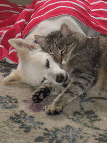
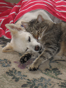
Keep the babies together as long as they are about the same age; this will help socialize them to their own species and will help in keeping them warm. Try to assemble the following equipment.

  1. The nest box in which the babies live should have tall sides so that they cannot climb out by mistake and become chilled. A cardboard pet carrier is perfect as it is portable, dark inside, and closable. These are inexpensive and should be available from a pet store or from your veterinarian.
  1. Place towels in the bottom of the box and cover them with a disposable diaper so the babies rest directly on the diaper. Most diapers have elastic leg holes and may have to be trimmed so they will lie flat. Expect to change the diaper several times daily. Keeping the babies clean and dry is very important, and a diaper is perfect for wicking away wetness.
  2. Place a heating pad under the nest box so that only half of the nest box is warmed. This way, the babies may crawl off the warm side of the box if they feel too hot. Alternatively, a water bottle filled with warm water can be buried in the blankets as a heat source as long as the babies have room to move away from it if they are too hot.

If the baby’s temperature drops below 94⁰F, the heart rate drops and intestinal motion ceases. Death occurs if this is not corrected.

Initial warming when babies have gotten cold should take place over an hour or two to avoid shock.

The Diet

  1. There are several brands of orphan formula on the market. For kittens, KMR® by PetAg and Just Born® by Farnam Pet Products come in both a powder and a liquid. The powdered form seems less associated with diarrhea than the liquid plus with the powdered form, the water content can be increased in the event of dehydration. For puppies, PetAg makes a canine formula called Esbilac® plus there is a canine version of Just Born formula. Again, both powder and liquid forms are available.
  2. Mix up the powder according to the directions on the can. If you are using the liquid form, you may want to dilute it with one part water for every two parts of formula. As the babies get older, less water may be used whether you are mixing up the powder or the liquid. If diarrhea occurs at any time, you should add more water to the formula to make up for fluid lost as diarrhea.
  3. Store the can of powder in the freezer after opening. Do not mix up more than a day’s worth of formula. Use a blender to mix the formula several hours ahead to allow time for the bubbles to settle. 

Makeshift Formula Until Commercial Formula can be Obtained

Puppies   
1 cup whole milk
1 tsp. salad oil
1 drop multi-vitamins (if you have any)
2 egg yolks

Kittens
1/2 cup whole milk
1 egg yolk
1 drop multi-vitamins
3 crushed Tums (antacid)

For either one, then mix in a blender.

The Bottle

  1. Obtain a pet nurser bottle from a pet store or your veterinarian.
  2. Use fine scissors or a hot needle to make a hole in the nipple. The hole should be big enough that formula will slowly drip out if the bottle is held upside-down and gently squeezed. The nipple should not collapse when the baby is sucking.
  3. Warm the bottle in a cup of hot water. Always test the formula before giving it to the babies. Taste it to be sure it is not sour. Do not use a microwave oven to heat the bottle as it may not heat evenly – some areas of the bottle can be scalding hot.

How / When To Feed

  1. Expect to feed them every two to three hours during the day. If this is done, the babies should be able to sleep through the night.
  2. Do not wake the babies at feeding time. Let them sleep. When they wake up hungry, they will let you know.
  3. During feeding be sure to tip the bottle so that no air is swallowed. It is more natural to feed them right side up rather than on their backs like human infants.
  4. Play with/rub them after feeding to burp them.
  5. Occasionally small amounts of formula will come out of the nose. The baby is drinking too fast. If excessive amounts of formula appear to be coming out the nose or if you are concerned, call your veterinarian.

Tube feeding involves using a syringe and lubricated tube to introduce formula through the mouth and directly into the stomach. Your veterinarian will need to show you how to do this should it become necessary. A baby who rejects three meals in a row will need tube feeding.

Urination / Defecation

  1. Infant animals are unable to take care of these matters alone and must be given help. Normally their mother’s tongue does the job as she washes them. Use a cotton swab, tissue, or your finger to gently rub the baby’s genital area. Have a tissue ready to catch the urine.
  2. Rubbing the anal area as well may also be necessary if the babies do not seem to be defecating as much as expected. Watch for diarrhea. Normal infant stool is loose but should not be watery.
  3. It is normal for baby kittens and puppies to have round bellies. If you think the baby might be constipated, usually taking the temperature rectally with a normal rectal thermometer provides enough extra stimulation to get things back to normal. Only the bulb of the thermometer tip needs to be inserted rectally.

Expect the baby to need stimulation for urination/defecation for the first three weeks of life.

Bathing

  1. Using kitten or puppy shampoo and warm water, bathe the babies a couple of times daily. Urine will burn their tender skin and caked feces can lead to infection, so keeping the babies clean is important.
  2. Take care not to submerge the infant in water. Be careful that it cannot drown or choke on the water and be sure the water temperature is acceptable.
  3. Gently blow dry the babies when the bath is over. Do not allow chilling.

How to be Sure You Are on Track

Starting Solid Food

The best way to be sure everything is going well is to track weight gain in your new babies. A postal scale or food scale (ideally one that measures weight in grams) will be helpful. A puppy or kitten should gain 10% of the birth weight every day and should be drinking 22 to 26 cc of formula per 100 grams of body weight over the course of the day. Puppies are variable in growth expectancy depending on breed, but kittens are more predictable and should gain 50 to 100 grams weekly. Kittens weighing less than 90 grams (approx three ounces) at birth have a high (59%) mortality rate.

  • When the babies start biting and chewing at their bottle instead of sucking (three to four weeks of age), they may be started on some finely textured canned food. At first it may be necessary to mix solid food with a little formula and /or smear a little around their mouths gently with a finger. Prescription recovery diets have a very soft texture and are readily accepted but any pate style canned food should do well. Your vet’s office will most likely have a recovery formula diet available.
  • Between ages four and six weeks, they should begin readily accepting solid food. New homes may be found for them at age eight weeks.

A Note on Colostrum

Colostrum is the first milk produced by the mother shortly after giving birth. It is rich in antibodies which will protect the babies for the first several months of life. Colostrum is only produced for a day or two and the baby is only able to absorb its antibodies for a day or two.

Without colostrum (if their mother did not nurse the kittens during the first two days of life) the babies have a serious immunological disadvantage. Especially great care should be taken in cleanliness and the babies can be vaccinated as early as age two weeks depending on circumstances. They may require a plasma transfusion to make up for the colostrum. There is no substitute for a real mother.

See more information on raising orphan kittens.

Many foster parents have difficulty distinguishing male and female kittens at this early age. See some guidelines and (illustrated) instructions.

4126881

Omega Three Fatty Acids for our Pets

Did you know that skin diseases account for as much as 25% of the cases seen by small animal veterinarians?

Skin problems typically faced by pets and their owners include:

  1.   Itching
  2.   Dandruff
  3.   Blackheads
  4.   Odor
  5.   Crusting
  6.   Redness
  7.   Rashes
  8.   Oiliness

The nutritional aspect of skin disease is quite broad. There are true nutritional deficiencies that affect the skin and other skin diseases that can be made dramatically better by using supplements.

Just because a condition responds to a nutrient does not necessarily mean that there is a deficiency of it.

Everyone wants their pet to have a lustrous beautiful coat and would like to do what it nutritionally possible to ensure this. Recently essential fatty acids have received a great deal of press. A brief primer follows. 

What is a Fatty Acid?

Biochemically, a fatty acid is what we just call fat. When we talk about different types of fatty acids, we are talking about different types of fat. A fatty acid consists of a long carbon chain (say 20 or so carbons in length) with biochemical acids called a carboxyl group at one end and a methyl group on the other end.

Saturated Vs. Unsaturated

Each carbon has four binding sites. In the carbon chain, two sites will be taken up by other carbons (i.e., the two adjacent carbons on the chain). In a saturated fat, the other two sites are taken up by hydrogen atoms. Saturated fats are typically solid at room temperature, like lard and butter, and are usually made from animals. Saturated fats are generally burned as fuel by our bodies. An example of a saturated fatty acid is depicted in the above illustration.

Unsaturated fats have two adjacent carbons held together by a biochemical double bond. These fats are generally liquid at room temperature and come from plants, such as olive oil, corn oil, etc.

Unsaturated fats can be classified as omega three fatty acids or omega six fatty acids, depending on the location of the double bond relative to the methyl group at the end of the chain. These types of fatty acids are essential, meaning that our bodies cannot make them; instead, we must eat them in our diet. These fats are not burned for fuel but are used as structural components.

The omega six fatty acids are used as the main structural components in our cells. Omega three acids are used in the retina and central nervous system.

For healthy skin and coat, the diet must contain adequate omega six fatty acids as these make up the surface of the skin.

Examples of omega six fatty acids (also called n-6 fatty acids): Linoleic acid, gamma linolenic acid, and Arachidonic acid. Evening Primrose oil is an excellent example.

Examples of omega three fatty acids, also called n-3 fatty acids, include: Alpha linolenic acid (ALA), Eicosatetraenoic acid (EPA), docosahexaenoic acid (DHA).

Cold water fish oils are an excellent source of DHA and EPA. A terrific source of ALA would be flax seed oil. DHA has anti-inflammatory properties, which is why it is so frequently recommended in inflammatory conditions. Many people prefer to use flax seed oil as their omega 3 source because flax seed oil does not taste fishy and people readily convert ALA to DHA. This is great for people but it turns out that pets are only able to convert about 10% of ALA to DHA, so for them cold water fish oils are better. Fortunately, most dogs and cats like the fishy taste. 

Should we Supplement Essential Fatty Acids?

There is no question that a diet must contain adequate omega 6 fatty acids to maintain optimal skin and coat quality. A diet found to be “complete and balanced” will have an amount of omega 6 fatty acids that should be optimal for a normal animal.  

But there’s more.

Omega 6 Fatty Acids and Dandruff

Research has shown that dogs with oily, dandruffy skin (seborrhea) have insufficient omega 6 fatty acids in their skin despite eating a diet that should be optimal. When omega 6 fatty acids are supplemented, the seborrhea improves. This finding supports the old-time remedy of adding a spoonful of corn oil to the diet to ensure a glossy coat. Seborrhea is a complex condition but animals with it may need more omega 6 fatty acids.

Anti-Inflammatory Effects of Omega 3 Fatty Acids

Omega 6 fatty acids constitute our cell membranes. During some biochemical situations, it is necessary to produce hormone-like substances called prostaglandins and leukotrienes. These substances are made from omega 6 fatty acids and the resulting prostaglandins and leukotrienes are not necessarily good for us. In fact, these substances are responsible for itching and inflammation leading to the clinical skin problems listed above. One way to address this is to supplement omega 3 fatty acids, which become incorporated into cell membranes along with the omega 6s. After a couple of months of supplementation, omega 3 fatty acids infiltrate cell membranes significantly. When it comes time to make prostaglandins, the omega 3s are mobilized instead of the omega 6s only in this case, the prostaglandins that result are not inflammatory. When omega 3 fatty acids are supplemented, itching can be substantially reduced and even arthritis pain improved.

One problem with this is that no one really knows how much omega 3 fatty acid to supplement. There is some evidence that a ratio of omega 6 to omega 3 fatty acids in the supplement is crucial. If this is so, clinical research becomes hugely complicated as the diets of pets cannot be standardized easily for study. If pets in a study eat different diets, then it is impossible to tell what overall omega 6:omega 3 ratio each is receiving. Essential fatty acids are being pursued as treatment for diseases of virtually every organ system; watch for new research in this area.

Conditions that Have Been Shown to Benefit from Omega 3 Fatty Acid Supplementation

  1.   Renal (kidney) insufficiency
  2.   Heart failure
  3.   Lymphoma
  4.   Airborne allergies

Research is ongoing. We know that supplementing with omega 3 appears to be benign with the potential to do a great deal of good. 

4128011

Osteosarcoma in Dogs

What is Osteosarcoma?

Osteosarcoma is by far the most common bone tumor of dogs, usually striking the leg bones of larger breeds. Osteosarcoma usually occurs in middle aged or elderly dogs but can occur in a dog of any age; larger breeds tend to develop tumors at younger ages.

  • Osteosarcoma can develop in any bone but the limbs account for 75-85 percent of affected bones. Osteosarcoma of the limbs is called appendicular osteosarcoma.
  • It develops deep within the bone and becomes progressively more painful as it grows outward and the bone is destroyed from the inside out. The lameness goes from intermittent to constant over 1 to 3 months. Obvious swelling becomes evident as the tumor grows and normal bone is replaced by tumorous bone.
  • Tumorous bone is not as strong as normal bone and can break with minor injury. This type of broken bone is called a pathologic fracture and may be the finding that confirms the diagnosis of bone tumor. Pathologic fractures will not heal and there is no point in putting on casts or attempting surgical stabilization.

How do we Know my Dog Really has an Osteosarcoma?

Radiographs (x-rays)

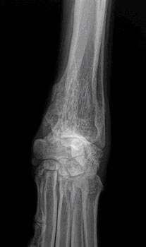
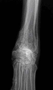
The lytic lesion looks like an area of bone has been eaten away. One of the first steps in evaluating a persistent lameness is radiography (x-rays). Bone tumors are tender so it is usually clear what part of the limb should be radiographed. The osteosarcoma creates some characteristic findings.

  1. The sunburst pattern – shows as a corona effect as the tumor grows outward and pushes the more normal outer bone up and away.
  2. A pathologic fracture may be seen through the abnormal bone.
  3. Osteosarcoma does not cross the joint space to affect other bones in the joint.

Radiography is almost completely diagnostic in most cases, but there are a few other far less common conditions that can mimic the appearance of a bone tumor, so a confirming test is going to be needed if one is to be complete. If a basic blood panel and urinalysis haven’t been done, this would also be a good time to do so as basic information about liver and kidney function will be needed for treatment regardless of whether this turns out to be a bone tumor or not; plus, a tissue sample from the bone is needed for confirmation (see later).

Tissue Sampling: Biopsy and Needle Aspirate

Radiographs are close to being confirmatory but still they are not definitive. Since life and death decisions are going to be made, it is best to obtain a tissue sample for confirmation. This can be done by either biopsy or by needle aspirate.

Biopsy

A small piece of bone can be harvested surgically. The bone is preserved, sectioned, and examined under the microscope to confirm the diagnosis of osteosarcoma. There are several problems associated with this diagnostic.  Sometimes a bone tumor is surrounded by an area of bone inflammation and it may be difficult to get a representative sample. The tiny hole that results when a core of bone is removed can create a weak spot and the bone can actually break. Even if the procedure goes well, often there is increased pain and lameness for the patient afterwards. With so many potential problems, most specialists have switched to needle aspirate for diagnosis.

Needle Aspirate

With needle aspirate, a large bore needle is inserted into the area of the tumor and cells are withdrawn for analysis. A full core of bone is not removed, just a sampling of cells. This is usually sufficient to confirm osteosarcoma. If there is ambiguity, certain stains can often settle any questions the pathologist may have.

With a Diagnosis Confirmed, Staging is the next Consideration

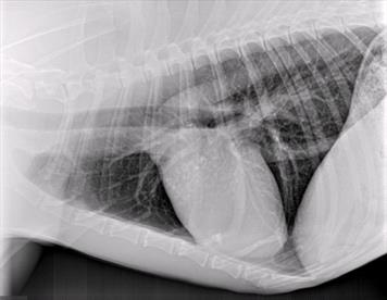
Osteosarcoma is an aggressive tumor and it can be assumed to have spread by the time it is first diagnosed; there is no possibility of preventing spread. That said, how well treatments can be expected to work depend on whether or not the tumor spread has progressed so as to be visible. Because osteosarcoma spreads to the lungs as one of its first stops, chest radiographs are important in checking for visible tumor spread. If there is already visible tumor spread at diagnosis, this changes what treatments are recommended.

Some specialists recommend nuclear imaging of the skeleton to identify any spread to other bones, which might also alter recommendations; however, this form of imaging is not readily available.

What if it isn’t Really an Osteosarcoma?

The location and radiographic appearance of the osteosarcoma in the limb are quite classic but there are a few outside possibilities that should at least be mentioned. Only a few other possible conditions cause similar lesions in bone: the chondrosarcoma, the squamous cell carcinoma, the synovial cell sarcoma, or fungal bone infection.

Chondrosarcoma

Chondrosarcoma is a cartilage tumor, possibly not as malignant as the osteosarcoma. The chondrosarcoma generally occurs on flat bones such as ribs or skull bones and is not usually found in the limbs. Still, should a chondrosarcoma occur in the limb, treatment recommendations still include amputation of the affected bone and many of the same treatments as for osteosarcoma.

Squamous Cell Carcinoma

The squamous cell carcinoma is a tumor of the external coating of the bone, called the periosteum. This is a destructive tumor locally but it tends to spread relatively slowly. Again, a bone suspected of malignant tumor should be amputated, and the tissue then analyzed and then treatment adjustments should be made thereafter. The squamous cell carcinoma tends not to arise in the same bone areas as the osteosarcoma; it tends to arise in the jaw bones or in the toe bones.

Synovial Cell Sarcoma This is a tumor of the joint capsule lining. Its hallmark is that it affects both bones of the joint. The osteosarcoma, no matter how large or destructive it becomes, will never cross over to an adjacent bone. Prognosis is much better with amputation with synovial cell sarcoma.

Fungal Bone Infection

Coccidiodes immitis is a fungus native to the lower Sonoran life zone of the southwestern United States. It is the infectious agent of the disease called San Joaquin Valley Fever or just Valley Fever. (More scientifically, the condition is called coccidioidomycosis.)  In most cases, infection is limited to a few calcified lymph nodes in the chest and possibly lung disease. In some rare cases, though, the fungus disseminated through the body and can cause a very proliferative bone infection.

The bone infection of coccidiodomycosis grows rapidly and lacks the lytic lesions that are typical of the osteosarcoma. Other fungi, such as Histoplasma capsulatum, also have potential for bone involvement. Pursuit of this possibility makes sense if you live in an area where these fungi are a concern. Disseminated fungal disease is serious and even though this diagnosis is not cancerous, amputating the limb is most likely going to be necessary.

Treatment of osteosarcoma involves two aspects: treating the pain and fighting the cancer’s spread.

How do we Treat the Pain?

Keep in mind that dogs are usually euthanized because of the pain in the affected bone. Treating the pain successfully will allow a dog to live comfortably and extend life expectancy by virtue of extending comfort.

There are two ways to address the pain: amputatating the limb and palliative radiotherapy (usually combined with periodic bisphosphonate infusion treatments).

For most patients, there is one tumor on a leg and no visible tumor spread in the lungs. These are the patients with the best potential results and they are good candidates for amputation. Patients with a lot of arthritis in the other legs or with tumor spread evident in the chest already are probably not candidates for amputation and it may be more appropriate to keep the leg and simply relieve the pain with radiotherapy.

Amputating the Limb

Since the tumor in the limb is the source of pain, it makes sense that amputating the limb would resolve the pain. In fact, this is true. Removing the affected limb resolves the pain in 100 percent of cases. Unfortunately, many people are reluctant to have this procedure performed because of misconceptions.

  • While losing a leg is handicapping to a human, losing one leg out of four does not restrict a dog’s activity level. Running and playing are not inhibited by amputation after recovery from surgery. That said, if the remaining legs are arthritic, the stress on them can pose a mobility issue.
  • While losing a limb is disfiguring to a human and has social ramifications, dogs are not self-conscious about their image. The dog will not feel disfigured by the surgery; it is the owner that will need to adjust to the dog’s new appearance.
  • Median survival time for dogs who do not receive chemotherapy for osteosarcoma is 3 to 5 months from the time of diagnosis regardless of whether or not they have amputation. Do you want your dog’s last 3 to 5 months to be painful or comfortable?

Read a letter about amputation from a veterinarian who sees too many owners who reject the option of amputation out of hand. His letter includes videos of two happy dogs without limbs.

Limb-sparing Surgery (removing the tumor but not the leg)
Limb-sparing techniques developed for humans have been adapted for dogs. To spare the limb and thus avoid amputation, the tumorous bone is removed and either replaced by a bone graft from a bone bank or the remaining bone can be re-grown via a new technique called bone transport osteogenesis. The joint nearest the tumor is fused (i.e., fixed in one position and cannot be flexed or extended.)

  1. Limb sparing cannot be done if more than 50% of the bone is involved by tumor or if neighboring muscle is involved.
  2. Limb sparing does not work well for hind legs or tumors of the humerus (arm bone.)
  3. Limb sparing works best for tumors of the distal radius (forearm bone).
  4. Complications of limb sparing can include: Bone infection, implant failure, tumor recurrence, and fracture.

While amputation can be viewed as a pain management strategy, limb-sparing is only performed in conjunction with chemotherapy. It is important to keep in mind that grafting of a new bone structure requires healing time and that a great deal of post-operative confinement time is needed (in a patient whose life expectancy is going to be measured in months). For the right patient, limb-sparing can be the best choice but be sure to understand all the details of post-operative care from the specialist.

Palliative Radiotherapy for Pain Control

Sometimes amputation is simply not the right choice and happily there is an effective alternative treatment. Radiation can be applied to the tumor in two, three or four doses, depending on the protocol. Improved limb function is usually evident within the first 3 weeks and typically lasts 2 to 4 months. When pain returns, radiation can be given again for further pain relief if deemed appropriate based on the stage of the cancer at that time.

There are a couple of caveats:

  • When pain is relieved in the tumorous limb, there is an increase in activity that can in turn lead to a pathologic fracture of the bone.
  •  Radiotherapy does not produce a helpful response in about 1/4 of patients. Remember, amputation controls pain in 100 percent of cases but if amputation is simply not an option, there is a 3 out of 4 chance that radiotherapy will control the pain.

Current standard treatment involves pairing palliative radiation with monthly infusions of medications called bisphosphonates. 

Bisphosphonates

This class of drug has become the standard of care in humans with bone tumors and have been found helpful in managing osteosarcoma pain in dogs as well. Bisphosphonates act by inhibiting bone destruction, which in turn helps control the pain and bone damage caused by the bone tumor. The most common bisphosphonate in use for dogs has been pamidronate, though a new drug zoledronate is taking its place gradually. Treatment is given as an IV drip over two hours in the hospital every 3 to 4 weeks. In humans, an assortment of potential side effects have emerged (fever, muscle pain, nausea all lasting 1 to 2 days in up to 25 percent of patients, renal disease in certain situations, low blood calcium levels, jaw bone cell death); these issues so far have not panned out as problems for dogs and cats. Bisphosphonates are important in managing bone tumor pain in patients that have no undergone amputation.

Analgesic Drugs

At this time there are numerous analgesic medications available for dogs with this tumor. No single medication, however, is a match for the pain involved in what amounts to a slowly exploding bone. A combination of medications is needed to be reasonably palliative and should be considered only as a last resort if amputation or radiation therapy will not be pursued. There are several types of drugs that can be combined.

  • Non-Steroidal Anti-Inflammatory Drugs (NSAIDs)
    These are anti-inflammatory pain relievers developed for dogs: carprofen, etodolac, deracoxib, meloxicam, firocoxib, and others. These are typically given once or twice daily in tablet form at home. The patient should have good liver and kidney function in order to take medications of this class.
  • Narcotic Pain Relievers
    While these drugs do not have anti-inflammatory properties, they are well-known analgesics and have been used in an assortment of forms for thousands of years. They are particularly useful in chronic pain because they do not interact negatively with other pain relievers. Drowsiness is a potential side effect.
  • Miscellaneous Supplemental Pain Relievers
    There are two drugs that have surfaced as adjunct pain relievers for animals with chronic pain: gabapentin and amantadine. Gabapentin works on neurologic pain and is rapidly surfacing in the treatment of arthritis, surgical pain, and other chronic pain states. Amantadine works by reducing what is called wind up, a phenomenon where nerves become sensitized to pain leading to the experience of pain from stimuli that normally do not cause pain.

These different drugs are often given together to create analgesia to the osteosarcoma patient when amputation and radiotherapy are not going to happen. It is important to realize that there is a limit to how much pain relief can be achieved against a bone tumor with only pills. It will not be long before the pain of this tumor, as evidenced by not using the leg, tenderness to the touch, etc., overpowers the effect of oral medications.

How do we Treat the Cancer?

 Osteosarcoma is unfortunately a fast-spreading tumor. By the time the tumor is found, it is considered to have already spread. Osteosarcoma spreads to the lung in a malignant process called metastasis. Prognosis is substantially worse if the tumor spread is visible on chest radiographs, so if you are contemplating chemotherapy, chest radiographs should be taken.

  1. Chemotherapy is the only meaningful way to alter the course of this cancer.
  2. Young dogs with osteosarcoma tend to have shorter survival times and more aggressive disease than older dogs with osteosarcoma.
  3. Elevations of alkaline phosphatase, one of the enzymes screened on a basic blood panel, bode poorly. These dogs have approximately 50% of the survival times quoted below for each protocol.
  4. A tumor in the lymph nodes local to the leg being amputated also bodes poorly. In the study by Hillers et. al published in the April 15th, 2005 issue of the Journal of the AVMA, median survival was significantly longer (318 days vs. 59 days) in dogs where the tumor was not evident in local lymph nodes at the time of amputation. 

Cisplatin (given by IV every 3 to 4 weeks for 3 treatments)

  1.   The median survival time with this therapy is 400 days.
  2.   Survival at 1 year: 30% to 60% 
  3.   Survival at 2 years: 7% to 21%
  4.   Giving less than 3 doses does not increase survival time (i.e., if one can only afford one or two treatments, it is not worth the expense of therapy)
  5.   Cisplatin can be toxic to the kidneys and should not be used in animals with pre-existing kidney disease.

Carboplatin (given by IV every 3 to 4 weeks for 4 treatments)

  •   Similar statistics to cisplatin but carboplatin is not toxic to the kidneys and can be used if the patient has pre-existing kidney disease.
  •   Carboplatin is substantially more expensive than cisplatin.

Doxorubicin (given IV every 2 weeks for 5 treatments)

  1.   The median survival time is 365 days.
  2.   10% still alive at 2 years.
  3.   Toxic to the heart. An ultrasound examination is needed prior to using this drug as it should not be given to patients with reduced heart contracting ability.

Doxorubicin and Cisplatin in Combination (both given IV together every 3 weeks for four treatments)

  •   48% survival at 1 year
  •   30% survival at 2 years
  •   16% survival at 3 years. 

What is Median Survival Time? 

There are a number of ways to statistically evaluate the central tendency of a group. The median is the value at which 50% of the group falls above and 50% of the group falls below. This is a little different from the average of the group, though more people are familiar with this term. When evaluating median survival times, you are looking at a 50% chance of surviving longer than the median and a 50% chance of surviving less than the median. 

What Does Chemotherapy Put my Dog Through?

Most people have an image of the chemotherapy patient either through experience or the media and this image typically includes lots of weakness, nausea, and hair loss. In fact, the animal experience in chemotherapy is not nearly as dramatic. After the pet has a treatment, expect 1 to 2 days of lethargy and nausea. This is often substantially helped with medications like Zofran, a strong anti-nausea drug commonly used in chemotherapy patients. These side effects are worse if a combination of drugs is used but the pet is typically back to normal by the third day after treatment. Effectively, you are trading 8 days of sickness for 6 to 12 months of quality life. Hair loss is not usually a feature of animal chemotherapy. In dogs, hair loss may occur in breeds that have continuously growing coats, such as poodles, Scottish terriers, and Westies. 

Axial Osteosarcoma

While osteosarcoma of the limbs is the classical form of this disease, osteosarcoma can develop anywhere there is bone. “Axial” osteosarcoma is the term for osteosarcoma originating in bones other than limb bones, with the most common affected bones being the jaws (both lower and upper). Victims of the axial form of osteosarcoma tend to be smaller, middle-aged, and females outnumber males two to one.

In the axial skeleton the tumor does not grow rapidly as do the appendicular tumors, thus leading to a more insidious course of disease. The tumor may be there for as long as two years before it is formally diagnosed. An exception is osteosarcoma of the rib, which tends to be more aggressive than other axial osteosarcomas.

Treatment for axial osteosarcoma is similar to that for the appendicular form: surgery followed by chemotherapy. There is one exception, that being osteosarcoma of the lower jaw. Because of the slower growth of the axial tumor and the ability to remove part or all of the jaw bone with little loss of function or cosmetic disfigurement, it has been reported that 71% of cases survived one year or longer with no chemotherapy at all.

Additional information can be found at Bone Cancer Dogs, Inc., a nonprofit corporation.

Not all veterinarians are comfortable treating osteosarcoma. Discuss with your veterinarian whether referral to a specialist would be best for you and your pet.

4128293

Otitis Media (Middle Ear Infection) in Dogs and Cats

The word otitis means ear infection, but that one word doesn’t indicate which part of the ear is infected. Otitis media is inflammation and/or infection of the middle ear, whereas otitis externa begins in the external ear canal. Some nerves are closely associated with the middle ear, so infection there can damage them, causing the neurologic signs often seen with this infection: a head tilt, lack of balance, nystagmus (back-and-forth eye movements). These symptoms are called vestibular signs. Middle ear infections can also cause paralysis of the facial nerve, leading to a slack-jawed appearance on that side of the face.

Otitis media most often develops after an existing otitis externa travels from the external ear canal through the tympanic membrane and into the middle ear. The infection in the external ear canal leads to inflammation and damage to the ear canal and tympanic membrane, allowing the infection to enter the middle ear. 

Otitis media has been found in 16% of dogs that have acute (rapid onset) otitis externa and in 52% of dogs that have chronic (long-term) otitis externa. In contrast, 63% of cats that had otitis media/interna did not have a previous history of ear infection. This is why keeping your pet’s ears clean and watching for infection are important to your pet’s health.

Other causes of otitis media can include infections in the nose and throat, trauma, foreign bodies, fungal infections, inflammatory polyps, cancer, etc. In addition, developmental abnormalities of the external ear canal and pharynx can lead to otitis media.

Otitis media does not affect any particular age or animal, and males and females are affected equally.

Dogs that have long, pendulous ears, such as beagles and basset hounds, are more likely to develop otitis externa, which can lead to otitis media.

The Cavalier King Charles Spaniel breed is known to have a condition called primary secretory otitis media (PSOM). Dogs with PSOM typically have mild to moderate pain in the head/neck, neurologic signs (e.g. ataxia, facial paralysis, head tilt), and itchy ears, but without otitis externa. Owners of this breed will want to keep this disease in mind. 

Diagnosis

Diagnosis includes physical examination, ear examination with an otoscope, bacterial/fungal culture, radiography, and possibly advanced imaging.

Physical Examination

During the physical examination, your veterinarian will be looking for some of these signs: head shaking, pawing at the ear, pain when opening the mouth, signs of otitis externa (ear odor, redness, etc.), head tilt, facial nerve signs, Horner syndrome, ataxia, etc. Not every affected pet will exhibit all of these signs. In addition, cats are more likely to have neurologic signs than dogs are.  

Ear Examination (Otoscopy)


In order to do a complete examination of the ear, a deep ear cleaning may be necessary. To keep your pet comfortable, your veterinarian may decide to sedate or anesthetize your pet for the procedure. Then the infection can be more easily seen with an otoscope, which can see the eardrum.

Culture and Sensitivity

Your veterinarian will usually take samples of the material in the ear and culture those samples to determine what medications will work best.

Radiographs

Your veterinarian may take radiographs, also called X-rays. Radiographs can help show changes (thickening, destruction) to the bony structures of the ear. However, even if the radiographs appear normal, that does not mean that your pet does not have otitis media.

Advanced Imaging

In some cases, your veterinarian may suggest using computed tomography (CT) and/or magnetic resonance imaging (MRI) to further evaluate the tympanic bulla. CT is usually better than MRI at detecting bony changes, while MRI is better at detecting soft tissue abnormalities.

Treatment

Cleaning both the external and middle ear canals is an important step in treating otitis media. Debris can inactivate some topical medications and prevent medications from contacting infected tissues. Anesthesia will be necessary in order to do the deep ear cleaning and ear flushing that will remove the debris.

Your veterinarian may need to infuse (fill) the ear with medication in order to deliver a high concentration of antimicrobial products or corticosteroids directly to the affected area.

Topical medications may need to be used to control an otitis externa.

Systemic therapy with antibiotics, antifungals, and corticosteroids may also be used, depending on the infection.

If severe, irreversible changes to the external ear canal are found, if there are masses in the inner ear, or if medical therapy has failed to resolve the otitis media, your veterinarian may advise surgery, such as an ear canal ablation. This surgery is not undertaken lightly, but it may be necessary to control the infection. An ear canal ablation involves removing the vertical and horizontal portions of the ear canal, along with the bones of the middle ear and eardrum.

Monitoring (Rechecks)


During treatment, your veterinarian will usually want to recheck your pet’s external ear canal and tympanic membrane every 10-14 days. (A ruptured tympanic membrane will generally begin to heal within 21-35 days.) Topical medications will be given until the external ear canal and the tympanic membrane are normal, and until cytology of the ear canal reveals no infection. Systemic therapy is typically given for at least 4-6 weeks.

Prognosis

In general, the prognosis for dogs is good if appropriate therapy is started in time. The prognosis is not as good if the infection is resistant to the medications; if the concurrent otitis externa is not managed adequately; if there is significant bone infection; or if there is no response to surgery. In addition, some neurologic signs (e.g., facial nerve problems, Horner syndrome) may be permanent.

Reminder

Keeping your pet’s ears clean is critical to preventing ear infections of any kind. However, It is not always possible to prevent every infection in some pets. 

6274132

Osteochondritis Dissecans (OCD) in Dogs

If your adolescent puppy becomes lame or stiff when rising or after play on one or more legs, there are several possible reasons for it. One reason is a disease called osteochondritis dissecans (OCD). OCD negatively impacts the cartilage in certain joints. Cartilage is connective tissue that provides cushioning on the joint surface to soften the impact when walking and running. When the cartilage is damaged, the joint cannot function properly; discomfort and pain become lasting effects.

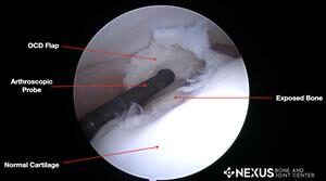
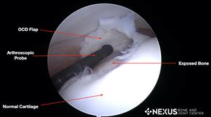
OCD is a condition where a cartilage flap forms during development. This flap will induce a painful inflammatory response within the joint.

In some situations, the flap separates, floating uselessly around the inside of the joint and causing pain and further destruction to the joint.

OCD is most commonly seen in the shoulder, but can also be seen in the knee, elbow, and ankle joints. OCD causes differing levels of pain and lameness, making it hard for your young dog to be active.

Generally speaking, OCD is a developmental disease seen in rapidly growing puppies of some but not all large breeds, including Great Danes, Bernese Mountain Dogs, Golden Retrievers, and German Shepherds. Some breeds are more likely to have it than others. It’s typically a disease of adolescence with onset often seen around the age of 4 to 9 months. In people, it is usually seen in children and adolescents. OCD is seen far more often in male dogs than females.

If your dog is stiff and sore after rising or heavy play, or is lame, it’s best to see the veterinarian as early as possible. Typically, when a lameness is noted in a young dog with an OCD lesion (the entire area affected), the lesion is chronic and warrants immediate treatment.

Causes

No one really knows what causes OCD, although various factors can contribute. A hereditary component is suspected in most dogs, especially Newfoundlands. Before you buy a puppy, ask your breeder specifically if the puppy’s parents have OCD. In general, it is considered multi-factorial in that a combination of factors contribute to it.

Along with genetics, diet also plays a role; in particular, puppies whose diet has too much calcium or vitamin D. Supplementation should be avoided in puppies. In addition, any effort to make the dog grow as big as possible as quickly as possible should be avoided. It’s healthiest to let the dog grow to the size nature intended without any dietary interference.

Other potential causes of OCD include injury, lack of blood flow and hormonal issues.

Signs

Dogs will initially show stiffness and soreness when rising or after heavy play. Overtime, this will turn into a limp in the affected leg or legs because they avoid bearing weight on a painful leg. If you press on that sore area, the dog may cry. Sometimes an affected joint is warm and swollen.

Lameness is a sign of pain. An OCD lesion can either be constant or once in a while.

Diagnosis

Most dogs are taken to the veterinarian because they are stiff after rising or play or are lame in at least one leg. Various causes of stiffness and lameness can occur in the joints of developing dogs. Diseases and problems such as elbow dysplasia, patella (kneecap) luxation, and growth plate fractures can have similar signs as an OCD lesion.  A thorough orthopedic examination and imaging should be performed by your veterinarian for a diagnosis and to rule out other diseases.

Radiographs (X-rays) should always be taken in a lame growing dog. Oftentimes sedation is needed because specific positioning is necessary, and several images must be taken. Radiographs are a great screening tool, but an OCD lesion is not always seen. In some cases, your veterinarian may prefer that the X-rays be reviewed by a veterinary radiologist. If there is a high suspicion of an OCD lesion, your veterinarian may recommend a CT scan or even an arthroscopy evaluation. A CT scan allows veterinarians to view the bone in more of a three-dimensional manner while an arthroscopic examination involves inserting a small video camera into the joint. This allows direct visualization and treatment of an OCD lesion. 

Treatment

Obviously, treatment depends on severity of the OCD lesion. Conservative management is only recommended for dogs less than 6 months of age with minimal clinical signs and no overt radiographic changes. Conservative management may consist of pain medications, exercise restriction, dietary changes, and formal rehabilitation therapy.

For any dog over the age of 6 months, conservative management of an OCD lesion is likely to be ineffective. Surgery is indicated to remove the cartilage flap and to stimulate the underlying bone bed to form scar tissue (fibrocartilage).  In some cases with large lesions, an artificial plug may be inserted into the bone bed.

Arthroscopic surgery is the gold standard for OCD treatment given the minimally invasive nature, better visualization, and quicker recovery. Total recovery time is about 8-12 weeks of exercise restriction to avoid running, jumping, and playing. The first 2 weeks after surgery activity is restricted to short, leashed walks – basically just enough for the dog to relieve themselves along with passive range of motion exercises. Beginning 2 weeks after surgery, a structured daily leash walking program and home exercise plan are begun along with formal rehabilitation guided by a rehabilitation therapist.

Prognosis

The earlier the problem is dealt with, the better the prognosis is. Any stiffness or lameness in a puppy warrants immediate evaluation by your veterinarian. More times than not, the stiffness or lameness may not be anything significant. However, the earlier your veterinarian can diagnosis the problem and recommended surgery, the better off your dog’s recovery will be.

Your dog’s prognosis depends on which joint or joints are affected, and how severely. Prognosis for full recovery is good if only the shoulder is affected. Other joint involvement has a more guarded prognosis, meaning progression of arthritis as well as possible periods of intermittent lameness.

Keeping your dog at a healthy weight helps keep the joints working properly. Your veterinarian can help you help your dog lose weight if that would be beneficial.

4477841

Nighttime Waking in Senior Dogs

Sleep is essential for dogs and people. Disturbances of the sleep-wake cycle occur in older pets and often disrupt the entire family. Interrupted sleep is a common problem in older pets and may occur for many reasons, including both medical and anxiety-related causes.

Diagnosis and Clinical Signs

There are many reasons an older dog may have difficulty sleeping at night. Dogs do get cognitive dysfunction, which is similar to dementia in people.  The hallmarks of these syndromes are progressive confusion, reversal of day-night wake-sleep patterns, and poor adaptability to new situations. Just like older people with early-stage dementia, the dog may function well in routine and familiar situations but be confused in new situations.

Waking at night may occur for reasons other than cognitive dysfunction, such as pain or discomfort. Dogs who are feeling discomfort may pace, pant, vocalize, or be restless at night but may not show any physical limitations during the day. The effects of pain or sore muscles may be more noticeable to the dog at night.

The urge to eliminate may wake many dogs. Senior pets are more likely to have medical conditions that would increase the pet’s urination or defecation frequency or decrease control of these bodily functions.

Many physical and medical conditions should be considered, including urinary tract infections, kidney disease, hormone imbalances, Cushing’s Syndrome, gastrointestinal (GI) upset, and neurological impairments.

Anxiety can cause nighttime waking. Dogs may be anxious because of weather-related noises such as thunderstorms or noises in the home from people or other animals.  The dog may also be anxious during the day but the severity is often increased at night since these noises are more pronounced and start when it is quiet. Furthermore, the dog’s reaction may be limited to restlessness or pacing, which is often not noticed during the day. Seizures may occur anytime but often seizures that occur at night are not seen by the family, so people are awakened by a dog in a confused state.

Thinking about the pattern of when the dog wakes can help figure out the cause and at least partially correct the problem. Some dogs have difficulty settling down or falling asleep while other dogs may go to sleep easily and then wake up in the middle of the night. Some dogs go to sleep, enjoy slumber much of the night, and then wake up early. A definitive diagnosis may be difficult because there are so many medical conditions to consider. Cognitive dysfunction is only diagnosed with certainty in the later stages, and there is no test for it.

Management

After evaluating possible medical causes, try to establish a predictable routine for nighttime slumber. The pet’s routine as a younger dog may provide some insight into his preferences but consider age-related changes. An older dog may require an orthopedic bed to ease pressure on bones and joints. Warmth may also be important as the dog may not be able to regulate body temperature and cold weather may affect sensitive joints.  Establish a bedtime routine that may include a late trip outside for elimination, a massage, and quiet time settling in a comfortable area. Most dogs prefer to be with family members but some prefer to rest alone. Quiet, soothing music may obscure sounds and a night light may help a dog with vision problems to walk in dark hallways with more ease.

Even though this condition may be frustrating, avoid punishing or scolding. After addressing your dog’s physical needs, try gently and calmly returning your dog to where he likes to sleep. Some dogs want to sleep in bed with people; this is a matter of personal preference and family tolerance. Unless it is unsafe due to aggression, it is acceptable for dogs to sleep with people. Be sure to consider if this pattern of slumber with canine companions will continue to be acceptable since once they have tried it, many dogs like to sleep on comfortable mattresses with comforters. Despite commonly perpetuated myths, this type of “spoiling” does not cause other behavior problems; it does, however, teach a dog it is nice to sleep in a person’s bed.

Nutraceutical and Pheromone Interventions

Cognitive dysfunction is a slow, degenerative, and progressive disorder. Supplements are available that may ease the severity and slow its course. Ask your veterinarian about diets designed for the unique needs of senior pets. Some of them provide antioxidants (vitamins C and E, beta carotene, selenium, flavonoids, and carotenoids) that may reduce the neurological damage caused by free radicals and fatty acids as well as dl-alpha-lipoic diet and l-carnitine. Others include botanic oils such as medium chain triglycerides (MCT’s) that provide an alternative energy source. These diets are shown to improve a senior dog’s performance on cognition tests.

Dietary supplements that contain phosphatidyl serine, ginkgo biloba, and grape extract (resveratrol) are likely to slow and minimize cognitive impairment. S-adenosyl-l-methionine, or SAMe, may reduce anxiety and increase awareness and activity in the senior dog. Furthermore, SAMe is safe or even beneficial for patients with compromised liver function. These supplements are most beneficial in the early stages of cognitive decline.

A dog-appeasing pheromone (such as Adaptil) may ease anxiety and promote a feeling of well-being. The diffuser may be especially useful to create an area and support the use of a desired area for resting. The collar is most useful for dogs who experience anxiety all the time.

Melatonin may help create a normal nighttime sleeping cycle. This supplement should be given at bedtime on a regular basis. Combined with a predictable nighttime routine, it can help create a normal sleep pattern.

Pharmacological Treatments

Sometimes medications may be helpful to induce sleep. Benzodiazepines (anti-anxiety medications) such as diazepam (Valium), alprazolam (Xanax), or oxazepam (Serax). Trazodone may also be helpful. These medications are relatively quick-acting but don’t last long so administration must be timed carefully. Medication should be combined with other recommendations described above.

Generalized anxiety may be reduced with medications such as SSRI’s or TCA’s but these medications require many weeks before positive effects are seen.  Cognitive dysfunction symptoms may be reduced with selegiline (Anipryl), which when given in the morning may help to keep the dog more alert and active during the daytime. Old age onset of anxiety may reflect the additive effect of lifelong learning or be a sign of progressive decline from cognitive dysfunction.

Expected Outcome

Nighttime waking may be caused by medical or behavioral changes common in senior dogs. Often these problems may be managed and good sleeping patterns can be re-established, though many of the underlying conditions may be persistent and worsen with age. Quick intervention is important since undesirable habits may form quickly and people may become frustrated when they’re sleep deprived.

4128250

Nebulizer Use for Dogs and Cats

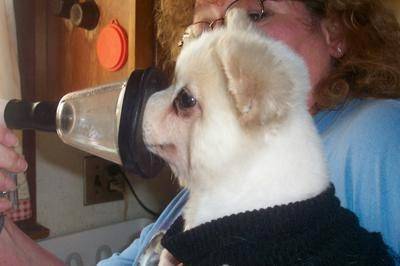
Dogs, cats, and other pets who develop lung diseases such as pneumonia, asthma, kennel cough, bronchitis, and so on can benefit from a nebulizer delivering saline, albuterol, antibiotics, or other drugs. A nebulizer is a machine that aerosolizes medicine into a fine mist; it’s like spraying medicine. A face mask is held over the pet’s muzzle (most pets can learn to accept the face mask) so that medication is inhaled deep into the respiratory tract.
 
The esophagus lies above the trachea and is protected by the epiglottis. Pneumonia may result from infections caused by kennel cough, viruses and bacteria. Pets with megaesophagus are prone to developing aspiration pneumonia because they tend to regurgitate food and fluids into the back of the throat and then aspirate the material into the lungs.

A nebulizer can be purchased from human medical supply stores, online, or from eBay. There are several good nebulizer brands, such as Pulmo-Aide, Omron, Devilbiss, etc. Make sure that you obtain the compressor (the machine), tubing, and the medication cup. The tubing, T-piece (a t-shaped tube that attaches the machine to the pet and to an exit) and medicine cup are called a nebulizer kit.

A vaporizer is not the same as a nebulizer. A vaporizer is typically used for sinus or upper respiratory issues rather than lung issues. Vaporized droplets are too big to get as deeply into the lungs the way nebulized particles

You can purchase an appropriately sized mask from a veterinarian, or make one yourself.

Using a Nebulizer

Follow these steps to use a nebulizer.

  1. Plug in the compressor.
  2. Insert one end of the tubing into the compressor and the other end into the cup.
  3. Place the correct dose of the medicine in the medicine cup.
  4. Start the compressor.
    a. Keep the cup up straight and hold the mask up to your pet’s face. You might want to practice placing the mask against the pet’s face before using the nebulizer
  1. b. Put a little canned dog/cat food, baby food, cream cheese or peanut butter inside of the mask and allow the pet to lick it. This can also distract a pet while the treatment is proceeding.
  2. After 5 to 10 minutes, you’ll see and hear that the medicine is running out.
  3. Shake the cup downward a few times to make sure all the medicine has come through.
  4. Take apart the plastic parts of the nebulizer and wash all the parts.

Cleaning the Nebulizer

Bacteria and viruses can live and reproduce in a nebulizer that is not routinely cleaned.

After each use, swish each piece in warm running water. At the end of the day, after you use it the last time, scrub each piece with warm soapy water. Rinse each piece under warm running water. Let each piece air-dry on a clean dish towel or paper towel. When the pieces are completely dry, put the nebulizer back together. If not using for a period of time, store it in a clean, dry bag.

Twice each week, or after the treatment is completed, mix a half cup of white vinegar with 1.5 cups of tap water. Soak each piece in this mixture for one hour. Rinse each piece under warm running water. Let each piece air-dry on a clean dish towel or a paper towel. When the pieces are completely dry, reassemble the nebulizer.

What do I do if I have Cleaned my Nebulizer and the Tubing is still Wet?

The easy way to dry the tubing is to attach it to the compressor. Then switch on the machine, and let it run for a minute or two. The blowing air will dry out the tubing. Use a paper towel or clean hand towel to dry the other parts.

What Medications can be Given with a Nebulizer?

A variety of drugs can be given using a nebulizer: bronchodilators like albuterol (open up the bronchioles allowing debris to more easily be removed from the lungs), steroids, water-soluble antibiotics, saline, N-acetylcysteine, etc. Albuterol opens up the bronchioles, making it easier to breath and allowing antibiotics to get deeper within the lungs. As albuterol can increase the heart rate, it should be used with caution in dogs with heart disease. Do not exceed three treatments per day unless advised by your veterinarian. The dose of albuterol may need to be adjusted based on the size of the dog. Following the albuterol, use the nebulizer with 2–3 cc of sterile saline. Some pets have side effects from albuterol although that is rare. Sometimes side effects are seen, such as nervousness, a rapid heart rate or excessive panting, and they will disappear within an hour or so. If this happens, report it to your veterinarian for a possible adjustment of dosage or change in frequency of treatments.

If your veterinarian has not previously written a prescription for albuterol, for which there is no veterinary version, here is an example of how to write it.

Albuterol 0.083% 2.5 cc vials × 25
sig: using a nebulizer, administer q8–24hr as directed

Albuterol comes in a box of 25 vials, each containing 2.5 cc.

  1. Your veterinarian will prescribe the dose of albuterol for your pet.
  2. Nebulize the computed dose of albuterol; if it is 1 cc or less, mix it with 1 cc of saline solution.
  3. Nebulize an additional 2–3 cc. of saline after the albuterol treatment is completed.

Warning: Albuterol should not be administered to pets by people who have heart or lung disorders. Ask your physician if it is safe for you to be exposed to the albuterol mist given off during nebulization.  

Saline decreases the thickness of mucus and helps moisturize the bronchioles, which helps the pet cough up debris blocking the lungs. Saline solution can be obtained from your veterinarian, or nebulizer saline can be obtained online.

Do not use saline solution intended for contact lenses.

Antibiotics such as gentocin or amikacin, which are intravenous antibiotics, can be mixed with saline. These drugs can be hard on the kidneys, so ideally it is best to monitor kidney function with blood and urine tests. These antibiotics are more kidney friendly when given with a nebulizer than when given intravenously.

Preventing Aspiration Pneumonia

Nebulizer treatments with either saline and/or albuterol can help prevent recurrence of aspiration pneumonia in pets with megaesophagus, pneumonia or bronchitis, and may be helpful in managing laryngeal paralysis by helping to clear aspirated debris from the lungs. Ask your veterinarian if your pet should have a preventive treatment daily, every other day or twice weekly.

6730102

Nasal Squamous Cell Carcinoma in Cats

Squamous cell carcinomas are tumors that are locally destructive and can spread aggressively in the area where they have arisen. That said, there are some circumstances where it is possible to either cure or achieve meaningful control of this tumor, and the nasal form is one of these situations.

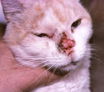
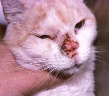
The squamous cell carcinoma is associated with sun exposure, and this means that lesions on the white-furred areas of the feline face are at risk. The ear tips and nose are particularly vulnerable, as one might expect, as these areas are not well-protected by fur.

These tumors begin as a small scabby area that may not attract much attention as it can be mistaken for a minor abrasion. This minor abrasion, however, does not heal and continues to get larger.

Because squamous cell carcinoma does not spread distantly in the body until late in its course, this provides a window during which it can be successfully treated. Once it gets too big, that window has closed even if the tumor has not yet spread.

Surgical Treatment

If the tumor is not too big, surgical removal is curative. That said, squamous cell carcinoma can spread to surrounding tissues, so a wide margin of normal tissue must be removed in order to get the whole tumor.  If the tumor has grown too large, there will not be enough tissue to get a good margin around it, and it will regrow, resuming its destructive course.

The surgery is ideally done with laser equipment to reduce bleeding; it is probably best to seek the skills of a surgery specialist or at least a veterinarian with a great deal of facial surgery experience. Your veterinarian may recommend a nerve block during surgery to help control pain after the procedure. 

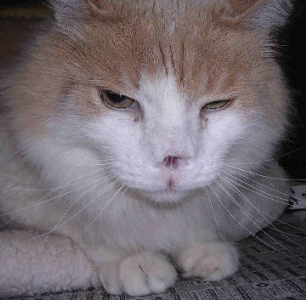
The leather of the nose is removed along with some of the surrounding haired skin. The nasal opening will heal cosmetically but will look a little unusual. Discuss with your veterinarian whether or not a referral to a specialist is in the best interest of you and your pet.
Surgical Recovery

Recovery can vary depending on the tumor size, location, and overall health of your cat. Your veterinary surgeon will give you post- operative instructions and can tell you what to monitor after taking your cat home, including what would be considered an emergency.

Preventing self-harm is crucial, and your cat may have to wear an e-collar or “cone” to prevent pawing at their face and harming the surgical site(s). Pain medications and antibiotics are commonly prescribed.

You may notice small drops of blood or nasal fluid coming from the nostrils right after bringing your cat home. They may also cough or have a “rougher” sounding voice from the tube placed during surgery which helps them to breathe while under anesthesia (endotracheal tube). Many cats don’t want to eat as much after surgery, and your veterinarian may recommend offering various foods to entice them, appetite stimulants, and surgery site cleaning under sedation (to allow them to smell their food).

Any excessive bleeding, difficulty breathing, changes in sleeping habits, or other situations after surgery should be discussed with your veterinarian.

Cryotherapy

Cryotherapy may be less invasive. This involves using a probe to freeze the tumor. Again, this can be curative if the tumor is small enough, but in freezing the tissue, the cells are damaged and can’t be analyzed by biopsy. The biopsy (lab analysis) of the tissue can confirm squamous cell carcinoma and  enables the pathologist to tell if the excision was complete. This is valuable information that we would prefer not to forgo for obvious reasons.

Side effects of cryotherapy are generally mild and include swelling, redness, and pain. For the next 3-6 months your veterinarian will monitor your cat for signs of tumor regrowth. Cats with tumors that have grown into surrounding tissues also need to be checked for cancers in other areas. This may include X-rays of your cat’s chest and lymph node exams. For cases of nasal squamous cell carcinoma caused by sun exposure, the ears and eyelid margins also must be closely monitored along with the nose for regrowth of cancerous areas.

Radiation Therapy

Radiotherapy is rapidly becoming the treatment of choice. Strontium plesiotherapy using strontium 90 involves applying a localized radiation source to the tumor directly for a brief period of time. (Imagine a wand with a radiation source on the end.) Radiation destroys tissue in a 3 mm diameter from the source, which allows for the destruction of small superficial tumors. As with surgery, if the tumor is too big, it cannot be cured.

Alternatively, external radiation beams can be aimed at the tumor under general anesthesia. In one study, 60 percent of irradiated cats had a complete response, and 33 percent had a partial response.

Radiation therapy obviously requires specialized equipment that is usually found only in specialty facilities. It is not available everywhere. A consultation with a veterinary oncologist will help determine if a given patient is a good candidate and what is involved in finding the nearest radiofacility.

Imiquimod 5% Creme

Imiquimod is an immunity-modifying topical crème that is palliative for tumors too large for curative removal or to treat the more superficial form of squamous cell carcinoma called Bowenoid carcinoma or squamous cell carcinoma in situ. Imiquimod is a relatively expensive product, and an upset stomach can result if the crème is licked (which it inevitably would be). 

NSAIDs

You may not know the term “cyclooxygenase,” but you are probably familiar with human anti-inflammatory cyclooxygenase inhibitors like aspirin, ibuprofen (Advil®), naproxen (Aleve®), and others. While these human anti-inflammatories are not safe for cats, there are some prescripton veterinary medications that are. This comes into play with nasal squamous cell carcinomas because there is potential for carcinomas to express enough cyclooxygenase that a feline-friendly non-steroidal anti-inflammatory pain reliever (such as piroxicam, meloxicam, or robenacoxib) may have anti-tumor effects. These medications are not completely without issues but may be helpful when other treatments are not an option, especially since they offer some pain relief.

Prognosis (Outlook)

Nasal squamous cell carcinomas usually do spread to tissues close by, but are slow to move to other areas in the body. Cats with superficial tumors (those that have not spread to deeper tissues in the nose or face) have better outlooks vs. large and deeply penetrating tumors.

Even though squamous cell carcinomas do not spread until relatively late in their course, they can still be extremely difficult to address. The nasal version is an exception to the poor prognosis rule as long as it is detected early and removed or treated with radiation promptly.

The potential for a full recovery depends on the size and location of the tumor, treatment procedures, and your cat’s overall health. Discuss your questions with your veterinarian. Also, talk about what you and your cat might experience. This will help you prepare for managing this condition as effectively as possible.

7750217

No Bones About It – Chewing Bones is Bad for Dogs’ Teeth

The Food and Drug Administration warned pet owners about bones and bone treats to include not only harmful bacteria (E.coli and Salmonella) that dogs can get and pass on to humans but the actual damage caused by the bone’s trauma to a dog’s teeth.

Other issues can occur with bones, such as blocked intestines, choking, wounds in the mouth, vomiting/diarrhea, and rectal bleeding (some of which could be fatal if not treated promptly).

Are Any Bones Safe for My Dog’s Teeth? 

No. Steak bones are too hard for teeth. Antlers are worse than bones because they’re even harder. Poultry bones – chicken, turkey, and duck – are awful because they are full of air and thus splinter easily. The pieces can get stuck in your dog’s palate (roof of the mouth) and cause infection or get stuck in the esophagus or intestines, most of which requires a surgical fix.

What Happens if My Dog’s Teeth Are Injured by Bones? How Will I Know if There is a Problem?

For many dogs, broken teeth do not show signs of discomfort even though they experience pain like humans. Some show apparent signs and symptoms of disease, including an open tract below the eye or under the chin that may be draining.

When the nerves die, the pain decreases until infection sets in. Signs relating to dental disease from a broken tooth include:

  1.  chewing on one side;
  2.  dropping food from the mouth when eating;
  3.  excessive drooling;
  4.  grinding teeth;
  5.  pawing at the mouth;
  6.  facial swelling;
  7.  fistulous tract below the eye or under the lower jaw;
  8.  regional lymph node enlargement;
  9.  shying away when the face is petted;
  10.  refusing to eat hard food;
  11.  refusing to chew on hard treats or toys.

Treatment for Broken Teeth

When presented with a tooth that is fractured and has pulp exposure (complicated tooth fracture), your veterinarian essentially has two choices:

  1.   Extract the tooth.
  2.   Refer to a veterinary dental specialist to perform treatment, which usually allows the tooth to be saved and returned to function. Specific treatment depends on the severity of damage to the tooth structure and if any other disease is affecting the tooth, and the functional significance of the tooth.

Types of Tooth Damage

  • Enamel fracture:  A fracture with loss of crown substance confined to the enamel 
  • Complicated crown fracture:  A fracture of the crown that exposes the pulp
  • Uncomplicated crown-root fracture:  A fracture of the crown and root that does not expose the pulp 
  • Complicated crown-root fracture:  A fracture of the crown and root that exposes the pulp 
  • Root fracture:  A fracture involving the root 

How Severe is the Damage to the Tooth?

Some fractures are limited to the enamel and require little or no therapy; others involve dentin and might not require endodontic care; others expose enamel, dentin, and pulp and require root canal care or extraction. The goal of endodontic care is to return the tooth to function if possible, and if not, extract the tooth to prevent further pain.

How Important is the Tooth?

The upper and lower canines (eye teeth) are the most common teeth broken, followed by the incisors and the upper cheek teeth.

Although endodontic care can be performed on any tooth, the canines and maxillary fourth premolars generally are the only teeth where endodontic therapy would be considered due to the importance of the tooth and ease of pulp chamber access.

Age of the Patient

The age of the patient is also essential when choosing endodontic therapy options. Canine teeth of patients younger than twelve months of age may have open root apices (the tip of the tooth’s root). Lower molar teeth generally have closed apices by seven months of age. Standard root canal therapy is not performed on teeth with open root apices because appropriate sealing of the apex cannot be assured.

Treatment options for teeth with open root apices include:

  1. Vital pulp therapy (partial coronal pulpectomy, direct pulp capping, and restoration) to promote the preservation of vital pulp tissue or
  2. A procedure called apexification is used to stimulate root development if the pulp is dead. Teeth with pulp exposure and closed root apices can be treated with standard root canal therapy.

Age of the Fracture

The age of the fracture affects endodontic treatment. Inflammation occurs less than two mm from the exposure site shortly after pulp exposure. In acute (sudden) fractures, the pulp appears pink or red at the fracture surface. The pulp of a long-standing fracture will appear brown or black. Healthy pulp tissue can be found several millimeters deeper within the pulp, which might respond to vital pulp procedures (i.e., vital pulp therapy).

 In the mature animal that has an acutely fractured tooth with a closed apex, standard root canal therapy results in a more predictable outcome compared to vital pulp therapy.

Aftercare Support

Tooth support is critical to the long-term success of endodontic treatment. If obvious periodontal disease is present before therapy, victory will be unlikely unless epic measures are taken, and strict home care is provided. 

Dogs love to chew. To find out what products are safe and effective in decreasing the accumulation of plaque and tartar, discuss with your veterinarian.

4128299

Neuropathic Pain in Dogs and Cats

Neuropathic pain is an abnormal painful response. It is caused by injury to or disease of the nerves, spinal cord or parts of the brain that are involved in receiving, conducting and interpreting painful stimuli. 

Both physical and chemical changes within the spinal nerves that convey pain contribute to neuropathic pain.

Such changes include: an increase in the chemicals and receptors that transmit signals between nerves; an imbalance between pain signals coming into the brain and the brain suppressing those signals; and a change that makes nerves that don’t normally relay pain signals into ones that do.

There are different types of neuropathic pain.  An exaggerated response to a painful stimulus, such as a pin prick, is called hyperalgesia.  Pain from a normal stimulus that shouldn’t be painful at all, such as a light touch, is called allodynia. Hyperpathia is increasing pain after repetitive stimulation, which includes continued pain after the stimulus is gone, and pain that radiates to adjacent areas.

People who experience  neuropathic pain describe it as “a pins and needles sensation,” tingling, burning, itching, numbness or cold, and sometimes feeling as if they had received a small electric shock.  Grades of neuropathic pain that have been established in human medicine are definite, probable and possible.

Pets cannot describe their pain to us but we believe it’s likely to be like what people have.  Neuroma formation in horses is known to cause definite neuropathic pain.  A neuroma consists of an abnormal sprouting of nerve endings from a severed nerve, and affected horses develop lameness or an abnormal gait. 

An example of probable neuropathic pain in dogs is the abnormal scratching behavior that is seen in King Charles Cavalier Spaniels with a spinal cord abnormality called syringomyelia. Cats with hyperesthesia syndrome incessantly lick and attack their back, and become extremely painful when petted. Neuropathic pain may be one of the causes for hyperesthesia syndrome.

Diagnosis

Diagnosing neuropathic pain in animals involves ruling out other painful conditions, such as osteoarthritis and bone cancer, as well a thorough neurological workup. A neurological examination evaluates the different parts of the nervous system (brain, spinal cord, nerves); if those parts are not normal, further diagnostic tests are indicated.  Laboratory tests, radiographs, CT or MRI imaging, and in some cases electrophysiologic testing may be recommended to look for diseases that are known to cause neurological injury and secondary pain.  Examples of such diseases include: diabetes mellitus, intervertebral disk herniation, spinal cord or nerve sheath tumors, cervical malformation, and syringomyelia.

Treatment

In some cases treatment is started early in conditions that are normally expected to be painful to try to prevent neuropathic pain from developing.  In other cases, neuropathic pain may be the first sign.

Combining drugs that reduce pain in different ways has proven to work better than using a single drug alone.  Depending upon the species, one or more drugs may be prescribed by a veterinarian.

Gabapentin, pregabalin, dextromethorphan, and amantadine are drugs that act on the relay centers for pain within the spinal cord. Amitriptyline acts both on the brain and within the spinal cord. The antibiotic minocycline has shown potential for modifying neuropathic pain.

These agents may be combined with one or more of the traditional analgesic drugs: tramadol and codeine, narcotic drugs that modulate the brain’s perception of pain; non-steroidal antiinflammatory drugs (NSAIDs) such as carprofen, deracoxib, and firocoxib; local anesthetics such as lidocaine and alpha-2 agonists; drugs such as medetomidine that modify pain, heart rate and blood pressure.

Some veterinarians have found success using combinations of herbs, diet, exercise, and acupuncture.

It is important to work closely with your veterinarian to manage neuropathic pain. Balancing the benefit of multiple medications against side effects can be challenging. Discussing the perceived effects of medications with your veterinarian is essential for the best outcome for your pet.