Tag: pets

6930337

Serotonin Syndrome in Dogs and Cats

Serotonin syndrome is a serious drug reaction to medications that build up high levels of serotonin in the body. A chemical that the body produces naturally, serotonin is needed for the brain and nerve cells to function properly. Medications such as those used to treat human depression, attention deficit hyperactivity disorder, seizures, and pain; illicit drugs such as LSD or ecstasy; dietary supplements including SAMe, St. John’s wort, ginseng, turmeric, Garcinia extract, and Garcinia cambogia; and even certain foods with high levels of tryptophan, such as salmon, spinach, poultry, milk, eggs, nuts, and soy products can all contribute to serotonin syndrome.

In veterinary medicine, serotonin-enhancing medications are often used to treat behavioral issues such as separation anxiety, fear of fireworks, storm phobia, compulsive disorders, aggression, and house-soiling behavior. Serotonin syndrome may occur with the start of a new medication, dosage change, or combination of medications. However, it is most commonly seen when a pet accidentally ingests medication prescribed to a human in the household.

Diagnosis is often based on the history of ingestion and clinical signs. Signs may appear anywhere from 1 to 12 hours after ingestion. Symptoms generally last about 24 hours but can extend for several days. Most animals will show GI upset, such as vomiting, diarrhea, and a loss of appetite.

Other common symptoms include:

  1. dilated pupils
  2. increased heart rate
  3. fever
  4. muscle tremors or jerking
  5. restlessness
  6. difficulty walking
  7. agitation
  8. excitation
  9. disorientation
  10. vocalization
  11. seizures

Serotonin syndrome can be a life-threatening situation; therefore, if you think your pet is developing it, visit a veterinarian immediately. Make sure to bring all the prescriptions that your pet may have ingested with you. Don’t forget to mention any over-the-counter dietary or herbal supplements your pet is taking because they could be a cause as well.

Treatment will vary based on clinical signs. Depending on the severity, hospitalization may be needed. Prognosis depends on what your pet swallowed, how much, and how long they have been symptomatic.

6058834

Senior Dog Health Issues

Aging is a natural process, not a disease. Just like people, dogs go through both mental and physical changes as they age. The muzzles of most older dogs turn gray and you can see a general decline in the quality of their coat; their skin becomes less elastic, causing hair loss and white hairs. Their eyes may also appear cloudy or bluish and pearl-like over time.

Older dogs tend to lose muscle and gain fat. As they age, their energy requirements decrease; they may not need the same number of daily calories they did as when they were young. Part of this is biological, part of this is due to increased sleeping and decreased activity. Many older dogs will not have the same endurance for play and exercise as they did in their youth. It’s a good idea to discuss their diet and energy levels with your veterinarian to avoid over-feeding and obesity.

Take your senior dog (see below for age ranges considered “senior”) to the veterinarian at least twice a year. Aging also increases your pet’s susceptibility to a number of different health issues. Regularly checking in with your veterinarian helps catch any potential issues sooner and provides an opportunity to address any changes you may have noticed in your pet. If you see a sudden change in your pet or notice a gradual worsening of their state, see your veterinarian.

Signs you don’t want to ignore:

  1. increased panting or difficulty breathing
  2. vomiting
  3. diarrhea
  4. change in appetite or thirst
  5. change in frequency of urination
  6. “accidents” in the house
  7. pain
  8. aggression

Defining Seniors

Where most veterinarians draw the line between an adult dog and a senior dog is different from where pet owners do. When a dog becomes a senior depends on their individual health and condition. The chart below provides a general guideline on when a dog is considered a senior based on their size. The ages veterinarians consider a dog to become a senior are much earlier than many owners think. It’s important to keep this difference in mind as your dog may be more likely to develop certain diseases earlier than you expect.

Dog sizeAge considered senior
Small breed (2-20 lbs.)7
Medium breed (21-50 lbs.)7
Large breed (51-90 lbs.)5
Giant breed (over 90 lbs.)5

Sensory Changes

Many older dogs experience decreases in their senses. Their ability to see, hear, taste, and smell may be affected. Partial or complete loss of hearing and vision can decrease your dog’s ability to sense their environment. As a result, your dog may not sense your approach and startle more easily. To avoid scaring your dog, try to announce your presence with a loud greeting and approaching from within your dog’s line of vision. Minimize rearranging or adding furniture to the areas your dog has access to as it will keep their environment familiar and make it easier for them to get around if their eyesight is failing. Your dog may also be less responsive to voice commands if they have some hearing loss; it may be beneficial to teach them hand signals before this occurs as this allows you to continue communicating with your dog regardless of their ability to hear you.

As dogs age, the number of taste buds they have decreases. That decrease coupled with a decreased ability to smell, may affect your dog’s desire to eat. Food becomes less tasty. Consult with your veterinarian if you note a decrease in appetite.

While sensory losses are a natural part of the aging process for many dogs, this may not be the case for every dog. There are several medical conditions and illnesses that cause visual, auditory and other sensory impairments in dogs. It’s important to have your veterinarian examine your dog to determine whether their changes are part of the aging process or whether there is an underlying disease or condition.

Behavioral Changes

It’s common for an older dog to slow down a bit and take more naps. It’s a mistake, however, to assume all changes in your dog are a natural part of the aging process. Behavior changes such as difficulty getting up or stiffness, anxiety, aggression and other abnormalities may mean something is wrong. There are many health issues that cause pain and discomfort in your dog which lead to their reluctance to move or increased irritability. It’s important to bring up behavior changes with your veterinarian as some conditions are treatable and doing so helps maintain a good quality of life for your dog.

Health Issues

In general, the most common health issues older dogs develop are dementia, arthritis and cancer. Other problems include heart, kidney, liver, and dental disease.

Dementia in dogs is called canine cognitive dysfunction (CCD). It is similar to the early stages of Alzheimer’s disease in people. About 14-35% of dogs over 8-years-old are affected, a percentage that increases significantly as dogs continue to age. Dogs with CCD have a slow progression of behavioral signs, such as aimless wandering/pacing, staring into space and more. If you notice any of these behaviors or the ones listed below bring it up with your veterinarian. They may be related to CCD or another disease.

Signs of cognitive dysfunction syndrome:

  1. decreased interaction with owner, other people and animals
  2. inappropriate or excessive vocalization
  3. changes in sleep/wake cycle
  4. restlessness
  5. disorientation
  6. confusion
  7. anxiety
  8. house-soiling
  9. altered appetite 
  10. irritability 
  11. aggression
  12. apathy

There is currently no cure for CCD but early diagnosis and treatment can improve your dog’s condition and slow progression.

Arthritis in older dogs results in pain and discomfort, which can lead to difficulty jumping up and down surfaces and decreased activity. If your dog is in significant pain, they may show signs of depression or irritability. There are many therapies to help alleviate discomfort including exercise, medication, nutritional support and complementary therapies.

Almost half of dogs over the age of 10 will develop cancer. Symptoms of cancer vary depending on the type of tumor and its location, but may include abdominal swelling, bleeding from body openings, difficulty breathing, non-healing wounds and sudden changes in weight. Your pet’s prognosis, like their symptoms, depends on their individual situation but it is generally better to catch the disease early on.

As dogs age, changes to their internal organs take place. These changes can increase the risk of developing heart, kidney and liver disease. Symptoms of heart disease include coughing, abdominal swelling and exercise intolerance while symptoms of kidney disease include changes to urination and thirst. Liver problems can show up as general signs of sickness, such as vomiting and depression. The same symptoms could be caused by many different diseases so your veterinarian may need to perform several diagnostic tests.

It’s important to keep up with oral hygiene as tartar buildup can lead to infection. Aside from problems directly affecting the mouth, bacteria can pass into the bloodstream and directly affect the health of the heart and kidneys. Discuss an oral health care plan with your veterinarian to keep your pet’s teeth in good shape.

Nutrition

The diet a dog is fed should be tailored to their individual condition and health. Not every dog will need to switch from an adult diet to a senior diet as they get older. Many dogs do well if fed the same good quality adult commercial diet they are used to. Some seniors, however, may benefit from changes to their nutrition. One important factor to consider is how much protein and what quality of protein they are getting.

Older dogs tend to lose muscle mass and feeding adequate protein can help prevent this. Another important factor to consider is the number of calories your dog actually needs on a daily basis. Senior dogs tend to have a slower metabolism and thus may not need as much food as before. Consult with your veterinarian to discuss how much to feed your dog and whether you need to feed your dog less or change them to a lower calorie diet.

There are many diets tailored to specific diseases dogs may develop. These include reduced sodium diets for dogs with congestive heart failure, urinary diets for dogs with kidney issues, and many others. Supplements such as antioxidants can help reduced inflammation and boost brain function.

Prevention and Maintenance

There are many things you can do to support your dog’s health as they age. Regular play and exercise are great for maintaining a healthy weight and active mind. Try teaching your dog new tricks and commands. Learning new things can help keep them mentally sharp and improve signs of dementia. With a combination of appropriate nutrition, social interaction and vigilance on your end for signs of illness, coupled with your veterinarian’s medical skill, your dog can continue to have a great life into their senior years. Remember that some age-related changes are not avoidable such as vision and hearing loss. It’s a good idea to check with your veterinarian to make sure there is not an underlying disease, but otherwise these changes are manageable and do not have to decrease your dog’s quality of life. Your dog may just need some extra patience and care.

6267326

Scottie Cramp in Dogs

Scottie cramp is a neurological disorder that temporarily affects a dog’s ability to move their limbs. The disorder has sudden episodes immediately following times of stress or exercise, lasting about 10 minutes. Affected dogs temporarily experience stiff limbs, muscle spasms, arching spine, a bunny hopping gait and are likely to fall over. Research shows Scottish terriers who lack a neurotransmitter called serotonin are affected because serotonin helps control muscle movement. This disease is lifelong but has no lasting complications to your dog besides the short and infrequent muscle spasm episodes.

Who Gets Scottie Cramp?

This disease occurs in Scottish terriers and Scottish terrier mixes. A similar disorder is recognized in West Highland White terriers, Cairn terriers and Cesky terriers and may have a common genetic cause.  Cats do not get Scottie cramp. There are similar types of muscle movement diseases in other dog breeds, such as Dalmatians, cocker spaniels, Norwich terriers, wirehaired terriers, and West Highland white terriers. However, they are different from Scottie cramp in their exact genetic cause.

  1. Gender: Females more likely than males
  2. Age: Juvenile onset (usually 1 month-18 months)
  3. Cause: Genetic serotonin deficiency
  4. Hereditable: Yes

Signs

Signs usually resolve within 10 minutes of episode onset. Episodes can last from 5 to 20 minutes.

  1. Arching spine
  2. Stiff limbs
  3. Falling over
  4. Bunny hopping
  5. Flexion of hips
  6. Downward flexed tail
  7. Inability to walk

Diagnosis

All blood and imaging test results are normal. Neurological and orthopedic tests are also normal because the signs of this disease are temporary. There is no genetic test available even though it is a genetic disease. Research suggests that electromyography (EMG) is a way to diagnose this disease, but dogs aren’t necessarily having an episode at appointment time. EMG looks at the electrical activity of the muscle during your dog’s episodes. However, diagnosis is mainly made by description and video recordings of episodes. A video of an episode taken on your cell phone is more diagnostic than anything else.

Treatment

Treatment is not always needed as episodes can be infrequent and improve with time. But when treatment is called for, it is life long and the prognosis is good for mildly affected dogs. You can monitor your dog’s disease by keeping a diary of triggers, signs, and duration of episodes.

  1. Fluoxetine or diazepam increase the serotonin available to prevent your dog’s stiff muscle signs.
  2. Reduce stress and excitement, which are triggers. Exercise is also a trigger, so talk to your veterinarian about how much exercise is appropriate for your dog.
  3. Chlorpromazine or acepromazine are sedatives that can help relax muscles

Prevention

The one and only method of prevention is not to breed affected dogs because the disease is heritable.

5064184

Schnauzer Comedone Syndrome

What is Schnauzer comedone syndrome?

In Schnauzer comedone syndrome, dogs develop multiple comedones (blackheads) on the back and neck. These small skin bumps develop when a pore, the skin’s opening for each hair, becomes clogged with skin oils and debris.  Despite the name, Schnauzer comedone syndrome can occur in non-Schnauzer breeds such as Cairn terriers and other rough-coated terriers. It is also called Schnauzer comedo syndrome, Schnauzer back, and Schnauzer back bumps.

How is it diagnosed?

Your dog’s veterinarian may need to take skin and other samples to diagnose and treat it. This is because many other skin conditions can cause comedones in dogs, including demodicosis or demodectic mange from Demodex mites.

What causes Schnauzer comedone syndrome?

No one really knows what causes Schnauzer comedone syndrome. However, it may be an inherited condition because it occurs more commonly in certain dog breeds.

How is Schnauzer comedone syndrome treated?

Schnauzer comedone syndrome cannot be curred but can be treated. To loosen and dissolve the comedones, your pet’s veterinarian may recommend a topical therapy, such as a medicated antiseborrheic shampoo or ointment. If comedones become infected, they may need to be treated with an antibiotic. Your pet’s veterinarian may take a sample for a skin culture (bacterial culture and sensitivity) to choose the antibiotic. Especially if the skin infection is itchy or painful, your pet’s veterinarian may do some tests to determine if your dog has other health conditions that are making the Schnauzer comedone syndrome bumps worse.

6499948

Sanitizing and Disinfecting the Environment after Parvovirus in Dogs

Canine parvovirus is a highly contagious virus. Young, old, unvaccinated, or immune-compromised dogs are particularly susceptible. Parvo, as it is typically called, or CPV, lives well in the environment, both indoors and outside. It is a non-enveloped virus, which means it lacks a membrane often called the envelope that is stronger than enveloped ones. It can survive indoors for months and outdoors it can live for months to years, especially in dark, moist environments.

Parvo can be easily spread by fomites, which are objects such as a doorknob or pet fur that can be contaminated by a virus. Dog-to-dog contact is not required for susceptible dogs to become infected. Dogs can become infected from contact with the remaining virus where an infected dog has been, or on objects an infected dog has used, or even from shoes and clothing carrying the virus. To make matters worse, non-enveloped viruses are not easily killed by most common household disinfectants and antibacterial soaps do not kill it. If you have had a dog with canine parvovirus in your home or yard, you need accurate information about what you can do to decrease or eliminate the risk of exposing other dogs while understanding that you cannot completely eliminate parvovirus from your home; it’s simply not possible.

First, Remove Organic Material

Sanitizing is a two-step process that involves both cleaning and disinfecting. Some surfaces and substrates are easier than others to sanitize. For instance, carpet and grass are difficult to properly sanitize, whereas sealed cement is easier.

Before disinfecting, you need to clean since many disinfectants do not work well on organic material, such as stool or urine. Clean up any organic material from the yard and in the house. Bedding and toys that are not heavily soiled can be washed through a soap/bleach cycle and dried on a hot setting. It is best to discard anything heavily soiled. All areas and items still need to be properly disinfected, but the first step is to mechanically clean them.

Water can also be used to spray down outside areas to help remove any dried poop or vomit. Diluting an area such as a lawn by spraying with water may also help, but only if drainage and temperature allow the yard to thoroughly dry afterward.

Bathe any dogs using regular dog shampoo who were exposed or infected and recovered, as well as other dogs who had contact with the infected dog or area. This will help to decrease the risk of fomite transmission from their fur.

Use an Effective Virucidal Disinfectant

When choosing a disinfectant, make sure it has been proven to kill parvovirus. A label claiming to kill viruses does not mean it kills all viruses, such as the hearty, non-enveloped parvovirus. Additionally, any disinfectant must be mixed to the proper dilution and remain saturated for the proper contact time to be effective against parvovirus. Independent studies or FDA approval backing label claims are the most reliable means of determining a disinfectant’s effectiveness.

Types of Disinfectants

Quaternary Ammonium Disinfectant

Despite its popularity for many years and label claims of efficacy against parvovirus, independent studies have shown this disinfectant to be unreliable against parvovirus. The label claims are based on diluting at 18 ounces per gallon, which is four times the typical concentration. Despite repeated reformulation and label claims, this is not seen as a reliable disinfectant against parvovirus and is not endorsed by independent studies or label-approved by the FDA. It must be rinsed off.

5% Sodium Hypochlorite (Bleach)

Bleach can kill parvovirus when it is used properly. It is readily available and relatively inexpensive but has some drawbacks. It can discolor or even ruin surfaces. The fumes can be irritating to the nose, eyes, and skin. Bleach is most effective on non-porous surfaces from which organic material has been thoroughly removed. For areas where you can safely use bleach, dilute household bleach (5% sodium hypochlorite) using 1 part bleach to 32 parts water (that’s 1/2 cup per gallon of water). The diluted bleach must have ten minutes of contact time with the surface, which means saturating the area or continually spraying for ten minutes. It must be rinsed off.

Potassium Peroxymonosulfate Disinfectant

This disinfectant has some detergent properties, meaning it is effective for mechanically removing organic matter, such as vomit and feces. It can be irritating to the skin and eyes. The label claims efficacy against parvovirus at 1% dilution, even when mixed with hard water or organic material. It can be used in carpet cleaners to clean carpets and furniture (spot test for staining first) and can also be sprayed on yard surfaces. The area must remain saturated for ten minutes. It can be left to dry and does not need to be rinsed.

Accelerated Hydrogen Peroxide (AHP) Disinfectant

This disinfectant has become widely used in animal shelters, where CPV is not uncommon. Its detergent properties make it effective even when there is some organic matter. It can be used in one step for mechanical cleaning and to kill the virus. As in the other disinfectants discussed, proper dilutions and contact time are crucial to ensuring it kills the virus. It should be diluted using one part AHP to 32 parts water (1/2 cup per gallon of water) with a ten-minute contact time. This can be irritating to breathe but is less corrosive to surfaces and mucous membranes than bleach. It can be used in carpet cleaners and sprayed on grassy surfaces as well. It can be left to dry and does not need to be rinsed.

The Yard

Careful and repeated removal of all visible feces is important. If possible, flush the yard with water and allow it to dry. Then apply a disinfectant that is effective with organic material, such as AHP or potassium peroxymonosulfate. These can be used with a pesticide sprayer or hose-end sprayer at the proper dilution to saturate the outside area. Diluted bleach may also be somewhat effective, but since it does not perform as well with organic matter it is less effective than the others. Bleach is also more dangerous to plant life and other surfaces. If your yard has dirt and grass, rather than just cement, there is no guarantee any disinfectant will be 100% effective. It is best to thoroughly clean, dry, disinfect with the proper contact time and dry at least twice. When possible, maximize exposure to sunlight and allow the area to dry completely.

In most home yard situations where there is grass and dirt, it is not possible to completely disinfect a yard. Out of an abundance of caution, you may want to avoid having unvaccinated dogs come to your yard for 6 to 12 months, even after cleaning and attempting to disinfect.

The House

It helps to understand the difference between porous and non-porous surfaces. Water can go through porous materials. Non-porous materials include stainless steel, glass, metals, rigid plastics, and painted or varnished surfaces. Granite countertops are porous, but quartz is not. Porous materials include unpolished wood, laminate, granite, drywall, carpeting, ceiling tiles, and unsealed cement.

As mentioned, the first step is thoroughly cleaning the house and removing any feces or vomit that can be seen. Once all areas have been mechanically cleaned, you are ready to disinfect. Non-porous surfaces can be disinfected with diluted bleach or AHP or potassium peroxymonosulfate. Make sure you follow the manufacturer’s instructions for the proper dilution and contact times for parvovirus.

For carpets and porous surfaces, AHP or potassium peroxymonosulfate disinfectants can be used in carpet cleaners to steam clean carpets and furniture. Understand that these areas are sometimes impossible to disinfect and heavily soiled items should be discarded if possible. Make sure you read the carpet cleaner instructions and spot-test any porous surfaces for possible discoloration. These surfaces will need to remain saturated for ten minutes to ensure the proper contact time. After disinfecting, the areas need to dry thoroughly.

Bedding, Porous Items, or Toys

Bedding and other fabric or porous items should either be thrown away, especially if heavily soiled, or washed thoroughly with detergent, hot water, and bleach and run through a hot dryer cycle if possible. If it is not possible to bleach the items, you could steam clean them with disinfectant as recommended for carpet and furniture. Consider if it is worth the risk to keep porous items such as bedding and toys that can’t be bleached, or if it is better to throw them away.

Exposed Bowls and Non-Porous Items

Metal or ceramic bowls and non-porous toys should be washed thoroughly with a detergent, rinsed well, and then disinfected using an effective disinfectant. Throw away any plastic bowls that have any signs of wear, scratches, or teeth marks, which create porous areas. Proper dilution and contact time are important. This can be achieved by soaking the cleaned items in the diluted disinfectant for ten minutes, then allowing the items to dry. After disinfecting, thoroughly rinse the items with water and allow them to dry.

When is my Home Safe?

Unfortunately, there is no guaranteed time frame. There has not been any research that definitively states canine parvovirus can be eliminated from a home or yard environment in a specific number of days. There are many factors that affect whether cleaning and disinfecting efforts will be effective inside the house and outside in the yard. After a parvovirus-positive dog has been in your home or yard, it is safest to only have animals visit who have completed their vaccination series.

4128526

Salmon Poisoning in Dogs

Salmon poisoning is also known as salmon disease, salmon poisoning disease (SPD), and Neorickettsia helminthoeca infection. It is an acute, and often fatal, infectious disease of dogs, coyotes, and foxes of the Pacific Northwest. This disease has been found in black bears and Malayan sun bears, but has not been reported in cats.

Despite its name, salmon poisoning does not involve a toxin. Salmon poisoning is an infection that develops when dogs eat raw or undercooked fish (salmon, trout, or steelhead) or Pacific Giant Salamanders that contain a fluke. A fluke, also known as a trematode, is an internal parasite. In this case, the fluke is Nanophyetus salmincola that contains a rickettsial organism called Neorickettsia helminthoeca.

Once the larval flukes reach the dog’s intestinal tract, they embed in the dog’s duodenal mucosa, and release the rickettsiae. The rickettsial organisms then spread through the bloodstream to the liver, lungs, brain, and lymphoid tissue. Clinical signs include, but are not limited to: fever; nausea; vomiting; bloody diarrhea; weight loss; ascites; nasal and eye discharge; enlarged lymph nodes; tachypnea; dyspnea; tachycardia; twitching; and seizures. Approximately 60 percent of cases have generalized lymph node enlargement. Unless the infection is treated, 50 – 90% of affected animals will die of the infection within 7 to 10 days.

Clinical signs usually begin about one week after the animal eats the infected raw fish, although delays of 33 days have been reported.

Geographic Distribution

Although infected fish can be found in the Pacific Ocean from San Francisco to the coast of Alaska, salmon poisoning is most commonly found in Washington, Oregon, northern California, and southern Vancouver Island (Canada). It is also seen inland, along the rivers of fish migration. The Pacific Northwest has the trematode’s first intermediate host, a small snail called Oxytrema plicifer.

Diagnosis

Although clinical signs are caused by the rickettsial organisms and not the fluke, presumptive diagnosis is usually made by identifying the fluke’s ova (eggs) in the feces. Ova are identified in 92% of cases. Direct smears of fecal material usually provide sufficient specimens for diagnosis, although other methods can be used.

Lymph node cytology and histopathology may provide a more definitive diagnosis, if necessary.

A history of access to raw fish also aids in diagnosis.

Treatment

To combat the hemorrhage, necrosis, and infection, affected animals usually need antibiotics, fluid therapy, blood transfusions, antiemetics, antidiarrheals, and anticestodals (drugs that kill flukes).

Prognosis

Many dogs respond well to antibiotic therapy, and resolution of gastrointestinal signs, fever, and thrombocytopenia can occur within 24-72 hours of starting therapy. However, if treatment is not started, death can occur 7-10 days after the onset of clinical signs. In one report, overall mortality rate was 14%. In another study of 43 dogs that were treated with antibiotics, 39 survived.

Preventive Measures

Dogs should not be allowed to eat raw or smoked-raw fish (salmon, trout, steelhead), or Pacific Giant Salamanders.

Dogs that survive salmon poisoning will be immune to re-infection with the same strain. However, infection with an alternate strain can occur because there is no cross-protection.

5836802

Salivary Mucocele in Dogs and Cats

A salivary mucocele, also known as a salivary gland mucocele or sialocele, is a swollen area associated with saliva (spit) leaking from a salivary gland into surrounding tissues. It can be caused by damage to either the salivary gland, which produces saliva, or the salivary duct, which is the passageway for saliva from the gland to the mouth.

What does a salivary mucocele look like?

Dogs and cats have several salivary glands, but the most common place for a salivary mucocele is on or beneath the lower jaw or under the tongue. If the mucocele is on/under the jaw, a large swelling will be seen underneath the skin in that area. The swelling may be hard or squishy (almost like a water balloon). If the mucocele gets too large, the pet may have trouble eating, swallowing, or breathing. Mucoceles under the tongue can be more difficult to see but will appear as a tumor-like bulge or bubble on the floor of the mouth.

This type of salivary mucocele is also referred to as a ranula. Mucoceles under the tongue may become so large that they can be seen next to the tongue or can prevent the mouth from closing properly. They can also cause trouble keeping food in the mouth and swallowing. 

Uncommon places a salivary mucocele may be seen is on the cheek, where it’s seen as a swelling below the eye. The eye on the mucocele side of the face may appear larger than the eye on the healthy side. A bulge in the roof of the mouth may be seen as well. 

How is a salivary mucocele diagnosed? 

For the most part, salivary mucoceles are easy to diagnose. All that is usually needed is to remove some of the fluid and cells by suctioning them out with a needle and syringe, a process called aspirating. The salivary fluid is examined under a microscope to confirm the type of fluid and to look for bacteria in case the mucocele is infected. 

What is the treatment for a mucocele? 

Surgery is often needed to remove the diseased gland. If the mucocele is caused by a damaged duct, sometimes surgically creating a new opening in the duct can solve the problem without removing the gland. Fortunately, though, there are multiple salivary glands in the mouth so removing one will not have a major effect on saliva production. Once in a while, multiple surgeries are needed to ensure all diseased salivary tissue is removed. 

Occasionally, the veterinarian will drain the mucocele to relieve some of the pressure on the neck and face. This is especially common when the mucocele is big enough to cause problems with eating, drinking, breathing, or swallowing. Almost all drained mucoceles refill with saliva again, so this is only a temporary solution. Antibiotics and anti-inflammatory medications may also be needed depending on if the mucocele is infected or if the pet has a lot of inflammation and pain. 

Most pets who undergo surgery do extremely well and recover normally. Salivary mucoceles are uncommon. It is unlikely for them to recur after a successful surgery.

4835953

Rattlesnake Bites in California

First, Some Venomous Snake Terminology

Venomous Snakes


Venomous animals secrete toxic substances that they use to immobilize and potentially kill their prey. There are many venomous animals including spiders, stinging insects, lizards, and, of course, snakes. There are four types of venomous snakes but the type that is responsible for 99% of venomous snake bites in North America are vipers.

Vipers can be “old world vipers” or “pit vipers.” In North America, we have pit vipers, chiefly rattlesnakes, cottonmouths (also called water moccassins) and copperheads.  Rattlesnakes are the most poisonous.

Pit Vipers (Also Known as Crotaline or Crotalid Vipers)

Pit vipers are named for their facial heat sensing organ, which is called a loreal pit. The snakes with hinged fangs, such as the rattlers, will strike delivering their poison, release the prey, and then look for the dead body. The prey is rapidly killed through an assortment of complicated mechanisms and the digestion process is actually initiated by the venom.

Snake venoms are highly complicated. At least 26 separate enzymes have been identified with 10 of these enzymes common to all snake venoms (though in different concentrations). All snake bites are not equal. The quality of venom depends not only on the type of snake but on the season, the geographical region, the age of the snake, and how recently it has released venom previously. Treatment depends on the type of venom but more on this later.

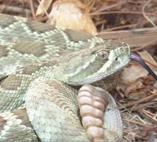
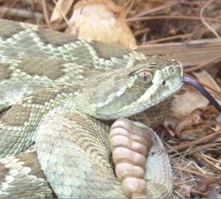
The only venomous snakes in California are the rattlesnakes and there are eight species: the Southern Pacific rattlesnake, the Northern Pacific rattlesnake, the Great Basin rattlesnake, the Western diamondback, the red diamondback, the sidewinder, the speckled rattlesnake, and the Mojave green rattlesnake. The Western rattlesnake has eight subspecies including the Southern Pacific rattlesnake, which is the only one native to the Los Angeles Basin. Ask your veterinarian what venomous species are native to your area.

The physical appearance of each snake species is variable and it is difficult to tell what species of snake you are looking at.

Some general principles in distinguishing poisonous snakes are:

  1. Broad, triangular head with a noticeable neck behind the head.
  2. Vertical pupils (non-poisonous snakes have round pupils), though hopefully you would not be close enough to evaluate this.
  3. As mentioned, the Crotalines are also called pit vipers because they have heat-sensing pits on their faces between the eye and nostril as shown above. The pits help them locate prey. If you see a pit, you are looking at a poisonous snake.

Rattlesnakes can be found in rural areas as well as suburban areas where there is sufficient natural habitat. In Northern California snakes will hibernate during cold months and are active March through September. In Southern California they are active all year round.

Dogs Vs. Snakes

Dogs encounter snakes in the snake’s natural habitat during play or work. Most bites to dogs occur on the face or extremities. The rattlesnake bite is generally hemotoxic, which means that it exerts its toxin by disrupting the integrity of the blood vessels. The swelling is often dramatic with up to one-third of the total blood circulation being lost into the tissues in a matter of hours. The toxin further disrupts normal blood clotting mechanisms, leading to uncontrolled bleeding. This kind of blood loss induces shock and finally death. Facial bites are often more lethal as the swelling may close the throat or impair ability to breathe.

The bite of this Mojave rattlesnake causes rapid paralysis. This includes paralysis of the respiratory muscles and suffocation. Because the Mojave rattlesnake venom is different, treatment may be different and snake venom vaccine (see below) is not protective against this venom.

The severity of a snake bite depends largely on these factors:

  • The species of snake
  • The size of the dog
  • The location of the bite
  • How much venom was involved.

The amount of venom injected (approximately 20-25 percent of bites are dry, meaning no venom has been injected; 30 percent of bites are mild meaning they cause local pain and swelling in the bite area and no systemic symptoms; 40 percent of bites are severe with approximately five percent being fatal.

Treatment

The faster the bite is recognized, the more effective the treatment is. Do not try to cut the bite wound open or suck out the poison. Pressure bandages have been advocated but while these will help keep the poison from moving centrally in the circulation, they also can increase the local tissue damage by concentrating the poison near the bite wound. It is best to seek veterinary care immediately for proper treatment.Seek veterinary care immediately for proper treatment, even if your dog has been vaccinated against snake venom.

IV Fluids

Since the most common mechanism of death from rattlesnake bite is circulatory collapse, IV support and monitoring for signs of blood pressure drop are important. Fluids may be started at a relatively slow rate if the patient is stable but if signs of impending trouble occur, circulatory volume replacement is as easy as opening a drip set valve. After a bite, 24 hours of observation is a prudent timeframe while giving IV fluids throughout.

Antivenin

There are numerous misconceptions about antivenin. The first is simply the name of the product. It is not “anti-venom.” It is not a single injection that provides the antidote to snake bite venom. Antivenin is a biological product consisting of antibodies made by horses in response to exposure to four common Crotaline venoms. The antibody serum is reconstituted into an intravenous drip that is run into the patient over at least 30 minutes or so.

Antivenin is expensive (at least $400 per vial) and a large dog with a severe bite is likely to require several vials. Because the product is of horse origin, often a scratch test to the ear flap is used to test for immunological sensitivity (i.e. to predict whether the patient is likely to have anaphylactic reaction to the antivenin once it is given by IV). The patient will likely always be sensitive to equine products after being given antivenin, which makes future snake bite treatment problematic.

A more purified antivenin of sheep origin has recently been marketed (Cro-Fab antivenin) but this is even more expensive at approximately $700 per vial. This product is made for human use but also works in dogs.

Other products include: Rattler Antivenin® (which actually includes antivenin against the Mojave green rattlesnake), VenomVet®, Antivipmyn Tri® (a Mexican product), and Polivet-ICP® (a Costa Rican product).

Antivenin is very helpful in the inactivation of snake venom but there is a narrow window during which it must be used. After about 4 hours post-bite, antivenin is of minimal use.

A separate antivenin is available for coral snake venom but this is not an issue for California.

Suctioning of the snake bite and using bands or tourniquets to prevent the venom from traveling are urban legends and should not be done. It may be helpful to remove collars or other items that could become constrictive once the swelling starts.

Vaccination

Red Rock Biologics has released a vaccination against the venom of the Western Diamondback (Crotalus atrox). This vaccine also has activity against the venom of six out of seven of the other California rattlesnakes (the Mojave Rattlesnake has such significantly different venom that it is not covered) and there is at least partial cross-protection against numerous other venomous snakes native to areas outside California. Hiking dogs and dogs that live in rattlesnake areas are good candidates for this product. That said, there is controversy about how well it works as the manufacturer does not wish to do efficacy testing that would involve a control group of unvaccinated dogs. Furthermore, a history of vaccination does not preclude the need for emergency treatment after snake bite; the goal of the vaccine is to lessen the poisoning. Efficacy studies thus far have involved mice.

Basics about the Vaccine

  1. Initial vaccination is given in two doses three to six weeks apart. Dogs over 100 pounds and dogs less than 30 pounds in body weight need three doses 3 to 6 weeks apart.
  2. Annual boosters are best given approximately one month before snake season starts in the spring. Dogs who live where snake season is year round or where they hike year round should have boosters every six months. If a vaccine is skipped, the initial vaccination protocol should be re-started.
  3. Vaccination is safe in pregnancy, lactation, and for puppies four months of age and older.
  4. Vaccination reactions occur in 0.27 percent of cases (27 per 10,000 doses given) and are largely limited to swelling at the vaccine site seven to 10 days after vaccination. This is particularly true for dogs with a past history of bee stings.
  5. Vaccinated dogs typically develop protection comparable to two to three vials of antivenin.

A snake bite should always be treated as an emergency even in a vaccinated dog. If your dog is bitten by a rattlesnake, seek veterinary attention immediately.

Snake Avoidance Training

Dogs can be trained to avoid rattlesnakes by scent, sound, and visual appearance. Training classes exist just for this purpose. If your dog hikes in snake territory, consider looking into one of these classes. Call training schools or ask your veterinarian about these specialized classes.

7486367

Rat Poison’s Effect on Dogs and Cats

The EPA has been at odds with Reckitt Benckiser (the makers of d-CON) since 1999 when they began seriously scrutinizing the risks of rat poison products to pets, children, and wildlife. At last, Reckitt Benckiser has aggreed to comply with the EPA directives. This means that second-generation rat poisons (which kill rats in one feeding) will be banned from residential use and that rat poisons for residential use must be secured within bait stations (not in open trays). The traditional d-CON products have not been manufactured since December 2014 and have not been distributed since March 15, 2015. Bromethalin-based rat poisons have since captured the residential market. Commercial exterminators may still use the anticoagulant rodenticides discussed here.

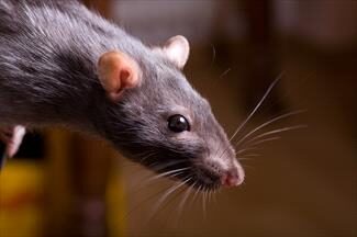
Mankind and the rat have been in conflict for thousands of years. Rats spread disease, eat our crops, leave droppings and make nests in our storage areas, and infest our homes. Rodent removal services are an important part of pest extermination even in this century. Rat poison can be obtained in most hardware stores, grocery stores, and even for free from city agencies in some areas. While you may want to get rid of rats, you certainly do not want to pose a hazard to the children or pets of the family.

Research continues to create a product that fits this bill but in the meantime be aware of the signs of rat poisoning, particularly if your pet travels with you to places outside the home where bait may be left out.

There are several types of rodenticides available. The traditional products are called anticoagulant rodenticides and are discussed here. If you intend to use a rodenticide, we encourage you to choose this type over others as there is a readily available antidote for the anticoagulant rodenticides. Other rodenticides are more toxic and no antidote is available. You should know what product, if any, you use at home. Common anticoagulant rodenticides are: brodifacoum, dopaquinone, warfarin, bromadiolone, and others.

Most of these products include green dyes for a characteristic appearance; however, dogs and cats have poor color vision and to them these pellets may look like kibbled pet food.

Anticoagulant rodenticides do not produce signs of poisoning for several days after the toxic dose has been consumed. Anticoagulant rodenticides cause internal bleeding. A poisoning victim will show weakness and pallor but bleeding will likely not be obvious externally.

Symptoms

Most of the time external bleeding is not obvious and you only notice the pet is weak and/or cold. If you look at the gums, they are pale. Sometimes bloody urine or stool is evident or nose bleeds may be seen. Signs of bleeding in more than one body location are a good hint that there is a problem with blood coagulation and appropriate testing and treatment can be started.

How Does Rat Poison Work?

You would expect the signs of a poisoning to be evident quickly but anticoagulant rat poisoning signs require at least 5 days to show up. Furthermore, unlike most poisons where treatment involves managing symptoms until the poison is out of the system, there is a true antidote for anticoagulant rat poison and it is actually a vitamin. How can this be? To understand what these poisons do, it is necessary to have some understanding of how blood clots.

A blood vessel is sort of like a pipe carrying rapidly flowing blood along its path. The pipe is lined by smooth flat cells called endothelial cells that facilitate the smooth flow of blood. If the pipe breaks, the structure of the pipe below the lining is exposed to the flowing blood inside.

From there the sequence of events is as follows:

  1. The blood vessel automatically constricts and spasms. This restricts the blood flowing to the damaged area and helps minimize blood loss.
  2. The exposed pipe attracts circulating platelets, cloud-like cells that circulate ready to assist in clotting should the need arise. Platelets clump together over the tear in the blood vessel, forming a plug within the first five minutes of the injury. This is all a good thing but the platelets will not stay in place unless a substance called fibrin can be made to bind them.
  3. Generating fibrin is complicated and beyond the scope of this article, but a cascade of activating proteins is needed to make the tiny protein threads (fibrin) that bind the platelets and makes a permanent platelet plug on the wound. Four of the proteins involved are called serine proteases, and these are the factors relevant to anticoagulant rat poisoning. These four factors must be able to work or there will be no fibrin, the platelets will not clump properly, and bleeding will continue without clotting.

About those Four Clotting Factors

The four clotting factor proteins are also called the K-factors because they depend on vitamin K for activation. After the clotting situation is under control, the used Vitamin K is recycled in the liver so it will be ready for the next time bleeding needs to be stopped. Anticoagulant rat poisons interfere with Vitamin K recycling. It takes several days to deplete the Vitamin K but after that is used up, there is no more and bleeding cannot be stopped. That is why it takes some 5 days for symptoms to show up and how it is possible to reverse this poison by giving more Vitamin K.

As long as there is plenty of vitamin K, the serine proteases can be activated and clotting can proceed normally. The anticoagulant rodenticides abolish vitamin K recycling. This means that as soon as the body’s active vitamin K reserves are depleted there can be no meaningful blood clotting.

In cases of poisoning you would expect symptoms to be nearly immediate but in the case of anticoagulant rodenticide poisoning, it takes several days to deplete vitamin K. After that, even the smallest of jostles and traumas can lead to life-threatening bleeds.

Testing

Testing can be done using clotting tests called a PTT (partial thromboplastin time) and PT (prothrombin time).  These tests evaluate the activity of the four K-factors. If both these tests are abnormal, there is an excellent chance that anticoagulant rat poison is in the patient’s system. The PT test in particular bears special mention as it tests the least stable of the K-factors called Factor VII. This means that the PT test becomes abnormal before the PTT test becomes abnormal. When it’s time to monitor a patient for recovery, the PT test must be normal in order to declare the poisoning resolved.

Another test called the PIVKA (Proteins Induced by Vitamin K Antagonism) test is more specific. The PIVKA test detects inactive serine proteases. An unusually high amount of inactive K-factors circulating indicates something is wrong with Vitamin K recycling.

Therapy

If the patient has only just ingested the poison, he or she may be made to vomit it up. Cathartics and adsorbents can be used to prevent the poison from entering the patient’s system. Still, it is best to use the antidote anyway. Certainly, if there is evidence that the patient is bleeding, the antidote obviously is required.

The antidote is simply vitamin K.

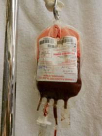
Vitamin K is generally started as an injection, and when the patient is stable, tablets are prescribed. The human formulation, available as a prescription drug at most drug stores, is a 5 mg tablet. The veterinary strength is a 25 mg tablet. Blood transfusions may be needed to stabilize a patient who has suffered significant blood loss.

There are different classes of anticoagulant rodenticides, and they remain in the body for several weeks. It is hard to know when to discontinue therapy, especially if the particular rodenticide is not known. After a couple of weeks of therapy, the medication is discontinued. Forty-eight hours later, a PT test is run. If there is still rodenticide in the patient’s system, the PT will be abnormal, but the patient will not yet have started to bleed. The results of the PT test will tell the veterinarian whether or not another couple of weeks of vitamin K are needed.

It is important to return for the recheck PT test on schedule. Waiting an extra day or two will allow internal bleeding to recur. There is no point in doing the PT test while the patient is still taking vitamin K. The test must be done 48 hours after discontinuing the medication.

When the PT test has returned to normal, it is safe to discontinue therapy.

Vitamin K1 Vs. Vitamin K2 Vs. Vitamin K3

There are three forms of vitamin K but only vitamin K1 is used therapeutically. Vitamin K1 is a natural form of vitamin K that is found in plants and absorbed nutritionally. Its more technical name is phylloquinone. Vitamin K2 (menaquinone) is also natural and is produced by the body’s intestinal bacteria but apparently not in amounts adequate for rescue from the anticoagulant rodenticides. Vitamin K3 (menadione) is a synthetic version that may be injected or taken orally. You may even see it available as a vitamin supplement tablet.

Within the body vitamin K1 and vitamin K3 are converted to vitamin K2. Vitamin K3 might seem like an inexpensive way to treat a pet with rat poisoning but unfortunately, K3 is sometimes toxic and can actually lead to red blood cell destruction. Inexpensive vitamin K3 pills on the drugstore shelf for over-the-counter sale are not acceptable antidotes. Vitamin K1 is used because it is absorbed early in the GI tract and concentrates directly in the liver, which is where the K-factors are activated.

It is only vitamin K1 that should be considered to be the antidote for anticoagulant rodenticide poisoning.

Other Rodenticides

While anticoagulant rodenticide poisoning is a life-threatening event, at least there is an antidote readily available. Other rodenticides are not as readily reversed. These rodenticides include:

  • Quintox, Rat-B-Gone,  Bromethalin (see an additional article)
  • Strychnine, gopher bait
  • Zinc phosphide, gopher bait 

Pets Who Eat a Poisoned Rodent

We are commonly asked about the risk to a cat or dog who eats a rodent that has been poisoned with an anticoagulant rodenticide. The rodent might have already died or simply not have died yet given that several days are required to feel the effect of these poisons. The fact is that when you’re talking about the newer generation anticoagulant rat poisons, such as diphacinone, the risk is real. A greedy rat can eat enough poison to kill 20 rats before he starts to feel sick, and if this was a second generation rodenticide it will accumulate in the rat’s liver ready to poison the cat that eats the rat’s liver. Fortunately, second-generation rodenticides have been banned for residential use. First-generation rodenticides are no longer in the rat’s body after several hours, making pet poisoning less of a concern. Furthermore, most rats do not overindulge in poison. The usual patient for secondary poisoning is a pet or predator that depends heavily on rats for food (a barn cat, for example). There is some controversy over how often this actually happens, as most pets do not consume numerous rats.
4902154

Rat Poison (Bromethalin-Based) in Dogs and Cats

The War Against Rodents 

Rodent control has been paramount in human civilization for centuries if not millennia. Wild rodents consume our crops, destroy our fabric and paper products, bring disease, and are generally unsanitary in our homes. Rat poisons, as well as traps, are widely used to control rodent populations in human communities all over the world.

In the past, anticoagulant rodenticides, which were effective and available in virtually every hardware and garden store, were the most common rat poisons in the U.S. residential market. These products were readily consumed by rodents, and after five days or so, they induced internal bleeding and death. Problems with them included toxicity when they were accidentally consumed by pets or by predatory wildlife. Either group of animals might consume the poisons outright or might become poisoned by eating an already intoxicated rodent. The EPA demanded that these types of poisons be phased out, and presently, they are only available to commercial exterminators. Demand for safer products rose.

Bromethalin was supposed to represent a better choice. Because only small amounts are needed to kill a rodent, a predator is unlikely to be harmed by consuming a bromethalin-poisoned rodent. This is good news for owls, hawks, and other rat-consuming wildlife. For dogs and cats, however, this is only a small advantage, as pets commonly consume the rodenticide directly.  Anticoagulant rodenticides have an antidote in case of accidental poisoning, while bromethalin has none, and bromethalin products are readily palatable to dogs and cats.

With anticoagulant rodenticides becoming less available, bromethalin has captured the home rat poison market and has created a new hazard for pets to contend with.

What does Bromethalin do?

Bromethalin has been around since 1985, and it is sold as bars, blocks, and pellets to be used against rats and mice, as well as in the shape of earthworms to be used against moles. The product can be sprinkled on the ground or, for a safer presentation, provided inside a plastic bait station accessible to rodents but not to pets.

Bromethalin acts by uncoupling oxidative phosphorylation in the brain. What that means is that bromethalin makes it impossible to maintain aerobic metabolism in the brain. Oxygen cannot be used. Without the ability to use oxygen, the brain cannot make enough energy to fuel itself. The brain swells, and the victim dies of cerebral edema.

Peak levels of bromethalin are achieved a mere four hours after the poison is eaten.

What Does This Mean for a Pet?

There are two forms of poisoning recognized in pets: the high dose (or convulsant form) and the low dose (or paralytic form).

The Convulsant Form

If a high dose of bromethalin has been consumed, symptoms generally begin in a matter of hours. The patient will develop hyperexcitability followed by muscle tremors, increased body temperature, and seizures. Death is virtually inevitable.

The Paralytic Form

If a lower dose has been consumed, symptoms do not show up for one to four days. Vomiting and appetite loss result. The patient becomes weak in the rear and uncoordinated. The eyes begin to move involuntarily back and forth (nystagmus) and the pupils become unequal in size. Muscle tremors culminating in paralysis result. Treatment is still possible with this form, but it must be aggressive.

Treatment

Treatment for this poisoning must begin immediately, ideally within an hour of eating the poison. The patient is made to vomit and then must receive several doses of activated charcoal to bind any poison in the intestinal tract before the patient’s body absorbs it. Use of activated charcoal can continue for days.

Muscle contractions are treated with sedatives and muscle relaxants. Brain swelling is treated with diuretics. One study in rodents found that if rats were given ginkgo biloba immediately, their toxicity symptoms were greatly reduced but no such research has been performed in dogs or cats.

Survivors may have permanent neurologic deficits and it make take weeks for recovery to plateau so that permanent damage can be assessed. Many patients will not eat during treatment and nutritional support, such as an esophagostomy tube, must be employed.

If you use a bromethalin-based rat poison, be sure pet access is impossible as there is no antidote for this poison and signs are rapid and not easily treated.

Anti-coagulant rodenticides may have their problems, but at least treatment is straight-forward and there is an antidote. 

If you must control rodents, consider the least toxic methods if there are pets in the vicinity.