Asthma in Cats
What is Asthma?
Asthma is a recurring respiratory compromise featuring the constriction (narrowing) of the lung’s airways.
There are three features that define asthma:
• Airway inflammation
• Airway hyper-responsiveness
• Reduced airflow (which is at least partly reversible)

More simply, this means the airways of the lungs are inflamed, plus they are extra reactive to mental stress or respiratory irritants, and the flow of air through the lungs is reduced (either sometimes or all the time). When all three features are present, we can be comfortable diagnosing asthma.
What Exactly is Happening in the Lungs with Asthma?
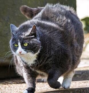
First, excess mucus forms in the airways. After that, the airway walls swell with inflammation and can actually ulcerate. Finally, the airway muscles go into spasms leading to constriction. Airway constriction leads to an inability to draw a deep breath, intolerance to exercise, coughing, and musical sighing sounds called “wheezes,” though not all these symptoms may be apparent at the same time.
Sometimes a low-grade chronic cough is the only evident sign, but it should be remembered that a sudden asthmatic crisis can arise at any time and can represent a life-threatening event. Asthmatic airway constriction can happen spontaneously, in reaction to stress or anxiety, or as a type of allergic reaction. Relieving and preventing airway constriction is the main focus of treatment.

Is Feline Asthma the Same as Human Asthma?
The feline condition was named asthma because of the clinical features shared with the human disease. As of this time, it appears that all the diagnostic criteria needed to diagnose asthma in people are shared by cats, yet we are still working out the mechanics of this syndrome in cats and have a great deal to learn. For example, in humans, we know that while actual symptoms of asthma occur in episodes, the airways of the lung are diseased all the time. We do not know yet if feline airways are also diseased all the time or if the actual airway structural changes occur only when there are clinical signs of distress.
How is the Diagnosis Made?
Because of the constricted airways, the actual volume of air this patient can move in and out of the lungs with each breath is reduced. There is often a great deal of effort seen in the cat’s breathing. The abdomen appears to be working to push air out and the breaths are shallow and rapid. The cat may even be breathing with its mouth open in an effort to move the largest possible amount of air.
The next step toward making a diagnosis of feline asthma is the chest radiograph, assuming the cat is not in too much distress to hold still for this procedure. Classically, this radiograph will show what is called air-trapping, meaning that the small airways have constricted so that inhaled air cannot be exhaled. The lungs are larger in appearance than normal as they are over-inflated. The diaphragm may seem flattened due to this over-inflation.
It is possible to view the tiny airways in much higher detail on radiographs. This is partly because they are made more prominent because of the mucus and inflammatory fluid inside them (the doughnuts and tramlines, as mentioned below) and partly because they are surrounded by more air than usual (air-trapping).
Inflammation and mucus build up within the airways, causing their walls to appear thickened in the radiograph. The terms used for such airway appearance are doughnuts (when viewing the airway end-on) or tramlines (when viewing the airway from the side). You may hear your veterinarian use these terms, and they are classical findings in airway disease.
Some Asthmatic Cats Have Normal Radiographs
Since visible changes are not always evident on radiographs, which can lead to diagnosing asthma when it is not there. Indeed, diagnosing asthma can be complicated, and it is famously underdiagnosed because up to 23% of asthmatic cats can have completely normal radiographs.
The coughing cat with normal chest radiographs poses a diagnostic dilemma. It is hard enough to sort out potential issues when there actually are radiographic changes, as many conditions can mimic others. Because of the nature of feline heart disease, where the heart muscle can thicken without the overall external shape of the heart changing, heart disease should be considered. In heartworm-endemic areas, feline heartworm disease should be considered. If the patient is thought not to have too high of an anesthetic risk, a tracheal wash and/or bronchoscopy can be considered but often have indefinite results. CT (cat scan) imaging is emerging as a helpful diagnostic tool, but it is not yet available in many areas.
Response to Therapy as a Diagnostic Test
One important asthma feature is that the airway constriction it engenders is reversible. In an emergency situation, a small dose of epinephrine (adrenalin) or terbutaline (which expands airways) can reverse an asthmatic crisis in as little as 15 minutes. Response to an injectable corticosteroid (such as dexamethasone) generally yields a positive response within 30 minutes.
Sometimes, diagnostic tests still leave room for questions, and you have to simply go with medical treatment for asthma and regard response to therapy as evidence that the diagnosis is correct. See below for a list of medications commonly used in the long-term management of this problem.
Treatment Options
Oral and Injectable Steroids
It is crucial to realize that the underlying problem in the airway is inflammation. Inflammation is what is responsible for constriction. To resolve inflammation, corticosteroid medications have been the cornerstone of therapy. These can be given in pills, by injection, or, more recently, through a metered dose inhaler. Usually, treatment is started with either an oral corticosteroid such as prednisolone or a long-acting injection such as Depo-Medrol. These medications are relatively inexpensive, and a good response to them helps confirm the diagnosis, as mentioned. A better strategy might be to use oral or injectable steroids to ensure that they work and if they do, change to a metered dose inhaler (see below), as there is much less potential for corticosteroid side effects with an inhaler.
If the response to oral corticosteroids is good, they may be continued long-term, supplemented with some of the other medications mentioned below, or used only during flare-ups. Long-term corticosteroid use has some potential for side effects, though feline patients tend to be resistant to these problems. If giving pills is too difficult, especially in a cat that is stressed and having some trouble breathing, a long-acting corticosteroid injection can be used periodically to control asthma symptoms. Cats are more sensitive to potential side effects from these injections as they are much stronger than oral corticosteroids. This sensitivity means that injections can only be given periodically without the risk of developing diabetes mellitus. If the cat seems to require an injection more frequently than every other month, a metered dose inhaler should be considered (see below for details).
When using oral corticosteroids, it is important to taper the dose over time so as to find the minimum dose needed to control symptoms. If higher doses are used indefinitely, asthma symptoms may become resistant to steroids. This resistance is more of a problem with injectable steroids and manifests as a shorter and shorter interval between the need for the injection. Again, if this is seen, consider changing to an inhaled form of medication.
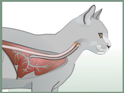
Metered Dose Inhalers
Human asthmatics have enjoyed the benefits of portable inhalers for years. These handy devices deliver medication locally to the airways, thus minimizing drug side effects to the rest of the body while maximizing desired response.
There were two problems with using these devices in cats:
- Cats cannot be told when to inhale.
- Cats tend to object strongly to the spray puff delivered by the device.
Interestingly these same problems apply to human infants with asthma and a device called a pediatric spacer was invented for them. This tubular chamber is attached to the inhaler on one end and a face mask on the other. The inhaler is actuated into the spacer and the infant simply breathes in the spacer’s contents.

The discovery that the pediatric spacer could also be used for cats has solved the feline inhaler problem as well. The setup is the same as described above for young children and the cat takes 7 to 10 breaths from the face mask to be properly dosed.
Corticosteroid inhalers, Flovent® being the most popular, are typically used twice daily long term while airway dilator inhalers such as albuterol-containing Proventil® or Ventolin® are used for flare-ups.
Cats are generally started on a combination of oral prednisone and the metered dose inhaler and gradually maintained on only the metered dose inhaler.
Pediatric equipment is generally available from most human pharmacies, however, a device created specifically for feline use can be ordered. It comes with a spacer and an appropriately sized face mask. Your veterinarian will need to prescribe the metered dose inhaler usually from a regular human pharmacy.
See the Aerokat device.
Are there other Treatment Options?
Other medications that might be helpful include:
Allergen-Specific Immunotherapy
Since allergy is generally a part of the asthma picture, it makes sense that “allergy shots” might be helpful. In fact, they are helpful to many cats. It is important to realize that this form of therapy takes months before benefits are obvious so it is not a method for fast relief but more for the “bigger picture.” The owner must be able to give injections to the patient at home regularly. A veterinary dermatologist is generally needed to oversee this type of therapy.
Airway Dilators
Terbutaline (Brethine®) and theophylline are airway dilators commonly used in the management of asthma. It makes sense that if constriction is an important feature of this disease that eliminating constriction would be therapeutically helpful. Terbutaline is available orally or as an injectable. Some veterinarians encourage owners to keep a bottle of injectable terbutaline at home in case of a crisis and show them how to give it. If you are interested in this, let your veterinarian know. Theophylline is an oral medication usually given once a day at bedtime.
Cyclosporine
The use of cyclosporine in asthma is relatively new. Cyclosporine is an immunomodulator often used in organ transplant patients. It has been used occasionally in cats where adequate suppression of inflammation has not been possible with combinations of the other medications listed or when the cat is unable to take corticosteroids for other reasons (concurrent diabetes mellitus, infection, history of calcium oxalate bladder stones, etc.). Cyclosporine should not be prohibitively expensive since cats, being small, require relatively small doses but blood levels of cyclosporine may be periodically recommended which increases the expense of this treatment significantly.
Don’t Forget…
Minimizing irritants in the air is always helpful to an asthmatic cat.
- Do not allow cigarette smoke in the cat’s environment.
- Use dustless cat litter.
- Consider non-topical insecticides. No sprays, either.
- Regularly replace air filters at home.
It is important to realize that asthma can culminate in a respiratory crisis that can become life-threatening if ignored. If your cat begins to breathe with an open mouth or if you see excessive abdominal movement during breathing and the cat is not purring, you may have an emergency situation. Contact your veterinarian immediately.
For more details, including a video on the use of the inhaled system, we recommend visiting Fritz the Brave, a website set up by one family devoted to their asthmatic cat. It has grown into a detailed instructional site for both pet owners and veterinarians interested in the details of inhalers for feline asthma treatment.