Tag: pets

7613596

Hyperesthesia Syndrome in Cats  

Feline hyperesthesia syndrome (FHS) is known by several names including “rolling skin disease”, “neurodermatitis”, neuritis, psychomotor epilepsy, and pruritic dermatitis. Hyperesthesia means “too much feeling.” It can be difficult to determine whether the cause is behavioral or medical without additional diagnostics, such as an MRI or EEG.

Clinical signs of feline hyperesthesia syndrome (FSH) can range from mild to self-mutilating.

Common behaviors seen include:

  1. rippling or rolling of skin and muscles over the back, ears, head, or tail;
  2. excessive staring at their tail, then attacking their tail or sides;
  3. biting at the base of their tail, front legs, and paws;
  4. running around while meowing in a distressed manner;
  5. aggression towards people or other cats. Some cats may show an increase in affectionate behavior;
  6. big, dark, round (dilated) pupils.

To diagnose FHS, other conditions related to the skin and nervous system must be ruled out first. These include:

  1. dermatologic causes: flea, food, environmental, or seasonal allergies; skin infections or parasites, auto-immune diseases, anal sac disease;
  2. neurological conditions such as seizures or neuromas (painful nerve bundle) which may form in cats after declaws or tail injuries;
  3. pain: frostbite and orthopedic conditions such as arthritis, injury to the tail, hips, or back;
  4. compulsive disorders: may be a primary behavior problem starting from one of the above conditions, then worsening over time;
  5. toxins: Pyrethrin/pyrethroid, Organophosphate/carbamate toxicosis;
  6. gastrointestinal diseases that are accompanied by changes to appetite, vomiting or diarrhea, flatulence or constipation;
  7. urinary conditions, particularly in male cats with a urinary blockage.

To rule out underlying medical causes, expect your veterinarian to do a full physical, orthopedic, and neurological exam to determine the cause. Diagnostics may include radiographs, blood work or urinalysis, skin swabs or skin scrapes.

Treatment for FHS depends on the cause. Your veterinarian may recommend medications, supplements, weight loss or rehabilitation. Treatment of skin conditions may require antibiotics, medications for itch and inflammation or diet change.

If a physical cause cannot be found, antidepressant and anti-anxiety medications may be prescribed.

In addition to medications, there are some treatments that apply to all cats with clinical signs of FHS such as:

  1. avoid known situations that cause the behavior in your cat, such as petting;
  2. avoid punishing the behavior verbally or physically. This causes conflict and is not likely to stop the behavior;
  3. provide a calm, predictable environment for your cat;
  4. provide different types of enrichment;
    •    vertical spaces and areas to hide;
    •    offer toys to chase;
    •    encourage hunting with food dispensing and puzzle toys;
    •    train with positive reinforcement techniques.
5107512

Haws Syndrome in Cats

Haws syndrome is an elevation of the third eyelid in both eyes. The third eyelid, or nictitating membrane, is a transparent eyelid some animals have that moistens and covers the eye for protection. In Latin, nictare is to blink. Haw is what the third eyelid in horses are called, although in cats it’s a nictitating membrane. Haws syndrome is not associated with diseases inside or outside of the eyeball, nor the socket. It is also called bilateral third eyelid protrusion, bilateral prolapse of the third eyelid, and bilateral prolapse of the nictitating membrane.

The condition has not been reported in dogs.

The cause is not known. However, the condition is frequently associated with gastrointestinal (GI) inflammatory disorders. The elevation of the third eyelid may be due to a change in the way nerves are supplied to the third eyelid.

A cat’s vision is normal with this condition, but may be functionally decreased simply because the elevated third eyelids are blocking the cat’s vision. Affected cats may have watery diarrhea that began before the onset of elevation. 

Diagnosis

Diagnosis is primarily made on finding classic clinical signs and eliminating other diseases with a thorough ophthalmic and physical examination. The main sign is a protrusion of both third eyelids. Thankfully, the eyes are not painful.

Your veterinarian may do a phenylephrine challenge. Phenylephrine eye drops are applied to the eyeballs to see if the third eyelids return to their normal position within 20 minutes; If they do, then Haws can be confirmed. 

Treatment

This condition is usually self-limiting, so no specific treatment is necessary.

Any diarrhea a cat has should be treated as that may be a contributing factor.

Monitoring and Prognosis

The prognosis is good because the condition is often self-limiting. In one report about cats, the average time for the third eyelids to return to normal was 47 days; however, the number of cases evaluated was limited. In a separate report, 17 out of 45 cats had a third eyelid protrusion that persisted for more than 4 weeks. If the condition persists, your veterinarian may suggest further diagnostic tests, especially if your cat has GI signs.

5458065

Tail Pull Injuries in Cats

First, an Anatomy Lesson

Tails are wonderful, expressive body parts used by cats for communication purposes as well as for balance. The tail consists of a varying number of vertebrae (called caudal vertebrae; caudal means near or at the tail) and voluntary muscles with ligaments and tendons holding it all together. The tail attaches to the body at an area called the tail head. The first caudal vertebra attaches to a backbone called the sacrum, which connects the tail and lower back (lumbar) vertebrae.

The “Horse’s Tail”

The spinal cord does not extend down to the tail (it typically ends at the level of the fifth lumbar vertebra), which means that tail injuries do not damage the spinal cord. Unfortunately, injuries to the tail can still cause serious nerve damage.

Cats get their tails pulled or broken through an assortment of traumas: A child might pull a tail or it might get caught in a closing door. A tail can get bitten during a cat fight and, of course, automobile accidents can easily lead to dislocated or broken tails. One might think a tail break would involve an obvious external wound but usually this is not the case; instead, signs of nerve damage from stretching of the cauda equina nerves are often the only tip off.

In many cases, the tail pull injury is eclipsed by bigger injuries. In one study presented at the World Small Animal Veterinary Association Congress in 2016, 46% of tail pull injuries were accompanied by pelvic fractures or sacroiliac dislocations, 18% were accompanied by broken rear legs, and 11% had additional spinal fractures. Only 21% had the tail pull as the only injury.

Because the spinal cord ends so high up, nerves exiting the cord to provide control and sensation to the tail, hind legs, urinary bladder, large intestine, and anus must extend long branches to reach their destinations.  When you look down at these nerves (an aerial view if you will), these nerves are said to look like a horse’s tail, and thus anatomists call this area the cauda equina (which literally means horse’s tail).

What Might be Noticeable at Home After a Tail Pull Injury

Cats with a tail pull injury might show any of these symptoms:

  1. A tail that drags or is never held high.
  2. Involuntary dribbling of urine.
  3. A dilated, flaccid anal sphincter with or without diarrhea or fecal incontinence.
  4. Incoordination of the rear legs.

Any of these symptoms might lead the cat into the vet’s office for an evaluation. Some additional findings the veterinarian might notice include:

  1. A distended bladder that is relatively easy to express manually (in other words, the bladder is full but can be emptied with careful squeezing.)
  2. Bloody urine (if the tail trauma is recent).
  3. Painful tail head.
  4. Loss of sensation in the tail.

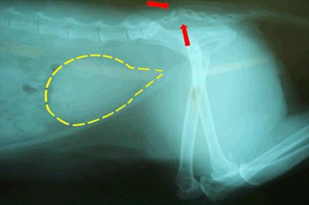
A radiograph will often show a break in the tail or a dislocation, although in some cases the tail bones are intact. How permanent these signs are largely depends on whether the cauda equina nerves have been over-stretched or actually torn. 

Classification of the Injury

In a 1985 study by Smeak and Olmstead, sacrocaudal fractures in cats were divided into five groups.

Group One: Cats With Painful Tail Heads as their Only Sign
These individuals are minimally affected and, while they may or may not have a chronically sensitive tail head, they are expected to otherwise have a full recovery.

Group Two: Cats With Lack of Tail Mobility and Sensation as Their Only Sign
These cats are expected to recover tail function completely and have an excellent prognosis.

Group Three: Cats With Lack of Tail Mobility and Sensation Plus Urine Retention as Their Only Signs
Most cats will have a complete recovery.

Group Four: Cats With Lack of Tail Mobility and Sensation and Diminished Anal Tone
This group of cats has about a 75% recovery rate, meaning 75% of the cats that fit this description should recover.

Group Five: Cats With Lack of Tail Mobility and Sensation Plus a Dilated/Flaccid Anus (i.e., Zero Anal Tone)
This group of cats has about a 50% recovery rate, meaning about half of the cats with this description will eventually recover.

If it is not clear whether a cat has partial anal sphincter tone or not, it may be possible to have a test called an electromyogram. The muscles of the anal sphincter and tail can be tested to see if they are receiving any nerve input at all. If they are, there is reasonable potential for recovery. The EMG test is highly specialized and not readily available except at practices with a neurology specialty. Referral will probably be necessary.

In a more recent study (Nov 2009, Journal of Small Animal Practice) researchers found that an excellent predictor of bladder function return is pain detection at the tail head. In the 21 cats with sacrocaudal fracture/dislocation, all 11 cats that had pain sensation at the tail head on the first day after the injury had regained bladder function within 3 days. Absence of pain sensation in this area on the first day did not necessarily mean that bladder control would not return; 60% of cats without tail head sensation on the first day had recovered bladder control by 30 days after the injury.

Should the Tail be Amputated?

If the tail is not expected to recover mobility or sensation, there are some reasons to consider amputation. If the cat cannot lift his tail, he may soil it regularly, creating an infection issue or simply an odor issue. Further, the weight of the tail dragging may further stretch the cauda equina nerves. Whether or not tail amputation is helpful or speeds recovery is controversial but certainly amputation could solve a soiling problem.

Bladder Management

It is important not to allow the bladder to over-stretch. The fine muscles of the bladder can become so stretched out that even if the nerves recover, the bladder may still remain unable to empty fully. This means the cat’s bladder must be gently squeezed three or four times daily to keep it from over-distending. Medications can also be used to assist the bladder’s own ability to contract (bethanechol chloride) or to relax the urinary sphincter (prazosin or phenoxybenzamine).  Since urine retention tends to promote infection, patients will periodically require some sort of monitoring urine tests. If constipation is a problem, periodic enemas or stool softening medications can be given regularly.

Recovery Time

Nerves heal notoriously slowly. It has been said that a good six months must pass before one can say the maximum recovery has occurred and no more positive progress can be expected. Most cats who are going to recover function do so in a one-week period, and most cats who do not recover urinary control after a month probably will remain incontinent. Tail function and sensation tends to take longer.

4128626

Tooth Resorption in Cats

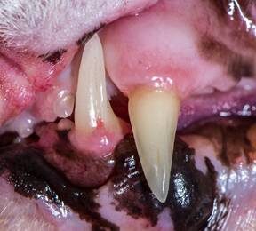
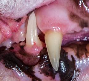
A common feline oral malady is tooth resorption (TR). Greater than half of all cats older than three years old will have at least one tooth affected by resorption; it affects dogs less frequently.   These tooth defects have been called cavities, neck lesions, external or internal root resorptions, feline odontoclastic resorption lesions (FORLs), and cervical line erosions.

Tooth resorptions are usually found on the outside of the tooth where the gingiva (gum) meets the dental hard tissue. The lower jaw premolars are mostly affected, however tooth resorption can be found on any tooth. 

The cause is unknown, but theories supporting an autoimmune response, calicivirus, and metabolic imbalances relating to calcium regulation have been proposed.  The resorption generally starts at the cemento enamel junction (gum line) and progresses inward, eroding sensitive dentin. Once the resorption is exposed to the oral cavity, bacteria invade causing pain and jaw spasms. Increased salivation, oral bleeding, or difficulty eating are other signs. Unfortunately, most times there are no outward signs. It is up to the veterinarian or astute owner to diagnose tooth resorption.

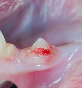
There are five recognized stages and three types of tooth resorption. Initially in stage 1, only an enamel defect is noted. The lesion is usually minimally sensitive because it has not entered the dentin. In stage 2, the lesion penetrates enamel and dentin. When resorption progresses into the pulp chamber (nerve) stage 3 has occurred.  In stage 4, large amounts of the tooth’s hard structure have been destroyed. By the time stage 5 has occurred, most of the tooth has been resorbed, leaving only a bump covered by gum tissue.

Intraoral radiographs (X-rays) are essential to evaluate all the teeth to determine the best course of therapy. Depending on what is seen on the visual patient exam and the intraoral X-rays (tooth resorption type 1, 2, 3), treatment involves following the resorption until it is exposed to the oral cavity, extraction of the entire tooth and roots, or a partial tooth extraction.  In cats affected by stage 5 without inflammation, treatment is not necessary.

3096810

Tritrichomoniasis in Cats  

Tritrichomoniasis is a major cause of diarrhea in cats and is caused by Tritrichomonas blagburni. The disease is seen most commonly in young cats and kittens.

T. blagburni is a type of protozoa, which are microscopic, single-celled parasites. They are not related to bacteria or viruses. T. blagburni is transmitted to cats when they come into contact with the organism directly or accidentally eat the infected feces of another cat. Infections are common in crowded environments such as catteries or animal shelters. Thankully, T. blagburni is not contagious to humans or other types of animals, so only cats get it.

Symptoms

Chronic (i.e. long term), voluminous, watery diarrhea is the most common symptom. Diarrhea can come and go so that the cat seems fine sometimes, which can help delay a diagnosis. Cats can also experience excess gas, going more often, foul odor to the stool (we mean more than usual!), and/or trouble defecating.

In general, cats infected with T. blagburni are otherwise normal, have a healthy weight and appetite, and are well hydrated. Sometimes, cats can become dehydrated or develop a poor appetite if the diarrhea is severe or excessive.

What to Expect at a Veterinary Visit

If you suspect your cat has tritrichomoniasis, bring some fresh feces to the veterinary appointment with you. Sandwich-sized plastic zippered bags are great for carrying the diarrhea sample.

Your veterinarian will start with a thorough physical examination, and then check for signs of dehydration and tummy pain. There will likely be an examination of your cat’s feces for intestinal parasites or increased levels of bacteria by looking at the feces under a microscope. Some cats are infected with multiple types of parasites at one time, so it is important that all issues be diagnosed and treated properly.

Unfortunately, T. blagburni is difficult to identify just by looking at the feces. Laboratory tests may be needed and/or blood tests to get an accurate diagnosis. Occasionally, a biopsy may be needed, but this is not common.

Treatment

Medications are available to treat tritrichomoniasis, but they are not always effective.  In some cats, especially in those less than one year of age, infections will improve without specific treatment. However, this can take several months.

Supportive care is important to keep your cat healthy during the infection. Feline diets high in fiber can help firm up the stools. Probiotics may also be useful to improve and maintain normal gut/GI flora. Any additional intestinal parasitic diseases need to be treated appropriately as well.

Control and Prevention

Keep your cat’s environment clean and free of feces. Minimize stress where possible by providing plenty of exercise and individual resting spaces. Scoop litterboxes daily and disinfect them regularly. All cats in the household should be tested for tritrichomoniasis because some cats may be infected without showing symptoms. Uninfected cats can be separated from the infected ones to prevent the disease from spreading, although that’s difficult in a home setting.

testimonials-goldberg-pet

Glomerulonephritis in Dogs and Cats

(Urinary protein loss due to kidney inflammation)

When a patient, human or non-human, is said to have kidney failure, renal insufficiency, or even chronic renal failure, what most people are talking about is a toxin build up when the kidney cannot adequately remove the body’s harmful wastes. This toxic state is called uremia and is associated with nausea, appetite loss, weight loss, listlessness, and other unpleasant issues.  It is also not the kind of kidney disease we are going discuss here. 

Glomerular disease is a completely different kind of kidney disease and may not involve any toxin build up at all. Glomerular disease is one of protein loss. 

What is a Glomerulus Anyway?

Consider for a moment what an important resource protein is to your body. Your blood, for example, is full of necessary circulating proteins handling clotting, fluid balance, transporting other chemicals etc. Your body went to a lot of trouble to build those proteins and you can’t afford to waste them. If you were to lose them, your body would have to break down muscle in order to recreate them because that is how important they are.

On the other hand, your blood carries an assortment of metabolic wastes that you need to get rid of. You need to filter out these bad materials without losing what is valuable. The millions of glomeruli you have are in charge of keeping your blood proteins where they belong — in the blood — while allowing for smaller wastes and extra fluid to filter out and be made into urine. There are other valuables in your blood besides cells and protein, but different areas of your kidney handle those.

The illustration to the right shows the nephron, which is the functional unit of the kidney. There are millions of these making urine every moment of every day. Only about 30 percent of them must be working in order to maintain normal kidney function. The rest form a back up system so that we will have plenty of extra nephrons should some of them get plugged with debris, damaged by scarring or infection, or starved for oxygen during a traumatic event.

The glomerulus, which in a way looks like a little dandelion tuft, is where our interest lies today. Blood flows through an afferent arteriole into the glomerulus. Inside the glomerular tuft, the blood vessel narrows into a complicated spiral of tiny capillaries, so small that the blood cells pass through single file. The capillaries are gripped by cells called podocytes. Like hands, the podocytes have tiny fingers (ironically called foot processes) that encase the capillaries. Fluid and small molecules can flow in between the fingers while cells and large molecules like proteins cannot pass through.

The cells and large molecules/proteins exit the glomerulus through an efferent arteriole and return to normal circulation. This first step in filtration is driven both by blood pressure as well as by the protein content of the blood.

Now imagine what would happen if there were holes punched in that filtration system so that protein can pass through the fingers. This is what happens in glomerular disease.

How does the Glomerulus get Leaky?

Sources of chronic inflammation are believed to be the ultimate cause of the problem. The chronic inflammatory state leads to the circulation of antigen: antibody complexes in the blood and these complexes stick in delicate glomerular membranes like flies in fly paper. Once stuck there, they call in other inflammatory cells and soon a hole is eaten into the membrane by the ensuing reaction. The holes in the filtration membranes are big enough for proteins to traverse.

There are many possible sources of chronic inflammation that could be generating antigen: antibody complexes. Chronic ear or skin infections could be the cause. Long-standing dental disease could do it. A latent, more internal infection might be the cause (such as heartworm, Lyme disease, prostate infection, or Ehrlichiosis). Even a tumor might generate enough of the immune system’s attention to lead to this sort of reaction.

If it is at all possible to identify and resolve the underlying cause of inflammation, this should be done as other therapy is unlikely to fully resolve the protein loss.

How is the Diagnosis Made?

There are several common scenarios that might lead to the diagnosis but they all boil down to one or both of two findings: excess urine protein found on a routine urinalysis and/or low albumin found on a blood test.

Let’s start with excess urine protein found on a routine urinalysis.

A urinalysis examines a urine sample for some of its chemical contents and properties. Protein content is one of the parameters that is checked and reported as a small, medium or large amount. On a urinalysis report this will be designated as “+,” “++,” or “+++.”

This seems like it would be easy enough to interpret but unfortunately there is more to the story. A small amount of protein in a well-concentrated sample may be  normal while the same amount of protein in a dilute sample would be highly significant. How dilute or concentrated the urine is depends on the patient’s water consumption, and we need a method to examine urine protein that is independent of the water consumption.

Further, we need to determine if any protein in the urine is truly coming from the kidneys; after all, a bladder infection or other bladder condition might generate urine protein. To help distinguish renal protein loss, the rest of the urinalysis will be helpful. When your veterinarian is confident that other issues with the urinary tract have been excluded, it is time for a urine protein:creatinine ratio (we will come back to this).

Low Blood Albumin Level found on a Blood Panel

Albumin is one of those proteins that the body really wants to conserve and here’s why. There are plenty of substances the body needs to circulate that simply are not water soluble. How do we circulate these things if they won’t dissolve in water? We bind them to a carrier protein that will circulate and carry them as if they were commuters on a city bus. The albumin molecule is that city bus, carrying important biochemicals around your body.

There’s more. Albumin also is important in keeping water in the bloodstream. This sounds odd but blood is basically a liquid and without enough water, it sludges and clots abnormally. Furthermore, if water is not held in the vasculature, it leaks into other body cavities such as the chest and abdomen, filling these cavities with liquid.

Your body prioritizes the maintenance of its albumin levels and will not allow them to drop. When the albumin levels are down, a serious protein loss is afoot. It could be intestinal or liver-related, but glomerular protein loss is going to be one of the first conditions to rule out. If there is no protein in the urine, the focus shifts to other organs but if there is protein in the urine, it must be quantified and that means there is a urine protein:creatinine ratio.

Interpretation of the Urine Protein: Creatinine ratio

The urine protein:creatinine ratio compares the amount of protein in the urine to the amount of creatinine, one of the metabolic wastes filtered by the kidneys. By using this ratio, it does not matter how dilute the urine is or how concentrated it is. The ratio allows for protein loss to be quantified and then we can tell how significant the protein loss actually is. If the urine protein: creatinine ratio is found to be abnormal, ideally it is repeated in 2 to 4 weeks to be sure that the protein loss is persistent, but this depends on how high the ratio is and whether or not there is a known inflammatory condition that would be expected to damage the glomeruli.

Determining how serious a patient’s protein loss is depends on overall kidney function as well. In other words, a protein-losing kidney that is effectively removing the daily load of toxins and wastes is in less trouble than a protein-losing kidney that is failing.

The International Renal Interest Society (IRIS) considers a urine protein:creatinine ratio of greater than 0.5 for dogs or 0.4 for cats to be abnormal, and if it is persistent, then further diagnostics and treatment are recommended.

Depending on how your pet responds to the therapies above, a biopsy may be recommended. Biopsy is most commonly recommended for patients with UPC ratios more than 3.5 or with significant proteinuria combined with low albumin levels or high blood pressure.

The goal of treatment is to reduce the UPC ratio to below 0.5 or to reduce it by at least 50%. Higher reductions are sought for cats (see later).

Ratios greater than 3.5 are particularly concerning and require more aggressive treatment and more extensive diagnostics. These patients have an increased risk of abnormal blood clotting and generally have more extensive kidney damage.

The urine protein: creatinine ratio varies by up to 30% above or below baseline as a matter of course. A significant change in the ratio caused by disease progression (up) or response to therapy (down) must be greater than 30%.

If Intervention is Recommended what Does that Mean?

Adding omega 3 fatty acids to the regimen appears to improve the protein loss situation and supplementation is recommended. Most renal diets are already fortified with these anti-inflammatory fats but additional use is felt to be beneficial.

Low Protein, Low Sodium Diet

Most commercial renal diets would fit in this category. It seems paradoxical that a disease that causes body protein to be lost would be treated with a protein-restricted diet but, in fact, supplementing protein causes albumin to drop faster.

ACE Inhibitor

These medications have been shown to reduce renal protein loss. Typically enalapril is recommended for dogs and benazepril is recommended for cats. These medications inherently reduce blood flow to the kidneys so care must be taken in patients with elevated creatinine ratios to be sure the uremia does not worsen. Lower doses are used and monitoring becomes more important.

Omega 3 Fatty Acid Supplementation

Most commercial renal diets are fortified with omega 3 fatty acids. These anti-inflammatory fats have been shown to improve survival of dogs with renal disease. It is still unclear how helpful they are for cats but studies are ongoing.

Angiotensin II Receptor Blockers (ARBs)

Angiotensin II receptor blockers are becoming more popular in human medicine and their use is trickling down to manage canine glomerular disease. These medications work with ACE inhibitors to further help reduce urinary protein loss though they can also be used alone. Like the ACE inhibitors, they not only reduce urine protein loss but also lower blood pressure as well and seem to have some effect on reducing the clotting tendency. They are new to veterinary medicine and protocols are still being worked out. The two commonly used medications are losartan and telmisartan.

Spironolactone

Aldosterone is the hormone that acts on the kidney to retain sodium and water and get rid of potassium. Spironolactone is an antagonist of this hormone, which means it increases urine production, retains potassium and removes sodium. In humans, it has been found to reduce urine protein loss by 34 percent, which makes it an attractive medication for this situation especially in patients with nephrotic syndrome (see below). In dogs it might be used when ACE inhibitors or ARBs have not controlled the proteinuria. It is not a medication for cats.

The goal in managing urine protein loss is a 50% reduction in urine protein:creatinine ratio for dogs and a 90% reduction in urine protein:creatinine ratio for cats. A combination of the above medications is likely to be prescribed, and urine and blood test monitoring will be periodically (at least quarterly) recommended in hope of achieving and finally maintaining these results.

Nephrotic Syndrome

In severe cases, a complication called nephrotic syndrome can result due to the extreme urinary protein loss. Nephrotic syndrome is defined as the combination of: 1) significant protein loss in urine; 2) low serum albumin; 3) edema or other abnormal fluid accumulation; or 4) elevated blood cholesterol level. This is a severe complication of glomerular disease and suggests a poor prognosis, especially if creatinine levels are elevated in the blood. 

High blood pressure is a common complication of nephrotic syndrome. Patients also tend to form inappropriate blood clots (embolism) that can lodge in small blood vessels, causing loss of circulation to entire organs or sections of organs. Nephrotic syndrome is an advanced state of urinary protein loss and must be treated aggressively.

Biopsy the Kidney?

There are pros and cons to this relatively invasive test. The kidney receives 25 percent of the blood supply at any given time, which means it is highly vascularized and can bleed in an extreme way. Blood transfusion is needed for 10 percent of dogs and 17 percent of cats having this procedure, and a three percent mortality rate has been reported. So why take the chance on this procedure? The main reason is to obtain information on prognosis.

There are different types of glomerular disease and glomerular inflammation, all of which may have different associated expectations. There is a type of glomerular disease called amyloidosis that involves abnormal protein (called amyloid) infiltrating the kidneys and has a much more progressive and damaging course. Approximately 50 percent of glomerular disease patients have diseases that can benefit from immune-suppressive therapy but the only way to identify these patients is with a biopsy. 

Conclusion

When the kidney cannot retain blood proteins, the body loses its ability to carry out normal blood functions. In an attempt to replace these proteins, muscle is broken down and the patient becomes debilitated. Maintaining proper nutrition and using medication to reduce the protein loss are crucial to managing this form of kidney disease. It is important for the pet owner to keep up the monitoring schedule and to stay in contact with the veterinarian as to the pet’s progress and response to therapy. 

In Summary

  1. Glomerular disease is one broad type of kidney disease in which the primary problem is loss of renal proteinuria. Glomerulonephritis is one broad classification of kidney inflammation. It usually results in protein loss in urine. There are subtypes that can be determined by specialist pathologists based on renal biopsy.
  2. It is not typical Glomerular disease differs somewhat from “classic” kidney failure renal disease, although it glomerular disease can lead to chronic kidney disease if undetected.
  3. In the kidney, there are a million nephrons that make urine every minute of the day and send that urine to the bladder and out of the body through a filtration system. A body only needs approximately 30% of those nephrons working correctly for normal kidney function, at least as far as veterinarians could detect with typical lab tests. The rest are a backup system called the functional reserve.
  4. Inflammatory cells punch small holes along the filtration route. Those holes are big enough for proteins to pass through.
  5. Sources of chronic inflammation are believed to cause the problem, possibly stemming from issues such as chronic ear or skin infections, dental disease, heartworm disease, vector-borne diseases like Lyme disease, or feline immunodeficiency.
  6. Diagnosis is suspected by excess urine protein found in urine, and/or low albumin found on a blood test. Definitive diagnosis of glomerulonephritis and its specific type requires a kidney biopsy.
  7. When the kidney cannot retain blood proteins, the body cannot carry out normal blood functions. In an attempt to replace these proteins, muscle is broken down and the patient becomes debilitated.
  8. The urine protein:creatinine ratio (UPC) found in a urine test compares the amount of protein in the urine to the amount of creatinine, a metabolic waste filtered by the kidneys. The ratio tells what the magnitude of the protein loss actually is. A persisting, reproducible urine protein:creatinine ratio of greater than 0.5 for dogs or 0.4 for cats is too high. Mild elevations can be due to causes other than glomerulonephritis.
  9. Determining how serious the protein loss also depends on overall kidney function. A protein-losing kidney that is still effectively removing the daily load of toxins and wastes is in less trouble than one that is failing. Once the urine protein:creatinine reaches a certain ratio, a biopsy may be recommended. Ratios greater than 3.5 (typical US units) are particularly concerning.
  10. The best treatment is when a specific cause is found, such as a systemic infectious disease, and that disease can be successfully treated. However, sometimes despite extensive searching with imaging and blood tests, no underlying cause can be found.
  11. One goal is to reduce the urine protein:creatinine ratio to below 0.5 or to reduce it by at least 50% in dogs and 90% in cats.
  12. Potential interventions expected to help specifically with proteinuria include: omega-3 fatty acid supplementation; a controlled protein, low sodium diet; and RAAS inhibition (with drugs called ARBs or ACEIs). Accompanying hypertension, when present, may require additional treatment.
  13. In severe cases, a complication called nephrotic syndrome can result due to extreme urinary protein loss. The prognosis for this complication is comparatively poor, although it does depend on whether the problem developed acutely or chronically.
  14. A kidney biopsy can be informative about a prognosis and suggest whether additional treatments are expected to help. Since immunosuppressive treatments can also do harm, biopsy is usually recommended to justify trying that type of medication. Biopsy can be expensive and requires pre-planning to make it safe. For sample evaluation to be truly valuable, the specimens are analyzed by a specialist veterinary nephropathologist.
  15. It is important for you to keep up the monitoring schedule and stay in contact with your veterinarian.

8226812

Hairballs (Trichobezoars) In Cats

“He’s throwing up hairballs all the time. I’m tired of stepping on slimy hair.”

To the client, the problem may be simple.

But to the veterinarian, it’s much more complicated.

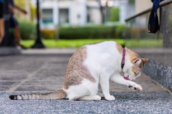
When cats groom themselves and their feline friends, hair gets caught on the barbs of the tongue. That hair then is swallowed. In a “healthy” cat, that hair will go through the stomach and intestines (gastrointestinal tract) and come out in the feces.

So, if a cat is throwing up or regurgitating hairballs, there is more going on than most owners realize.

Grooming

Grooming allows cats to keep themselves clean. They remove old hair, surface dirt, and foreign bodies when they groom themselves and other cats.

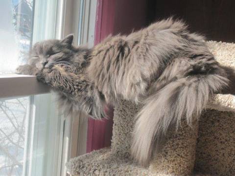
But if a cat is long-haired, he has more hair to remove. (Mother Nature designed felines with short hair. Life is easier for wild cats when they have short hair.) So, long-haired cats who have to do all the grooming themselves — who aren’t being groomed (brushed) by their owners — are ingesting more hair than Mother Nature planned. The elimination of the extra hair may be too much for a healthy long-haired cat’s digestive system.

Also, if grooming becomes excessive, then more hair is entering the digestive tract, whether the cat is a short-haired or a long-haired cat.

Over-grooming can occur when a cat is stressed. Anxiety, emotional stress, etc., can cause a cat to groom himself more often and for longer times. It’s a coping mechanism, like the same way many children suck their thumbs to seek solace.

Over-grooming can also occur in cats experiencing pain. They may lick and groom the painful area to try to make it “feel better” and/or to reduce the stress. Urinary tract pain, musculoskeletal pain, and abdominal pain may cause over-grooming by the unhappy cat.

But hairballs also occur in animals who aren’t “putting too much hair” into their digestive tracts.

What Are Other Potential Causes?

  1. Intestinal motility problems
  2. Stress and stereotypic behaviors
  3. Skin diseases
  4. Abdominal problems
  5. Urinary tract problems
  6. Musculoskeletal problems

If the stomach and small intestine don’t allow the ingested hair to move through normally, the cat will produce hairballs.

If the cat has inflammatory bowel disease, hair – and food – won’t pass through it at a normal speed, or at all. The stomach and intestinal tissue is damaged and doesn’t provide normal motility. It also doesn’t produce the normal digestive enzymes or lubricants that allow the intestinal contents to move along.

The same is true of gastrointestinal cancer. The contents of the tract (hair and food) don’t move along the way they should.

To find the more specific causes of hairballs, your veterinarian may need to explore various body systems and consider conditions that may lead to hairballs. This may include megaesophagus, diaphragmatic hernia, gastrointestinal neoplasia, ileus, intestinal parasites, inflammatory bowel disease, pancreatitis, gallbladder disease, cystitis, bladder stones, kidney stones, arthritis, boredom, frustration, skin parasites, fungal skin diseases, and more.

This is not quite as simple as the problem the client discussed.

Diagnostic Tests

Your veterinarian may recommend tests to look for causes of hairballs including a complete blood count, blood chemical analysis, fecal examination, urinalysis, ultrasonography, radiography, contrast radiography, skin cytology, histopathology, behavior evaluation, etc.

Finding and treating the primary problem is always preferable to just treating the symptom (hairballs). The cat and the client will be happier if everything can be put back to normal. In addition to treating the inciting problem, the veterinarian may recommend diet changes, more brushing by the owner, more play time with the owner, etc.

With diagnostic skills and a little luck, the veterinarian can make the owner happier and the cat healthier and more content.

portfolio-single-swiper-2

Workups for Itchy Dogs and Cats

Pruritus, or itching, in dogs and cats, can involve scratching and licking, chewing, or biting the skin. Pruritus is the most common symptom of skin disease in veterinary medicine. Many conditions can cause pruritus, including allergies, fleas, and other skin parasites. Some skin problems are more common in younger animals, while others are diagnosed more often in older pets. 

Medications are available to manage pruritus, but unless the underlying cause of the pruritus is diagnosed and addressed, it will return once the medication is stopped. Some anti-pruritic medications, such as steroids, can result in other health issues. Providing your dog or cat’s medical history can sometimes help your veterinarian determine the cause of the itching. 

A lack of response to some medications can help your pet’s veterinarian narrow down the possible causes. Additionally, some medications can actually cause itching.

When Did the Pruritus Start?

It can be helpful to know when the itching started. If allergies cause itching, it is beneficial to know if the problem is worse at different times of the year.  

Other Health Problems 

Sometimes, skin conditions develop secondary to another health problem. Your veterinarian will ask questions about other symptoms that could point to a specific cause of pruritus. For example, pets with food allergies may have frequent bowel movements or soft stools.

Location of Skin Lesions

Some pruritic skin conditions cause skin lesions more commonly in one body region than another.

Type of Skin Lesions

Although most pruritic skin conditions look the same to pet owners, there are actually many types of skin lesions, and some skin lesions occur more commonly with specific skin conditions. However, if the pruritus has become chronic, many pruritic skin conditions look similar to each other. Your veterinarian may need to run some tests to help determine the cause of the pruritus.

What Tests are Used to Diagnose the Cause of Pruritus?

Below is a list of some common tests for diagnosing the cause. Your veterinarian will select tests based on your pet’s medical history and examination findings. 

Skin Scrapings

To look for skin parasites, a scalpel blade is used to gently scrape the skin layers to collect cells and any skin mites. The sample is then examined under the microscope.

Skin Cytology

To look for bacteria and yeast organisms, skin cells and debris samples are collected and placed on a glass slide for examination under the microscope. This test can also help determine what types of cells are in the skin.

Parasite Therapy Trial

For some parasites, the only way to determine if they are the cause of the itching is to treat the parasites. A decrease in pruritus after the anti-parasite therapy would be consistent with the parasite causing the pruritus.

Ringworm Testing

To determine if a dog or cat’s skin is infected with ringworm (a disease caused by a fungus called a dermatophyte, not actually a worm), your veterinarian may start by examining your pet under a special ultraviolet lamp called a Wood’s Lamp. If the hairs glow (fluoresce), this may mean your pet has ringworm  Those hairs are collected for further testing.

In a fungal culture, hair and skin samples are placed on a fungal culture plate. If a dermatophyte grows, the dog or cat is diagnosed with ringworm. In a dermatophyte PCR test, (a test that looks for viral diseases) the hair and skin samples are checked for dermatophyte DNA. 

Food Trial

A prescription or home-cooked diet is fed for several weeks with no other foods or treats to diagnose food allergies. Unfortunately, there is no reliable skin or blood test in dogs and cats to diagnose food allergies.

Allergy Testing

Skin or blood allergy testing is used in dogs and cats that have had other causes of pruritus ruled out through other tests. It is mainly used to select allergens for a type of allergy treatment called allergen-specific immunotherapy. There are no reliable hair or salivary tests to diagnose allergies.

Skin Biopsy

A sample of skin can be obtained through minor skin surgery. Skin biopsy is especially helpful for diagnosing some less common causes of pruritus, such as skin cancer. However, a skin biopsy cannot be used as an allergy test and cannot diagnose the cause of a pet’s allergies.

team-single-fazenda

Xylitol Poisoning in Dogs  

Sugar substitutes are big business. Less sugar can mean weight loss, improved health, diabetic control, and even reduced tooth decay. The quest for products that can sweeten and cook like sugar is ongoing. Xylitol (also known as birch sugar) is a common sugar substitute, especially when it comes to sugarless gum, toothpaste, and more recently certain brands of peanut butter for bodybuilders. It is worth noting that even though birch sugar sounds natural and xylitol sounds like a chemical both are toxic to your dog.

Not only does xylitol offer sweetness with 40% of the calories of sugar, but it also has antibacterial properties in the mouth so it can reduce periodontal disease and has been found to have far-reaching health benefits in other areas of the body. Xylitol may help with osteoporosis, prevent ear and throat infections, and may reduce the risk of endometriosis, uterine fibroids, and even breast cancer.

Sounds wonderful and maybe it is if you are a human. If you are a dog, xylitol (birch sugar) is potentially lethal.

Two Deadly Effects of Xylitol

Hypoglycemia

In dogs, the pancreas confuses xylitol with real sugar and releases insulin to store the “sugar.” Even worse, the canine pancreas releases 3-7 times the amount of insulin that it would release to address a similar amount of actual sugar. Blood sugar levels plummet resulting in weakness, disorientation, tremors, and potential seizures.

It does not take many sticks of gum to poison a dog, especially a small dog (see below for toxic doses). Symptoms typically begin within 30 minutes and can last for more than 12 hours but, since xylitol can be absorbed into the body slowly, symptoms may not begin until 12 hours after the xylitol was eaten. Symptoms begin with vomiting and then progress to incoordination, collapse, and seizures.

Hepatic Necrosis

The other reaction associated with xylitol in dogs is the destruction of liver tissue. How this happens remains unknown but the doses of xylitol required to produce this effect are much higher than the hypoglycemic doses described above. Signs take longer to show up (typically 8-12 hours) and surprisingly not all dogs who experience hepatic necrosis will have had hypoglycemia first. A lucky dog experiences only temporary illness but alternatively, a complete and acute liver failure can result in death. Internal hemorrhage and the inability of blood to clot are commonly involved.

How Much Xylitol is Dangerous?

The hypoglycemic dose of xylitol for dogs is considered to be approximately 0.075 – 0.1 grams per kilogram of body weight (about 0.03 – 0.045 grams per pound). Chewing gum pieces have surprisingly variable amounts of xylitol depending on their flavor. (For example, most flavors of Orbit gum have about 0.009 grams of xylitol but their strawberry mint flavor has over 0.3 grams per piece). A small dog can easily be poisoned by a single stick of gum depending on the gum, how much the dog ate, and the size of the dog. It is important for you to bring the packaging for the xylitol product in question to the veterinarian’s office so that the amount of xylitol consumed can be estimated.

Treatment

Ideally, the patient can be seen quickly (within 30 minutes) and can be made to vomit the gum or candy. Beyond this, a sugar IV drip is prudent for a good 24 hours. Liver enzyme and blood clotting tests are monitored for two to three days. Blood levels of potassium are ideally monitored as well. Elevated blood phosphorus levels often bode poorly, and patients that develop hepatic necrosis usually do not survive.

What about Cats?

Xylitol toxicity appears to be limited to dogs. Cats have no significant ill-effects from xylitol.

What about Xylitol-Containing Mouthwashes for Pets?

The oral health benefits of xylitol do seem to hold true for dogs if appropriately low doses of xylitol are used.

Several products have been marketed for canine oral care, specifically for dogs that do not tolerate other methods of dental home care. These products are mixed in drinking water to provide antibacterial benefits.

Aquadent®, for example, comes in a 500 cc (a half liter) bottle that contains a total of 2.5 grams of xylitol. It also comes in small packets to mix in drinking water. Similarly, Breathalyser Plus® from Ceva is similarly packaged. If you follow the dosing instructions on the bottle or packet, there should be no problems.

Trouble could occur if there are animals of different sizes drinking from the same water bowl or if a dog is on medication or has a disease that causes excessive water consumption.  One should dose for the smallest animal to use the bowl to be sure overdose is not possible. Alternatively, a dental water additive that does not contain any xylitol can be used. Ask your vet what he or she recommends. A dog finding the bottle and chewing it up, drinking a substantial quantity of the undiluted product could easily be poisoned, depending on the dog’s size.

6350599

Using Medication to Lower Veterinary Visit Stress in Dogs and Cats

A pet’s mental health is as important as physical health, which is why it may not be possible for your pet’s veterinarian to complete an examination or treatments when you bring your pet in.  If your pet is showing significant levels of FAS (fear, anxiety and stress), your veterinarian may give you medication to be given before your pet’s next appointment.

Why does my pet need this medication? 

High levels of FAS can have negative effects in many ways.  It isn’t healthy for your pet.  Elevated stress hormone levels can suppress the immune system, change lab work results, and over the long term may even shorten your pet’s life.

Every time your pet is in a stressful situation such as going to the veterinarian, they learn to associate it with feeling anxious or scared.  That means that each time they go back to the clinic or a groomer, or you try to do something that they find scary, they get more and more scared.  They also anticipate something bad happening, so the anxiety starts sooner and sooner.

If they stay at a high level of FAS, your pet may learn that the only way to make people stop doing the things that scare them is to be aggressive.  This adaptation could result in a situation where your pet is sick or injured and making sedation risky but necessary because of aggression due to repeated negative experiences.  This risk can limit your veterinarian’s ability to provide necessary medical care to your pet.

My pet isn’t aggressive, can’t you just do what they need?

Physically we might be able to do these things, but it would have a negative impact on your pet’s mental health.  We don’t want them to learn to be more afraid at the vet’s office or start being afraid of strangers or you or in other situations.

He didn’t need medication last time, why are you saying he needs it now?

FAS often increases because of memories of bad experiences.  Giving medication to lower FAS also changes what your pet is learning so they don’t continue to become more and more anxious or aggressive. 

What if my pet is still fearful after taking these medications?

The medications you give at home may not be enough for your veterinarian to do full procedures but may allow injectable sedation to be given without causing undue stress.  If they don’t need urgent care, you may need to try a different combination of medications or different doses. 

What else can we do to lower my pet’s FAS?

Happy visits

If your pet travels in a carrier, use treats so they’re happy about going in and out of the carrier at home and take them for car rides in it so it isn’t a scary thing. Take your pet to the clinic when nothing is needed so they can learn that it’s a good place to be.  Start out just giving treats in your car or out in the parking lot.  Call ahead; if it’s a quiet time, staff may be able to come out and give your pet treats or have them come inside for some.

Home exams

If your dog or cat is fearful at the veterinarian’s, but is not aggressive or extremely fearful at home, you can get a pulse and respiratory rate and take their temperature at home.  You can also take pictures of their teeth in case they need to be muzzled, in which case teeth can’t be examined.

Basket muzzle training for dogs

For the safety of your clinic staff and ultimately of your dog, many fearful dogs require muzzles.   If the muzzle is introduced properly, your dog will associate it with treats and good things, so it will lower anxiety when worn.  If it is not introduced properly, putting a muzzle on a fearful dog can increase anxiety and aggression.