Tag: pets

4128698

Nicotine Poisoning in Pets

Everyone knows the Surgeon General’s warning about cigarette smoking but what about cigarette eating?

Nicotine poisoning is a real concern anywhere that a pet may find cigarettes, cigarette butts, chewing tobacco, nicotine gum, patches, or e-cigarettes. Dogs, particularly puppies, tend to chew things up first and ask questions later. Cats may find a cigarette butt to be a nicely sized pouncing toy worthy of chewing.

Luckily for pets and small children, tobacco tastes terrible. Even chewing tobacco must have flavorings added to make it worthy of oral enjoyment. 

Still, cigarettes have plenty of nicotine and even a small cigarette butt can mean serious illness or even death for a small pet.

The toxic dose for nicotine in pets is 0.5 to one mg per pound of pet body weight while the lethal dose is four mg per pound of pet body weight. So how does this translate to nicotine-containing products?  A cigarette contains between 9 & 30 mg of nicotine depending on the type of cigarette. When the cigarette is smoked, the nicotine concentrates down in the oral end meaning that a small nub of a cigarette butt will retain 25% of the nicotine contained in the original cigarette. Smoking a cigarette yields 0.5-2.0 mg of nicotine to the smoker but eating the cigarette (or other nicotine product) is a whole different ballgame as all of the nicotine becomes available for absorption into the body. Consider that a 2-lb dog would only need 10 mg of nicotine to become poisoned and a 40-lb dog would need only 1cc (less than a quarter teaspoon) of e-juice.

  • Cigarettes: 9-30 mg of nicotine
  • Cigarette butts: 2-8 mg of nicotine
  • Cigars: up to 40 mg of nicotine
  • Chewing tobacco: 6-8 mg of nicotine per gram
  • Nicotine gum: 2-4 mg of nicotine per piece
  • Nicotine patches: 8.3-114 mg of nicotine
  • E-cigarette cartridges: 6-36 mg of nicotine
  • E-juice/E-fluid (to refill E-cigarettes): up to 36 mg of nicotine per ml

Some good news is that nicotine is not absorbed directly in the acid environment of the stomach; the nicotine must move past the stomach into the small intestine for absorption. One of the first things nicotine does in the body is to stimulate the vomiting center of the brain, thus inducing vomiting that may save the patient’s life if there is more cigarette material in the stomach.

Symptoms of Nicotine Poisoning

Signs begin as quickly as one hour post-ingestion. Symptoms include:

  1. Tremors
  2. Constricted pupils
  3. Drooling
  4. Auditory and visual hallucinations
  5. Excitement
  6. Vomiting and diarrhea
  7. Twitching, possibly progressing to seizures
  8. Racing heart rate but slow heart rate with small doses
  9. High blood pressure but at higher doses, there is a circulatory collapse

It is easy to confuse nicotine poisoning with other poisonings such as strychnine, chocolate, organophosphate insecticides, and certain molds. Hopefully, there will be cigarette materials in the vomit to give away the diagnosis. It is also worth mentioning that some nicotine gums contain xylitol as a sweetener. This material is toxic to dogs and can add an entirely new dimension to nicotine poisoning.

Treatment

If not too much time (an hour or so) has passed since consumption of the nicotine product, vomiting can be induced. Washing out the stomach to get rid of any remaining cigarette materials is helpful but is likely to require sedation as well as activated charcoal to bind the nicotine and prevent it from entering the body. Obviously, this would be performed at the veterinarian’s office.

Treatment is basically supportive after that. Intravenous fluids support circulation while the body gets rid of the nicotine. If seizures or tremors are occurring, medications will be needed to suppress them.  If the pet survives the first four hours, the prognosis is felt to be good. Nicotine is inactivated by a healthy liver and its metabolites are excreted in the urine. After 16 hours, the nicotine ingested should be gone.

The ASPCA National Animal Poison Control is available 24 hours a day at 888-426-4435. Expect an initial consultation fee of around $100.00 and additional follow-up is at no charge. You will be assigned a case number your veterinarian can use to communicate with a toxicology specialist before beginning treatment.

5718924

Nutrition for Ferrets

A ferret is an obligate carnivore, and must eat appropriately for the species to maintain health. Adult ferrets have 34 teeth. Ferrets have a very short intestinal tract; the GI transit time is approximately 3 hours, a time so short that absorption of nutrients is not that efficient. Because of this timeframe, 3 hours is adequate when fasting a ferret before surgery. Transit time in young ferrets can be as short as one hour. Ferrets tend to eat small amounts at frequent intervals. Young ferrets particularly need high-quality diets while they are growing.

Pregnant ferrets will require extra protein (not fat) during gestation. Lactating (nursing) ferrets will require extra caloric intake. Abandoned or orphaned kits may be hand raised with either puppy or kitten milk replacer with cream added to raise the fat content. Ill, anorexic ferrets can develop hepatic lipidosis and hypoglycemia. There are good dietary supplements commercially available for assist-feeding anorexic ferrets. Ferrets with insulinomas, a common problem, should be encouraged to eat every few hours.

Whole prey diets (chicks, mice, and/or rats) are fine for ferrets and are popular in certain parts of the world although not so much in North America. Cat food is often fed to ferrets, but ordinary grocery store cat food is a poor choice for them. Premium dry cat foods or balanced commercial ferret diets are better choices. The ideal ferret diet is high in protein (30-35%) and fat (15-30%), and low in fiber. The protein source should be meat-based, rather than grains. Grain-based diets have been associated with urolithiasis in ferrets. Ferrets fed dog food will not do well. Meat or poultry or their by-products should top the ingredient list of any suitable ferret food. A diet in which the first main ingredient is corn is not a suitable diet for ferrets. The feeding of table scraps should be kept to a minimum. There are several brands of supplemental nutritional products for ferrets that are commercially available. These products are unnecessary in most ferrets. Older ferrets may require less protein than when they were younger.

Fruits and vegetables have little nutritional value for ferrets. An adult ferret needs approximately 43 grams of dry food per kilogram of body weight, and 200-300 calories/kilogram per day. Calcium: phosphorus ratios in ferret diets should be minimally 1:1. Dilated cardiomyopathy (DCM) occurs in ferrets, and although it has not been associated with taurine deficiency, taurine is generally added to ferret diets as is the case with cat food. Commercial ferret diets have vitamins and minerals added in adequate amounts. Water should be made available at all times; most ferrets will prefer a bowl to a sipper bottle. They also like to play with their water. Ferrets tend to not be fond of changes in their diet after they are used to a certain kind. Ferrets imprint by smell on the food they like at a very young age. Most ferrets will be content to eat one brand of commercial diet that they enjoy for their entire lives.

Treats should not consist of more than 10 percent of the daily diet. They love raisins; however, while there is no formal study on ferrets and raisins, there are anecdotal reports of raisin toxicosis in ferrets, so raisins are best avoided. Egg can be a good supplement, as can small amounts of liver or hamburger.

8297847

Neutering Rabbits is Best

The word neuter refers to the removal of the reproductive organs of either a male or a female of a species, although people frequently refer to the surgery in a female as a spay. The scientific terminology for neutering in the male is castration and in the female is ovariohysterectomy. Let’s take a look at the issue of neutering and whether or not it is appropriate for your pet rabbit.

Reasons for Neutering Rabbits 

Prevention of Pregnancy

This is the most common reason that rabbits are neutered, particularly if there are both male and female rabbits living together in a household. There are certainly enough rabbits in the world and too many are neglected or abandoned. One should not consider breeding these pets just for fun or education. Be a responsible pet owner and do not breed your pet unless you are well educated on the topic and are prepared to take on all the responsibilities such activity entails.

Prevention of Uterine Cancer

This is the most compelling medical reason to neuter female rabbits. In some rabbit populations the rate of uterine adenocarcinoma (a malignant uterine cancer) can approach 80% of the females. It is believed that the incidence may be related to the rabbit’s genetic makeup. Since we usually don’t know the genetic background of most of our rabbits, it is best to have the surgery done as a preventative for this cancer. Uterine adenocarcinoma can spread rapidly to other organs of the body such as the liver, lungs and even the skin and it is not treatable once it metastasizes outside of the uterus. We see many cases of this disease each year and sadly these rabbits could have avoided this problem. Rabbits under two years of age rarely develop this disease so it is best to get your female spayed before this age.

Prevention of other Uterine Disease

Although cancer is the most common disease of the rabbit uterus, we see many cases a year of other uterine disease such as pyometra (infected uterus full of pus), uterine aneurism (uterus full of blood) and endometritis (inflamed uterine lining). Like uterine cancer, these conditions are all more common in female rabbits over two years of age.

Prevention of False Pregnancies

Female rabbits can go into a hormonal state triggered by their ovaries where the body acts as if it is pregnant but there is in fact no pregnancy. Although this is not medically harmful, it can be stressful for the rabbit who goes through all the activities of being pregnant including nest building, milk production and aggressive protection of her territory. This aggression can be taken out on the caretakers and cage mates and can make the pet difficult to handle during this period. Some rabbits experiencing false pregnancy will develop a decreased appetite and have gastrointestinal disturbances as well.

Prevention of Mammary Gland (Breast) Disease

Mammary gland cancer is not common in female rabbits, but when it occurs it can spread rapidly and be difficult to treat. It is preventable if the pet is neutered before two years of age. It is interesting to note that the most common type of mammary cancer is a malignant form called mammary carcinoma and it is almost always associated with uterine cancer. The other common mammary gland disease is mammary dysplasia or cystic mammary glands. This is a benign condition, where the mammary glands fill with a cystic material. It can be uncomfortable to the pet. Neutering a female rabbit before two years of age will prevent both of these diseases.

Prevention of Aggressive Behavior

Both male and female rabbits can display aggressive behavior when they are sexually mature. Many rabbits are sweet and easy to handle as little babies, but when the teenage years hit at around six to twelve months of age…watch out! They can become little Frankensteins almost overnight! They don’t want to be touched or picked up and they act like they want to destroy everything in sight. This is their way of learning to protect themselves, their territory and potential future families and to establish their social position in the big wide rabbit world. However, they can often take out their aggression on you or their cage mates. There may be more biting, striking, lunging and chasing. It is best to neuter just before or shortly after sexual maturity to keep this behavior to a minimum.

Prevention of Urine Spraying


Both male and female rabbits can spray urine on vertical surfaces to mark their territory. Intact mature males do this at least 10 times more frequently than females. In addition, the urine from a sexually mature male rabbit can have a strong odor that is unpleasant to many humans. If this behavior is allowed to continue for a long period of time, it may be impossible to completely stop this behavior. Therefore, it is best to nip it in the bud and get the little guys neutered just prior to or shortly after sexual maturity.

Prevention of Testicular Disease
Disease of the testicle is uncommon in the male rabbit, but it can occur. Most commonly we see abscesses (usually the result of bite wounds from other rabbits), hematomas (blood filled areas) and cancer.

Age to Neuter

The best age to neuter either a male or female rabbit is just before or shortly after sexual maturity. Depending on the breed, this time could range from four to six months in the small to medium sized breeds and up to nine months in the giant breeds. We do not recommend neutering rabbits younger than four months of age because the surgery may be more difficult due to the size and position of the reproductive organs. There is no health benefit to neutering earlier than four months of age. However, there is a benefit in females of neutering before two years of age to reduce the incidence of uterine and mammary gland disease.

Your rabbit should be examined by a veterinarian prior to surgery to make sure he is in good condition and ready for neutering. Sexual maturity can be gauged a number of ways including; visualizing testicles in the scrotal sacs, a well developed vulva, a mature body condition, and by behavioral changes such as urine spraying and increased aggression. Your veterinarian may recommend some simple tests prior to surgery, particularly if your pet is older or has had other medical problems. We do not recommend performing routine neutering procedures on obese animals or those with other disease because these rabbits are at higher risk for surgical complications. The weight should be reduced and any disease conditions managed prior to having a major elective surgical procedure performed.

What Happens at Neutering

When a male rabbit is castrated, the testicles are completely removed. There may either be one incision in front of the testicles through which both are removed, or there may be two incisions, one over each scrotal sac. The incisions may be left open which is acceptable if scrotal incisions were made, or closed with suture or surgical glue if the incision was made in front of the scrotal sacs. The scrotal sacs will swell within 24 to 48 hours after surgery but in another seven to ten days the swelling should be gone. The scrotal sacs will eventually shrink to a very small size over time. It is important to note that neutered males should not be put in contact with intact females for at least 3 weeks after neutering. Male rabbits can still have living sperm in ducts within the spermatic cord called the vas deferens, which cannot be removed during surgery. The sperm in these ducts can live for two weeks. Testosterone blood levels drop slowly after neutering and male rabbits will still try to mate with female rabbits for several weeks after the testicles are removed. After three weeks the sperm are completely dead. Since the testicles are gone, no new sperm are being produced so it is safe to put a male and female rabbit back together again. However, whenever you put two rabbits together, regardless of the circumstances you must watch closely for signs of aggression. Aggressive rabbits left unattended can cause serious and sometime fatal injuries.

When a female rabbit is neutered, the ovaries, the oviducts, the uterus and often both cervices are removed. Rabbits have a uterus that is made up of two long tubes with an ovary at one end and a cervix at the other. They have two cervices unlike cats, dogs, humans and many other species which only have one. An incision is made just below the umbilicus (belly button) and the uterus and associated structures are gently pulled out from the abdomen through this incision. The blood vessels supplying the uterus and ovaries are tied off with suture material, surgical clips or a laser and the reproductive organs are removed. The incision is sutured with two to three layers of suture material. Since rabbits have incisors that are excellent at cutting through many materials, we find it beneficial to bury the final row of sutures under the skin so they are not accessible. In this way the rabbit has nothing to chew on or pull out. These sutures dissolve eventually over several weeks and there are no external sutures to remove.

Some veterinarians are now advocating the removal of only the ovaries in female rabbits less then 2 years of age.  These veterinarians argue that uterine cancer usually occurs after two years of age and the removal of only the ovaries is a shorter and less invasive surgery and thus less stress on the rabbit.  This is still a controversial topic because it is difficult to know at exactly what time uterine cancer may start at the microscopic level.  Exotic animal veterinarians will be looking carefully over the next few years at these ovariectomy cases to see if uterine cancer was prevented during the life of the rabbit.  Consult with your veterinarian on this topic so you can make an informed decision.  However, if you have acquired an adult rabbit and you do not know the exact age of the rabbit (such as when adopting from a rescue), I highly recommend having both the uterus and the ovaries removed to be on the safe side.

Postsurgical Care 

It is important after any surgery to check the surgical site at least twice a day for any signs of bleeding, unusual swelling, discharges or opening of the wound. Many rabbits will be off feed for 24 hours after surgery, but this should gradually return to normal over the next two to three days. In addition, some rabbits will have unusual stools for a day or two including soft stools, clumped stools, and irregularly shaped or small stools. If your rabbit is acting very uncomfortable, is extremely lethargic, is not eating at all, is not producing any stools or is unwilling to move, contact your veterinarian immediately. Your veterinarian should prescribe a post-surgical pain medication for one or more days for your pet, which will help ease discomfort and shorten the recovery time. It is usually not necessary to use an antibiotic after a routine neutering. 

5999162

Myxomatosis in Rabbits

What is Myxomatosis?

Myxomatosis is a contagious and deadly viral disease of wild European and pet rabbits. Its fatality rate can be devastating, reaching nearly 100% in some cases. Ultimately, though, this percentage depends on the species and breed of rabbit affected and the strain of the virus causing disease. Cases of myxomatosis in the United States are largely limited to the West Coast.

What Causes Myxomatosis?

Myxomatosis is caused by the myxoma virus. This virus occurs naturally in Sylvilagus species (cottontails/related rabbits) in the Americas, but is generally harmless to these rabbits. It was purposely introduced into such places as Europe and Australia to reduce numbers of wild European rabbits. The virus can spread from these wild sources and cause disease in any breed of pet rabbit.

There are many strains of the myxoma virus, some of which are more severe and fatal than others. For example, the Californian myxoma virus in the Pacific states of the U.S. and the Baja Peninsula in Mexico is particularly deadly, estimated to kill over 99% of rabbits it infects. There are similarly severe strains in Europe and Australia, but also weaker strains with death rates around or below 50%.

How is Myxomatosis Transmitted?

  • Bites by blood-sucking insects (e.g., mosquitoes, fleas, flies, fur mites)
  • Direct contact with an infected rabbit
  • Contact with material contaminated by an infected rabbit (e.g., food, bedding, water bottles)

What are the Signs of Myxomatosis?

  1. Reddened eyelids and eyes
  2. Discharge (may be milky) from the nose and eyes
  3. Swollen eyelids, nose, lips, ears, face, anal/genital region
  4. Skin nodules or lumps
  5. Drooping ears
  6. Purple spots on the skin
  7. Lethargy
  8. Fever
  9. Reduced appetite
  10. Weight loss
  11. Trouble breathing
  12. Convulsions
  13. Coma
  14. Death

Note: While some rabbits may show obvious and severe signs of illness, other rabbits may show only minor or even no symptoms, but could still die.

How is Myxomatosis Diagnosed?

  • Clinical signs
  • Lab tests, which include:
    • Examining affected tissues under microscope
    • Detecting myxoma virus in biopsies or swabs from eyes/nose

How is Myxomatosis Treated?

Because myxomatosis tends to be a severe and fatal disease, veterinarians will often recommend euthanasia of a sick rabbit. In some situations attempts may be made to treat the patient, but unfortunately these efforts are rarely successful. 

Treatment may consist of:

  1. Keeping rabbits warm
  2. Assisted feeding
  3. Intravenous fluids (fluids administered through the blood)
  4. Antibiotics to treat secondary infections
  5. Pain relievers
  6. Isolating the sick rabbit from other rabbits

The rabbit’s progression of symptoms, response to treatment and quality of life will all be monitored and play a role in decisions to continue treatment or euthanize.

How can Myxomatosis be Prevented?

  • Vaccines are available in some countries (not in the U.S.); 100% protection is not guaranteed
  • Quarantine new rabbits
  • Prevent contact between pet rabbit and insects/wild rabbits by:
    1. Keeping pet rabbit inside
    2. Using insect screens in outdoor rabbit enclosures
  • Isolate rabbits suspected or confirmed to have myxomatosis
4129016

Husbandry and Health of Rats

Rats have been kept as pets since the late 19th century and have become quite tame and domesticated over time. They’re intelligent and can be taught tricks and interactive games. They have also been used for research as working animals for odor detection, for landmine and tuberculosis detection, raised for food, and are revered in some cultures.

In others, they’re associated with zoonotic disease and are most renowned for being carriers of the bubonic plague, or Black Death.

Wild rats are known to carry pathogens including leptospirosis, toxoplasmosis, Yersinia, and Campylobacter in addition to other conditions. Fortunately, zoonotic disease transmission from pet rats is uncommon.

Despite those different perceptions, rats are quiet and easy to care for, have minimal odor, and are affectionate and friendly animals. With adult supervision, they make excellent pets for children. However, they’re not maintenance free and bringing a pet rat into your home comes with a commitment to provide a large enough cage; time to regularly clean and launder the cage accessories; daily social time, and a high-quality diet.

As pets, with care and attention, rats are gentle, calm, mild mannered, and rarely bite. Although short lived, they are appropriate first pets for young families providing that there is always adult supervision. The most common rat kept as a pet is a domesticated brown rat known as the fancy rat. Over time, breeding has resulted in a variety of different coat colors and types, and fancy rat breeders and clubs are common.

Also, particularly when the rat is to be a pet for a child, mature animals are less likely to explore the world with their mouths; in other words, they tend to be a little less nippy, and are often not quite as fast moving, and therefore, a little easier to handle. On the downside, animals coming from a previous home may come with bad habits including poor dietary preferences, and some retraining may be necessary.

Rats are social animals living in groups in the wild. They appear to derive great enjoyment from social interactions with one another as well as with humans and appear most content when housed in small groups.

Habitat

Unfortunately, many of the basic husbandry needs of rats are overlooked, harkening back to the days of small plastic hamster cages with tubes, wood cedar bedding and food bowls filled with seed and dried fruit. Sadly, this traditional care has led to countless diseases in our pet rats over the years and resulted in significant suffering and pet mortality. In my practice, we spend a generous amount of time counseling our rodent owners on proper husbandry and care of their animals. This hopefully helps not only the animal at hand but generations of pets that these owners may eventually have.

Habitat size must be increased to accommodate group size, but even an enclosure housing a pair of rats can result in a content rat family without requiring too much room. Rats will sometimes exhibit aggressive behavior towards one another, particularly when originally introduced. However, a bonded pair will sleep together, play together, wrestle, groom one another, have squabbles, and develop a variety of communication skills.

Without good basic care, rat health is a house of cards that will easily be upset with even the most minor of infections.

Rats need a significant amount of room to play and be active within a cage environment in addition to being allowed to have regular out-of-cage playtime. A minimum cage size for a single rat should be 8 cubic feet, such as a 2 x 2 x 2-foot cage. Multiple rats will need more space in order to be comfortable. Rats enjoy multi-story cages with ramps and complete floors, allowing them to climb and play with minimal risk of falling.

Cages should not be plastic or glass, both of which significantly limit ventilation and may contribute to respiratory tract infections. I prefer wire mesh cages that have been covered with soft bedding such as fleece blankets, old clothing, and customized bedding to fit the cage.

This helps prevent the feet from injury on the wire bottom of the cage, and allows the rat to create a comfortable nesting area. Solid floors are ideal, if available. 

There should be multiple sleeping areas including enclosed sleep boxes, hammocks, and soft, blanketed material. Many rats will readily litter train and each story of the cage should have a corner box available for them. The litter boxes or cage base should be covered with paper-based recycled bedding such as CareFRESH bedding. Avoid wood-based and scented bedding as they can contribute to respiratory disease as well as elevate liver enzymes.

A rat cage that is kept clean will not have a strong odor, and if you feel the need to have scented bedding, this is a sign that the cage is likely not kept clean enough. Bedding should be washed at least weekly and more often if there are multiple rats. Boxes are best changed daily and the remainder of the cage spot cleaned as needed. Rats in general are very clean creatures and will thrive in a clean environment. 

As we have discussed briefly, rats are social creatures. Not only do they bond with their human owners, but they also establish a complex and rich series of community relationships and behaviors. Although rats are frequently kept as singleton pets, they are likely to be more content and enriched when living in small groups. A bonded community of three to four individuals generally appears to be ideal. This can be either as single-sex environment or a mixed group of altered animals.

When intact animals are housed together, particularly males, they need enough room to prevent fighting and establishing territorial aggression. Some dominance is to be expected in any group of animals, but in general most rats appear to settle their disputes amicably.

Wrestling and grooming behavior within a group is common and should not be mistaken as aggression. Adequate space is a solution to many inter-rat problems, and so when in doubt, provide the largest cage you can and as much out-of-cage social interaction time as possible. 

What to Feed

Rats are nutritional omnivores and can eat and digest a wide variety of foods. Unfortunately for the rats, pet food manufacturers have taken advantage of this fact to provide a wide variety of foods that are appealing but do not provide the necessary nutritional basics; in other words, it’s junk food.

In general, the staple of a rat’s diet should be made up of high-quality pelleted food such as that made by Mazuri. These foods are most commonly referred to as ‘rat block’ or ‘rodent block’. They should not contain any colored pellets, nuts, dried fruits or vegetables, or seed bits.

Rat block should comprise 80% to 90% of the overall caloric intake for an animal. Many rats are fed some variety of seed, dried fruit, and nut-based diets. Unfortunately, these diets are often high in fat, and rats can become obese even when eating a healthier diet. Over time, these diets can lead to organ disease, a weakened immune system, skin disease, and obesity.

The higher quality rat food brands have been formulated to completely meet the rat’s nutritional needs with no significant excesses or deficiencies, and they do not allow the rat to pick and choose its favorite diet components. Although not visually appealing to us and perhaps not as popular as the seed-based junk foods marketed for rats, these pelleted diets are vastly superior nutritionally, the equivalent of a salad instead of candy.

Have a thorough diet review with your veterinarian so you can help prevent illnesses wherever possible. The risk of both kidney disease and obesity-related diseases can be reduced strictly by calorie and protein control alone.

Not only is the content of the diet important in the health and wellbeing of the pet rat, but the amount is also important. Rats who can eat whenever they want have been shown to have higher incidences of mammary, pituitary and pancreatic cancers. Moderate caloric restriction has been demonstrated to reduce the likelihood of cancer, particularly with tumors that have an endocrine influence. Therefore, a moderately restricted, high-quality diet yields multifactorial benefits in the health of pet rats.

A variety of healthy treats and supplements should be offered to the pet rat. When you begin to offer these extras, do so slowly in order to avoid digestive upset. Healthy fresh foods that can be given include small amounts of cooked beans, peas, corn, dry or cooked pasta, squash, carrots, green leafy vegetables, breads, and small amounts of other fresh fruits and vegetables. Occasional snacks of nuts, lean meats, and eggs are also appropriate, but rats are sensitive to excesses of proteins in their diet and high-protein snacks should be kept to the occasional treat only.

Portions should be appropriate for the size of the rat: a pinky fingernail-size portion of food is similar to an entire plate of food for an adult human. Although rats adore sugary and salty treats, avoid them.

Basically, if it would be considered a healthy snack for a human, it will likely also be a healthy snack for a rat as long as portion control is appropriate. Rats are truly an example of ‘you are what you eat.’ With their already too-short life spans and a tendency towards disease, we owe it to them to provide as much of a nutritional head start as we possibly can.

Health

Despite the emphasis on overall husbandry and preventative medical care, the realistic truth is that the vast majority of rats seen by veterinarians are pretty sick. That being said, many of the conditions our little rat friends are prone to are controllable and some are even curable. They are surprisingly sturdy patients and can tolerate a wide variety of both medical and surgical procedures.

Although rats in general are hardy little creatures, they are certainly prone to a number of conditions. Some of these, such as renal disease and obesity-related conditions, can be prevented with good husbandry and diet. Others, such as tumors and respiratory diseases, are a combination of genetic predisposition, exposure, and environmental causes. Even though an animal may have a large and dramatic tumor, there are often options to improve the animal’s quality of life even when there is no cure.

Mammary adenomas are the most common mammary tumors found in rats and can be seen in males and females. Spaying can significantly reduce their occurrence in females.  Although they have been reported in rats of all ages, they are most common in animals older than 18 months of age. Some of these adenomas can be extremely large and carry a rich blood supply. Fortunately, this completely benign and encapsulated mass is significantly more common than its cousin, the mammary adenocarcinoma. Mammary adenomas can be deceptive since rats are essentially walking mammary glands. There is mammary tissue from the chin to the base of their tail. Any skin mass could potentially be a mammary adenoma.

Fortunately, rats do very well when having these tumors surgically removed. As a rule, fibroadenomas tend to remain localized and are not invasive. They can, however, be extremely large, sometimes approaching the size of the rat itself. Tumors of this size can be a surgical challenge since essentially you are removing the rat from the tumor. If the tumor is not completely removed, they will often regrow. Still, most rats will benefit from debulking large tumors.

Spaying just as you would any dog or cat at a young age and then restricting calories or not allowing a free-fed diet can go a long way toward preventing mammary fibroadenomas. 

Mammary tumors are dependent on estrogen and prolactin concentrations and spaying markedly reduces their incidence. Early age spaying may virtually eliminate the incidence of later development of mammary masses, and even spaying at the time the tumor develops may prevent a recurrence.

Free-fed rats have a higher incidence of pancreatic, mammary, and pituitary tumors than rats fed in identical diets with moderate quantities. High-caloric intake also enhances tumor growth and the endocrine-based tumors, particularly mammary masses and pituitary masses, appear to be the most influenced by caloric intake. Therefore, feeding a calorically restricted, high-quality diet is crucial to preventing mammary masses and their recurrence. Practicing preventative medicine makes a positive difference.

Fortunately, when it comes to fibroadenomas, we have good treatments to help most pets. Rats make surprisingly hardy surgical candidates. They do, however, tend to self-mutilate their incisions. Surgery is the easy part of treatment. It’s the two weeks after the surgery that’s the hardest part. When we make it to suture removal, I consider the treatment to have been a success.

Pituitary masses are relatively common in rats and appear most frequently in female animals between 13 and 24 months of age. These are occasionally seen in male animals or younger animals but are much more frequently seen in females. Even in the normal animal, the female has a larger and heavier pituitary gland. Females that have been spayed don’t have pituitary masses as often as rats who are intact. Signs are extremely variable and often continue to change. Most commonly, however, these tumors are benign; they rarely metastasize and are a relatively slow-growing tumor. A number of factors influence pituitary tumors including age, genetics, hormonal status, spaying and neutering, injury, infections and so on. Unfortunately, the location of the tumor and the pituitary gland makes surgical removal a poor option. However, targeting the symptoms of most clinical concern for the patient can lead to significant palliation of the clinical signs and improve quality of life for the patient.

Respiratory diseases are one of the most common reasons pet rats to see the veterinarian. Infections are endemic in the rat population and many animals are infected at the time of purchase or adoption by the owner. Periods of immunosuppression, stress, or concurrent infections can result in the sudden appearance of clinical signs. There are several common underlying causes for infection, including bacterial, viral, fungal, and parasitic diseases. Infections frequently have significant and severe secondary results including pneumonia, abscesses, empyema, pleural effusion, and bacteremia. Non-infectious causes of pneumonia, such as aspiration pneumonia or chemical irritants resulting in inflammation and pneumonia, are seen.

Of all the reasons for pneumonia in rats, the most common is the bacteria Mycoplasma pulmonisMycoplasma differs from the most common bacteria because they lack cell walls but are enclosed by a lipid protein cell membrane. They’re not considered to be either gram-positive or gram-negative. Because of these facts, some antibiotics such as penicillin and cephalosporins don’t against mycoplasmas.

Clinical signs vary depending on the virulence of the strain involved, the site of infection, the age of the animal, and concurrent disease. For patients experiencing upper respiratory tract disease, clinical signs are generally mild and include sneezing, snuffling, squinting, and porphyrin staining around the eyes and nose. In fact, many owners take their rat to the doctor thinking that there’s eye disease, complaining that the animal is squinting and bleeding from the eyes. These rats may also have signs associated with an inner ear infection including head tilts, rolling, face and ear rubbing, and ear pain. Patients may also have signs of lower respiratory disease including rattling or moist breath sounds, labored breathing, chattering, coughing, and gasping. These animals are often clinically ill and may appear lethargic and/or have a poor appetite, poor coat, weight loss, hunched posture, and grumpy behavior. Some rats don’t have any signs at all. Pups may be infected before birth.

At this time, there is no cure for mycoplasmosis, and it requires long-term management. At best, we try to minimize the clinical signs, reduce concurrent diseases, improve husbandry, and look for the overall quality of life of the patient.

8151637

Health and Husbandry of Mice

Mice are intelligent and playful, and in the right situation can be affectionate pets. Mice are easy to care for, quiet, and friendly animals when handled daily and carefully. With adult supervision, they make excellent pets for older children.

However, they’re not maintenance-free, and bringing a pet mouse into your home comes with a commitment to purchase a large enough cage, provide regular cleaning of the cage and its accessories, daily social time, and a high-quality diet.

They have excellent hearing and a good sense of smell but unfortunately have poor eyesight, and therefore are prone to falling if left unattended in areas with ledges.

Mice are largely nocturnal and are often swift-moving creatures, which makes them inappropriate pets for most small children. Additionally, they may jump quickly and without warning. Daily handling is important from a stimulation standpoint and will help to tame them and make them more friendly. 

The average life expectancy for the pet mouse is approximately 18 to 24 months. Unfortunately, as a prey species, they tend to hide illnesses. Often, by the time any trouble is apparent to you, it’s probably been going on for a while. Because of this, most mice are already significantly ill when they get to a veterinarian.

Housing

Female mice generally live well housed in small groups if provided with a large enough cage space.  Males are best housed alone since they may fight if kept in groups. Mice have a fairly significant odor that is much more prominent in males than females. Small groups of females do better than mixing in the boys with them.

Males and females housed together lead to baby mice. To avoid adding up to 140 baby mice per year to your family, it is best to house males and females separately even if they are from the same litter!

Pet care, Fancy mouse, a colored mouse sits in a house made of coconut on a white background with copy space

Cages should be large and well-ventilated. Wire caging with narrow bars allows for good ventilation and helps prevent escape. Many people choose aquariums for no-escape safety reasons, and these must have wire mesh tops to allow for good ventilation. A single mouse should be housed in nothing smaller than a 15-gallon tank, but a 20-gallon tank would be an appropriate size for two mice.

The bedding should be deep and absorbent but not made from wood-based materials and unscented only. Recycled paper bedding is ideal. Scooping out waste materials daily and changing the entire cage weekly will help keep the environment healthy and minimize odors.

Mice prefer sleeping in secluded areas. They enjoy nesting in plastic hideaways or flowerpots laid on their sides. They will often take treasured items into their sleeping or nesting areas. Torn paper towels are often prized bedding material and it’s inexpensive to replace when it becomes soiled.

Diet and Enrichment

Commercial pelted rodent block is an appropriate diet and can be supplemented with small amounts of healthy people foods. It is wise to avoid high-fat extras such as seeds and large amounts of nuts. Introduce all foods slowly to avoid diarrhea. 

Playful and intelligent, mice like enrichment. Enrichment items are things that help mentally and physically stimulate animals and may mimic things in their natural environment. A well-outfitted mouse home includes numerous items for play and enrichment, such as a solid wheel (wire wheels can cause legs to break), an enclosed sleeping box, chew toys, and climbing toys. Toilet tissue rolls, sisal rope climbs, and cornstarch baths are favorites. Chew toys such as wood from apple trees and dog rawhides are popular.

Health Conditions

New mice should be quarantined for an extended period, even as long as four weeks, before integrating into an otherwise healthy group.

Skin diseases are common in mice and can have several underlying causes. One of the more common includes bite wounds from cage mates. Taking care of the underlying reason for the squabble will usually end this behavior. This problem may require separating the mice, providing a larger enclosure, or neutering the mice. Most commonly, infections of these wounds occur secondary to Staphylococcus species. The wounds are generally treated with antibiotics and pain medication. However, these wounds can occasionally abscess and become chronic problems that require surgery.

In groups of mice, barbering of fur is common. Affected animals show bare patches of fur although the underlying skin looks normal. Barbering appears to have hierarchal indications within the group and removing the barber often results in resolving the symptoms in the barbered mouse. Some controversy exists as to whether it is the dominant mouse being barbered or doing the barbering.

Mite infestation is another common problem. These parasites can result in the mouse scratching itself and causing severe skin irritation on the head, neck, or shoulder area. Skin scrapings can diagnose the mites and your veterinarian may see the mites on the fur. The mite’s entire life cycle is completed on the mouse, but transmission between animals is common and reinfection happens. Medical treatment, in addition to addressing any secondary bacterial infection, is generally curative.

Occasionally, a mouse appears to scratch severely for no obvious reason. This has been referred to as obsessive-compulsive itching disorder in mice. Vitamin A supplementation can help in addition to controlling any secondary bacterial infections. Ringtail is most commonly associated with low humidity conditions in young animals resulting in constrictions around the tail. In severe cases, tail amputation is often indicated.

Pneumonia frequently happens in mice as well and is often caused by a bacterial infection. Signs include upper respiratory signs such as sniffling and sneezing, and lower respiratory tract signs including labored and noisy breathing. Signs of overall illness include squinting, red-brown tears, and a rough coat. Some cases may also have neurologic signs including a head tilt.

This disease is very contagious and similar to rats with mycoplasmosis. Mice may be carriers of mycoplasmosis and yet be asymptomatic, which means that all mice in contact that show respiratory disease should be considered infected.

In addition to medical therapy, replacing their bedding daily with material unlikely to irritate the respiratory tract such as unscented recycled paper bedding can reduce clinical signs of respiratory conditions. Unfortunately, mycoplasmosis is a common and severe disease that often leads to death in pet mice.

Mammary adenocarcinomas occur in mice and can be found anywhere on the body due to the large amount of mammary tissue in mice. Unfortunately, these are more aggressive than those in rats and although they can be surgically removed, they do have a more significant amount of local tissue invasion as well as a likelihood of spreading to the lung.

Lymphosarcoma is also a common cause of tumors. Chemotherapy tends to work, but a remission time of weeks is considered a reasonable expectation.

Lastly, benign respiratory adenomas are seen in some strains of mice and older animals. White lesions may be observed on the surface of the lungs that can be large enough to show up on X-rays or other imaging.

Since mice are commonly used as models in research, much information is readily available about the cancer tendencies of this species. Unfortunately, much less information is available about the treatment options for an individual mouse.

Summary

Mice can make excellent pets for older children through adults and are affectionate, curious, and energetic pets although mostly nocturnal. Just like other pets, they have specific diet, enrichment, and housing needs that must be met for them to be happy and healthy. Talk to your veterinarian any time you have general wellness or medical concerns with your mice.

4476518

Hamsters as Pets

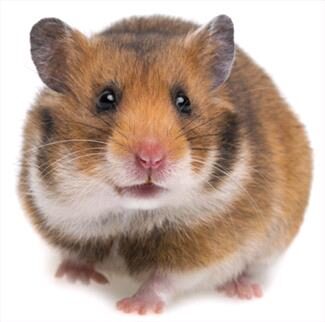
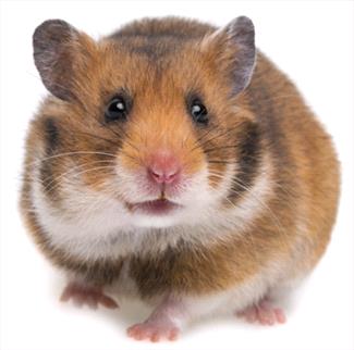
Hamsters are peculiar little rodents with large cheek pouches and short stubby tails. They have gained popularity as pets and research animals since the 1930s. The Syrian hamster’s (golden hamster) wild habitat extends through the Middle East and Southeastern Europe. In 1930, a litter of eight baby hamsters was taken to Palestine and raised as research animals. Virtually all domesticated hamsters sold in the pet trade and research are descendants of three of the survivors of this litter. Hamsters were introduced first into the United States in 1938.

Since their domestication, several color and hair coat varieties of the Syrian hamster have arisen through selective breeding. The three basic groups which now exist include the common ‘golden’ hamster, colored short-haired ‘fancy’ hamster, and long-haired ‘teddy bear’ hamster. All three varieties are popular as pets, while the research community generally employs the basic golden hamster.

On occasion, one may encounter other species of hamsters, but these are much less common than the Syrian hamster. The smaller, dark brown Chinese hamster (dwarf hamster) is often used in biomedical research, and they are sometimes acquired as pets. These hamsters are recognized for their small size, dark brown color and black stripe down the back. The Armenian (grey) hamster and European hamster are two other species occasionally used in research, but seldom kept as pets. The information here pertains particularly to Syrian or golden hamsters because they are by far the most popular.

Diet

As with any pet, good quality food and clean, fresh water must be provided at all times. The precise nutritional requirements of hamsters have not been fully determined. In the wild, these animals feed on plants, seeds, fruit and insects. Current recommendations for feeding in captivity are pelleted rodent ration containing 15% – 20% protein. These rations are typically processed as dry blocks or pellets designed for rodents. Seed diets are also ‘formulated’ and sold for hamsters, but these diets should only supplement the basic rodent pellet. Seed diets contain high levels of fat which can easily become rancid if improperly stored. In addition, when fed alone, these diets often lead to obesity and potential nutritional deficiencies, especially calcium. Other supplements to the diet may include sugarless breakfast cereals, whole wheat breads, pasta, cheese, cooked lean meats, fresh fruits and vegetables; all fed in moderation. Hamsters eat approximately 12 grams of food daily, and usually consume the majority of this at night. Hamsters are like little pack rats that often hoard their food in a corner of their cage, making it seem as though they eat a lot more than they really do.

Water is easily provided in water bottles equipped with sipper tubes. This method also helps keep the water free from contamination. Always make sure that the tubes are positioned low enough to allow the pet easy access. Juvenile hamsters need special consideration to make sure they are strong enough to use the sipper tube as well as reach it. The average hamster drinks approximately 10 ml of water per 100 grams body weight (average adult size). Although this amount is only a fraction of the total bottle volume, fresh water should be provided daily, not only when the bottle empties.

Diseases

Proliferative Ileitis (Wet Tail)

The most commonly encountered bacterial infection recognized in hamsters is ‘wet tail’. The precise cause of the disease is not fully understood, but underlying infections with the bacteria Campylobacter fetus subspecies jejuni have been reported. Similar Campylobacter sp. are responsible for serious intestinal diseases in other animal species, such as swine, dogs, ferrets, primates and even humans. Although this agent is suspected to be an underlying cause of this syndrome, pure cultures of the bacteria cannot reproduce the disease, suggesting other predisposing factors or agents. Such contributory factors include improper diet, sudden dietary changes, overcrowding and other stresses.

This disease most often affects weanling hamsters between the ages of 3 to 6 weeks, but hamsters of all ages are susceptible. Since this is the age at which most hamsters are sold, this is a common disease encountered in recently acquired pets. The long haired ‘teddy bear’ hamster seems to be more vulnerable than the other varieties.

Death may result within 1 to 7 days after the onset of watery diarrhea. Other signs include matting of the fur around the tail, unkept hair coat, hunched stance, loss of appetite, dehydration, emaciation and irritability. Blood from the rectum and rectal prolapse may be noted in some serious cases. This is a very serious disease, with death being the most likely outcome. Due to the severity, any hamster exhibiting these signs must be examined by a veterinarian as soon as possible. Antibiotics, fluid therapy and anti-diarrheal medications will be administered to the patient. Supportive care will also be instituted. Despite all the best efforts, treatment is often unrewarding with death occurring within a couple days in many cases.

Hair Loss (Alopecia)

Hair loss can occur for a number of reasons in hamsters. This loss of hair can be due to both disease and nondisease conditions. Continual rubbing on feeders or sides of the cage as well as protein deficiency and barbering (hair chewing by cage mates), are examples of nondisease causes of alopecia. Infestation with demodectic mites is one of the most common infectious causes of patchy alopecia and scaling in hamsters. Other conditions that lead to hair loss include adrenal tumors, thyroid deficiency, and chronic renal disease. Some of these conditions may be correctable, while others are not.

Demodex mites are the most common external parasite causing problems in hamsters. The mite lives within the hair follicles and certain skin glands of their host. The presence of these mites result in dry, scaly skin and subsequent hair loss, especially over the back and rump. This disease is rarely a problem by itself. Demodectic mange in hamsters is often associated with chronic, debilitating diseases or other underlying problems. For this reason, a thorough examination must be performed on any hamster presented with mites. To confirm the presence of mites, the veterinarian may perform a skin scraping for microscopic observation. Treatment for the mites is often possible, but remember that there may be another problem, often more severe, underlying this one which must also be addressed.

Old Age Diseases (Geriatric Conditions)

Hamsters tend to have relatively short life spans when compared with other species. The average life expectancy of a hamster is between 2 and 3 years of age. For this reason, spontaneous aging diseases are not uncommon in these animals, typically after the age of one year. Two of the most common geriatric diseases of hamsters are amyloidosis (protein deposition in various organs) and cardiac thrombosis (blood clots in the heart). Treatment of these conditions involves managing clinical signs because a cure is not possible. A diagnosis of virtually any geriatric disease carries a poor prognosis.

Amyloidosis is a condition whereby proteins produced by the body are deposited in various organs, primarily the liver and kidneys. Kidney and liver failure often occurs as a result of this protein deposition. Many other organs are also affected, and the changes are irreversible. Signs of this condition include swollen abdomen, urinary problems, dehydration, poor appetite and rough hair coat. Supportive care is the only treatment since this condition is eventually terminal.

Blood clots within the heart occur at a relatively high frequency in older hamsters. This condition is known as cardiac thrombosis, and typically occurs in the left side of the heart. Many factors are involved in the formation of these clots including clotting disorders, heart failure, circulating bacterial infection and amyloidosis. Many other old age diseases occur in hamsters over the age of one year. Liver and kidney disease is not uncommon in middle age to old hamsters. Other conditions commonly encountered are gastric ulcers, tumors, and dental diseases.

Facts

  • Average life span: 2 – 3 years
  • Adult body weight: 100 – 150 grams (3.5 – 5 ounces)
  • Environmental temperature range: 65 – 80°F
  • Relative humidity range: 40 – 70%
  • Age at first breeding:  males 10-14 weeks; females 6-10 weeks
  • Gestation period: 15.5 – 16 days
  • Litter size: 5 – 10 (average)
  • Weaning age: 21 – 25 days

Handling

Hamsters handled frequently from a young age usually remain docile and seldom bite. These animals of a docile nature can be picked up gently by cupping in one or both hands and held against one’s body. Beware that even docile hamsters may bite if surprised or abruptly awakened from sleep.

Other hamsters, however, may not have received a lot of attention and handling throughout their lives, and thus may be more apprehensive and aggressive. Any animal whose personality is not fully known must be approached cautiously. The use of a small towel or gloves can assist the handler in capturing and restraining such a pet. Another method of capture involves coaxing the animal into a container (such as a can or tube), which can then be removed from the cage. Once removed from the cage, biting hamsters can be restrained by grasping a large amount of skin at the scruff of the neck. As much skin as possible must be grasped using this method because their skin is very loose. If lightly scruffed, the hamster can easily turn around within its skin and bite the handler.

Housing

Several types of cages are available that are suitable for housing hamsters. Many of these units come equipped with cage ‘furniture’ such as exercise wheels, tunnels, and nest boxes as added luxuries. Such accessories, as well as sufficient litter depth within which to burrow, are desirable for the pet’s psychological well-being. Cages should be constructed with rounded corners to prevent chewing. Hamsters will readily chew through wood, light plastic, and soft metal; so recommended caging materials are wire, stainless steel, durable plastic, and glass. Beware that glass and plastic containers drastically reduce ventilation and can lead to problems with humidity, temperature and odor concentration. These materials make suitable cages when at least one side of the enclosure is open for air circulation. In addition, make sure that the enclosure is escape proof because these little rodents are known escape artists.

Hamsters do very well in solid bottom cages with deep bedding and ample nesting material. Bedding must be clean, non-toxic, absorbent, relatively dust free, and easily acquired. Shredded paper or tissue and processed corn cob are preferred bedding. Be sure that the ground corn cob is free from mold, mildew, or other contamination before using. Shredded tissue paper makes excellent nesting material.

Adult hamsters require a minimum floor area of a 10-gallon aquarium (approximately 20 inches by 10 inches) and a cage height of a minimum of 6 inches. Female breeding hamsters require much larger areas. Optimal temperature range for hamsters is between 65 to 80°F, with babies doing best at 70 to 75°F. The relative humidity should be between 40% and 70%. Twelve hour light cycles are preferred, with hamsters being more active during the night.

Pet hamsters are generally housed singly. Mature female hamsters tend to be very aggressive towards one another and should never be housed together. Females are also larger and more aggressive than males, thus males usually need to be separated immediately after breeding. Males may also fight when housed together, but tend to be less aggressive than females.

The cage and accessories should be cleaned thoroughly once to twice weekly.  An exception to this schedule is when newborn babies are present; wait until they are at least two weeks old to disturb the cage. Other factors that may require increased frequency of cleaning are the number of hamsters in the cage, the type of bedding material provided, and the cage design and size. Clean the floor and walls with soap and water weekly. Change the bedding weekly.

Cages should be sanitized with hot water and a nontoxic disinfectant or detergent, then thoroughly rinsed. Water bottles and food dishes should be cleaned and disinfected daily. Most of the time just hot water and a mild detergent, and rinsing it afterward, is fine. Bleach kills most of the major pathogens that a hamster would create or be in contact with, so for a more thorough cleaning once in a while use a solution of 1 part bleach in 10 parts water; wipe it on, let it sit for 15 minutes, and then rinse it off thoroughly. Water bottles and bowls can be soaked in this bleach solution for 15 to 30 minutes but must be rinsed well before use. Cleaners are sold at pet stores that can be used but most are not as effective for deep cleaning as bleach.

Reproduction

The sex of hamsters can be easily determined. Mature male hamsters possess large, prominent testicles, which often alarm owners who first notice their size and mistake them for tumors. In addition, the genitourinary to anal separation is much wider in males than females, making it possible to sex young hamsters.

Male hamsters should be first bred at 10 to 14 weeks of age. Females can be bred at the age of 6 to 10 weeks. As the female comes into ‘heat’ she will begin assuming a breeding stance with her back swayed and body stretched out. When petted over her back, she will remain motionless and sway her back even further. A thin mucus will be noticed coming from her vulva on the next day of the ‘heat’ cycle, which indicates that estrus will occur two days later. For breeding, place the female into the male’s cage about one hour before dark. Closely observe the pair for mating activity or fighting. Females can be very aggressive towards males and can cause serious injuries. At the first sign of aggression by the female, remove the male; then try again the next night. Also, remove the male shortly after a successful mating has taken place.

Pregnancy is of very short duration in hamsters; lasting only 15 to 16 days. Just before delivery, the expectant mother will become restless and may discharge a small amount of blood from her vulva. Do not handle or disturb her at this time. It is wise to clean her cage two weeks following breeding, so her cage is relatively clean when babies arrive. Litter size ranges from 5 to 10 pups; larger litters are not uncommon. The pups are born hairless with their eyes and ears closed. However, they do already have their front teeth, the incisors.

Provide ample nesting material and bedding for the new mother and young. Plenty of fresh food and water should be available before the babies are born. DO NOT disturb the mother and young for any reason during the first week or two after birth. If a mother hamster seems threatened for any reason, she typically will kill and cannibalize the young. In other instances, she may stuff the young into her cheek pouches and frantically carry them around the cage looking for a safe place to establish a nest. Occasionally, pups will suffocate as a result of this activity, especially if the disturbance is prolonged.

Young hamsters usually begin eating solid food at 7 to 10 days of age, but are not weaned until 21 to 25 days. Provide food on the cage floor for the young and mother, and also have soaked, softened pellets available for them as well. Make sure that the water bottle is low enough for the weanlings to use, and that they are strong enough to use it; or provide an alternative water source during this time.

5908508

How Does Anesthesia Work?

Pet owners may shy away from surgical or dentistry procedures for their pets because they fear the risks of anesthesia. While it’s a good to consider the risks and benefits of general anesthesia for elective procedures, anesthesia is now safer than ever. If the proposed procedure will improve your pet’s quality of life, then fear of anesthesia shouldn’t prevent you from moving forward.

This was not always the case. In the 40s, 50s and 60s — well over half a century ago — death from anesthesia was about 1 in 400 for dogs and 1 in 260 for cats. These numbers are from referral hospitals, which are generally anesthetizing sicker patients, and that could increase the numbers over what you would expect to see in healthier pets in a general practice. There is a study from the early 2000s in the U.K. that followed mortality in a variety of practices (from general practices all the way to referral hospitals) and included dogs, cats, birds and pocket pets; in it, they found a death rate from 1 in 588 for dogs and 1 in 416 for cats. The risk has continued to drop, so that today those fatality numbers are around 1.7 in 1,000 dogs and 2.4 in 1,000 cats.

Although only statistics for dogs and cats have been created, the same improvement in risk applies to other pets such as rabbits, rats, hedgehogs, bearded dragons, and even goldfish as equipment and teaching new methods of anesthesia have improved over the years.

How does it work?

General anesthesia is given through an IV or an inhaled gas, or sometimes a combination of the two. It is similar, on a smaller scale, to a medically-induced coma. Oftentimes a sedative is given first.

It works by interrupting nerve signals in your pet’s brain and body. It keeps them temporarily unconscious: asleep, relaxed, and pain free during a procedure or surgery. It slows down automatic functions like breathing, heart rate, and circulation. During that coma-like period, brains cannot process pain, nor will the pet remember what happened during surgery.

During the procedure, the veterinarian or veterinary anesthesiologist will check some basic functions while your pet is under, such as breathing, temperature, heart rate, blood pressure, blood oxygen level, and fluid levels (as would happen in a human surgery). Monitoring doesn’t have to be done with expensive equipment, as having a trained person monitoring the depth of anesthesia as well as certain physical parameters can be just as valuable. Computerized anesthesia monitors can be used to enhance, but not replace, that person’s ability.

If those functions aren’t where the veterinarian wants them to be, adjustments in the level of anesthesia can correct them.

Monitoring doesn’t stop when surgery is over, or even when the equipment is removed. About half of the anesthesia deaths in small animals happen during the first 3 hours after surgery. The causes are usually breathing or heart problems. That makes this time critical for patient safety. Careful monitoring by a trained observer of physical variables, especially oxygenation and temperature, can help make this time period safer.

What main factors influence anesthetic risk?

Not unsurprisingly, the first is the health of the animal. Healthy patients, or patients with mild, controlled disease, are at less risk than patients who are sick. For healthy dogs, about 1 in 2,000 are expected to have a fatality and about 1 in 1000 cats would have one. In sicker patients, it would be closer to 13 dogs and 14 cats out of 1,000.

Having anesthesia in an emergency can also increase risk. That makes sense if you think about it. When you have time to plan a procedure, stabilize your patient and can schedule surgery at a time that is optimal for everyone, things go better. Having said that, the risk of anesthetic death with an emergency procedure, while higher than regular procedures, is still low.  Emergencies are emergencies because they can’t wait, and there are times that the pet will die if surgery is not done as soon as possible, such as with bloat.  

Veterinarians can decrease risk by focusing on monitoring and supportive care before, during, and after anesthesia. Preoperative planning is done with a good history, physical examination, and often baseline lab work to check liver and kidney function, among other things. Any abnormalities identified before surgery can be corrected if needed.

Follow all of your veterinarian’s pre-operative instructions exactly, including when to stop giving food the night before. Anesthetized patients lose their ability to swallow. If there is food in the stomach, the pet could vomit while under anesthesia or soon afterwards. If vomiting occurs before the swallowing reflex comes back, the vomited material can enter into the lungs, causing aspiration pneumonia. For emergency surgery, this factor is overlooked, but not for elective surgery.

While anesthetic risk is not zero and never will be, the risks are low and shouldn’t prevent any pet owner from providing health care for their pet whether it’s a dental cleaning or major surgery.

5171558

Head Tilt in Pet Rabbits

Head tilt in rabbits is seen with some frequency and can be caused by a variety of diseases.  Another common name for head tilt is wry neck.  The correct medical term is vestibular disease, which can include other signs besides a head tilt. Another term that is often used is torticollis. Rabbits with vestibular disease can have a head position that ranges from a few degrees to 180 degrees off the normal position.  They can fall over, circle, have difficulties standing and develop eye injuries because the prominent eye globe (especially of the “down” eye, the one facing the ground when the head is tilted) is prone to trauma.  The cardinal signs of true vestibular disease in the rabbit are a persistent head tilt and a loss of balance. 

Anatomy

First let’s look at the anatomy and function of some vital areas in order to understand what does and does not contribute to head tilt in the rabbit.

External Ear

Disease in this area alone can cause head shaking, drooping ear and pain but does not cause a persistent head tilt or loss of balance. 

Middle Ear

Disease of the middle ear can cause head shaking, drooping ear and pain as well as deafness but does not cause a persistent head tilt. 

Inner Ear

The inner ear controls balance and hearing.  Signs of disease of the inner ear include deafness, head tilt, loss of balance and horizontal or rotary nystagmus. Proprioception (the ability to sense where the feet and legs are) and postural reactions (the ability to try to return to a normal standing position) are normal.  The rabbit will not act weak in other parts of the body and will continue to try to maintain normal body position even if it is difficult and the head is tilted. 

Brain

A specific area of the brain stem contains the vestibular nuclei, the origin of the vestibular nerve in the inner ear. The vestibular nuclei serve as the body’s central balance control. Signs of disease to this tiny area of the brain stem include head tilt, loss of balance, circling toward the affected side, rolling, vertical nystagmus, positional nystagmus, delayed or absent proprioception and loss of postural reactions.

Spinal cord

Head tilt is not a sign of primary spinal cord disease. 

Diseases Resulting in Head Tilt

A major differentiation that has to be made when diagnosing the cause of head tilts is whether it is peripheral (affecting areas other than the brain) or central (involving the brain and most specifically the vestibular nuclei).

Otitis Interna (Inflammation of the inner ear)

Causes can include the following: infectious disease, foreign bodies, trauma, neoplasia, and toxins. Signs of otitis interna include persistent head tilt toward the affected side, circling, nystagmus, ataxia (inability to walk normally), and deafness. The most accurate way to diagnose otitis interna is with a CT scan or MRI. A negative finding on an x-ray may not rule out otitis interna. 

Treatment for otitis interna depends on the primary cause, but since the majority of head tilts in rabbits are likely caused by bacterial otitis interna, it is advantageous to use a long-term course of antibiotics (3 to 6 weeks up to several months).  It is currently recommended to avoid the use of corticosteroids in rabbits. Rabbits may be more sensitive than other animals to developing immunosuppression when taking corticosteroids, either topically, orally or parenterally.

Otitis Media (Inflammation of the middle ear)

This is also a common disease of rabbits and may occur along with or even be the cause of otitis interna.  However, disease in this area alone does not cause a persistent head tilt.  Signs of otitis media include periodic head tilting and shaking. Diagnosis and treatment are generally the same as for otitis interna. 

Brain Stem Disease

Disease at the brain stem, specifically the vestibular nuclei, can cause similar signs as seen with inner ear disease.  Because the vestibular nuclei are deep in the brain, it is likely that disease affecting this area will also affect surrounding brain tissue.  Therefore, additional neurologic signs may be seen such as loss of appetite, mental dullness, paralysis and sudden death.  If the disease is also affecting the cerebrum, additional signs such as seizures can be seen. Bacteria, fungi and viruses can affect the brain stem.

Encephalitozoon cuniculi is a one-celled organism called a microsporidium that can infect rabbits. There is an ongoing controversy over the prevalence of E. cuniculi as a cause of primary head tilt in the rabbit.  It has been extremely difficult to demonstrate a definitive correlation between head tilt and active E. cuniculi infection.  Serological testing for E. cuniculi has some value but is not definitive and, if not interpreted appropriately may be misleading.  The only way to diagnose E. cuniculi as the definitive cause of a head tilt is to take brain tissue samples from the rabbit and find the organism and its damage in the microscopic samples.  No one has yet proven this correlation because a brain biopsy is dangerous for the rabbit and the E. cuniculi organism can be difficult to find in brain tissue. There are few, if any, case reports or studies definitely proving E. cuniculi is a significant pathogen in the rabbit nervous system. If a rabbit shows signs compatible with central vestibular disease, has a positive test for E. cuniculi, and all other diseases have been ruled out, some veterinarians will choose to treat for E. cuniculi empirically.  Proper and effective treatment for E. cuniculi is controversial. Some of the medications that have had been used to treat infection with E. cuniculi include albendazole, fenbendazole and oxibendazole.

The most common parasite associated with head tilt in a rabbit is the raccoon roundworm Baylisascaris procyonis.  Signs observed in rabbits with Baylisascaris may include head tilt, tremors, weakness, blindness, seizures or sudden death. Prevention of exposure to the parasite eggs is clearly the best way to counteract this disease.

Other causes of head tilt in rabbits may include cerebrovascular accident (stroke), cancer, trauma, toxins, metabolic disease, and heat stroke.

Diagnostic Approach to Head Tilt

History

A detailed history is of vital importance to determine the cause of disease. 

  1. History of any prior illness
  2. History any prior bouts with head tilt, weakness or incontinence
  3. Possibility of exposure to environmental toxins or parasites
  4. Exposure to other rabbits that are or were ill (particularly with neurological disease)
  5. Possibility of trauma
  6. Possible contact with human with active herpes viral infection

Physical Exam

A thorough physical exam and a thorough neurological exam is essential to diagnosing the cause of head tilt.

  1. Mental attitude: Is the rabbit still alert and active, or dull and depressed?
  2. Head tilt:  Look at persistence, side to which rabbit tilts, is circling involved?
  3. Balance:  Does the rabbit try to right itself if given support?
  4. Gait:  Any abnormalities in gait?
  5. Nystagmus: If present, is it spontaneous or positional, is it horizontal or vertical?
  6. Ear exam:  Are there signs of external or tympanic membrane disease?  Are there signs of fluid, blood or pus beyond the tympanic membrane?
  7. Respiratory:  Are there signs of respiratory disease?
  8. Systemic: Any other neurological signs, weakness (particularly hind limb), paralysis, incontinence, behavioral changes, external signs of trauma, particularly around the head and neck?   

Blood Tests

  • Complete Blood Cell Count:  This test may be helpful to determine if there is anemia or infection.  
  • Serum biochemistries:  These tests are helpful to rule in or out a number of metabolic diseases
  • Serology for E. cuniculi – These tests are of limited use in definitively diagnosing active disease.
  • Blood testing for heavy metals – These tests are particularly important if heavy metal intoxication is suspected.

Bacterial Cultures

Unfortunately, it is frequently not possible to safely or easily collect a sample to culture from a rabbit with vestibular disease.

Endoscopy

Endoscopy of the ear canal may be useful if middle ear infection is present, or possibly used to obtain cultures through the tympanic membrane with a surgical technique called myringotomy. 

Radiographs (X-rays)

Radiographs are useful to detect any heavy metal in the GI tract and for diagnosing head trauma.  Radiographs are also helpful in screening for disease of the tympanic bulla where the middle ear is housed

CSF (cerebrospinal fluid) Analysis

This may be useful if central disease such as encephalitis is suspected.

Biopsy

If it is possible to obtain a sample of the affected tissue, then a microscopic analysis can be extremely helpful in making a diagnosis.

CT scan or MRI

These imaging techniques are the most accurate and safest means of diagnosing disease of the inner and middle ear.

Treatment and Nursing Care

  1. If a definitive diagnosis of the cause cannot be made (very common situation), but peripheral disease is the suspected due to physical exam, history and whatever diagnostics could be performed, the rabbit should be put on a course of broad spectrum antibiotics for an extended period of time ranging from 3 weeks to several months.
  2. Generally, corticosteroids (cortisone-like drugs) should be avoided if possible, because rabbits may be especially sensitive to the immunosuppressive qualities of these drugs, and their use may cause further complications. 
  3. If a diagnosis of E. cuniculi infection is strongly suspected based on multiple signs of central disease, serology, and ruling out other disease,  a short-term use of oxibendazole,  fenbendazole, or albendazole, can be considered. 
  4. Non-steroidal anti-inflammatory drugs should be considered to reduce inflammation and control pain that may be present.  These drugs may be needed only at the beginning of therapy.
  5. The use of anti-nausea drugs is controversial as there is no clear evidence that rabbits experience feelings of nausea.  There is no substantiated evidence that the use of anti-nausea drugs helps improve the condition of rabbits with head tilt.  Some veterinarians feel anti-nausea drugs, like diphenhydramine or meclizine, are useful in a rolling rabbit or one who is not eating.
  6. Eye lubrication is useful, particularly in those animals that have a severe head tilt.  The down eye is prone to injury due to the protruding nature of rabbit eyes.  Rabbits do not blink often and this eye may become dry, abraded or infected.  Daily attention is necessary.
  7. Fluid therapy and nutritional therapy (assisted feeding) may be necessary in any rabbit with vestibular disease.
  8. Rabbits with vestibular disease from any cause often cannot access their cecotropes.  These nutrient-rich droppings can be collected while still moist and placed in a rabbit’s food bowl along with the pelleted food. 
  9. It is essential to modify the environment of a rabbit with severe vestibular disease. This involves providing an enclosed padded or smooth-sided cage or enclosure. 
  10. Aside from occasional anecdotal reports or testimonials, there is no evidence that any kind of physiotherapy or acupuncture will reduce the length of time a head tilt persists or will resolve a residual head tilt.
  11. It is important to remember that the course of vestibular disease, even with the best prognosis, can take many weeks to months of committed care before improvement is seen. 
  12. If a rabbit shows a continual decline or continued mental depression, loss of appetite or other weakness over a 2 to 3 week period, then the prognostic outlook is fairly grim and euthanasia should be a consideration. 

Prognosis

The prognosis for recovery from vestibular disease is variable, depending on the cause.  For most rabbits with peripheral disease, the prognosis is good to guarded. Some rabbits will have a lifelong residual head tilt even if the inner ear disease is cured. For rabbits with central vestibular disease the prognosis becomes guarded to poor for recovery to a sustainable state.

Key Points

  1. Persistent head tilts accompanied by nystagmus and loss of balance are either peripheral (inner ear) or central (vestibular nuclei of brain).  
  2. Peripheral vestibular disease is probably the most common cause of head tilt and is usually confined to head tilt, spontaneous nystagmus, circling and loss of balance.  The majority of cases are still mentally alert, maintain an appetite and do not exhibit other signs of weakness, gait abnormalities or seizures.
  3. Peripheral vestibular disease is most commonly caused by inflammatory disease of the inner ear with bacterial disease being the most common.  There is no current evidence that E. cuniculi causes disease of the inner ear.
  4. Peripheral vestibular disease carries a good to guarded prognosis for clinical recovery.  There is often a residual head tilt, but the rabbit can learn to re-establish balance and live a relatively normal life. 
  5. Central vestibular disease is less common, and also includes head tilt, positional nystagmus, circling and loss of balance. 
  6. Rabbits with central vestibular disease may also have histories of other signs compatible with central disease, potential exposure to toxins, parasites, or trauma.
  7. Central vestibular disease may be caused by a variety of conditions including bacterial infections, E. cuniculi, parasites and trauma, and carries a guarded to poor prognosis for recovery. 
  8. Radiographs are necessary to rule out trauma and may detect middle ear disease.  However, in many cases there will be no radiographic change even in middle or inner ear disease. Therefore, a negative x-ray is not proof that this disease does not exist.
  9. CT scan or MRI is the most accurate and safe means of detecting inner ear disease as well as some types of central disease.
  10. It is probably best at the minimum to treat rabbits with strictly peripheral signs that are confined exclusively to head tilt, nystagmus, circling and loss of balance with appropriate antibiotics because bacterial disease of the inner ear is common.
  11. Non-steroidal anti-inflammatory drugs should also be considered in many head tilt cases to reduce inflammation (since inflammatory disease is so common in both peripheral and central disease) and reduce any pain. 
  12. Appropriate nursing care for a rabbit with vestibular disease is crucial and requires a long-term commitment to both environmental and patient management.
  13. The sooner you get veterinary care for a rabbit with vestibular disease, the greater the chances for successful resolution with a relatively short recuperative period.
4128972

Histopathology in Pets

What Is Histopathology?

Histopathology is the microscopic examination of stained tissues to look for a potential cause or the size of a diseased area. Staining is the process that applies dyes to the slide to highlight cells and make them easier to see.

What Is a Biopsy?

A biopsy is the removal and/or examination of cells or tissue from the body to figure out a disease’s presence, cause, or extent.

Benefits of Histopathology

Histopathology can be a powerful diagnostic tool. There are many diseases that cause vague and nonspecific clinical signs. The results of histopathology can help your veterinarian know the specific disease your pet is suffering from and the required treatment.

In some cases, histopathology is the only way to find out for sure what condition your pet may have. Many diseases require histopathology to be confirmed. Some of these are various inflammatory, congenital (a condition existing from birth), and cancerous diseases.

Examples of inflammatory diseases that require histopathology to be diagnosed include inflammatory bowel disease, glomerulonephritis (inflammation of the glomeruli, which are part of the kidneys), and hepatitis (inflammation of the liver).

Unusual or severe skin abnormalities are instances when your veterinarian may initially recommend histopathology when making a diagnosis. Histopathology can be useful in a pet who isn’t displaying obvious signs of sickness. Some veterinarians may recommend taking biopsies of masses based on their size, location, duration, appearance, or if seemingly painful.  

Cancer is a type of disease that often requires histopathology for diagnosis. Histopathology is often needed to correctly identify the mass as cancerous or not. This identification is important for detecting your pet’s cancer and deciding the best treatment plan.

Biopsy Procedure

A tissue biopsy is obtained by removing a small piece of tissue. How that tissue is taken depends on the type of tissue and where that tissue is located.

Some methods of obtaining a biopsy do not require invasive surgery. Your pet may only need local anesthesia (numbing the area) and sedation in these cases. For example some skin lesions are often investigated with a simple skin punch biopsy.

Other biopsy methods may require general anesthesia, where your pet is “put under” and unconscious for the procedure. This option is usually for larger superficial masses or internal biopsies, like those of the intestines, and may involve the use of an endoscope (a small camera on a long flexible wand to see within the body) or exploratory surgery, where a body cavity like the chest or abdomen is opened to look for masses or other abnormalities. 

Your veterinarian will explain how they will get a biopsy sample and decide whether your pet is stable enough to undergo general anesthesia if needed. Depending on the procedure, your veterinarian may also refer you to a veterinary specialist to obtain the biopsy and to help recommend further treatment.

Once a biopsy is obtained, it is sent out to a pathologist. A veterinary pathologist will examine or “read,” the tissue sample and report their findings. In most cases, the biopsy sample will take some time to ship to a laboratory and to be evaluated. Your veterinarian will give you an estimate of when to expect results. 

Recovery

Your pet will have a wound where the tissue was removed and may have stitches. Your veterinarian should provide you with post-surgical instructions and may provide pain relief medications as needed.

The biopsy site needs to be kept clean and dry; your pet should not be able to lick it or irritate the wound. Your veterinarian may recommend an Elizabethan collar (cone) to help prevent your pet from touching it. If you have questions at any time about how your pet is healing, always contact your veterinarian.

Limitations of Histopathology

While histopathology can provide valuable information, it has limitations too. It helps your vet to find the diagnosis most of the time. Errors can happen when tissue sampling is not good enough (e.g., the biopsy was not representative of the affected area) or if there is a lack of clinical history. Your veterinarian can prevent many of these errors by collecting and handling the tissue appropriately and working closely with the pathologist.

Your veterinarian may request a second opinion on the biopsy if the results come back inconclusive or are unexpected, and they may recommend repeating a biopsy. Depending on the disease, more specialized lab tests may be needed, including genetic and immunology testing.

Histopathology is not always needed or recommended. It should only be pursued if your pet is stable enough to undergo the procedure needed to obtain the biopsy, if the results will affect the treatment plan, and if you are willing to pursue treatment.

If your veterinarian has a high suspicion for a specific disease, they may recommend treatment without a definitive diagnosis. This can resolve your pet’s problem without the need for a biopsy. However, a biopsy may be needed if your pet does not respond well to the treatment.

Not all lumps and bumps require histopathology; some may be able to be diagnosed with a less invasive test called cytology, which involves taking cells from a mass or affected organ with a needle (a fine needle biopsy), a sample of a body fluid, or a washing, brushing, scraping or swab collection. If there is low suspicion for disease, your pet is not showing discomfort associated with an abnormality, or a mass is small, your veterinarian may recommend cautious monitoring.

Results of Histopathology

Your veterinarian will let you know when your pet’s histopathology report comes back from the pathologist, and will then discuss potential next steps with you. While the results may not always be good news, knowing exactly what is affecting your pet is one step towards ensuring they receive the best treatment and care. Hopefully, with the right information, you can help your pet be as healthy as possible.