Tag: Keratoconjunctivitis Sicca

4128550

Dry Eye (Keratoconjunctivitis Sicca) in Dogs and Cats  

(Dry eye is formally known as keratoconjunctivitis sicca or KCS)

Why Tears are Good

We can all imagine the discomfort of dry, irritated eyes and the soothing that is provided by lubricating eye drops. Tears are essential to the comfort of our eyes but they do more than just provide lubrication. Tears contain anti-bacterial proteins and salts and serve to flush away the irritants and infectious agents that are constantly getting in our eyes. In addition, since the outer portions of the eye do not have a blood supply to remove metabolic waste, it is up to the tears to provide this service as well. 

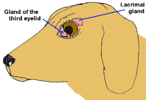
Diagram shows the two lacrimal (tear-producing) glands of the canine eye. Graphic by marvistavet.com

Tears consist mostly of water, but also of oil and mucus secreted by their respective eyelid glands. The water portion of tears is secreted by two lacrimal glands in dogs and cats: one just above the eye and another in the third eyelid (or so-called nictitating membrane).

Dry eye with the classical ropey discharge.

(Photo courtesy of Dr. Michael Zigler)

Without tears, eyes become irritated, the conjunctival tissues around the eyes get red, the cornea itself in time will turn brown in an effort to protect the eye, and a gooey, yellow discharge predominates. Blindness can result.

Keratoconjunctivitis sicca is a fancy way of saying the eye is dry. “Kerato” refers to the cornea or clear covering of the eye that faces the outside world.

“Conjunctivae” are the moist pink membranes of the eye socket. “Itis” means inflammation and “sicca” means dry. Keratoconjunctivitis sicca, abbreviated KCS, means there is an inflamed, dry cornea and conjunctiva. It occurs when there is a deficiency in the water portion of the tear film, which normally accounts for 95% of the tear volume. Without water, one is left with oil and mucus; hence, the gooey yellow eye discharge characteristic of this condition.

Why do Eyes Become this Dry?

There are many causes of dry eye. Some are:

  • Canine distemper infection attacks all body interfaces with the environment including the eyes. Dry eye is part of the constellation of symptoms that can occur with canine distemper infection. 
  • In cats, herpes upper respiratory infection can lead to chronic dry eye (see more on herpes conjunctivitis).
  • There could be a congenital lack of tear-producing gland tissue (as described in certain lines of Yorkshire terriers).
  • Exposure to sulfa-containing antibiotics, such as trimethoprim-sulfa combinations, can lead to dry eye. It can be either temporary or permanent and occurs unpredictably.
  • Anesthesia will reduce tear function temporarily (thus eyes are lubricated with ointment by the attending nurse).
  • During surgery for cherry eye, removal of the third eyelid tear-producing gland, instead of replacing the gland in its proper location, can lead to KCS. So can too much damage to the gland prior to proper gland replacement.
  • A knock on the head in the area of one of the tear-producing glands can lead to KCS.
  • The most common cause of KCS appears to be immune-mediated destruction of the tear-producing gland tissue. We do not know what causes this type of inflammatory reaction but certain breeds are predisposed: the American Cocker Spaniel, the Miniature Schnauzer, and the West Highland white terrier.

How We Make the KCS Diagnosis

When KCS is in an advanced state, the situation is pretty obvious but early on in the case, it may look like a simple case of conjunctivitis. You may also notice a dry nose or nasal philtrum (area at the bottom of the nose). In either case, it is important to measure the tear production to determine how dry the eyes are.

The test that accomplishes this is called the Schirmer Tear Test.

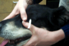
To perform the test, a strip of specific paper is put just inside the lower eyelid in the outer corner of the eye and left for 60 seconds.

The moisture of the eye will wet the paper. At the end of the 60-second period, the length of the moistened area on the paper is measured. A length of 15mm or more is normal. A length 11 to 14mm is a borderline result. A height of less than 10mm is dry. A height less than 5mm is severely dry.
How do we Treat this Condition?

Not that long ago, all we had to treat this condition was tear replacement formulas and mucus-dissolving agents. These are still helpful but require an impractical frequency of administration. A breakthrough came with the discovery of cyclosporine topical therapy to control immune-mediated gland destruction.

Cyclosporine is an immunomodulating drug used to prevent organ transplant rejection in people and treat certain immune diseases in dogs and cats. When applied as an eye drop or ointment, it suppresses the immune destruction that is the most common cause of KCS, and tear production is restored.

The success of this treatment plus its convenient dosing interval (1 -3 times daily) has made this medication the primary treatment for KCS.

Animal hospitals used to make their own cyclosporine eyedrops out of oral cyclosporine and vegetable oil, but this largely ended when Optimmune® eye ointment (containing 0.2% cyclosporine) came out. Occasional patients simply do not show a good response to cyclosporine ointment but will respond when the concentration is increased. Higher-concentration products can easily be formulated by compounding pharmacies or one of the alternative medications listed below can be used. Treatment is almost always required for the lifetime of the pet.

After beginning cyclosporine eye drops or ointment, a recheck in three to four weeks is a good idea to check for improvement. If the Schirmer tear test is still showing poor results, the dosing frequency can be increased to three times a day; similarly, if excellent results are seen, the medication can be dropped to once a day.  Periodic rechecks are needed for dose adjustment and some dogs take as long as three to four months to show a response. Dogs with Schirmer tear tests as low as 2 mm still have an 80 percent chance of responding to cyclosporine. This medication has been a miraculous breakthrough in the treatment of KCS.

Tacrolimus is another medication that is an immune modulator. No commercial products are available for use in the eye, so they must be obtained from a compounding pharmacy. It is often tried in cases that are unresponsive or poorly responsive to cyclosporine. It is used in a manner similar to cyclosporine and is generally similar in cost.

Pilocarpine is a cholinergic drug, which means it works on the autonomic nervous system (the part that controls automatic functions such as glandular secretion). This medication can be given for the particular form of dry eye known as neurogenic KCS. In these cases, neurogenic stimulation of the tear gland is absent, so the pilocarpine is given in an attempt to stimulate the gland. Although the drug comes as an eye drop, for KCS it is actually given orally at an increasing dose until side effects are seen (diarrhea, drooling, vomiting). If side effects are encountered, the dose is reduced to that which the animal tolerates. It is continued indefinitely or until the neurogenic KCS subsides, usually twice daily. Neurogenic KCS typically affects only one eye.

Artificial tear solutions, gels, and ointments can be purchased in most drug stores. These can be combined with other therapies and are soothing. Their use is particularly important early in therapy until cyclosporine or tacrolimus takes effect and in eyes that do not respond to these latter medications. Over-the-counter products may be recommended two-12 times daily, depending upon their formulation and the severity of the KCS.

Topical antibiotics are often needed, especially when starting treatment for KCS because secondary infections are common with inadequate tears. These products do not increase tear production but help relieve the thick discharge.

Topical steroids may be beneficial in decreasing the inflammation associated with KCS. Typically they are combined with topical antibiotics in the same solution or ointment, especially when given to dogs.

Surgical Solutions

Parotid duct transposition is a surgical solution to unresponsive, severe KCS, although it is a delicate procedure, usually done by a veterinary ophthalmologist. The parotid duct is the salivary gland on either side of the face/cheek. It produces saliva that is carried to the mouth via a long duct. This duct can be carefully dissected out and moved to the outer corner of the inside of the lower eyelid in order to deliver saliva to the eye. Saliva is a reasonable substitute for tears, although in time some mineral deposits may form on the eye surface. Mineralization of the cornea can range from mild to severe and there is no good way to predict whether mineralization will be a problem prior to the transposition of the duct. The eye may water uncontrollably when the dog is fed and facial wetting may be objectionable in some cases.
5313942

Brachycephalic Airway Obstruction Syndrome in Flat-Faced Dogs  

Dogs with Short Faces

Most people are not familiar with the term brachycephalic, but if you own a Pug, Boston Terrier, Pekingese, Boxer, Bulldog, Shih Tzu, or any one of the other breeds with pushed-in or short faces, you should become familiar with this word. The word comes from the Greek roots brachy, meaning short, and cephalic, meaning head.

Brachycephalic dogs have been bred so as to possess a normal lower jaw that is in proportion to their body size and a compressed upper jaw. In producing this cosmetic appearance, we have compromised these animals in many important ways, and you, as an owner, must be familiar with the needs of your pet.

The Respiratory System and Brachycephalic Airway Obstruction Syndrome

Brachycephalic breeds make a lot of snorting respiratory sounds, as a matter of course, simply because of the way their throats and faces are shaped. These sounds are generated from a combination of several anatomic deformities that will be described below. These deformities occur to varying degrees of severity. Most brachycephalic dogs are not hampered by their anatomy on a day-to-day basis, but they do have limitations that must be recognized. Severely affected individuals require surgical intervention.

Stenotic Nares

This is a fancy name for narrowed nostrils. The brachycephalic dog’s respiratory passage begins with very small, often slit-like, nasal openings for breathing. This leads to a great deal of open-mouth breathing and panting as the nasal breathing route is limited by its small opening. In selecting a puppy for adoption, it is a good idea to look at the nostril sizes on each member of the litter and look for the widest opening. Stenotic nares can be surgically corrected after the age of five months.

Enlarged Tongue (Macroglossa)

The brachycephalic dog’s tongue can be particularly thick and large, which contributes to the overall obstruction. The French and English bulldogs seem particularly predisposed to this issue.

Elongated Soft Palate

It is difficult to fit the soft tissues of the canine mouth and throat into the brachycephalic’s short face. As a result, the soft palate, which separates the nasal passage from the oral cavity, flaps loosely down into the throat, creating snorting sounds. Virtually all brachycephalics suffer from this, but actual respiratory distress is rare except in English Bulldogs. The English Bulldog tends to have more severe symptoms in almost all aspects of brachycephalic syndrome. Excess barking or panting may lead to swelling in the throat, which can, in turn, lead to trouble. Again, the soft palate can be surgically trimmed.

Tracheal Stenosis/Hypoplastic Trachea

The trachea is also known as the windpipe. The brachycephalic dog’s windpipe may be dangerously narrowed in places. This condition creates tremendous anesthetic risk and should be ruled out by chest radiographs prior to any surgical procedures, as anesthetic safety depends on the placement of a tube in the windpipe to secure air passage. If the trachea is too small, intubation may not be possible, and it is important to know this in advance of any planned procedure. The English bulldog is particularly predisposed to this anatomical defect.

Everted Laryngeal Saccules

The normal larynx has two small pockets called ventricles or saccules. When a dog has increased effort to breathe, over time, these little pockets will actually turn inside out inside the throat. When this occurs, it obstructs the throat. This condition can be corrected surgically; however, in many cases, it resolves on its own after the stenotic nares are corrected, so surgery is generally only performed in severe situations. The pug is especially predisposed to this situation.

Heat Stress, Excitement, and Exercise 

Because of all these upper respiratory obstructions, the brachycephalic dog pants inefficiently. A dog with a more conventional face and throat is able to pass air quickly over the tongue through panting. Saliva evaporates from the tongue as air is passed across and the blood circulating through the tongue is efficiently cooled and circulated back to the rest of the body.

In brachycephalic dogs, so much extra work is required to move the same amount of air that the airways become inflamed and swollen. This inflammation leads to a more severe obstruction, distress, and further overheating, which is potentially a life-threatening problem. It should always be a consideration during stressful situations, especially air travel. 

The American Veterinary Medical Association has travel guidelines for brachycephalic dogs.  

The American College of Veterinary Surgeons has further information on brachycephalic obstructive airway syndrome.

Brachycephalic dogs are the most likely candidates for heat stroke.

Altogether, the upper airways of the brachycephalic dog compromise the ability to take in air. Under normal conditions the compromise is not great enough to cause a problem; however, an owner should take care not to let the dog become grossly overweight or get too hot in the summer. Be aware of what degree of snorting and sputtering is usual for your pet, and should your dog require general anesthesia or sedation, your vet may want to take extra precautions or take radiographs beforehand to assess the severity of the syndrome. Anesthetic risk is higher than usual in these breeds, though under most circumstances the necessary extra precautions are readily managed by most animal hospitals.

To be clear, brachycephalic syndrome can be progressive if it is not corrected at an early stage. Severely affected dogs can experience collapse of the larynx and require a permanent tracheostomy (a hole in the throat for breathing).

Gastrointestinal Tract

There appears to be a constellation of stomach and swallowing issues that correlates to the respiratory obstruction. In brachycephalic breeds, the esophagus (the tube that conducts swallowed food from the throat to the stomach) experiences reflux of stomach contents backward. 

This reflux generates pain and inflammation. Compounding this problem is the retention of food in the stomach for prolonged periods. The pooling of food in the stomach creates a sensation of nausea and increases the potential for vomiting and/or stomach acid reflux. The English bulldog seems particularly predisposed to GI complications in this way. This sounds like a separate issue from the respiratory syndrome, but in fact, the reflux, regurgitation, and even herniation of part of the stomach into the chest cavity stems from the extreme inhalation efforts made against the upper airway obstruction that comes from the shape of the brachycephalic head. Medications are helpful for intestinal issues as is weight control, but often correcting the respiratory obstruction corrects the intestinal problems as well.

Eye Problems

With most of the nasal bones compacted, brachycephalic dogs tend to have trouble with the way their eyes seat in their heads. The eye sockets are shallow making the eyeballs especially prominent and vulnerable. Often a nasal fold of skin rubs on the eyeball and/or there are eyelashes rubbing on the eye.

Lagophthalmos

Sometimes, the eyes are so prominent that the lids cannot close all the way over them. This will lead to irritation and drying of the center of the eye unless surgical correction is performed. If you cannot tell by watching your pet blink, watch as your pet sleeps to be sure the eyelids close all the way. If the eyelids cannot protect the eyes, blinding pigmentary changes will form where the eyes become irritated.

Nasal Fold Irritation

Many brachycephalic dogs have a fold of skin between the nose and eyes. This skin fold may need regular cleaning as it tends to collect skin oil and moisture but it can also be prominent enough to rub on the actual eyeball. Chronic irritation will show as a pigmented area on the eye surface, especially on the side nearest the nose. This is hard to see without a bright light but if it is noted, a search for the cause is warranted. Depending on the location of the pigmentation, surgery may be recommended.

Entropion (Turned-In Eyelids)

The shortened face leads to rolling of the eyelids in such a way that the eyelashes or even haired skin can rub the eye. This is not only uncomfortable but will damage the eye. Some dogs have eyelids that droop or turn out in one area but turn inward in another area (usually the corner of the eye). Surgical correction may be needed to protect the eye and restore comfort.

Dry Eye (Keratoconjunctivitis Sicca)

Brachycephalic breeds tend to get more than their share of tear production problems. In this situation, inadequate tears are produced so a thick, goopy, yellow eye discharge results. In response to the irritation, the eye becomes pigmented and can become blind if treatment is not instituted. This condition is treatable with medication so it is important to recognize it before it progresses to a point where vision is lost and the goal is simply restoring comfort. 

Risk of Proptosis

Recognize the prominence of the eyes on brachycephalic. The bony eye sockets are very shallow. This means that any blow to the back of the head, even a fairly minor one, can cause an eye to pop from its socket (proptosis) and require surgical replacement or even removing the eye. This can happen also with too much pulling against the leash if the pet is wearing a collar. You may wish to consider a harness for your pet. Traumatic proptosis is frequently blinding to the eye that was popped out.

Other Concerns

The normal dog has 42 teeth in the mouth. The brachycephalic dog also has 42 teeth but a lot less space to fit them in. This means that the teeth will be crowded and growing in at odd angles which, in turn, traps food debris and leads to periodontal disease at a far younger age than in non-brachycephalics. The earlier you begin using home care dental products, the longer you will be able to postpone full dentistry under general anesthesia.

As mentioned, skin fold infections are common amid the facial folds of the brachycephalic breeds. Be sure to examine these areas periodically for redness. Even if the fold does not rub on the eyeball, it will likely need regular cleaning and topical disinfection.

The broad-headed nature of these breeds makes reproduction a tricky matter as a Caesarean section is frequently needed. Difficult labor is common and, as surgical assistance is often necessary, it is important not to breed females with tracheal stenosis (see above). Brachycephalic breeding is not for the inexperienced. 

Altogether, the brachycephalic breeds show plenty of personality and intelligence, just as all dogs do, but because of their specific needs, their owners need extra knowledge. If you have any questions about your brachycephalic dog, don’t hesitate to call your veterinarian.