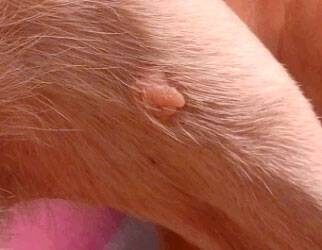
Vaginal Protrusions in Dogs and Cats
When dogs or cats develop masses protruding from their vaginas, it can be alarming for an owner. Fortunately, they are not common and are rarely seen in spayed pets.
Is it hyperplasia, swelling, prolapse, or a mass?
Many of these can look similar – a pink, fleshy mass in the cat or dog’s vaginal area. Although vaginal hyperplasia, swelling, and prolapse are similar and often related, they are not the same. Keeping it simple, hyperplasia means that there’s more tissue than there should be due to more cells being present.
Technically, this is different from swelling, which occurs for reasons other than an increase in the number of cells, such as an increase in the amount of fluid in between cells. A prolapse is where the vagina is everted (turned inside out) of the body. Imagine something from the inside of the body pushing the vagina outward, like turning a sock inside out. The tissue will often swell when combined with a prolapse.
Vaginal hyperplasia is more uniform than a mass. Think of vaginal hyperplasia as the difference between a thin cotton sock and a thick wool one. The “walls” of the wool sock (the vagina) are thicker than that of the cotton sock. If the sock couldn’t stretch outward for some reason, perhaps tight shoes, it would lessen the amount of room inside the sock for your foot, and the sock would feel tight.
Now, if that hyperplasic vagina should prolapse, a round, tongue- or doughnut-shaped mass may be easily seen; it’s often more in the neighborhood of “you can’t miss it” because the dog is paying so much attention to that area. The prolapse generally starts out smooth and shiny but eventually dries out a bit, after which cracks called fissures develop. It’s basically caused by an overreaction to estrogen and tends to occur just before she goes into heat (proestrus) or while she’s in heat (estrus).
Generally speaking, it only happens in dogs and cats who have not been spayed because spayed dogs and cats do not have enough estrogen to cause it. That said, if a spayed pet is exposed to estrogen from outside of her body, like what can happen to a dog that licks estrogen cream off her owner’s arm, there is the possibility of a prolapse developing then as well. Occasionally, difficult labor and delivery may lead to a prolapse, such as if the vagina everts outward as part of the pressure and forces involved in giving birth. Due to a slight increase in estrogen prior to the date of labor and delivery, it occasionally will happen then, too.
During delivery of a litter, if you can see any type of abnormal vaginal protrusion, it is a medical emergency.
Vaginal hyperplasia can interfere with sex while breeding; before the hyperplastic vagina prolapses outside the body, a reluctance to breed or difficulty urinating may be the only signs.
Occasionally, the discomfort continues throughout pregnancy or just recurs when the puppies are born.
Breeds predisposed to vaginal hyperplasia and prolapse include the boxer, English bulldog, mastiff, German shepherd dog, Saint Bernard, Labrador retriever, Chesapeake Bay retriever, Airedale terrier, English springer spaniel, American pit bull terrier, and Weimaraner. Because it tends to run in some family lines, it’s best not to breed dogs if they have had a prolapse even though the genetics and heritability of the condition are not fully worked out yet.
A mass is a collection of cells. Sometimes, masses will form inside a dog’s (or, rarely, in a cat’s) vagina and grow into a larger mass that eventually pushes itself outside the vagina. Vaginal masses, whether benign or malignant, are not common in cats and dogs, particularly if spayed. Continuing with our sock analogy, imagine a burr inside your sock, where the burr is like a vaginal mass. The burr protrudes into the middle of the sock where your foot sits. You’re wearing your sock as usual. If you stick your finger inside your sock, you can feel the burr.
If it’s a really big burr or you turn your sock inside out, you will see it when protruding from the sock. Masses can be pedunculated or sessile. If pedunculated (on a stalk) they are nearly always vaginal polyps, which occur more commonly in intact vs spayed bitches but can occur in both. Usually, the bitch is older. Polyps can be single or occur in groups. If the mass is sessile it is more likely a leiomyoma or leiomyosarcoma, which are differentiated by an incisional biopsy. Occasionally, other neoplasms can occur in the vaginal vault.
Growths called Transmissible Venereal Tumors may sometimes cause vaginal protrusions in dogs.
Treatment for Vaginal Prolapses
With vaginal prolapses, unless the prolapse is extreme, it will generally resolve on its own as the dog’s heat cycle moves along or after the dog is spayed. In minor cases, the dog only needs cleaning and an ointment to keep the tissue moist, so it doesn’t dry out.
If minimal tissue damage has occurred, your veterinarian can push it back in with a gloved hand. It is first cleaned appropriately, and swelling is reduced by applying hypertonic dextrose or sugar. Sutures can then be put in to keep it in place.
If the tissue is dead (necrotic), it has to be removed surgically. Spaying her will prevent another occurrence and can be done at the same time as removing the dead tissue.
Dogs with difficulty delivering litters due to the protrusion will likely need a C-section.
Sometimes supportive therapy involving an E-collar to prevent self-trauma, a diaper with a lubricated pad, and hormone treatment can be given to make ovulation occur faster. However, dogs don’t generally have a good response to hormones, and it’s ineffective if given after ovulation, so it’s not usually helpful.
After treatment, the intact dog should be monitored in case of a relapse.
The only prevention is spaying.
Treatment for Vaginal Masses
Treatment for any kind of mass in the vagina will depend on many factors including the type of mass it is (such as a benign mass or cancer), the exact location, the extent of it, and whether or not it has metastasized to any other location. If your pet has a vaginal mass, talk with your veterinarian to discuss treatment options.
In summary, if you notice a pink mass protruding from your cat’s or dog’s vagina, notify your vet. If your cat or dog seems to be in discomfort or having trouble urinating, contact your veterinarian or a veterinary emergency clinic right away.


