Arthritis, also known as osteoarthritis or degenerative joint disease (DJD), is a degenerative, progressive, and irreversible condition of the joints. It is characterized by the progressive loss of joint cartilage, bony spurs/growths, and the thickening and scarring of connective tissue around the joint, usually as a result of injury.
Approximately 25 percent of dogs are diagnosed with arthritis in their life time, and as many as 60 percent of dogs have radiographic evidence of it.
Degenerative joint disorders are probably as common in cats as in dogs but are less likely to be associated with obvious clinical signs, such as lameness. In one study, 90 percent of cats over 12 years of age had radiographic signs of osteoarthritis.
Arthritis is classified as primary or secondary. Primary arthritis is associated with aging, due to years of wear and tear on the joints. Secondary arthritis is the result of an external event or force (e.g., trauma, poor joint alignment, etc.) that once damaged the joint cartilage.
Arthritis can affect any age, sex, and breed of dog and cat. Most predispositions to it relate to underlying causes, such as elbow dysplasia, hip dysplasia, osteochondrosis, and so forth.
Diagnosis
Diagnosis is based on what is found in the physical examination, X-rays and other diagnostic imaging, joint taps, cytology, force plate gain analysis, therapeutic drug trials, and other tests.
Physical Examination Findings in Dogs
Lameness is the most common sign. It may happen once in a while (episodic) progressive (gets worse over time), or be persistent. Stiffness is common after periods of rest. Stiffness and lameness may decrease when the dog warms up a bit with some activity. Lameness often gets worse after periods of overexertion. Pain, swelling, and decreased range of motion may be seen. Thickened joints, excess fluid in the joint space, and muscle weakening are likely to occur.
Physical Examination Findings in Cats
As opposed to the visible lameness seen commonly in dogs, many cats simply become less active, may hide, or develop behavioral changes, such as irritability, decreased grooming, or difficulty getting into position in the litterbox. Cats also may have joint swelling/thickening, too much fluid in the joint space, and decreased range of motion. There may or may not be pain when the cat’s affected joint is moved by you or your veterinarian.
Diagnostic Imaging
Radiographs (X-rays) and CT scans may show the excess fluid in the joints; the bony spurs; signs of an underlying disorder, such as elbow dysplasia, osteochondritis dissecans, hip dysplasia, or cruciate ligament rupture; and so forth.
Kinetic Tests
Force plate gait analysis in dogs can help find where the lameness is within the joint. It can also show the severity of lameness, both before and after therapy. A force plate is mounted to the floor, and the dog walks over it. Measurements are taken to see which areas use the least force (more affected) and vice versa. The gait analysis hasn’t been used as often in cats.
Therapeutic Trial
In some cases, a therapeutic trial of nonsteroidal anti-inflammatory drugs (NSAIDs) may help confirm the diagnosis if the lameness is less noticeable after taking them.
Treatment, Management, and Prevention
It is not possible to cure arthritis.
The goals are to alleviate your pet’s discomfort, to minimize further degenerative changes to the joint, and to restore the joint’s functionality. Multiple types of treatment are usually necessary to relieve pain, stiffness, and discomfort.
Managing your pet’s weight is important. Excess weight increases stress on the joints and muscles. If your pet is obese, your veterinarian will want your pet to lose weight. Daily, low-impact activities, such as walking and swimming, will not only help your pet with losing some pounds but can also improve joint mobility, muscle mass, and exercise tolerance.
Joint supplements known as chondroprotective agents will help support the cartilage and will have some anti-inflammatory effects. These agents will slow the breakdown of cartilage and/or provide the building blocks that can help build it. Some agents also increase joint fluid secretion and thus decrease inflammation.
The main components of chondroprotective agents are polysulfated glycosaminoglycan (PSGAG), glucosamine, and chondroitin sulfate. Oftentimes there is no improvement using chondroprotective therapies. The failure may be due to too little cartilage left in the affected joint as there’s bone on bone; unresponsive joint inflammation; variable bioavailability of the drug between different products; and the lack of analgesia.
Although the injectable PSGAGs are more expensive, they provide a faster and longer-lasting response than the oral forms. If there is no response after 4-6 weeks, your veterinarian may discontinue use of PSGAGs.
Diets containing omega-3 fatty acids may help reduce inflammation. Some studies have found that dietary supplementation with fish oil omega-3 fatty acids can improve the clinical signs of arthritis in dogs, and may allow the NSAID dose to be reduced.
NSAIDS are an important component of arthritis therapy in dogs. Side effects that you should be aware of include stomach upset, elevated liver enzymes, and potential worsening of chronic kidney disease. Few NSAIDs are licensed for use in cats; your veterinarian will advise you about what NSAID options are available for your cat.
Analgesics such as tramadol, gabapentin, and amantadine may provide pain relief in dogs.
Alternative therapies such as acupuncture, stem cell therapy, platelet-rich or conditioned plasma, physical therapy, rehabilitation therapy (e.g. radial shock wave therapy, pulsed signal therapy), green-lipped mussel supplements, vitamin E, and zoledronic acid may be beneficial in some canine patients. Studies to support their use are still being done.
Surgery
Your veterinarian may consider surgical options if your pet’s response to medical treatment is low. In addition, your veterinarian may suggest surgery for certain underlying causes of arthritis, such as cranial cruciate ligament rupture, elbow dysplasia, osteochondritis dissecans, joint incongruity, intra-articular fractures, and joint instability. Reconstructive procedures can eliminate joint instability and correct the anatomic defects. If your pet has severe hip dysplasia, your veterinarian may suggest a total hip replacement and femoral head/neck ostectomy. If the arthritis is in the wrist or ankle (then joint fusion may be considered; this surgery is usually well tolerated and can result in reasonable functionality.
Monitoring and Prognosis
Your veterinarian may need to do periodic physical examinations every 1-4 months to monitor your pet’s response to therapy and the progression of the disease. In addition, if your pet is on an NSAID, blood tests including complete blood counts and biochemistry profiles, should be done every few months to ensure there are no side effects impacting the liver or kidneys.
With therapy and careful monitoring, arthritis can be managed in many dogs and cats, resulting in a good quality of life that you and your pet will appreciate.
The terms chronic kidney disease, chronic renal failure, renal insufficiency, chronic kidney failure, and kidney insufficiency all mean the same thing. For simplicity’s sake in terms of this article, the term kidney failure will be used to reflect all these terms or conditions.
The kidney is an amazing organ. It is designed to do a lot more than just make urine and get rid of toxins. It also manages hydration, blood pressure, red blood cell production, pH balance in the blood, calcium, and phosphorus balance, and more. Kidneys are made up of millions of filtration units called “nephrons” and we are born with vastly more nephrons than we need to keep healthy. Over time and age, however, nephrons get damaged through the wear and tear of living and they die off. We only need enough nephrons to fill up about 1/3 of one kidney to manage normal kidney function and we start with two full kidneys packed with working nephrons at first but there may come a time when we just don’t have enough anymore.
When we don’t have enough working kidney tissue to maintain normal function, we are said to have “renal insufficiency,” with “renal” being the medical term for the kidney. Kidneys with insufficient function need help to get their work done and a long-term diseased kidney, regardless of its function or lack thereof, has “chronic kidney disease.” Chronic kidney disease patients are divided up by what stage of insufficiency they are contending with and what symptoms they have.
Treatment is focused on preventing progression of the insufficient function in earlier stages and on maintaining good life quality in later stages.
In the past, we used the term “chronic kidney failure” instead of “renal insufficiency” but this wording sounded far too dramatic and evoked images of ongoing sickness, expensive hospitalization, and doom when management could be as simple as diet change depending on the stage at which the disease is discovered. Early detection is paramount so let us review what the kidneys do and where they might need some help.
What our Kidneys Do/What Insufficient Kidneys Cannot Do
First, it is important to know what normal kidneys do. Kidney function goes far beyond simply making urine. Kidneys are made of millions of processing units called nephrons. These tiny little units are responsible for separating the chemicals you want to keep in your body from the chemicals you need to dispose of. The chemicals to remove are dissolved in water and make up the fluid we all know as urine. So here is what your kidneys do for you and for your pet, what they become unable to do in renal insufficiency, and some of the parameters your veterinarian will want to track.
Water Conservation
Hydration of the body depends not only on water consumed but on water removed. In times of dehydration, the kidney must respond by conserving water. This means that all the materials that the body needs to get rid of still need to be removed, but the kidney needs to do so using the smallest amount of water possible.
Similarly, if you drink too much water, the kidney needs to efficiently remove it to prevent dilution of the bloodstream. A pet with insufficient kidney function will not be able to make concentrated urine and will need to drink extra water to process the body’s waste chemicals. For this reason, excessive water consumption is an important early warning sign and should always be investigated.
When we analyze a urine sample, one of the most important parameters is the specific gravity. It is a measure of how concentrated a urine sample is. Water has a specific gravity of 1.000. A dilute urine sample has a specific gravity of less than 1.020 (often less than 1.010). A concentrated urine sample would have a specific gravity over 1.030 or 1.040. A failing kidney, by definition, cannot make concentrated urine and the patient must drink excessively to get enough water to get rid of the day’s toxic load.
Toxin Removal
The kidneys remove our metabolic wastes for us. If there is inadequate circulation going through the kidneys or if there are not enough functioning nephrons to handle the waste load, toxins will build up. When the toxins build up and exceed the normal range, a condition called azotemia occurs. If the toxins build up to a level where the patient actually feels sick, they have a condition called uremia. If we can keep our azotemic patients below the uremia level, they will feel pretty normal and have good life quality.
The most important marker of uremia is called creatinine. Creatinine is a by-product of muscle breakdown and is always in small amounts in the bloodstream. The kidney removes it continuously unless there is a kidney function problem. A newer parameter called SDMA (symmetrical dimethylarginine) becomes abnormal much earlier than creatinine does and is becoming more commonly tracked. We are able to stage a patient’s kidney disease based on creatinine blood level and SDMA level (see the staging section below). Another marker is the BUN, which stands for blood urea nitrogen. This parameter is similar to creatinine but is influenced by dietary protein as well as kidney function. These three markers are central to determining the severity of a kidney problem.
Calcium/Phosphorus Balance
The balance between calcium and phosphorus in the blood is really important. Too much of one or the other will lead to crystals forming in body tissues and bones to the extent that they may actually become rubbery. The kidney plays an important role in this balance and when kidney function is lost, phosphorus levels begin to rise. Therapy for insufficient kidney function requires monitoring phosphorus levels and the use of diet and medications to keep phosphorus levels in a reasonable range.
Sodium/Potassium Balance
The kidney plays a major role in controlling electrolyte balance as well. In particular, conserving potassium is an important aspect. Insufficient kidneys lose their ability to conserve potassium and potassium levels begin to drop, leading to weakness. Potassium supplements are commonly needed in the treatment of kidney failure.
Blood Pressure Regulation
Blood pressure sensors in the kidney help regulate blood pressure in the body. When these are damaged, hypertension (high blood pressure) can result and can damage the kidney even more. Blood pressure is commonly measured in kidney failure patients.
Protein Conservation
There are a lot of important proteins circulating in the bloodstream and it is crucial that they are not lost in the urine. The nephron possesses a filtration system that keeps the protein in while removing harmful wastes. If this filtration system is damaged (glomerular disease) then a much more severe form of kidney failure results. Screening for this damage is an important aspect of staging kidney failure and a test called a urine protein: creatinine ratio is often included in the testing profile to assess this condition.
Red Blood Cell Production
The kidney produces a hormone called erythropoietin. This hormone tells the bone marrow to make more red blood cells. In the absence of this hormone, non-responsive anemia occurs and can get so bad that a transfusion is necessary. Erythropoietin can be given by injection to treat this problem but there are some potential pitfalls in doing this as will be discussed in a later article. Hematocrit and packed cell volume (PCV) are measures of red blood cell volume commonly monitored in kidney patients.
pH Balance
Metabolic processes require a narrow pH range for efficiency. The kidney also regulates this balance and if it cannot, intervention, usually in the form of fluid therapy, is necessary.
That’s a lot of tasks for a pair of kidneys to handle! With chronic kidney disease/renal insufficiency, chances are at least one of these tasks is not moving along smoothly. There may or may not be any symptoms when the disease is discovered but the goal is to slow the progression to the point where symptoms develop. If there already are symptoms, the goal becomes controlling the symptoms and maintaining good life quality.
Once kidney disease has been discovered, the next step is staging and evaluating any other aspects of the above function list that may not have been covered. Typically animals need special food and possibly medications depending on symptoms. Very advanced cases will likely need hospitalization, intravenous fluids, and possibly assisted feeding to get to where they can resume a more normal life at home. As mentioned, early screening is especially important as it is much easier to slow the progression of disease than it is to reverse it.
STAGING KIDNEY DISEASE
Kidney disease is either discovered via wellness screening tests of apparently healthy animals or during the course of the evaluation of patient illness. As mentioned, what we do for the patient will depend on the stage of the disease. The International Renal Interest Society (IRIS) has determined a staging system based on the creatinine level and SDMA level found on a blood test (see Toxin Removal section above). Patients in Stage 1 or 2 rarely have any symptoms related to their disease and those in Stage 3 or 4 usually have symptoms to control.
The chart below stages dogs and cats according to their Creatinine and SDMA levels. Once the patient has been staged then further testing for “sub-staging” begins.
Table 1: Creatinine Values for Each IRIS Stage listed in US, International, and SDMA Units
Units
Stage I(pre-failure)
Stage II(mild failure)
Stage III(moderate failure)
Stage IV(severe failure)
Dog
mg/dl μmol/l SDMA μg/dl
<1.4 <125 <18 mcg/dl
<1.4-2.8 <125-250 <18-35 mcg/dl
<2.9-5.0 <251-440 <36-54 mcg/dl
>5.0 >440 >54 mcg/dl
Cat
mg/dl μmol/l SDMA μg/dl
<1.6 <140 <18 mcg/dl
<1.6-2.8 <140-250 <18-25 mcg/dl
<2.9-5.0 <251-440 <26-38 mcg/dl
>5.0 >440 >38 mcg/dl
Sub-staging of kidney disease involves screening for urine protein loss and measuring blood pressure. A urine protein: creatinine ratio is performed on a urine sample and the protein amount is classified as either non-proteinuric, borderline proteinuric, or proteinuric. Blood pressure is checked and the patient is classified as normotensive (normal), borderline hypertensive, hypertensive, or severely hypertensive. The ultimate classification for the patient will reflect all of these things (“Stage 3, non-proteinuria, normotensive” would be an example). Further testing and monitoring are determined based on what the actual parameters are. For more details on these substages, see the articles on glomerular disease, and hypertension.
Stage I is mostly for pets that are known to have kidney disease based on abnormal ultrasound or their slightly elevated SDMA. We check their blood pressure and urinary protein levels, and screen for urinary tract infections. If abnormalities are found, they are usually checked quarterly for any sign of progression.
Stage 2 is where diet change to a renal diet is typically recommended with the idea being that the earlier this is started, the more likely the pet will accept it. The same type of screening is performed as with Stage I, only now we need to start watching the phosphorus levels so they don’t rise above 4.6 mg/dl. Also, potassium levels may begin to drop especially in cats. Both of these factors are helped by using a renal supportive diet but if that proves inadequate, medications may become needed.
In Stage 3, the patient is likely to be showing some symptoms such as reduced appetite or occasional nausea. In addition to the tests of Stages I and 2, it is in Stage 3 that we start paying attention to red blood cell numbers and blood pH. This is a good time to consider the use of calcitriol (active vitamin D supplementation) if one is going to do so.
In Stage 4, the patient is almost certainly showing symptoms and it becomes important to be sure the patient is eating enough calories to maintain a healthy weight. Similar concerns exist as for the first three stages but a phosphorus level of 4.6 mg/dl is probably unrealistic at this point and a better goal is 6.0 mg/dl. Fluid support at home is a good idea. This may mean adding water to the food, giving fluids under the skin (subcutaneous fluids), or even a stint of hospitalization and IV fluids to drive the numbers down.
Many patients are not diagnosed with kidney disease until they are far past a creatinine of 5.0 mg/dl. It is not unusual for sick patients to come to the vet and have creatinine values of 10 mg/dl or higher. At this point, more aggressive treatment will be needed to stabilize the patient and the goal is going to be improving the dysfunction to a level where the patient has acceptable life quality and comfort at home. Transitioning to a new lifestyle will take some time. (For example, appetite and comfort must be restored but changing to a renal support diet may be something for further down the treatment path when more stability has been achieved).
A Note on Pyelonephritis
Pyelonephritis is another name for kidney inflammation and usually means kidney infection. Patients get kidney infections when a bladder infection goes unnoticed or incompletely treated long enough and the bacteria go up the ureters into the kidney where they set up shop and cause damage and pain. This form of kidney failure has the most potential to reverse or partially reverse, so it is important to culture the urine for it early. Urinalysis is frequently unable to detect infection in this situation as the patient drinks so much water that the visible markers of infection are diluted out and cannot be found. The infection also will produce a significant amount of urinary protein so the patient will be sub-staged incorrectly if an infection is not ruled out.
A Special Note on Ureteral Stones
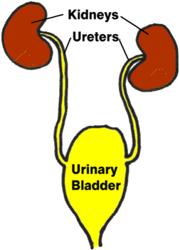
The ureter is the tube that connects the kidney to the bladder and urine should constantly be flowing through it. Often in age (plus with calcium/phosphorus imbalance), mineralization will occur in the kidney. This may or may not be a problem depending on how big and how plentiful the mineral deposits are but sometimes a mineral deposit will break off and get lodged in a ureter. This can obstruct the ureter completely and the backlog can destroy the kidney. Or it can partially obstruct the ureter, causing obstruction and then the relief of obstruction as it rolls down the ureter and (hopefully) into the bladder where it will not cause trouble. A spike in creatinine can be a clue that this has happened and if one acts quickly intervention may be possible. For this reason, some kind of imaging of the kidneys is recommended early in the course of kidney disease and periodically after.
The bottom line is that there is a wide range of treatments when a patient has insufficient kidney function and a lot of monitoring is needed to keep on top of all the aspects. Each patient will have a different stage and a different constellation of issues to contend with. Some will need only a diet change, and some will need ongoing fluid therapy at home or hospitalization. The prognosis depends on all the factors involved. Your veterinarian will let you know what your particular pet needs and will be watching many different lab tests to keep control of your pet’s condition.
In Summary:
From a practical standpoint, the terms chronic kidney disease, chronic renal failure, renal insufficiency, chronic kidney failure, and kidney insufficiency all mean the same thing.
The kidney’s job is more than just to make urine and get rid of toxins. Additionally, the kidney manages hydration, blood pressure, red blood cell production, pH balance in the blood, calcium and phosphorus balance, and more.
Over time, kidney cells can become damaged. When there is not enough working kidney tissue to maintain normal function, it is called “renal failure”. A long-term diseased kidney, regardless of its function or lack thereof, has “chronic kidney disease.”
Hydration of the body depends not only on water consumed but on water removed. In times of dehydration, to keep this balance, the kidney will remove the toxins using the smallest amount of water needed.
If over-hydrated, the kidney must remove excess water to prevent dilution of the bloodstream.
A failing kidney cannot make concentrated urine, and the patient must drink excessively to get enough water to get rid of the day’s toxic load. Excess water consumption by your pet could be an early warning sign of kidney disease.
If there is inadequate circulation going through the kidneys or if there are not enough functioning kidney cells to handle the waste load, toxins will build up.
If concerned about possible kidney disease and toxin levels, your veterinarian will order a urinalysis and blood work for your pet. Markers in the urine and blood will show the presence and severity of a kidney problem.
Levels of kidney disease are labeled by “stages,” i.e., I (pre-failure), II (mild failure), III (moderate failure), IV. (severe failure). How far the disease has progressed will determine treatment.
Treatments can be combinations of fluid therapy, diet changes, medications, and possibly vitamin supplementation. On-going examinations and laboratory tests will be a part of the treatment as your pet is monitored for changes to their condition.
Prognosis depends on the stage of the disease and factors involved.
Congestive heart failure (CHF) is the clinical syndrome of fluid retention due to severe heart disease. In dogs and cats, the fluid is retained in specific parts of the body, depending on the side of the heart that is diseased.
If the left side of the heart is diseased (e.g. mitral valve disease, most dilated cardiomyopathies, hypertrophic cardiomyopathy, and most common congenital cardiac defects), fluid is mostly retained in the lungs or within the pleural cavity (the chest space around the lungs).
If the right side of the heart is diseased, fluid is retained mostly in the belly or within the pleural cavity.
Why Does CHF Occur?
CHF occurs because the pressure in the veins and capillaries draining into the diseased side of the heart increases to the point that fluid leaks out of the veins and capillaries. The capillaries and veins that drain into the left side of the heart are those coming from the lungs, so fluid leaks into the lungs. The capillaries and veins that drain into the right side of the heart are those coming from the body, so fluid leaks into the belly (ascites) or creates swelling in the legs, the skin under the belly, etc.; swelling of the legs or the skin is uncommon in dogs and cats with right-sided CHF, but much more common in humans and horses.
It can be useful to think of this like a garden soaker hose. If water is flowing through the hose under low pressure, only a little bit of water leaks through the soaker hose. However, if we increase the pressure inside the hose, water starts to emerge from the soaker hose at a much greater rate and floods your garden. The same happens with the capillaries – under low pressure, only a little fluid leaks out of them (and is resorbed). But if the pressure is too high, a large volume of fluid leaks out into the surrounding tissue, and overwhelms the resorption mechanisms.
What Causes CHF?
CHF requires severe heart disease that increases the resting (diastolic) cardiac pressure. For example, diseases that cause leakage of valves (mitral or tricuspid valve disease) can result in CHF if the leak is big enough. Cardiomyopathies also cause the resting cardiac pressure to increase and can result in CHF. Pericardial diseases can prevent the heart from relaxing and stretching properly, which can result in right-sided CHF. Heartworm disease can cause right-sided CHF. Many congenital defects that shunt blood can result in CHF (e.g. patent ductus arteriosus, ventricular or atrial septal defects).
Importantly, congenital diseases that obstruct outflow from the heart, such as pulmonic stenosis or aortic stenosis, do not normally cause CHF.
What Are the Clinical Signs of CHF?
This depends on the side of the heart that is diseased. Let’s start with left-sided CHF, which causes pulmonary edema and, sometimes, pleural effusion. As fluid starts to deposit fluid into animals’ lungs, the amount of oxygen in the blood decreases because it can’t be absorbed from the lungs. Human patients describe this as shortness of breath, initially during exertion, such as climbing stairs, and eventually even when stationary. However, animals cannot tell us if they are short of breath. Sometimes, owners will notice a decrease in exercise ability, such as not going as far on walks and getting winded on walks more quickly. However, many things can cause exercise intolerance. Therefore, we tend to look for an increase in breathing (respiratory) rate, especially when sleeping.
Coughing has been described as a feature of CHF in dogs, but there is some doubt as to whether CHF really causes coughing or not. Again, many dogs and cats cough for reasons other than heart disease. As the fluid continues to build up, dogs and cats will also show difficulty in breathing as well as a faster respiratory rate. This is especially true of cats, who can hide their disease until it becomes really advanced. Consequently, many cats see veterinarians with labored, open-mouth breathing that is almost gulping for air, which appears to have started all of a sudden. In dogs, the clinical signs can be more gradual and subtle.
With right-sided CHF, the most common presentation is a swelling of the abdomen (ascites), making the dog appear pregnant. The discomfort from a belly full of fluid results in difficulty getting comfortable or breathing comfortably when lying down. These dogs will even resort to sleeping in a sitting position at times! Appetite often decreases slightly because of the abdominal pressure. If there is a buildup of fluid in the chest cavity, the animal might show difficulty in breathing.
How Do Veterinarians Diagnose CHF?
The diagnosis of CHF relies on pairing the clinical signs of increased respiratory rate and difficulty in breathing with the severe heart disease that is responsible for these clinical signs. Many times, a murmur can be heard for the first time. The pet’s heart rate will be elevated, their respiratory rate will be elevated. Some cats come in with a low body temperature because they are somewhat shocky from inadequate oxygen.
If veterinarians suspect CHF, they will generally take chest X-rays to see if there is evidence of (A) severe heart disease that appears as an enlarged heart; and (B) opacity (areas through which light does not pass) in the lungs consistent with pulmonary edema, or fluid in the chest cavity consistent with pleural effusion. If they are unsure or require additional information, they might perform or recommend a cardiac ultrasound. Those are often performed by specialists, typically veterinary cardiologists or radiologists, who have the necessary equipment.
How Do we Treat CHF?
Treatment is directed at both the underlying heart disease and the accumulation of fluid. If possible, the cause should be corrected. For example, closing a congenital shunt, such as a patent ductus arteriosus, will immediately correct the problem permanently. Repairing a leaking mitral valve will also correct the problem almost immediately, although this procedure is currently very expensive and performed by a limited number of surgeons.
The mainstay of medical treatment of left-sided CHF is the use of diuretics called loop diuretics, which include furosemide or torsemide. Diuretics reduce blood volume and consequently reduce the pressure in the veins, forcing the fluid out into the lungs or the abdomen.
Other drugs that are commonly used to treat CHF include angiotensin-converting enzyme inhibitors, pimobendan, thiazide diuretics, and spironolactone. These are much less effective than loop diuretics and should never be given as the only drugs for managing CHF. Indeed, if a dog or cat does not require a diuretic, they most likely do not have CHF.
The fluid buildup in right-sided CHF often requires repeated manual removal, using large catheters to make the patient feel better. In many dogs, this can be done as frequently as every week or two. Ideally, when coupled with medical treatment, the frequency of belly taps can be reduced somewhat.
How Do we Monitor CHF?
The most important thing to monitor with left-sided CHF is respiratory rate. Provided the respiratory rate when sleeping is in the normal range, we can be reasonably confident that we have good control of the CHF. When the sleeping respiratory rate starts to increase, adjustments in treatment might be necessary; a veterinarian will evaluate the patient to determine exactly what to do in such instances. (See monitoring videos below.)
Many veterinarians will monitor bloodwork to make sure that the medications being given are not causing problems with kidneys or elsewhere.
If a pet appears to destabilize after a period of control, additional x-rays might help better evaluate the situation.
What is the Prognosis?
This depends somewhat on the underlying disease. With the more common diseases, such as mitral valve disease in dogs or hypertrophic cardiomyopathy in cats, once CHF is diagnosed and treatment instituted, survival is generally less than two years. With mitral valve disease, approximately 50 percent of dogs will succumb to their disease within 8 to 10 months, and only 20 percent live for 18 to 24 months. With hypertrophic cardiomyopathy survival data are less clear, but some older studies suggest that 50 percent of cats will succumb to the disease within 7 to 10 months. As is always the case, a few individuals will live longer than expected.