Tag: Dogs and Cats

6029135

Causes of Blindness in Dogs and Cats

Owners may notice their pet is disoriented, bumping into objects and struggling to find food and toys. These all may be signs of blindness.  There are many potential causes of blindness in dogs and cats. However, before discussing what leads to vision loss, it helps to understand how the eye functions.

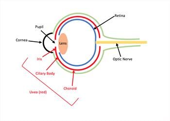
The eye acts like a camera that takes pictures and sends them to the brain for interpretation. Light reflecting off an object enters the eye through the cornea, which is like the eye’s windshield and helps focus light onto the retina. It then passes through a central black hole called the pupil. 

The iris, which is the colored part of the eye, surrounds the pupil and regulates how much light passes through it. Behind the iris and pupil is the lens, a transparent structure that also focuses light onto the retina. The retina converts the light into nerve impulses, which travel to the brain through the optic nerve. The brain interprets these impulses into an image. Abnormalities in these and other structures of the eye may lead to blindness.

Some of the more common causes of blindness in dogs and cats include the following.

  • Uveitis: Uveitis is a painful condition in which the uvea becomes inflamed. The uvea is a blood vessel-rich tissue consisting of the iris, the ciliary body (which produces fluid inside the eye) and the choroid (which nourishes the retina). Causes of uveitis include infections such as feline infectious peritonitis (FIP) and tick-borne ehrlichiosis, tumors, immune-mediated conditions, eye trauma, toxins and eye irritants.
  • Cataracts: A cataract is a cloudiness in the lens. The lens is supposed to be transparent to allow light to pass through, but cataracts impair this function. Cataracts may affect only a small part of the lens initially, but progress to affect more of the lens over time. The most common cause of cataracts in cats is uveitis, while genetics and diabetes are the two most common causes in dogs. Other causes include eye trauma, toxins, nutritional deficiencies, radiation, electric shock and age-related degeneration.
  • Glaucoma: Glaucoma is increased pressure in the eye. In a healthy eye, the volume of fluid that goes in and out of the eye is balanced to maintain a normal eye pressure. In glaucoma, a problem draining that fluid causes the eye pressure to rise, a condition that can quickly and painfully cause loss of sight. Primary glaucoma results from genetics while secondary glaucoma results from conditions such as uveitis, eye tumors and anterior lens luxation (see section on lens luxation). 
  • Retinal detachment: The retina is a 10-layered structure that converts light signals into nerve impulses that are sent to the brain for interpretation into an image. When specific layers of the retina separate or detach from one another, the retina can no longer carry out this function properly, resulting in impaired vision. Causes of retinal detachment include genetics, trauma, tumors, infections, immune-mediated conditions, uveitis, eye surgery and high blood pressure. 
  • Progressive retinal atrophy (PRA): PRA is an inherited disease that occurs in dogs and more rarely in cats. In pets with PRA, the retina degenerates over time, leading to blindness. In most cases, the pet is initially blind only in low-light conditions, but will eventually become blind in all conditions. Cataracts may also accompany PRA in some dogs.
  • Sudden acquired retinal degeneration syndrome (SARDS): This syndrome is a disease in dogs in which the retina rapidly and irreversibly deteriorates, leading to blindness within days to months. The cause of SARDS is unknown.
  • Optic neuritis: Optic neuritis is inflammation of the optic nerve, which carries impulses from the eye to the brain to be interpreted as images. Dogs and cats affected by this condition generally end up with partial or complete blindness. Causes of optic neuritis include such infections as distemper in dogs and cryptococcosis, a systemic fungal infection, in cats. Inflammation, immune-mediated conditions, toxins such as rodenticides, eye trauma and tumors are other potential causes.
  • Lens luxation: The lens is normally held in position by fine ligaments. If these ligaments fail, then the lens shifts – or luxates – from its normal position. A forward shift can block circulation of the eye’s fluid and quickly lead to painful glaucoma. A backward shift is not as immediately harmful, but secondary glaucoma and retinal detachment can occur. Terrier breeds are predisposed to primary, inherited luxations. Lens luxation may also occur due to cataracts, glaucoma, trauma, tumors, and uveitis.
  • Corneal diseases such as keratoconjunctivitis sicca, pigmentary keratitis, and pannus will completely scar the cornea if untreated.

If you notice any signs of blindness or changes in your pet’s eyes, take your pet to the veterinarian. Oftentimes, older dogs and cats develop an age-related cloudiness in their eyes that is usually harmless to vision. However, it can look similar to a cataract, so it is still worth getting checked by a veterinarian to make certain it’s harmless.
7902272

Cannabidiol Use in Dogs and Cats

What is Cannabidiol?

Plants make many different chemicals called phytochemicals, many of which have positive medical benefits. Cannabis sativa, commonly known as marijuana, produces over 500 other phytochemicals. The most recognized is tetrahydrocannabinol, also known as THC.

Drugs that affect the mind are called psychoactive, and THC is one of these. This is one reason marijuana is used recreationally by humans. It is also why marijuana is a Schedule I controlled substance in the United States and many other countries. THC is not the only phytochemical made by the marijuana plant.  Of the over 500 phytochemicals that can be made, at least 100 are other phytocannabinoids.

Cannabidiol, or CBD, is another commonly recognized cannabinoid produced by Cannabis sativa. However, unlike THC, CBD is not psychoactive. The United States Congress recently passed a law allowing industrial hemp to be grown. Hemp is a strain of Cannabis sativa that still has CBD but has very little THC. Cannabidiol derived from hemp is not a controlled substance.

Cannabis is a Schedule I substance in the US, which means it has a high chance of abuse (due to the THC, which is the psychoactive substance in marijuana) and has no currently accepted medical use. Nevertheless, research is showing that THC may help many diseases.

Some synthetic versions of THC for humans are approved as drugs and are federally legal, but none are currently approved for pets. However, CBD is approved in humans as a drug, so your veterinarian may be able to prescribe it if considered necessary for treating your pet.

How Cannabinoids Work

One of the many systems working to keep the body healthy is the endocannabinoid system in the brain. This system is made of naturally produced chemical compounds produced by neurons (cells that make up nerves) that are then sent on to direct many other cells.

The endocannabinoid system includes receptors located on different body cells, especially the nervous system. Phytocannabinoids may work at receptors like endocannabinoids.

Scientists are increasingly finding more diseases can be treated by targeting the endocannabinoid system.

The endocannabinoid system includes receptors located on different body cells, especially the nervous system. Phytocannabinoids may work at receptors like endocannabinoids. A common endocannabinoid, anandamide (AEA), is called the “bliss” endocannabinoid because it helps relieve stress.

THC interacts with cannabinoid receptors like AEA does, causing the same positive psychoactive effects in most people. CBD also interacts with the endocannabinoid system, but not at the same sites that THC works, which is why it does not affect the brain the same way THC does. 

Both THC and CBD can potentially interact with other receptors in the body. Because CBD may have many of the medical benefits of marijuana but does not have psychoactive effects, CBD is emerging as the cannabinoid most used medicinally in animals.

CBD as a Potential Treatment for Some Diseases

Because cannabinoids are mainly present in the brain, diseases involving the nervous system have been the focus of study for positive benefits. Some studies in humans have not found a clinical effect of CBD, but that does not mean that CBD does not work.

CBD appears to help other drugs control pain in humans, especially if it is chronic. Studies have shown that pain associated with osteoarthritis in dogs may be better controlled if CBD is added to drugs such as nonsteroidal anti-inflammatories (e.g., carprofen, robenicoxib, grapiprant, etc.).

Another potential indication for CBD is epilepsy, or seizures, that have not responded sufficiently to other antiseizure medications.

The use of CBD in treating diseases involving the nervous system, such as anxiety and pruritis (itchy skin), needs further research but may be promising. Studies involving cancer are also underway. CBD for treating various cat diseases has not yet been studied. 

CBD as a Supplement  

Animals must receive CBD orally. It is commonly given to pets as a treat, in oil, or as a capsule supplement. In the last decade, many supplements containing CBD have been marketed for use to treat a wide variety of diseases in animals. While CBD may prove to help treat some of these diseases, using these supplements to treat a pet can be a problem for several reasons.

It is important to understand that supplements are not like drugs approved by the Food and Drug Administration (FDA). Approved drugs have undergone a rigorous assessment to ensure that the drug is safe and effective for the disease being treated. The label guarantees the product contains the correct medication in listed amounts, strength, and quality. Any additional ingredients are clearly listed.

Supplements, including those containing CBD, are not held to the FDA standard. There is no guarantee that what is listed on the label is correct.

CBD products can be inconsistently labeled. Hemp oil is not the same as hemp extract, and the amount of CBD in the product can vary.

Labels can list the amount of CBD in ounces, by ml, in the total container, per dose, or per serving. This can make it difficult to ensure you give your pet the correct amount of CBD needed.

Another issue with CBD supplements is that they may contain other compounds added by the manufacturer. The compound might be a drug or, worse, a synthetic cannabinoid. Added synthetic cannabinoids are also Schedule I products and are illegal but hard to identify. This includes metabolites of THC. They are much more likely to be toxic compared to phytocannabinoids.

Yet another concern is how the CBD is “delivered” to your pet. Some manufacturers are trying to increase CBD absorption by changing what it is in. However, CBD may be absorbed differently if mixed in hemp oil, sesame seed oil, or other fats or added to a cookie or biscuit.

A final consideration is to be aware of the cost of CBD in these supplements, which can vary greatly. Higher cost does not equate to a better product.

One of the ways to avoid most of these concerns is to ask your veterinarian to prescribe a CBD that has been approved by the FDA for human use. Because it is approved, your veterinarian is legally allowed to prescribe it.

Is Cannabidiol Safe for Your Pet?

CBD appears very safe in dogs and is not likely to cause behavior changes. Studies have shown that CBD in oil, even at very high doses, does not cause any more side effects than oil alone in dogs or cats.

However, longer-term studies need to be done because there is some suggestion that the liver might be affected. This may be more likely if CBD is combined with other drugs.

Another safety consideration is whether CBD might change how other drugs work in the body. Some drugs inhibit the metabolism (or processing) of other medications, making it possible for other medications to become toxic.

In most cases, studies show that the effects of CBD are seen around two weeks after beginning treatment at the dose recommended by your veterinarian. However, the body often develops a tolerance for drugs used in treating the nervous system.

If tolerance develops, a higher dose may be necessary to control the clinical signs.

You must discuss with your veterinarian any other drug or supplement your pet may be receiving and make sure you know the potential benefits, dosing suggestions, and any cautions to look for when giving CBD to your pet.

4128352-1

Atrial Fibrillation in Dogs and Cats

What Makes a Normal Heart Beat?

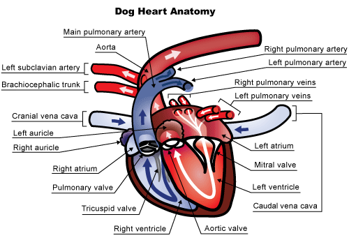
The heart is a large muscle with four chambers. There are two top chambers (left and right) called the atria and two bottom chambers (left and right) called the ventricles. These chambers fill with blood from the body and then contract to circulate or deliver poorly oxygenated blood to the lungs (right side) or well-oxygenated blood to the rest of the body’s tissues (left side).

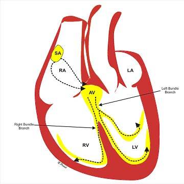
The heart muscles contract after they are stimulated by an electrical impulse. A small area of special tissue in the right atrium called the sinoatrial (SA) node starts an electrical impulse (it’s like the heart’s spark plug) that travels out through the atrial muscle, stimulating that muscle, and, simultaneously down special electrical tracts (AV node, Bundle Branches, Purkinje Fibers – the heart’s “wiring”) to the ventricles and ultimately out into the ventricular muscle (see Figure 2 below).

The electrical impulses cause the heart to contract in a coordinated fashion: the atria contract first, because they get stimulated first. As they contract, they push blood into the ventricles. Then, as the electrical signal reaches the ventricles, they also contract and push blood to either the lungs or the rest of the body. This coordinated electrical activity can be recorded on an electrocardiogram (ECG or EKG). There are several components to the normal ECG, called waves or complexes. They are labeled P, QRS and T. The P waves represent the atrial stimulation, while the QRS waves represent the ventricular stimulation. The T waves represent the re-setting of the ventricular muscles, so that they can be stimulated by the next impulse. The short pause between the P waves and the QRS waves is the time that the impulse is traveling along the wiring system from the atria to the ventricles.

Atrial fibrillation (sometimes called “A fib”) is an arrhythmia, an irregularity of the heart’s rhythm.  Instead of the SA node (the spark plug) starting and coordinating the electrical signal, many areas within the atria send out electrical impulses in a rapid, disorganized manner.  This whirlwind of electrical impulses or wavelets spreads over the atrial tissue causing the atrial muscle to quiver or fibrillate, instead of contracting in an organized, structured manner (Figure 3). Only a fraction of the electrical impulses are able to jump onto the “wiring system” and get across the AV node and down into the ventricles to make the ventricles contract.  The pattern with which all these abnormal atrial impulses are conducted through the AV node is irregular making the ventricles beat irregularly. 

What Causes Atrial Fibrillation?

In humans, atrial fibrillation can occur for no apparent reason (often in athletes or older people). This is because the size of our hearts is large enough to allow such abnormal rhythms to develop (veterinarians also see this type of atrial fibrillation in horses, which have large hearts). However, most pets have much smaller hearts than humans, so for atrial fibrillation to occur in your pet, the atrial chambers must usually be bigger than normal. This enlargement of the atrial chambers occurs with many types of heart disease, such as degenerative valve disease of the mitral valve or cardiomyopathy, dilated cardiomyopathy and hypertrophic cardiomyopathy. Generally, the smaller your pet, the more significant the heart disease must be to cause atrial fibrillation.  Thus, atrial fibrillation is much less common in cats than in dogs. And it’s less common in small breed dogs than large breed dogs.

Occasionally, giant and large-breed dogs develop atrial fibrillation without any other heart disease, similar to what we see in humans and horses (let’s face it, some Great Danes are almost as big as horses!). This is termed “lone” or primary atrial fibrillation.  In some cases, this is “lone” atrial fibrillation turns out to be the first indication of dilated cardiomyopathy; in other cases, the arrhythmia exists without underlying heart disease.

How Is Atrial Fibrillation Diagnosed?

Often, the first idea that a pet has atrial fibrillation is when your veterinarian detects an erratic heart rhythm while listening to the heart with a stethoscope. Many veterinarians describe the sound as bongo drums or shoes in a dryer. Your pet’s pulse quality may also be irregular. However, to confirm the diagnosis an electrocardiogram (ECG) needs to be performed (because other arrhythmias can sound like atrial fibrillation. There are specific ECG criteria that must be met prior to making the diagnosis.

At times, a pet with severe heart disease may suddenly become weaker, or have a relapse of heart failure upon development of atrial fibrillation, prompting you to visit your veterinarian. Again, the veterinarian will auscultate the abnormal rhythm and diagnose atrial fibrillation via an ECG examination.

In giant breeds with lone atrial fibrillation, the arrhythmia may be detected on routine examination – often the owners are unaware that any problem exists. Some dogs may develop mild exercise intolerance when they develop atrial fibrillation.

What Happens When a Pet Develops Atrial Fibrillation?

People with lone atrial fibrillation typically require treatment to re-establish a normal heart rhythm because they usually have exercise intolerance or shortness of breath, symptoms of palpitations, and are at an increased risk of having a stroke. If a normal rhythm cannot be re-established, people are often placed on “blood thinners” to reduce the risk of stroke. However, dogs with lone atrial fibrillation rarely show exercise intolerance or shortness of breath and do not suffer from strokes, so treatment is rarely required. Their heart rate remains normal in most cases.

However, in a pet with serious heart disease, atrial fibrillation causes a worsening of clinical signs, and can further damage the heart. Cardiac output, or the amount of blood pumped out of the heart to the rest of the body, is reduced and often congestive heart failure develops or recurs. These pets usually have very fast heart rate (more than 200 beats per min) which, if not controlled, can lead to additional heart muscle damage and dysfunction.

How Is Atrial Fibrillation Treated?

Treatment in people is aimed at re-establishing and maintaining a normal rhythm, because this greatly reduces the risk of strokes. This is usually achieved by delivering a “shock” to the heart (termed electrocardioversion), or by using antiarrhythmic drugs. Recent studies in people suggest that re-establishing a normal rhythm is not essential and medical therapy might suffice if clinical signs are tolerable or absent.

In pets, cardioversion is generally not feasible. Several studies have examined electrocardioversion or chemical cardioversion in dogs with lone atrial fibrillation – none has shown consistent success. Newer studies are being conducted that may ultimately provide a technique that allows a normal rhythm to be established in these pets.

Pets with severe heart disease are generally not candidates for cardioversion. In these pets, a normal rhythm can rarely, if ever, be re-established. Instead, the veterinarian attempts to reduce the high heart rate with antiarrhythmic drugs. By reducing the heart rate (which is often more than 200 beats per minute), the veterinarian allows the heart to pump more efficiently, and prevents some of the damage that the high heart rate can produce. Additionally, the clinical signs associated with the development of atrial fibrillation (that is, congestive heart failure) resolve when the rate is controlled. The drugs that decrease the heart rate need to be given indefinitely to maintain rate control and will generally be added to drugs already being administered. Studies have shown that dogs in which the heart rate is reduced to 120 beats per minute (average over a day) do better than dogs in which the heart rate is not as well controlled. In other words, the closer to “normal” your veterinarian can bring the heart rate, the better the outcome.

People with atrial fibrillation are usually treated with anti-coagulants, such as warfarin, or other similar drugs (commonly, but incorrectly, termed blood thinners), to reduce the incidence of strokes, which can be devastating or fatal. However, dogs with atrial fibrillation do not appear to have increased risk of strokes and are therefore not treated with anti-coagulants. Cats with atrial fibrillation usually have severe heart disease, and are often treated with anti-coagulants whether they have atrial fibrillation or not.

What is the prognosis for a pet with atrial fibrillation?

Lone atrial fibrillation, like we see in large or giant-breed dogs, is well tolerated, even if we cannot convert it back to a normal sinus rhythm. In dogs with underlying severe heart disease, 50 percent of those with higher heart rates (more than 125 beats per minute) in one study had died from their heart disease within four months. On the other hand, 50 percent of those with lower heart rates (less than 125 beats per minute) lived to at least three years. Whether or not these numbers reflect what will happen with all dogs is hard to say, but we can say that bringing the heart rate close to normal improves survival.

4128968

Ataxia in Dogs and Cats

When your pet is stumbling and staggering around, almost as though drunk, the condition is called ataxia. It is an inability to make normal, coordinated, voluntary movements but is not caused by muscle weakness, involuntary spasms, or too little strength to move. Generally speaking, ataxia is a symptom caused by some type of central nervous system problem in which the brain cannot correctly tell the body what to do.

Ataxia affects a pet’s ability to coordinate their head, legs, and body. Perhaps the pet cannot place their foot appropriately while walking, resulting in the pet knuckling their feet and dragging their toes on the ground. Since the pet does not realize where their feet are, they cannot just flip the feet over to walk normally. Dragging knuckles on the ground can damage the tops of their feet. Some pets with ataxia do not knuckle their toes, but their foot placement is exaggerated.

Ataxia needs to be differentiated from lameness or weakness due to musculoskeletal disease or generalized illness.

Three types of ataxia can be seen in dogs and cats, depending on whether the inner ear, brain, and/or spine is involved: 

  • Cerebellar (brain)
  • Proprioceptive (brain or spine)
  • Vestibular (inner ear)

Cerebellar

The cerebellum is the part of the brain that coordinates voluntary movement, allowing your pet to reach toward the water bowl or you to move your arm appropriately when you want to pick something up. Common causes of cerebellar ataxia in pets are congenital defects and inflammatory diseases. Other causes include degenerative neurological diseases, brain tumors, and strokes.  

The pet’s head and torso sway and stagger (see video) and their feet step up high and wide in an exaggerated walk as though they are about to go up steps even though they’re not. They may have head tremors.

Proprioceptive

Proprioception is an awareness of where your body is positioned in space, strengthening coordination. It tells us how much force to use when pushing or pulling. Proprioception is how your pet knows without looking if their tail or foot is up or down, and you know if you are standing up straight or slanted. Proprioceptive ataxia is caused by a problem in the cerebral cortex of the brain or the spine. Common causes include brain tumors, infectious diseases of the brain or spinal cord, traumatic brain injuries, injured disks, and strokes. 

The pet’s unsteady body is seen swaying, knuckling, feet crossing over, and with a wide-based stance. You would see an inability to get the limbs into their normal positions. This form can affect one or more limbs and the torso.

Vestibular

Vestibular ataxia is caused by a problem in the vestibular system in the inner ear or within the balance center in the brain, both of which contribute to balance. The vestibular system helps your pet stay upright when walking forward and lets you walk or run on uneven ground without falling. The vestibular systems signal to the brain to let the eyes and extremities know what they are supposed to do. Here, the ataxia is characterized by leaning and falling to one side or, less commonly, both sides. It affects the head, trunk and limbs. It’s often the easiest form of ataxia to recognize and often is worse if the animal is removed from contact with the ground.

Pets with vestibular ataxia may have a head tilt, walk like they are going in a circle rather than straight ahead, and may have odd back-and-forth eye movements called nystagmus. Drooling, leaning, rolling, and falling are common. Nausea or vomiting may occur as a result of dizziness.

Diagnosis

Your veterinarian will start by taking a detailed history of what has been happening to identify likely causes and will do a neurological examination (neuro exam) to determine where the problem is located. A neuro exam is quite simple and has two objectives: (1) Confirm that the pet has a neurological condition, and (2) see where the problem is located. A neuro exam may sound like you need to see a specialist, but you do not as the exam does not require special knowledge or expensive equipment.  Your veterinarian will watch your pet walk, move, check reflexes, see what happens when they lift your pet’s leg, and other tests to see what your pet’s physical reaction is. The neuro exam checks on:

  • Mentation (level and quality of consciousness)
  • Posture of head, limbs, and body
  • Gait (how the pet walks)
  • Postural reactions (incoordination)
  • Spinal reflexes (normal, diminished, or increased)
  • Cranial nerves (e.g., vision, eye movements, facial sensation, etc.)
  • Palpation of the spine to look for swelling or atrophy
  • Nociception (ability to sense pain)

A neuro exam will suggest the location of the problem but cannot by itself determine which disease your pet has. The history you provide is the most important information in determining which diseases are likely causes of abnormal signs.

Treatment

Treatment depends on what is happening with your pet. Your veterinarian needs to figure out where the problem is located, and which diseases are most likely, to determine which tests and treatments to recommend. Hospitalization with some medication to help the pet’s specific signs, such as vomiting, and some IV fluids may be of most use, especially early in the game. Good nursing both at the clinic and at home can be helpful. 

The prognosis for pets with ataxia depends on where the lesions are located and what the specific disease is that is causing the abnormal signs. Some diseases are benign, and recovery only requires time and supportive care. Other diseases are serious and may be difficult to resolve.