Tag: Dogs and Cats

testimonials-goldberg-pet

Glomerulonephritis in Dogs and Cats

(Urinary protein loss due to kidney inflammation)

When a patient, human or non-human, is said to have kidney failure, renal insufficiency, or even chronic renal failure, what most people are talking about is a toxin build up when the kidney cannot adequately remove the body’s harmful wastes. This toxic state is called uremia and is associated with nausea, appetite loss, weight loss, listlessness, and other unpleasant issues.  It is also not the kind of kidney disease we are going discuss here. 

Glomerular disease is a completely different kind of kidney disease and may not involve any toxin build up at all. Glomerular disease is one of protein loss. 

What is a Glomerulus Anyway?

Consider for a moment what an important resource protein is to your body. Your blood, for example, is full of necessary circulating proteins handling clotting, fluid balance, transporting other chemicals etc. Your body went to a lot of trouble to build those proteins and you can’t afford to waste them. If you were to lose them, your body would have to break down muscle in order to recreate them because that is how important they are.

On the other hand, your blood carries an assortment of metabolic wastes that you need to get rid of. You need to filter out these bad materials without losing what is valuable. The millions of glomeruli you have are in charge of keeping your blood proteins where they belong — in the blood — while allowing for smaller wastes and extra fluid to filter out and be made into urine. There are other valuables in your blood besides cells and protein, but different areas of your kidney handle those.

The illustration to the right shows the nephron, which is the functional unit of the kidney. There are millions of these making urine every moment of every day. Only about 30 percent of them must be working in order to maintain normal kidney function. The rest form a back up system so that we will have plenty of extra nephrons should some of them get plugged with debris, damaged by scarring or infection, or starved for oxygen during a traumatic event.

The glomerulus, which in a way looks like a little dandelion tuft, is where our interest lies today. Blood flows through an afferent arteriole into the glomerulus. Inside the glomerular tuft, the blood vessel narrows into a complicated spiral of tiny capillaries, so small that the blood cells pass through single file. The capillaries are gripped by cells called podocytes. Like hands, the podocytes have tiny fingers (ironically called foot processes) that encase the capillaries. Fluid and small molecules can flow in between the fingers while cells and large molecules like proteins cannot pass through.

The cells and large molecules/proteins exit the glomerulus through an efferent arteriole and return to normal circulation. This first step in filtration is driven both by blood pressure as well as by the protein content of the blood.

Now imagine what would happen if there were holes punched in that filtration system so that protein can pass through the fingers. This is what happens in glomerular disease.

How does the Glomerulus get Leaky?

Sources of chronic inflammation are believed to be the ultimate cause of the problem. The chronic inflammatory state leads to the circulation of antigen: antibody complexes in the blood and these complexes stick in delicate glomerular membranes like flies in fly paper. Once stuck there, they call in other inflammatory cells and soon a hole is eaten into the membrane by the ensuing reaction. The holes in the filtration membranes are big enough for proteins to traverse.

There are many possible sources of chronic inflammation that could be generating antigen: antibody complexes. Chronic ear or skin infections could be the cause. Long-standing dental disease could do it. A latent, more internal infection might be the cause (such as heartworm, Lyme disease, prostate infection, or Ehrlichiosis). Even a tumor might generate enough of the immune system’s attention to lead to this sort of reaction.

If it is at all possible to identify and resolve the underlying cause of inflammation, this should be done as other therapy is unlikely to fully resolve the protein loss.

How is the Diagnosis Made?

There are several common scenarios that might lead to the diagnosis but they all boil down to one or both of two findings: excess urine protein found on a routine urinalysis and/or low albumin found on a blood test.

Let’s start with excess urine protein found on a routine urinalysis.

A urinalysis examines a urine sample for some of its chemical contents and properties. Protein content is one of the parameters that is checked and reported as a small, medium or large amount. On a urinalysis report this will be designated as “+,” “++,” or “+++.”

This seems like it would be easy enough to interpret but unfortunately there is more to the story. A small amount of protein in a well-concentrated sample may be  normal while the same amount of protein in a dilute sample would be highly significant. How dilute or concentrated the urine is depends on the patient’s water consumption, and we need a method to examine urine protein that is independent of the water consumption.

Further, we need to determine if any protein in the urine is truly coming from the kidneys; after all, a bladder infection or other bladder condition might generate urine protein. To help distinguish renal protein loss, the rest of the urinalysis will be helpful. When your veterinarian is confident that other issues with the urinary tract have been excluded, it is time for a urine protein:creatinine ratio (we will come back to this).

Low Blood Albumin Level found on a Blood Panel

Albumin is one of those proteins that the body really wants to conserve and here’s why. There are plenty of substances the body needs to circulate that simply are not water soluble. How do we circulate these things if they won’t dissolve in water? We bind them to a carrier protein that will circulate and carry them as if they were commuters on a city bus. The albumin molecule is that city bus, carrying important biochemicals around your body.

There’s more. Albumin also is important in keeping water in the bloodstream. This sounds odd but blood is basically a liquid and without enough water, it sludges and clots abnormally. Furthermore, if water is not held in the vasculature, it leaks into other body cavities such as the chest and abdomen, filling these cavities with liquid.

Your body prioritizes the maintenance of its albumin levels and will not allow them to drop. When the albumin levels are down, a serious protein loss is afoot. It could be intestinal or liver-related, but glomerular protein loss is going to be one of the first conditions to rule out. If there is no protein in the urine, the focus shifts to other organs but if there is protein in the urine, it must be quantified and that means there is a urine protein:creatinine ratio.

Interpretation of the Urine Protein: Creatinine ratio

The urine protein:creatinine ratio compares the amount of protein in the urine to the amount of creatinine, one of the metabolic wastes filtered by the kidneys. By using this ratio, it does not matter how dilute the urine is or how concentrated it is. The ratio allows for protein loss to be quantified and then we can tell how significant the protein loss actually is. If the urine protein: creatinine ratio is found to be abnormal, ideally it is repeated in 2 to 4 weeks to be sure that the protein loss is persistent, but this depends on how high the ratio is and whether or not there is a known inflammatory condition that would be expected to damage the glomeruli.

Determining how serious a patient’s protein loss is depends on overall kidney function as well. In other words, a protein-losing kidney that is effectively removing the daily load of toxins and wastes is in less trouble than a protein-losing kidney that is failing.

The International Renal Interest Society (IRIS) considers a urine protein:creatinine ratio of greater than 0.5 for dogs or 0.4 for cats to be abnormal, and if it is persistent, then further diagnostics and treatment are recommended.

Depending on how your pet responds to the therapies above, a biopsy may be recommended. Biopsy is most commonly recommended for patients with UPC ratios more than 3.5 or with significant proteinuria combined with low albumin levels or high blood pressure.

The goal of treatment is to reduce the UPC ratio to below 0.5 or to reduce it by at least 50%. Higher reductions are sought for cats (see later).

Ratios greater than 3.5 are particularly concerning and require more aggressive treatment and more extensive diagnostics. These patients have an increased risk of abnormal blood clotting and generally have more extensive kidney damage.

The urine protein: creatinine ratio varies by up to 30% above or below baseline as a matter of course. A significant change in the ratio caused by disease progression (up) or response to therapy (down) must be greater than 30%.

If Intervention is Recommended what Does that Mean?

Adding omega 3 fatty acids to the regimen appears to improve the protein loss situation and supplementation is recommended. Most renal diets are already fortified with these anti-inflammatory fats but additional use is felt to be beneficial.

Low Protein, Low Sodium Diet

Most commercial renal diets would fit in this category. It seems paradoxical that a disease that causes body protein to be lost would be treated with a protein-restricted diet but, in fact, supplementing protein causes albumin to drop faster.

ACE Inhibitor

These medications have been shown to reduce renal protein loss. Typically enalapril is recommended for dogs and benazepril is recommended for cats. These medications inherently reduce blood flow to the kidneys so care must be taken in patients with elevated creatinine ratios to be sure the uremia does not worsen. Lower doses are used and monitoring becomes more important.

Omega 3 Fatty Acid Supplementation

Most commercial renal diets are fortified with omega 3 fatty acids. These anti-inflammatory fats have been shown to improve survival of dogs with renal disease. It is still unclear how helpful they are for cats but studies are ongoing.

Angiotensin II Receptor Blockers (ARBs)

Angiotensin II receptor blockers are becoming more popular in human medicine and their use is trickling down to manage canine glomerular disease. These medications work with ACE inhibitors to further help reduce urinary protein loss though they can also be used alone. Like the ACE inhibitors, they not only reduce urine protein loss but also lower blood pressure as well and seem to have some effect on reducing the clotting tendency. They are new to veterinary medicine and protocols are still being worked out. The two commonly used medications are losartan and telmisartan.

Spironolactone

Aldosterone is the hormone that acts on the kidney to retain sodium and water and get rid of potassium. Spironolactone is an antagonist of this hormone, which means it increases urine production, retains potassium and removes sodium. In humans, it has been found to reduce urine protein loss by 34 percent, which makes it an attractive medication for this situation especially in patients with nephrotic syndrome (see below). In dogs it might be used when ACE inhibitors or ARBs have not controlled the proteinuria. It is not a medication for cats.

The goal in managing urine protein loss is a 50% reduction in urine protein:creatinine ratio for dogs and a 90% reduction in urine protein:creatinine ratio for cats. A combination of the above medications is likely to be prescribed, and urine and blood test monitoring will be periodically (at least quarterly) recommended in hope of achieving and finally maintaining these results.

Nephrotic Syndrome

In severe cases, a complication called nephrotic syndrome can result due to the extreme urinary protein loss. Nephrotic syndrome is defined as the combination of: 1) significant protein loss in urine; 2) low serum albumin; 3) edema or other abnormal fluid accumulation; or 4) elevated blood cholesterol level. This is a severe complication of glomerular disease and suggests a poor prognosis, especially if creatinine levels are elevated in the blood. 

High blood pressure is a common complication of nephrotic syndrome. Patients also tend to form inappropriate blood clots (embolism) that can lodge in small blood vessels, causing loss of circulation to entire organs or sections of organs. Nephrotic syndrome is an advanced state of urinary protein loss and must be treated aggressively.

Biopsy the Kidney?

There are pros and cons to this relatively invasive test. The kidney receives 25 percent of the blood supply at any given time, which means it is highly vascularized and can bleed in an extreme way. Blood transfusion is needed for 10 percent of dogs and 17 percent of cats having this procedure, and a three percent mortality rate has been reported. So why take the chance on this procedure? The main reason is to obtain information on prognosis.

There are different types of glomerular disease and glomerular inflammation, all of which may have different associated expectations. There is a type of glomerular disease called amyloidosis that involves abnormal protein (called amyloid) infiltrating the kidneys and has a much more progressive and damaging course. Approximately 50 percent of glomerular disease patients have diseases that can benefit from immune-suppressive therapy but the only way to identify these patients is with a biopsy. 

Conclusion

When the kidney cannot retain blood proteins, the body loses its ability to carry out normal blood functions. In an attempt to replace these proteins, muscle is broken down and the patient becomes debilitated. Maintaining proper nutrition and using medication to reduce the protein loss are crucial to managing this form of kidney disease. It is important for the pet owner to keep up the monitoring schedule and to stay in contact with the veterinarian as to the pet’s progress and response to therapy. 

In Summary

  1. Glomerular disease is one broad type of kidney disease in which the primary problem is loss of renal proteinuria. Glomerulonephritis is one broad classification of kidney inflammation. It usually results in protein loss in urine. There are subtypes that can be determined by specialist pathologists based on renal biopsy.
  2. It is not typical Glomerular disease differs somewhat from “classic” kidney failure renal disease, although it glomerular disease can lead to chronic kidney disease if undetected.
  3. In the kidney, there are a million nephrons that make urine every minute of the day and send that urine to the bladder and out of the body through a filtration system. A body only needs approximately 30% of those nephrons working correctly for normal kidney function, at least as far as veterinarians could detect with typical lab tests. The rest are a backup system called the functional reserve.
  4. Inflammatory cells punch small holes along the filtration route. Those holes are big enough for proteins to pass through.
  5. Sources of chronic inflammation are believed to cause the problem, possibly stemming from issues such as chronic ear or skin infections, dental disease, heartworm disease, vector-borne diseases like Lyme disease, or feline immunodeficiency.
  6. Diagnosis is suspected by excess urine protein found in urine, and/or low albumin found on a blood test. Definitive diagnosis of glomerulonephritis and its specific type requires a kidney biopsy.
  7. When the kidney cannot retain blood proteins, the body cannot carry out normal blood functions. In an attempt to replace these proteins, muscle is broken down and the patient becomes debilitated.
  8. The urine protein:creatinine ratio (UPC) found in a urine test compares the amount of protein in the urine to the amount of creatinine, a metabolic waste filtered by the kidneys. The ratio tells what the magnitude of the protein loss actually is. A persisting, reproducible urine protein:creatinine ratio of greater than 0.5 for dogs or 0.4 for cats is too high. Mild elevations can be due to causes other than glomerulonephritis.
  9. Determining how serious the protein loss also depends on overall kidney function. A protein-losing kidney that is still effectively removing the daily load of toxins and wastes is in less trouble than one that is failing. Once the urine protein:creatinine reaches a certain ratio, a biopsy may be recommended. Ratios greater than 3.5 (typical US units) are particularly concerning.
  10. The best treatment is when a specific cause is found, such as a systemic infectious disease, and that disease can be successfully treated. However, sometimes despite extensive searching with imaging and blood tests, no underlying cause can be found.
  11. One goal is to reduce the urine protein:creatinine ratio to below 0.5 or to reduce it by at least 50% in dogs and 90% in cats.
  12. Potential interventions expected to help specifically with proteinuria include: omega-3 fatty acid supplementation; a controlled protein, low sodium diet; and RAAS inhibition (with drugs called ARBs or ACEIs). Accompanying hypertension, when present, may require additional treatment.
  13. In severe cases, a complication called nephrotic syndrome can result due to extreme urinary protein loss. The prognosis for this complication is comparatively poor, although it does depend on whether the problem developed acutely or chronically.
  14. A kidney biopsy can be informative about a prognosis and suggest whether additional treatments are expected to help. Since immunosuppressive treatments can also do harm, biopsy is usually recommended to justify trying that type of medication. Biopsy can be expensive and requires pre-planning to make it safe. For sample evaluation to be truly valuable, the specimens are analyzed by a specialist veterinary nephropathologist.
  15. It is important for you to keep up the monitoring schedule and stay in contact with your veterinarian.

4126909

Urinary Incontinence in Dogs and Cats

When a house pet develops urinary incontinence, many owners fear the worst. Assumptions that incontinence signifies senility or irreparable age-related change may lead to delay in medical consultation, relegation of the pet to an outdoor life, or even euthanasia.

In reality, urinary incontinence is usually one of the easiest problems to solve, so it is crucial that veterinary assistance be sought before an owner’s patience is completely worn out and before any permanent decisions about the pet’s future become topical.

Causes of Incontinence

It is important to differentiate incontinence (involuntary urine leakage) from behavioral urinary issues (submissive urination), simple lack of housetraining, territorial marking of anxious cats or unneutered males, or the senile loss of house training from canine cognitive dysfunction. Animals may urinate in the house voluntarily, and this is different from incontinence. Watch your pet closely to be sure what you are seeing is really incontinence, and if it is, the good news is that most cases are easily resolved with simple, inexpensive medications.

There are several important causes of incontinence, and most of these are ruled in or out with a urinalysis and urine culture. The urinalysis reveals cell types and biochemical elements in the patient’s urine, while the culture isolates the bacteria growing in the urine. The bacterial species grown are identified and tested for their sensitivity towards different antibiotics, the end result being confirmation of the presence of infection and a list of appropriate antibiotics.

Most cases of incontinence are due to:

  1. Infection of the urinary tract (usually bladder infection)
  2. Excessive consumption of water
  3. Weak bladder sphincter (especially common in female dogs)
  4. Spinal cord disease.

Other conditions that may need to be explored could include bladder stones and even tumors in the urinary tract.

Bladder Infection

This is a common cause of urinary incontinence in female dogs of all ages and in geriatric cats. This condition is usually easily diagnosed by urine culture, though signs of infection, such as white blood cells or bacteria, are actually visible in the urinalysis. A urine culture will confirm the infection, identify the organism, and list several antibiotics that will be effective. An antibiotic is selected based on expense, potential for side effects, and convenience of usage. After a short course (generally somewhere between 1 and 3 weeks) of medication, ideally, a second urine culture or urinalysis is done to confirm that the infection has truly been cleared up.

If a bladder infection is the cause of incontinence, most patients show improvement in their incontinence and comfort after only a few doses of antibiotics (but it is still important to finish the entire course so as to avoid recurrence). 

Excessive Water Consumption

Some animals drink so much water that their bladders simply overflow too easily. While some owners have noticed that their pets seem to be drinking more than usual, our experience is that most owners are surprised when the urinalysis shows excessive water consumption.

Dilute urine is obvious on the urinalysis through a measurement called specific gravity that compares the amount of dissolved biochemicals in the urine to that of pure water (which has no dissolved biochemicals).

A urine specific gravity nearly the same as water, confirms excessive water consumption; blood tests may be indicated to go with the urine tests to determine the cause.

Causes of excessive water consumption include:

  1. Diabetes mellitus
  2. Cushing’s syndrome
  3. Hyperthyroidism (cats)
  4. Bladder infection (see above)
  5. Diabetes insipidus
  6. Kidney insufficiency

There are other causes as well but 90% are ruled in or out by a blood panel and urine culture.

Weak Bladder Sphincter

Aging, obesity, reduced sensitivity of neurologic receptors in the sphincter and possibly other factors all contribute to this condition which is especially common (up to one in five affected) in female dogs. Once other more serious conditions have been ruled out, the weak sphincter may be treated symptomatically with one of several medications.

Estrogens

It is not entirely clear how estrogens are helpful in this treatment. Originally, estrogens were given to post-menopausal women with urinary incontinence and the treatment was simply extrapolated to dogs. It is possible that estrogens are important in the maintenance of neuroreceptors in the bladder sphincter and without estrogens the receptors become unresponsive to the transmission of the storage message from higher neurologic centers. (In other words, the message from the brain to hold the urine does not get through to the bladder.)

In dogs, diethylstilbestrol (DES) has traditionally been the most common estrogen used, though it is now only available through compounding pharmacies. Other estrogens that have been used include estriol (Incurin®) and conjugated estrogens such as Premarin®.  Regardless of which product is used, the basic process is the same. A higher dose is utilized to begin therapy and, if it is effective, the dose is tapered to the lowest dose needed to maintain effect. For DES, dosing every couple of days is typical while with estriol most dogs end up dosed once daily.

In male dogs, testosterones seem to be more effective than estrogens, possibly through action on the prostate which sits at the neck of the bladder and incorporates the sphincter.

Alpha-Adrenergic Agonists

These medications act by enhancing release of the neurotransmitter chemicals that act on the receptors of bladder sphincter. Effectively, they turn up the volume dial on the “hold it” message from the high neurologic areas. The usual medication for canine use is phenylpropanolamine in chewable tablets, which is typically given two or three times daily.

Ephedrine and pseudoephedrine, common decongestants, are sometimes recommended alternatively. Side effects can include irritability, appetite suppression (phenylpropanolamine was the active ingredient in many human diet pills until recently), and blood pressure changes. Most dogs, male and female alike, tolerate phenylpropanolamine uneventfully. For especially resistant cases of incontinence, estrogens and alpha-adrenergic agonists can be used together.

Anticholinergics

Anticholinergic drugs are medications that work, not on the sphincter of the bladder, but on the rest of the bladder where urine is stored, relaxing the muscle fibers thus facilitating storage. An example of such drug would be imipramine, an anti-anxiety medication commonly used in humans. It has anticholinergic properties and can be used in combination with phenylpropanolamine in the treatment of animal incontinence. While phenylpropanolamine and DES are commonly used medications for this condition, imipramine is not but it does represent another option when a patient does not respond to the first two medications combined. Other anticholinergic drugs that might be used included oxybutynin or flavoxate.

Gonadotropin-Releasing Hormone (GnRH)

Several studies have been performed using analogs of this hormone (such as leuprolide) in incontinent dogs for which DES and phenylpropanolamine have failed.  More than half of the dogs tested regained complete continence while still more achieved improvement.

Surgical Therapy

Medication works for most patients with weak sphincters but when medication fails there are some surgical options to consider: colposuspension and cystourethropexy.

Colposuspension, for females only, is the most commonly performed procedure. Here, the vagina is tacked to the bottom of the belly wall entrapping and compressing the urethra. In one study of dogs that had failed on medication, there was complete resolution of incontinence in 53% with colposuspension. An additional 37% became less incontinent and 25% of those without full resolution gained complete continence when therapy with phenylpropanolamine was added. Another recent study reported that complete continence lasted for 1 year in only 14% of affected dogs, although many dogs improved.

Cystourethropexy is the modification of the above procedure that can be performed in either males or females. Since there is no vagina to use in the male, the ductus deferens are tacked down to compress the urethra. Fibers from the urethral muscles can also be tacked down (in either male or female patients). Complications include an increased frequency in the need to urinate (occurring in 2/3 of the patients in one study) and straining to urinate (in about 1/3 of patients).

Urethral lengthening works for patients for whom incontinence is caused by a short urethra. A short urethra causes a full bladder to be displaced into the pelvis and makes the urethra too short for either of the other two surgical procedures. This is a newer surgical procedure that so far has had good reports in the small number of dogs in which it has been studied. Further studies should be forthcoming.

Medications listed above are used in conjunction with surgery. Surgery alone improves approximately 50% of patients but often incontinence returns unless oral medication is restarted.

Collagen Injections: The Newest Therapy

In this procedure an endoscope is inserted in the urethra and several injections of collagen (the same kind used for cosmetic injections in humans) are deposited. In a study of 40 dogs who had failed to become continent on medication alone, 27 became continent for an average of 17 months. Furthermore, 10 dogs that experienced only partial improvement after the procedure became completely continent when oral medication was added, even though medication alone had been ineffective.

The procedure can alternatively be performed with medical grade collagen, with Teflon® (which does not last as well but is apparently considerably less costly) or with a reconstructive product called ACell®. Unfortunately, at this time there are few facilities that perform this procedure as special equipment (cystoscopy) is required.

Unusual Causes of Incontinence

The list of causes of incontinence presented above is by no means exhaustive.

While uncommon, other causes should not be entirely counted out. Some possible causes include:

  1. Ectopic ureter (instead of connecting to the urinary bladder, the ureter transporting urine from the kidney connects to the vagina or rectum so that there is no storage of urine. This condition is typically noted in a puppy that simply cannot be housebroken and leaks urine. The condition can be solved surgically.)
  2. Spinal damage, especially in the lower lumbar area. Spinal surgery may be indicated to decompress an area of nerve pressure that is interfering with the nerves of urination control.

Your veterinarian is in the best position to determine if it is worthwhile to pursue a rare disease or not. Do not hesitate to ask your veterinarian questions regarding your pet’s incontinence and the treatments or procedures described above.

5261039

Retained Baby Teeth in Dogs and Cats Need Surgical Extraction

Puppies normally have 28 deciduous (baby) teeth that erupt during the first six months of life, and adults have 42 permanent teeth. Kittens have 26 baby teeth, and adult cats have 30.

In order for the baby teeth to fall out, specific cells resorb (dissolve) the roots of the primary tooth. This allows the permanent teeth to erupt normally. We don’t understand what triggers the resorption, so we don’t know why some baby teeth don’t fall out the way they are supposed to. 

When the baby teeth don’t fall out to make way for the permanent teeth, they are called retained deciduous teeth or persistent teeth. They should be extracted soon after they are discovered because otherwise they will cause dental problems (overcrowding in the mouth, plaque buildup, malocclusion, etc.).

As dentists like to say, two teeth should never try to occupy the space intended to contain one tooth.

When both deciduous (persistent) and permanent teeth are trying to occupy the same spot, this double row of teeth overcrowds the mouth, and food gets trapped between the teeth. This trapped food causes periodontal disease. In addition, the double set of roots can prevent the tooth socket from developing normally and eventually erode support around the adult tooth.

These difficulties can be prevented by extracting the retained deciduous teeth as soon as they are seen. If the tooth is extracted early enough, the adult tooth usually will move to its correct position; if it isn’t, there is a greater chance that the adult tooth will be in the wrong position, which can cause damage to the tongue, palate, mandible, etc.

It is much easier to position the permanent tooth while it is erupting, not after it has erupted. Repositioning after eruption requires orthodontic care to keep your pet’s mouth healthy.

4128293

Otitis Media (Middle Ear Infection) in Dogs and Cats

The word otitis means ear infection, but that one word doesn’t indicate which part of the ear is infected. Otitis media is inflammation and/or infection of the middle ear, whereas otitis externa begins in the external ear canal. Some nerves are closely associated with the middle ear, so infection there can damage them, causing the neurologic signs often seen with this infection: a head tilt, lack of balance, nystagmus (back-and-forth eye movements). These symptoms are called vestibular signs. Middle ear infections can also cause paralysis of the facial nerve, leading to a slack-jawed appearance on that side of the face.

Otitis media most often develops after an existing otitis externa travels from the external ear canal through the tympanic membrane and into the middle ear. The infection in the external ear canal leads to inflammation and damage to the ear canal and tympanic membrane, allowing the infection to enter the middle ear. 

Otitis media has been found in 16% of dogs that have acute (rapid onset) otitis externa and in 52% of dogs that have chronic (long-term) otitis externa. In contrast, 63% of cats that had otitis media/interna did not have a previous history of ear infection. This is why keeping your pet’s ears clean and watching for infection are important to your pet’s health.

Other causes of otitis media can include infections in the nose and throat, trauma, foreign bodies, fungal infections, inflammatory polyps, cancer, etc. In addition, developmental abnormalities of the external ear canal and pharynx can lead to otitis media.

Otitis media does not affect any particular age or animal, and males and females are affected equally.

Dogs that have long, pendulous ears, such as beagles and basset hounds, are more likely to develop otitis externa, which can lead to otitis media.

The Cavalier King Charles Spaniel breed is known to have a condition called primary secretory otitis media (PSOM). Dogs with PSOM typically have mild to moderate pain in the head/neck, neurologic signs (e.g. ataxia, facial paralysis, head tilt), and itchy ears, but without otitis externa. Owners of this breed will want to keep this disease in mind. 

Diagnosis

Diagnosis includes physical examination, ear examination with an otoscope, bacterial/fungal culture, radiography, and possibly advanced imaging.

Physical Examination

During the physical examination, your veterinarian will be looking for some of these signs: head shaking, pawing at the ear, pain when opening the mouth, signs of otitis externa (ear odor, redness, etc.), head tilt, facial nerve signs, Horner syndrome, ataxia, etc. Not every affected pet will exhibit all of these signs. In addition, cats are more likely to have neurologic signs than dogs are.  

Ear Examination (Otoscopy)


In order to do a complete examination of the ear, a deep ear cleaning may be necessary. To keep your pet comfortable, your veterinarian may decide to sedate or anesthetize your pet for the procedure. Then the infection can be more easily seen with an otoscope, which can see the eardrum.

Culture and Sensitivity

Your veterinarian will usually take samples of the material in the ear and culture those samples to determine what medications will work best.

Radiographs

Your veterinarian may take radiographs, also called X-rays. Radiographs can help show changes (thickening, destruction) to the bony structures of the ear. However, even if the radiographs appear normal, that does not mean that your pet does not have otitis media.

Advanced Imaging

In some cases, your veterinarian may suggest using computed tomography (CT) and/or magnetic resonance imaging (MRI) to further evaluate the tympanic bulla. CT is usually better than MRI at detecting bony changes, while MRI is better at detecting soft tissue abnormalities.

Treatment

Cleaning both the external and middle ear canals is an important step in treating otitis media. Debris can inactivate some topical medications and prevent medications from contacting infected tissues. Anesthesia will be necessary in order to do the deep ear cleaning and ear flushing that will remove the debris.

Your veterinarian may need to infuse (fill) the ear with medication in order to deliver a high concentration of antimicrobial products or corticosteroids directly to the affected area.

Topical medications may need to be used to control an otitis externa.

Systemic therapy with antibiotics, antifungals, and corticosteroids may also be used, depending on the infection.

If severe, irreversible changes to the external ear canal are found, if there are masses in the inner ear, or if medical therapy has failed to resolve the otitis media, your veterinarian may advise surgery, such as an ear canal ablation. This surgery is not undertaken lightly, but it may be necessary to control the infection. An ear canal ablation involves removing the vertical and horizontal portions of the ear canal, along with the bones of the middle ear and eardrum.

Monitoring (Rechecks)


During treatment, your veterinarian will usually want to recheck your pet’s external ear canal and tympanic membrane every 10-14 days. (A ruptured tympanic membrane will generally begin to heal within 21-35 days.) Topical medications will be given until the external ear canal and the tympanic membrane are normal, and until cytology of the ear canal reveals no infection. Systemic therapy is typically given for at least 4-6 weeks.

Prognosis

In general, the prognosis for dogs is good if appropriate therapy is started in time. The prognosis is not as good if the infection is resistant to the medications; if the concurrent otitis externa is not managed adequately; if there is significant bone infection; or if there is no response to surgery. In addition, some neurologic signs (e.g., facial nerve problems, Horner syndrome) may be permanent.

Reminder

Keeping your pet’s ears clean is critical to preventing ear infections of any kind. However, It is not always possible to prevent every infection in some pets. 

4128250

Nebulizer Use for Dogs and Cats

Dogs, cats, and other pets who develop lung diseases such as pneumonia, asthma, kennel cough, bronchitis, and so on can benefit from a nebulizer delivering saline, albuterol, antibiotics, or other drugs. A nebulizer is a machine that aerosolizes medicine into a fine mist; it’s like spraying medicine. A face mask is held over the pet’s muzzle (most pets can learn to accept the face mask) so that medication is inhaled deep into the respiratory tract.
 
The esophagus lies above the trachea and is protected by the epiglottis. Pneumonia may result from infections caused by kennel cough, viruses and bacteria. Pets with megaesophagus are prone to developing aspiration pneumonia because they tend to regurgitate food and fluids into the back of the throat and then aspirate the material into the lungs.

A nebulizer can be purchased from human medical supply stores, online, or from eBay. There are several good nebulizer brands, such as Pulmo-Aide, Omron, Devilbiss, etc. Make sure that you obtain the compressor (the machine), tubing, and the medication cup. The tubing, T-piece (a t-shaped tube that attaches the machine to the pet and to an exit) and medicine cup are called a nebulizer kit.

A vaporizer is not the same as a nebulizer. A vaporizer is typically used for sinus or upper respiratory issues rather than lung issues. Vaporized droplets are too big to get as deeply into the lungs the way nebulized particles

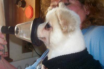
You can purchase an appropriately sized mask from a veterinarian, or make one yourself.

Using a Nebulizer

Follow these steps to use a nebulizer.

  1. Plug in the compressor.
  2. Insert one end of the tubing into the compressor and the other end into the cup.
  3. Place the correct dose of the medicine in the medicine cup.
  4. Start the compressor.
    a. Keep the cup up straight and hold the mask up to your pet’s face. You might want to practice placing the mask against the pet’s face before using the nebulizer
  1. b. Put a little canned dog/cat food, baby food, cream cheese or peanut butter inside of the mask and allow the pet to lick it. This can also distract a pet while the treatment is proceeding.
  2. After 5 to 10 minutes, you’ll see and hear that the medicine is running out.
  3. Shake the cup downward a few times to make sure all the medicine has come through.
  4. Take apart the plastic parts of the nebulizer and wash all the parts.

Cleaning the Nebulizer

Bacteria and viruses can live and reproduce in a nebulizer that is not routinely cleaned.

After each use, swish each piece in warm running water. At the end of the day, after you use it the last time, scrub each piece with warm soapy water. Rinse each piece under warm running water. Let each piece air-dry on a clean dish towel or paper towel. When the pieces are completely dry, put the nebulizer back together. If not using for a period of time, store it in a clean, dry bag.

Twice each week, or after the treatment is completed, mix a half cup of white vinegar with 1.5 cups of tap water. Soak each piece in this mixture for one hour. Rinse each piece under warm running water. Let each piece air-dry on a clean dish towel or a paper towel. When the pieces are completely dry, reassemble the nebulizer.

What do I do if I have Cleaned my Nebulizer and the Tubing is still Wet?

The easy way to dry the tubing is to attach it to the compressor. Then switch on the machine, and let it run for a minute or two. The blowing air will dry out the tubing. Use a paper towel or clean hand towel to dry the other parts.

What Medications can be Given with a Nebulizer?

A variety of drugs can be given using a nebulizer: bronchodilators like albuterol (open up the bronchioles allowing debris to more easily be removed from the lungs), steroids, water-soluble antibiotics, saline, N-acetylcysteine, etc. Albuterol opens up the bronchioles, making it easier to breath and allowing antibiotics to get deeper within the lungs. As albuterol can increase the heart rate, it should be used with caution in dogs with heart disease. Do not exceed three treatments per day unless advised by your veterinarian. The dose of albuterol may need to be adjusted based on the size of the dog. Following the albuterol, use the nebulizer with 2–3 cc of sterile saline. Some pets have side effects from albuterol although that is rare. Sometimes side effects are seen, such as nervousness, a rapid heart rate or excessive panting, and they will disappear within an hour or so. If this happens, report it to your veterinarian for a possible adjustment of dosage or change in frequency of treatments.

If your veterinarian has not previously written a prescription for albuterol, for which there is no veterinary version, here is an example of how to write it.

Albuterol 0.083% 2.5 cc vials × 25
sig: using a nebulizer, administer q8–24hr as directed

Albuterol comes in a box of 25 vials, each containing 2.5 cc.

  1. Your veterinarian will prescribe the dose of albuterol for your pet.
  2. Nebulize the computed dose of albuterol; if it is 1 cc or less, mix it with 1 cc of saline solution.
  3. Nebulize an additional 2–3 cc. of saline after the albuterol treatment is completed.

Warning: Albuterol should not be administered to pets by people who have heart or lung disorders. Ask your physician if it is safe for you to be exposed to the albuterol mist given off during nebulization.  

Saline decreases the thickness of mucus and helps moisturize the bronchioles, which helps the pet cough up debris blocking the lungs. Saline solution can be obtained from your veterinarian, or nebulizer saline can be obtained online.

Do not use saline solution intended for contact lenses.

Antibiotics such as gentocin or amikacin, which are intravenous antibiotics, can be mixed with saline. These drugs can be hard on the kidneys, so ideally it is best to monitor kidney function with blood and urine tests. These antibiotics are more kidney friendly when given with a nebulizer than when given intravenously.

Preventing Aspiration Pneumonia

Nebulizer treatments with either saline and/or albuterol can help prevent recurrence of aspiration pneumonia in pets with megaesophagus, pneumonia or bronchitis, and may be helpful in managing laryngeal paralysis by helping to clear aspirated debris from the lungs. Ask your veterinarian if your pet should have a preventive treatment daily, every other day or twice weekly.

4345752

Hemangioma in Dogs and Cats

Hemangiomas are a type of tumor of the blood vessels or the skin. They are benign, but the related hemangiosarcomas are a malignant cancer that also target the blood vessels. They come from the same type of cells and the only difference is that one is malignant.

Hemangiomas arise from a mutation in the cells and the cause is unknown. Research suggests that solar radiation through UV light may play a role when these tumors occur in the skin. Both dogs and cats can get hemangiomas. Depending on where the disease is and its progression, clinical signs can vary. There may be no clinical signs, dark purple blisters on the skin, or internal bleeding causing weakness and anorexia. Treatment and long-term outcome for animals vary depending on the type of tumor.

Who gets hemangiomas?

Both dogs and cats can get hemangioma.

Subcutaneous (under the skin) tumors tend to occur as a single mass. The masses may bleed and bruise easily, contain areas with ulcers and dead tissue, and be painful when touched. Approximately one-third of dogs with subcutaneous form have a history of tumor-associated illness that may include lack of appetite, lethargy, lameness, neurologic abnormalities, cough, voice change, and hemorrhages and/or bruises involving the mass.

Cats and dogs with the skin-related form typically have one or more red to purple skin bumps that are located in areas of sparsely haired, lightly pigmented skin. In dogs, these tumors most commonly occur on the chest and belly, sometimes because they like to sunbathe on their backs. In cats, lesions are most common on lightly colored pinnae (ear flaps) and other areas of the head. Lesions are usually small and nonpainful.

Dogs:

Breeds: American Pitbull Terrier, basset hound, Beagle, Boxer, Dalmatian, English Bulldog, English Pointer, Greyhound, Italian greyhound, Staffordshire terrier, Whippet . These breeds are mostly predisposed to the solar-induced form because they are light-skinned dogs with short hair, especially over the chest and belly – at least in those that like to sunbathe on their backs. However, any breed can get hemangiomas, especially the kind that are not related to the sun.

Sex: Both males and females are equally affected.

Age: Middle aged to older.

Cats:

Breeds: No breeds are predisposed.

Sex: Both males and female cats are equally affected.

Age: Middle aged to older.

Diagnosis

Definitive diagnosis usually requires surgical removal of all or part of a mass and its analysis at a laboratory. Blood tests, clinical signs, and predispositions (age, breed, hair coat color/type, sun exposure history) may suggest that your animal has hemangioma or hemangiosarcoma. Routine blood tests may show anemia. A fluid sample may show cancer cells, although many times these samples only show blood. Your veterinarian may suggest an ultrasound if they suspect hemangioma or hemangiosarcoma before surgery to look for more of an internal mass.

Treatment and Prognosis

The treatment options and long-term prognosis of your pet depends on the type of tumor they have. Once a benign hemangioma is removed surgically, your pet usually requires no additional treatment and is back to normal health. Dogs and cats with solar-induced hemangiomas may develop new hemangiomas (or other solar-induced tumors) at other sites of sun-damaged skin, potentially requiring additional surgeries to remove them.

4128327 (1)

Glaucoma in Dogs and Cats

Glaucoma is an eye disease where by the pressure within the eye becomes elevated. It can cause significant pain and lead to blindness. Dogs and cats with primary glaucoma are believed to have an anatomic or biochemical problem that affects how fluid drains from the eye, so the onset can be spontaneous. Secondary glaucoma results from another disease process within the eye. Glaucoma usually requires lifelong treatment unless the underlying cause can be reversed or treated successfully.

Inside the eye, a clear fluid called aqueous humor circulates in the front chamber between the cornea and the pupil. The aqueous humor leaves the eye through a small sieve-like structure between the base of the cornea and front of the iris. Normal pressure is a dynamic function of the balance between aqueous production and drainage. When the drainage sieve does not work properly or becomes clogged, or when the flow of aqueous through the pupil becomes obstructed, intraocular pressure can increase to an unsafe level. This is particularly true if aqueous production continues at a normal level. Glaucoma is never caused by over production of aqueous humor. It is always a drainage problem.

Glaucoma occurs far more frequently in dogs (0.675%) than in cats (0.197%). Primary, hereditary, breed-related glaucoma is most commonly seen in purebred dogs. Cats usually have secondary glaucoma that is associated with chronic inflammation of the iris (uveitis) or with intraocular tumors. Primary glaucoma in cats is very rare but Siamese and Burmese cats may be predisposed. Over forty different breeds of dogs are predisposed to glaucoma, with common ones being the cocker spaniel, beagle, basset, Akita, chow chow, Samoyed, Bouvier de Flandres, Shih Tzu and Chinese Shar Pei.

Glaucoma can be a very painful disease for pets – more so than for people – as the intraocular pressure can become rapidly elevated to levels much higher than typically occur in people. In people, the pain feels like a constant bad headache. Normal intraocular pressure in people is 12-22 mmHg. The normal pressure for dogs and cats is 10-25 mmHg. With glaucoma in cats and dogs, the pressure may go up to 30 mmHg and higher. Values above 50 mmHg rapidly cause blindness, are painful and may cause the eye to stretch and enlarge.

An affected eye may look normal to a pet owner when the glaucoma is mild. Early signs can include a bloodshot eye, cloudy cornea, dilated pupil, and squinting/holding the eye closed. Over time, the eye size can increase and it may bulge.

Unfortunately in cases of severe glaucoma, the eye is often permanently blind by the time of diagnosis. Pets can act normal with vision in just one eye, which is one reason glaucoma is often diagnosed late in the disease’s course.

Diagnosis and Treatment

Glaucoma can be detected by measurement of eye pressure using an instrument called a tonometer. Tonometry can be done by your veterinarian during the eye exam. Pressures are measured in both eyes and then compared.

If glaucoma is found, then numerous medications are available to lower the pressure. All glaucoma medications used in dogs and cats are medications that have been manufactured for human glaucoma. The treatment of glaucoma requires faithful, consistent application of the medications. These are not medications that can be skipped or forgotten without risk of the eye pressure rapidly deteriorating.

Some cases of primary glaucoma are also treated with surgery. A major emphasis in secondary glaucoma is to determine the underlying cause and to treat it, as well as the elevated pressure. If primary glaucoma is diagnosed in one eye, it is common for the other eye to be started on preventative medication because both eyes are prone to the disease.

Removal of the eye (enucleation) is sometimes recommended. Examples include a blind eye in which the glaucoma does not respond to medications; any eye containing a tumor; when the eye is blind and the owner cannot afford glaucoma medications; and when infection is the cause of the glaucoma and it does not respond well to appropriate therapy.

Prognosis

In most cases, glaucoma is not curable and once vision is lost it will not return. With primary glaucoma, vision is often lost over time, even with appropriate treatment. For secondary glaucoma, prognosis depends on the underlying disease and its response to treatment.

4128133

Gallstones in Dogs and Cats

The liver makes bile to help digest food and break down fats. The gallbladder is a sac-like organ connected to the liver. The gallbladder stores bile and releases it into the intestines through a bile duct. Sometimes, when things go awry, gallstones form inside the gallbladder. Gallstones are also called choleliths or cholelithiasis. For some pets they can be no trouble at all for the rest of their lives. For others, gallstones can be potentially fatal if the gallbladder bursts open.

Gallstones are often made up of parts of the bile, such as cholesterol, bilirubin, and calcium. Under the right conditions, these substances layer together inside the bile, essentially forming a hard blob that eventually results in a solid stone.

These stones form because either the gallbladder isn’t functioning properly or something is wrong with the bile. A common cause is when a tumor or especially thick bile blocks the bile’s movement into the intestines. Abnormal bile can also occur when the pet has high cholesterol, bilirubin, or triglycerides. Poor diets, such as those with too much fat or not enough taurine and protein, can also cause gallstones.

In some cases, gallstones don’t cause the pet any problems, and no symptoms will be noticed; however, gallstones can sometimes lead to cholangitis, a painful, inflammatory condition in the gallbladder. Gastrointestinal symptoms, such as vomiting, diarrhea or loose stool, poor appetite, and sometimes a yellow tinge to the skin and eyes (jaundice) may be seen.

Should gallstones block the bile duct, bile may overfill the gallbladder, causing it to eventually burst open (rupture) like a water balloon, which is an absolute emergency. The pain from a rupture is significant: it’s the kind of pain that would send you to the ER in the middle of the night in a blizzard. Pets in this kind of pain may bite, so be careful during transport to the emergency hospital. Dogs and cats may also become weak or faint quite suddenly.

Gallbladder rupture is a life-threatening emergency

A ruptured gallbladder is a worst-case scenario; just be aware that it is a possibility.

To diagnose stones before they ever get to the point of rupturing the gallbladder, a veterinarian will perform a thorough physical examination. The tummy will be checked for signs of pain around the gallbladder. Bloodwork will likely be run to check for liver changes; increased bilirubin, calcium, or cholesterol in the blood; and to look for evidence of inflammation, dehydration, or infection. Next, x-rays will be taken to see if any stones are visible. Unfortunately, only about 50% of gallstones can be seen on x-rays. An ultrasound of the belly may also be needed. This allows the veterinarian to check the size of the gallbladder; look for signs of bile inside the belly; confirm gallstones; make sure the liver looks healthy; and see if the bile duct is blocked. These tests can also help the veterinarian determine what caused the stones, so that steps can be taken to prevent more from forming.

In all cases, treatment will depend on what signs your pet is experiencing. If the pet is not showing any symptoms, there likely isn’t any treatment needed other than watching and waiting. The veterinarian will probably want to monitor the pet periodically with bloodwork, x-rays, and/or ultrasound to ensure the stones don’t change or cause discomfort.

If your pet is showing symptoms of duct obstruction or pain, surgery to either remove the gallstones or the entire gallbladder may be necessary. If the gallbladder bursts, emergency surgery to remove it and uncontained bile from inside the belly is needed immediately. (Remember, a ruptured gallbladder is a life-threatening emergency.) Hospitalization and intensive care are often required after gallbladder surgery.

Sometimes medications that protect the liver, such as ursodiol or SAMe, will be prescribed to help keep the liver healthy. Antibiotics may also be given if an infection is suspected or if the gallbladder ruptured. Treating the cause of the stones with diet change or dietary supplementation may also be recommended.

For pets that don’t show symptoms, the long-term risk for issues is low. Bile duct obstruction may occur, but it’s not set in stone (no pun intended). Your veterinarian may recommend a special diet or discuss the importance of cutting out any table scraps or people food. A well-balanced diet is important for pets with gallstones.

For pets that need surgery, the chance for improvement is good, but gallbladder surgery can be risky. If bile leaks into the belly, pets can become extremely ill and may die. For pets that need emergency surgery because their gallbladder ruptured, chances of improvement are much lower than those whose gallbladder didn’t burst. Pets that survive the surgery may need to be hospitalized for a long time before they can safely be sent home to continue to improve.

If you suspect your pet has gallstones, call your veterinarian for a checkup. Fortunately, gallstones are not common, but the symptoms could be caused by something else that needs to be taken care of. If your pet was recently diagnosed with gallstones, be sure to follow all your veterinarian’s instructions and give all medications as prescribed. Call your clinic with any questions or concerns, especially if your pet does not improve.

5211854

Diets and Heart Disease in Dogs and Cats

Can pet diets cause heart problems?

Yes. Thirty years ago, veterinarians recognized deficiency of an amino acid (taurine) as the most common cause of dilated cardiomyopathy in cats.  Cats are unable to make their taurine (just like people can’t make their own vitamin C) and must ingest it to stay healthy.  Taurine is plentiful in most animal tissues. 

Being carnivores, domestic, feral, and wild cats that hunt for food get adequate amounts of taurine from their diet.  However, some commercial cat foods at that time contained insufficient amounts of taurine causing dilated cardiomyopathy and other health issues.  This discovery resulted in pet food companies adding more taurine to their manufactured foods. Subsequently, taurine-associated dilated cardiomyopathy in cats has all but disappeared.  It is occasionally diagnosed when cat owners feed unbalanced diets (e.g., boiled poultry or vegetarian diets).

A few years later, certain breeds of dogs were found to also have dilated cardiomyopathy in association with taurine deficiency.  This was unexpected because dogs, unlike cats, can synthesize taurine from other sulfur-containing amino acids in their food.  Investigators suspected that these breeds were unable to synthesize taurine in amounts needed to replace losses. Taurine is typically efficiently recycled in the small intestine, but various dietary factors can affect this process. When supplemented with high doses of taurine, these dogs resolved their cardiomyopathy in many cases.

Since that time, specific categories of diets have been sporadically linked to heart disease.  One group of Newfoundlands that were fed a commercial lamb-and-rice diet developed taurine deficiency and reversible cardiomyopathy.  Other dogs have also been found to have a taurine deficiency when fed similar lamb-and-rice diets.  Occasionally, dogs fed vegan or vegetarian diets have been taurine deficient and suffer heart disease.

Most recently, Golden Retrievers have been identified as having a taurine-deficiency associated cardiomyopathy. 

Is diet-associated cardiomyopathy caused by taurine deficiency?

The evidence suggests that the recent “outbreak” of cardiomyopathy in dogs is likely not primarily related to taurine deficiency.  Blood assays of taurine in many affected dogs show normal taurine concentrations.  The observations suggesting this condition is related to taurine deficiency originate from populations where several of the affected dogs were Golden Retrievers (who are a special case, as described above).  It is hypothesized by some that being taurine deficient makes these dogs more susceptible to whatever is causing the diet-associated cardiomyopathy. Because of concerns from some veterinary researchers about the methods used to assess taurine status in the studied populations with few or no Golden Retrievers, we cannot completely rule out the role of taurine deficiency at this time in all breeds. Time will tell.

If it’s not taurine, what is the cause?

We don’t know.  The only common link that investigators have observed is “grain-free” diets that use lentils and other legumes (peas) as the “base ingredient.” There are many theories, but no definitive answers explaining how these diets cause cardiomyopathy. There is also a lack of reporting and consistent data collection on diet-related heart issues in pets. Cardiomyopathy is most likely caused by a combination of genetics, other medical conditions, and diet.

Are all “limited ingredient” or “grain free” diets at fault?

Some dogs are prescribed diets to diagnose and treat allergies (skin or gastrointestinal diseases).  Such diets might include a limited number of uncommon ingredients, such as salmon, kangaroo, potatoes, peas, etc.  At this point, no therapeutic diets, manufactured by the major pet food manufacturers (Hills, Purina, Royal Canin), have been associated with cases of diet-associated cardiomyopathy.

There is no medical or nutritional indication for “grain free” although some veterinary therapeutic diets recommended to diagnose and treat allergies are also grain free. They are chosen because the ingredients happen to be novel for the specific patient, but they are not used because they lack grains per se. Grain free is simply a marketing category and there is no specific benefit. 

In recent years, canine diets containing pulses, which are the dried seeds of foods like chickpeas, lentils, beans, and dry peas, have been linked to diet-associated cardiomyopathy versus just the presence or absence of grains (corn, rice, and wheat).

What should I do if my dog eats a grain-free legume-based or other implicated diet?

First, check the ingredient label.  If peas or lentils are the main ingredient (or main carbohydrate source), consider changing to a diet that contains grains.

Second, if you are reluctant to change the diet, consult your veterinarian about having a cardiac ultrasound (echocardiogram) to see if your dog has evidence of cardiomyopathy.  If your dog is found to be affected, even if it’s showing no clinical signs, change the diet to a grain-based commercial diet. Most nutritionists recommend using the WSAVA guidelines for the selection of commercial diets.  

Third, if you have a dog that is “at risk” for taurine deficiency (American Cocker Spaniel, Golden Retriever, Newfoundland, Dalmatian) and eating an implicated diet, have the blood taurine levels checked. Measure both whole blood and plasma collected at the same time to enable the most accurate interpretation of your dog’s taurine status. If those are low, determine if the dog has cardiomyopathy with a cardiac ultrasound, change the diet, and supplement taurine as directed by your veterinarian.

The more data collected, the more likely researchers will be able to resolve whether taurine deficiency plays a primary or secondary role in causing this diet-associated cardiomyopathy.

Your veterinarian or veterinary cardiologist is the one best able to advise you about the most appropriate course of action for your dog.

What about cats?

A few cat cases have been reported to the FDA, but the numbers are too small to say anything definitive. This appears to be primarily a dog problem. 

4435548

Cataracts in Dogs and Cats

The normal lens of the eye is a focusing device. It is completely clear and is suspended in position by tissue fibers (called zonules) just inside the pupil. The pupil opens and closes to control the light entering the eye so as to project an image onto the retina in the back of the eye, the way a projector projects an image onto a movie screen. The lens focuses the projected image in a process called accommodation. The focusing power of the dog’s lens is at least three times weaker than that of a human lens, while a cat’s lens is at best half the power of a human’s. 

(Dogs and cats have a sense of smell at least 1,000 times more accurate than ours and this is their primary means of perceiving the world.)

Anatomy First

Despite its clarity, the lens is in fact made of tissue fibers. As the animal ages, the lens cannot change its size and grow larger; instead, it becomes more compact with fibers. The older lens, being denser, appears cloudy. This condition is called nuclear sclerosis and is responsible for the cloudy-eyed appearance of older dogs. The lenses with nuclear sclerosis may look cloudy but they are still clear and the dog can still see through them; these are not cataracts.

The fibers making up the lens come together in the center of the lens, forming a “Y” shape that is sometimes visible when one looks into the eye. These Y-shaped lines are often called the sutures of the lens.

The lens is enclosed in a capsule that, if disrupted, allows the immune system to see the lens proteins for the first time, recognize them as foreign, and attack. The resulting inflammation (a form of uveitis) is painful and can be damaging to the eye. A cataract can result from this inflammation or from any of the numerous other reasons listed below.

A cataract is an opacity in the lens.

A Note on Diabetes Mellitus in Dogs

Cataracts can be congenital (born with it), age-related; of genetic origin (the most common cause); caused by trauma; dietary deficiency (some kitten milk replacement formulas have been implicated); electric shock; or toxins. The patient with a cataract is not able to see through the opacity. If the entire lens is involved, the eye will be blind.

Many things can cause the lens to develop a cataract. One cause is diabetes mellitus. In this condition, the blood sugar soars as does the sugar level of the eye fluids. The fluid of the eye’s anterior chamber is the fluid that normally nurtures the lens but there is only so much glucose that the lens is able to consume. The excess sugar is absorbed by the lens and transformed into sorbitol. Sorbitol within the lens unfortunately draws water into the lens, causing an irreversible cataract in each eye. Cataracts are virtually unavoidable in diabetic dogs no matter how good the insulin regulation is; diabetic cats have alternative sugar metabolism in the eye and do not get cataracts from diabetes.

What Else Could It Be?

Many owners cannot tell which portion of the eye looks cloudy. Cloudiness on the cornea, as caused by other eye diseases, can be mistaken for a cataract by an inexperienced owner. Also, in dogs, the lens will become cloudy with age as more and more fibers are laid down, as described above. Nuclear sclerosis, as described, can mimic the appearance of a cataract, yet the eye with this condition can see and is not diseased. It is a good idea to have your veterinarian examine your pet if you think there is a cataract, as you could be mistaken.

Why is it Bad to Have a Cataract?

The area of the lens involved by the cataract amounts to a spot that the patient cannot see through. If the cataract involves too much of the lens, the animal may be blind in that eye and there could be cataracts in both eyes, which means the pet could be rendered completely blind.

A cataract can luxate, which means that it can slip from the tissue strands that hold it in place. The cataractous lens can thus end up floating around in the eye, where it can cause damage. If it settles to block the eye’s natural fluid drainage, glaucoma (a buildup in eye pressure) can result, leading to pain and permanent blindness. A cataract can also cause glaucoma when it absorbs fluid and swells so as to partially obstruct fluid drainage.

Cataracts can begin to dissolve after they have been there long enough. While this sounds like it could be a good thing, in fact, it is a highly inflammatory process. The deep inflammation in the eye creates a condition called uveitis, which is in itself painful and can lead to glaucoma. If there is any sign of this type of inflammation in the eye, it must be controlled before any cataract surgery.

A small cataract that does not restrict vision is probably not significant. A more complete cataract may warrant treatment. Cataracts have different behavior depending on their origin. If a cataract is a type that can be expected to progress rapidly (such as the hereditary cataracts of young cocker spaniels) it may be beneficial to pursue treatment (i.e. surgical removal) when the cataract is smaller and softer, as surgery will be easier.

What Treatment is Available?

Cataract treatment generally involves surgical removal or physical dissolution of the cataract under anesthesia. This is invasive and expensive and is not considered unless it can restore vision or resolve pain. Pets with one normal eye and the other with a cataract can still see with their good eye and may not need surgery depending on circumstances.

Determining if a Dog is a Candidate for Cataract Removal

Obviously, the patient must be in good general health to undergo surgery; diabetic dogs must be well-regulated before cataract surgery. Also, it should be obvious that for a patient to be a good candidate for surgery, the patient must have a temperament conducive to getting eye drops at home. 

Pre-anesthetic lab work can be done with the patient’s regular veterinarian. Some ophthalmologists prefer that patients have their teeth cleaned before surgery to minimize infection sources in the eye.

A complete examination of the eye should be performed. If your veterinarian is not comfortable treating cataracts or does not have the appropriate equipment, your pet may be referred to a veterinary ophthalmologist.

It is impossible to see the retina through a cataract; an electroretinogram test can determine if the eye has a functional retina and can benefit from cataract surgery. Ultrasound of the eye can be used to look for retinal detachments. If the eye has a blinded retina, there is no point in subjecting the patient to surgery. Inflammation in the eye will require treatment before surgery. Sometimes, other eye drops are prescribed for a period before surgery depending on the veterinarian’s preference.

Cataract Removal: Phacoemulsification and Surgical Removal

Historically, removing the cataract meant surgically cutting into the eye and physically removing the lens. This is still done for older patients whose lenses are compact. For younger patients in whom the lens is soft, a technique called phacoemulsification is preferred.

This technique has become the most common method of removing cataracts in dogs. Here, the lens is broken apart by sound waves and removed with an instrument similar to a small vacuum cleaner.

In either case, the eye must be paralyzed during surgery to prevent eye movement or even blinking at a critical moment. Nerve blocks can be used to paralyze the eye, or specific medications can be used to paralyze the entire patient (in which case a mechanical ventilator is used to breathe for the patient during surgery).  After the lens is removed, an artificial lens is implanted. (Without the prosthesis, the dog’s vision will be approximately 20/800, and objects will appear to be reversed, as in a mirror.)

After surgery, the pet must wear an Elizabethan collar for a good three weeks, and eye drops to reduce inflammation will be needed for several months. A harness may be recommended for walks instead of a collar to reduce pressure on the head and eye from pulling. There will be a schedule of recheck appointments.

Complications

Some degree of uveitis (deep inflammation) is unavoidable. This can cause a pupil constriction reaction that can increase the risk of scarring within the eye. Eye drops to keep the pupil dilated are usually effective in preventing this but not always. Inflammation in the eye will resolve over weeks to months after surgery. The success rate is higher for cataract surgery if there is minimal inflammation in the eye prior to surgery, thus pre-operative anti-inflammatory eye drops are frequently prescribed.

Another complication involves the development of opacities on the remaining lens capsule. In humans, laser surgery is used to remove the lens capsule, but in dogs, the capsule is too thick for this. Some ophthalmologists prefer to remove the capsule as a preventive measure. The portion of the capsule that is involved in this reaction is present in young dogs but not in adult dogs.

Bleeding after surgery can be an enormous complication and can easily be caused by excess barking or activity after surgery. Small bleeds are of little consequence, but a large bleed could ruin vision.

Glaucoma can develop at any time after cataract surgery. This complication is not only blinding but painful as well. The risk of this complication has been decreased by placing a prosthetic lens (a formerly uncommon but now fairly standard procedure) but dogs who start off with hypermature (dissolving) cataracts or have an unusually long surgery time tend to have an increased risk for this complication.

Overall, a 95 percent vision rate is described immediately after cataract surgery with 80 percent having long-term vision success.

Before embarking on the adventure of cataract surgery, be sure to obtain a clear explanation from your veterinarian or ophthalmologist of exactly what the home care will involve.

What if the Cataract Goes Untreated?

A cataract by itself does not necessarily require treatment. If there is no associated inflammation or glaucoma and the only problem is blindness, it is perfectly reasonable to have a blind pet. Blind animals have good life quality and do well though it is important not to move furniture around or leave any hazardous clutter in the home. Some dogs, however, become anxious or even aggressive when they lose their vision. Restoring vision for the pet is weighed against risk and expense and is a decision for each owner to make individually. Many cataracts will progress to a hypermature state where they will begin to dissolve as described and anti-inflammatory eye drops are needed as mentioned. 

Can Eye Drops Dissolve Cataracts?

Products containing N-acetylcarnosine have been marketed to dissolve cataracts and have led to a great deal of false hope. N-acetylcarnosine is an antioxidant eye drop that may have beneficial effects on the eye but they do not include any sort of dissolution of a mature cataract. For smaller cataracts, it may be possible to dilate the pupil so that the pet can see around the cataract but there is some controversy about doing so as these medications have other effects on the eye.