Tag: dog & cats

1536066833

Coccidia Infects Intestines of Cats and Dogs

What on Earth are Coccidia?

Coccidia are single-celled organisms that infect the intestine. They are microscopic parasites detectable on routine fecal tests in the same way that worms are, but coccidia are not worms and are not susceptible to deworming medications. They are also not visible to the naked eye. Coccidia infection causes watery diarrhea that is sometimes bloody and can be a life-threatening problem to an especially young or small pet. There are many different species of coccidia but for dogs and cats, the most common infections are with coccidia of the genus Cystoisospora. Dogs and cats each have their own coccidia species and cannot infect each other, nor can they infect people.

Where do Coccidia Come from?

Oocysts (pronounced o’o-sists) are passed in the stool. In the outside world, the oocysts begin to mature or sporulate, which leaves them able to infect a new host. It only takes 12-36 hours for an oocyst to become infective and after that, any host that accidentally swallows the oocyst will probably be infected. Because the oocyst becomes infective in such a short time, it is important to remove stool as quickly as possible so that the pet’s environment does not become contaminated with infective oocysts.

Coccidia infection is especially common in young animals housed in groups (in shelters, rescue areas, kennels, etc.) This is a common parasite and is not necessarily a sign of poor husbandry.

Oocysts can also be swallowed by mice, flies, cockroaches or other insects. A dog or cat can be infected by eating an animal carrying an oocyst.

What Happens after the Host Consumes the Oocyst?

This gets a little complicated, but the short version is that the oocyst breaks open and releases eight sporozoites. These eight sporozoites go on to infect intestinal cells and, once inside these cells, they divide rapidly over and over until a new stage called a merozoite results. The merozoites divide and reproduce rapidly, filling up the intestinal cell until it bursts. A multitude of merozoites are released when the cell bursts and they go on to infect and similarly destroy more and more intestinal cells. When enough intestinal cells are destroyed, in three to 11 days, bloody watery diarrhea and disease result.

Merozoites divide and reproduce asexually but eventually a sexual generation of coccidia results in microgamonts, which are male, and macrogamonts which are female. They merge and create an oocyst, which is what started the whole thing in the first place. The oocyst passes in stool where it can infect a new host and the life cycle begins again.

How are Coccidia Detected?

A routine fecal test is a good idea for any new puppy or kitten whether there are signs of diarrhea or not as youngsters are commonly parasitized. This sort of test is also a good idea for any patient with diarrhea and is recommended at least once a year for healthy dogs and cats as a screening test. Coccidia are microscopic and a test such as this is necessary for diagnosis. Small numbers of coccidia can be hard to detect, so just because a fecal sample tests negative, this doesn’t mean the pet isn’t infected. Sometimes several fecal tests are performed, especially in a young pet with refractory diarrhea (one that won’t go away); parasites may not be evident until later in the course of the condition.

How is Coccidia Treated?

There are two common treatments used for Cystoisospora infections in pets: sulfa drugs (the traditional treatment) and coccidiocidal medications (newer treatment). The most common medicines used against coccidia are called coccidiostats. They inhibit coccidial reproduction. Once the numbers stop expanding, it is easier for the patient’s immune system to catch up and wipe out the infection. This also means, though, that the time it takes to clear the infection depends on how many coccidia organisms there are to start with and how strong the patient’s immune system is. A typical treatment course lasts about a week or two, but it is important to realize that the medication should be given until the diarrhea resolves plus an extra couple of days. Medication should be given for at least five days in total. Sometimes courses as long as a month are needed. In dogs and cats, sulfa-based antibiotics are the most commonly used coccidiostats.

There are newer medications that actually kill the coccidia outright: ponazuril and toltrazuril, both actually being farm animal products that can be compounded into concentrations more appropriate for dogs and cats. These medications are able to curtail a coccidial infection in only a few doses and have been used in thousands of shelter puppies and kittens with no adverse effects. Their use is becoming more popular used in kennels, catteries, and animal shelters and you may be pleasantly surprised to find one of them in stock at your regular veterinary office.

Can People or other Pets Become Infected?

While there are species of coccidia that can infect people – Toxoplasma and Cryptosporidium, for example – the Cystoisospora species of dogs and cats cannot infect people. Other pets may become infected from exposure to infected fecal matter but it is important to note that this is usually an infection of the young (i.e. the immature immune system tends to let the coccidia infection reach large numbers whereas the mature immune system probably will not.) In most cases, the infected new puppy or kitten does not infect the resident adult animal.

Decontaminating the home environment may also be recommended by your veterinarian. Since the oocysts are resistant to most commonly used disinfectants, prompt removal of all stool, then scrubbing any soiled areas with a detergent, will be the best environmental measure. 
4128649

Bad Breath in Dogs and Cats

What Is Halitosis?

Halitosis, also called bad breath, is as an offensive odor emanating from the oral cavity. Bad breath is a common presenting pet odor complaint. Common causes may be related to the mouth or, rarely, related to other health problems.

What Causes Halitosis?

The most common cause of halitosis is periodontal disease caused by plaque (bacteria). Bacteria is attracted to the tooth surface within hours of teeth cleaning. Within days, the plaque becomes mineralized producing calculus. As plaque ages and gingivitis develops into periodontitis (bone loss), bacteria changes from somewhat irritating strains to bone destroying types that produce hydrogen sulfide causing halitosis.

Other causes include eating malodorous food; metabolic disease (diabetes, uremia); respiratory disease (rhinitis, sinusitis, neoplasia); gastrointestinal (megaesophagus, neoplasia, foreign body); dermatologic (lip fold pyoderma); dietary (fetid foodstuffs, eating stool); non-periodontal oral disease (orthodontic, pharyngitis, tonsilitis, neoplasia); foreign bodies; trauma including electric cord injury; open fractures; caustic agents; infectious agents including bacteria, fungi, and viruses; autoimmune diseases; and eosinophilic granuloma complex.

What Are The Signs Of Halitosis?

Periodontal disease is painful. Some dogs and cats will have problems chewing hard food, others will paw at their mouths. Unfortunately most will not show any signs.

How Is Halitosis Diagnosed?

Halitosis is easily diagnosed by smelling your dog or cats breath. If there is a disagreeable odor, halitosis is present. A veterinary examination is necessary to diagnose the specific cause of bad breath. If the diagnosis is not obvious after oral examination, blood tests will be taken to check for internal disease.

How Is Halitosis Treated?

Halitosis treatment depends on the cause. There are four recognized stages of periodontal disease. The first two (early gingivitis and advanced gingivitis) are treated by professional teeth cleaning. As the disease advances bone loss occurs causing periodontitis, which may require surgery or tooth extraction.

Odor neutralization of hydrogen sulfide occurs with the use of zinc citrate.

What Is The Prognosis For Halitosis?

Once the underlying disease has been treated, halitosis will disappear. If due to periodontal disease, daily tooth brushing will help maintain good oral health and sweet breath.

5666579

Bladder Stones in Dogs and Cats

Bladder stones are solid mineral deposits that form inside the bladder of dogs and cats and are quite common. Stones start out as crystals that form in the urine. Crystals form when a combination of events take place, such as urine pH change (pH is a measure of acidity and alkalinity in urine), increased urine concentration, and changes in the mineral makeup of the urine produced by the body. Over time, the crystals combine and layer together to form bladder stones. The process of forming bladder stones is called urolithiasis or cystolithiasis, 

The number of bladder stones produced can range from one or two stones to hundreds. Some bladder stones are small and grit-like, while others can grow to be larger than two inches in diameter. Kidney stones, more common in humans, occur much less often in dogs and cats. Only 2% of stones found in the urinary tract of our pets are found in the kidney.

The two most common types of bladder stones are those made of calcium oxalate and those made of struvite (also known as magnesium ammonium phosphate). Urate, cystine, xanthine, calcium phosphate, and silica are other mineral types that can form bladder stones but are rare.

Types of Bladder Stones 

Struvite bladder stones can occur with bladder infections. Certain bacteria will change the urine’s pH to reproduce faster. This pH change causes the urine to be more alkaline, causing struvite crystals to form. Struvite stones can also form without an infection, which is seen more commonly in cats than dogs.

Less is known about why struvite stones form without an infection. They often play a role in idiopathic cystitis in cats (also known as feline lower urinary tract disease), a condition associated with stress and straining to urinate.

Calcium oxalate stones tend to form with a more acidic pH and are rarely caused by bacteria. Other types of stones can occur with toxins, such as antifreeze poisoning, or can be breed-related, as occurs in Dalmatians.

Clinical Signs

Signs related to bladder stones include:

  •       straining to urinate
  •       urinating small amounts more frequently
  •       dribbling urine
  •       urinating in unusual places
  •       vocalizing or crying when urinating
  •       licking the vulva or penis frequently
  •       urine may have a strong odor or show mucus or blood

Diagnosis

To diagnose bladder stones, your veterinarian will perform a thorough physical examination. Sometimes large stones can be felt by your veterinarian during an exam when they feel the belly (also called abdominal palpation) to check for normal organs within the abdomen. Urine can be obtained to look for crystals, pH changes, and evidence of infection. Radiographs, also called X-rays, are almost always necessary to confirm bladder stones. Unfortunately, some stones do not show up well on X-rays, so your veterinarian will also take into account any signs present during a physical exam and examination of your pet’s urine.

Ultrasound is sometimes used to look for bladder stones, especially those invisible on X-rays. It can also be used to view any damage to the urinary tract from bladder stones.

Determining the type of mineral in a stone is difficult when looking at its shape or appearance. Urine pH can provide clues, but this is not very accurate. To determine the stone’s composition, it must be sent to a laboratory that does urolith analysis, and results can take several weeks.

Treatment Options

Any infection will need to be treated. Depending on the type of stone, removing bacteria that change the pH can prevent new stones from forming. Very small stones may dissolve with normalized pH. Unfortunately, large stones create a perfect environment for bacteria to live, so antibiotics may not be able to completely kill off all the bacteria. Some stones are made of different layers of various minerals, so adjusting the urine pH by getting rid of bacteria may not have a big impact on these “combination” bladder stones. Also of concern, giving antibiotics in situations where all bacteria cannot be killed can potentially cause antibiotic-resistant bacteria. That can make infection harder to control, even after bladder stones are gone.

In some cases, usually with small stones and in combination with antibiotics for bacteria, struvite stones can be dissolved by feeding a therapeutic diet recommended by your veterinarian. This diet acidifies urine pH and restricts certain minerals such as magnesium and phosphorus. Dissolving the stones can take between one week to two months, depending on many factors. Calcium oxalate crystals cannot be dissolved with diet, but specific therapeutic diets can change the urine environment such that enlargement or new stone formation is less likely.  

Sometimes, bladder stones migrate along the urinary tract and become lodged in the urethra, which is the tube that leads from the bladder to outside your pet’s body (to the hole from which they urinate). In such cases, your pet is at serious risk of a urethral blockage, which prevents your pet from urinating. When this occurs, a method called retropulsion (also called retrograde hydropulsion or hydropropulsion) may be attempted. A urinary catheter is inserted into the urethra, and sterile saline (and sometimes lubricant) is injected into the catheter to try to push the urethral stones into the bladder to allow for easier removal.

Sometimes veterinarians will use retropulsion to try to remove small stones from the bladder itself (in which case it may be called voiding hydropulsion or hydropropulsion). The increased pressure of additional fluid in the bladder allows for small stones to pass easily and quickly out of the bladder and through the urethra with the extra fluid once the catheter is removed.

If hydropulsion does not work and diet change is not an appropriate option for your pet, surgery is needed to remove the bladder stones. This type of surgery is called a cystotomy. Your veterinarian will surgically open the abdomen and bladder to physically remove the stones. Sutures, also called stitches, or staples are used to close surgical openings.

After surgery, recovery can take two to four weeks. Pets will often need pain medication for at least one week. Pets are usually given antibiotics after surgery if they had a urinary tract infection prior to surgery. Remember, more stones may form if the infection is not cured. Your pet will need to be on restricted activity (this means no exercise, no running in the house, going outside only on a leash, even to use the bathroom) for one to two weeks following the surgery so they don’t damage the surgical sites or break internal sutures. Urine may be blood-tinged for several days following surgery. Straining to urinate should improve by two to three weeks after the surgery.

Lithotripsy, a method to fragment stones into a smaller size so they can be passed or removed through the urinary tract, is extremely uncommon in veterinary medicine. The procedure is only available at a few referral institutions and veterinary schools.

Increasing water intake by providing more water or adding canned food can also help with bladder stones. Increased water allows for increased flushing of the bladder and dilution of minerals within the urine.

Consequences

If left untreated, bladder stones can grow to the point that urinating is difficult or impossible. This difficulty is especially problematic if stones become stuck in the urethra. The inability to urinate is a life-threatening situation. Other issues associated with bladder stones are chronic pain and an increased risk for urinary tract infections.

The inability to urinate is a life-threatening situation.

Prevention

Once your pet has had bladder stones, therapeutic diets selected for that specific stone can help prevent recurrence. It is crucial that only the therapeutic food be given to your pet. No additional treats (unless specific to the diet), bones, or flavored chew toys can be given. Even a small change in the diet can change the pH and mineral content of the urine and lead to the stones reforming. Increase water consumption as much as possible to help dilute the urine to further decrease the chances of bladder stone formation. 

6618065

Alternative Diets for Dogs and Cats

You’ve spoken with your veterinarian and are thinking about personally making your pet’s meals. There are about as many diet options for your pet as there are for you, so which one is best? Breed, age, weight, allergies, medical conditions, and finances are some of the contributing factors that need to be considered when choosing an alternative diet.

If you decide to home cook your pet’s food, we highly recommend first consulting with a veterinary nutritionist to ensure that your pet’s nutritional needs are adequately met. Once there is a recipe available, it is important to follow it exactly. Any changes could affect its nutritional value.

Sometimes, especially in the case of dietary adverse responses, your veterinarian may recommend a limited ingredient home-cooked diet. The purpose is to identify those foods to which your pet is sensitive; there will be trial and error. When discussing your pet’s diet history with your veterinarian, be as thorough as possible to shorten the time that your pet has to be on a limited-ingredient diet.  During this period, do not give anything outside of the specific diet or the results will not be accurate.

When purchasing foods and/or ingredients, look at the nutrition label, just as you do for your own food. “Organic” is a USDA certification that refers to how a food is grown and processed. For example, organic vegetables may be grown without pesticides and organic meats may come from animals that were allowed to graze or are antibiotic/hormone free. “Natural” products are those without synthetic ingredients, added coloring, and often are minimally processed.

A common misconception is that corn is a filler and causes significant allergies in pets. However, a study was done in 2016 on 297 dogs. Of those,  only about four percent had a corn allergy. Corn is a source of carbohydrates, proteins, vitamins, minerals, fiber, and fats. Corn provides energy and helps digestion and in most cases is completely fine for dogs to eat.

“Grain-free” means that the product does not include grains such as rice, corn, or wheat. Therefore, the source of carbohydrates are peas, legumes, potatoes and/or lentils. Grain-free diets and their possible association with dilated cardiomyopathy (DCM) are continuously being researched. Caution is advised at this time before making any choices about grain-free diets.

Gluten is the protein in grain that is left over after all the starch is removed. Celiac disease is an intolerance to gluten. Celiac disease in dogs is incredibly rare (as it is in people) and has not been documented in cats as of November 2019. Dogs reported to have celiac disease were mainly from a single line of inbred Irish Setters in the United Kingdom. Unless your dog has a known sensitivity to gluten, there is no need to avoid it in their diet.

Raw pet food is commonly either frozen or freeze-dried. Prepackaged raw diets that are not marked as complete may need to have vitamin and mineral supplements added to provide a balanced meal. Do not exclude the parts of the meal that your pet selectively ignores, as this can also lead to deficiencies and subsequent clinical disease; make sure, one way or another, that the pet eats all of it.  Nutrient deficiencies can occur if a meal is not made correctly. Bones can potentially cause obstructions, constipation, and tooth fractures. A real risk of feeding raw food is bacterial contamination, especially by Salmonella and E. coli. Contamination can affect your pets, healthy humans, and can be devastatingly harmful to pregnant, young, old, or immunocompromised people in the house. Raw diets also increase the risk of exposure to parasites. Raw feeding is not recommended according to the official AVMA position, which strongly discourages raw feeding.

Some veterinarians recommend that cats be able to eat various foods, including non-dry, non-kibble food to provide moisture in their diets. If your feline arrives to your home as a kitten, make sure to feed a variety of foods and textures as kittens will imprint on what they were fed as kittens. If your feline arrives as an adult, do your best to encourage a variety of foods. For some medical conditions, feeding an all-canned diet may be recommended. 

Although raw feeding is not recommended, if you have made the decision to feed a raw or alternative diet, it is time to hit the store! A great way to make consistent meals for your pet is to print and laminate instructions from your veterinarian and veterinary nutritionist. You can purchase a cutting board and bowls/plates that are specifically for your furry friend’s food to lower the risk of cross-contamination. Many people find they can reduce their costs by purchasing large quantities of each ingredient and storing the meat in a freezer purchased specifically for pet food. Some butchers understand the needs of pet owners, and you may be able to arrange with them to have the best possible price.  Dedicate a part of your pantry and fridge to ingredients specifically for your pet; Your pet will thank you!

4128349

Anaplasmosis

Anaplasmosis is a tick-borne disease. Two forms of anaplasmosis are known: granulocytic anaplasmosis and infectious cyclic thrombocytopenia. Granulocytic anaplasmosis is more common. A dog can have both infections at the same time.

Transmission is via a tick vector (a vector is an organism that can passively carry and transmit disease). A tick needs to be attached for a minimum of 24 hours to transmit the organism. Incubation time is about 1 to 2 weeks. Reservoir hosts (a source of infection and possible reinfection that sustains a parasite) are usually small rodents, deer, etc.

Granulocytic Anaplasmosis

Granulocytic anaplasmosis is an infection of white blood cells. It is caused by Anaplasma phagocytophilum.

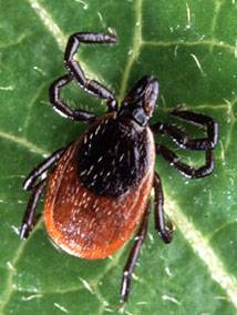
Anaplasma phagocytophilum is transmitted via a bite from an Ixodes tick. Ixodes scapularis (often commonly called the deer tick, blacklegged tick, or bear tick) is the primary vector in the Midwest and Northeastern United States; Ixodes pacificus (commonly called the Western blacklegged tick) is the primary vector in the Western United States; and Ixodes ricinus (commonly called the castor bean tick) is the primary vector in Europe.

Granulocytic anaplasmosis is seen most commonly in dogs in the Northeastern, upper Midwest, and coastal Western United States. Since granulocytic anaplasmosis requires the Ixodes tick as its vector, seasonal outbreaks of the disease can occur from spring through summer. Clinical disease is most often seen in adult dogs, and golden retrievers and Labrador retrievers appear to get it more than other breeds.

A. phagocytophilum can infect a wide range of mammals. In addition to dogs, the clinical disease has been documented in cats, cattle, sheep, goats, llamas, and humans.

Because of common vectors and rodent reservoirs, co-infection with Borrelia burgdorferi can be seen and can lead to more severe illness.

Clinical Signs and Prognosis

Many dogs exposed to granulocytic anaplasmosis do not get obvious signs of the condition. If signs are seen, they most often occur during the acute phase of infection, which is 1 to 2 weeks after transmission. The signs may be vague and include lethargy, lack of appetite, and fever. Some dogs may become lame because their joints are painful. Less common signs include vomiting, diarrhea, coughing, and difficulty breathing.

The prognosis for granulocytic anaplasmosis is quite good.

Infectious Cyclic Thrombocytopenia

Infectious cyclic thrombocytopenia is an infection of blood platelets. It is caused by Anaplasma platys.

Anaplasma platys transmission has not been fully determined, although tick vectors are probable. The organism has been found in Rhipicephalus and Dermacentor ticks.

Clinical Signs and Prognosis

Signs include lack of appetite, lethargy, fever, bruising on the gums and stomach, nosebleeds, and weight loss.

Many dogs with infectious cyclic thrombocytopenia have only mild clinical disease, so the prognosis is generally good.  

Diagnosis

Blood tests and urinalysis are the main diagnostic tools for anaplasmosis. The blood tests usually include a complete blood count, blood smear evaluation, biochemistry panel, serology to look for antibodies and polymerase chain reaction (PCR) assays. If the dog is lame, radiographs and analysis of joint fluid are usually included.

Treatment

Treatment includes antibiotics, pain relievers, and anti-inflammatory drugs.

Doxycycline is the most commonly used antibiotic. Most dogs respond within one to two days after they first take doxycycline. Other antibiotic options are tetracycline or minocycline.

Analgesia and anti-inflammatory drugs may be needed for joint pain. Let your veterinarian choose the anti-inflammatory, rather than choosing and dosing it yourself because dogs metabolize these medicines differently than humans do. Your veterinarian will have the most appropriate medication.

Disease Prevention

Because there is no vaccine for anaplasmosis, appropriate tick control is critical to preventing this disease. Preventing ticks from attaching and removing any ticks from your pet within a few hours of attachment is vital.

Note: Dogs with anaplasmosis may also be infected with other tick-borne organisms (Ehrlichia, Borrelia, etc.), so infected dogs should be screened for those diseases also.