Tag: dog

4536889

Wound Healing in Dogs and Cats

One thing is certain about life: we can all expect to experience some wounds. The good news is that we are fundamentally designed to heal.

While the statement above has philosophical implications, we will stick to the physical ones in this discussion. In particular, we will be considering the skin and the wounds experienced in the skin and underlying tissues.

The skin and its associated tissues exist in layers: the epidermis on the outside (layered in and of itself), the dermis below, the subcutis below that, and fat and muscle below that. When we are injured, these areas may be cleanly cut, punctured, scraped, ulcerated, or burned. 

These wounds can be sterile, unclean (relatively clean but not sterile), or even heavily contaminated. The body is designed to address all of these situations, and often, as caregivers, we can help.

The Healing Process Starts as Soon as the Wound is Inflicted

There are four phases of wound healing: Inflammation, debridement, repair, and maturation.

Inflammation (Starts Immediately)

This is the first phase of healing and is all about controlling bleeding and activating the immune system. Without going into too much biochemical detail, blood clots are forming, and blood vessels are constricting to limit blood loss in the area. This process also calls in “clean up” cells of the immune system to address contaminating bacteria and any dead tissue.

Debridement (Starts in a Few Hours)

Wound fluid, dead tissue, and immunologic cells form pus, which is designed to flow as a liquid from the wound and carry debris with it. The cells that were called to the wound in the inflammation phase are now actively working on consuming dead tissue and cleansing the area.

Repair (Starts in a Couple Of Days)

Collagen begins to fill in the wound to bind the torn tissues, a process that will take several weeks to complete. New blood vessels begin to grow into the area from the uninjured blood vessels nearby. The wound edge begins to produce granulation tissue, the moist pink tissue that will ultimately fill in the wound. The wound will shrink in a process called wound contraction so that new skin can form and cover it.

Maturation (Starts in 2-3 Weeks and can Take Months or Even Years)

Once plenty of collagen has been deposited, the final phase of scarring can form. The scar becomes stronger and stronger over time as new blood vessels and nerves grow in, and the tissue reorganizes. The final result will never be as strong as un-injured tissue but should ultimately achieve approximately 80% of the original strength

Spay Incision

A spay incision is an example of wound healing by primary intention.

Primary Intention

When the wound is a surgical incision with sutures in place, there is no area for the body to fill with granulation tissue. Instead, the wound margins are already held together and the two margins simply need to bond. New skin begins to form across the margin within two days. The four stages of healing continue as above but go much faster (10-14 days total) because there is no gap in the tissue to fill in.

Healing occurs across the wound margin, not down its length, which means long incisions heal just as fast as short ones.

Secondary Intention

If the wound cannot be closed with sutures (it is too big, there is too much tension on the wound margins pulling them apart, the wound is too infected, etc.), then a process called second intention comes into play. This is the part of wound healing where granulation tissue must form to fill in the gap. Once the wound is filling with granulation tissue, contraction soon follows, which means the wound will be getting smaller and smaller. Eventually, it can be allowed to simply close on its own or when it is small enough, the margins can be trimmed and the wound surgically closed with primary intention of a smaller scar and better fur coverage. In the right circumstances, skin grafts can be applied, but only if there is a healthy granulation bed.

We Love Granulation Tissue

It looks like it would be sore. Many people incorrectly feel granulation tissue is not supposed to be there when, in fact, it is a sign of a healthy healing wound. When a wound is cleansed of debris, scabs, or crusts (and often when a bandage is removed), granulation tissue is evident. Many people, especially those not familiar with wound management, find granulation to be disturbing: it is red or bright pink, moist, bleeds easily, and is often confused with underlying muscle.

Granulation tissue:

  1. Should be moist so as to allow better blood flow and a proper debridement phase.
  2. Bleeds easily as it is rich in blood vessels.
  3. Generally is not painful as nerves grow into granulation tissue late in the healing process.

How Can We Help?

The body is actually pretty good at healing, but there are some things that can go wrong, as well as ways that we can facilitate the healing process.

  • Deeper pus pockets must be drained. Eventually, these will probably burst out on their own. Depending on the size of the pus pocket (abscess), a large amount of tissue may slough off when the abscess bursts, so, if possible, the pocket is lanced, flushed, and drained before it gets to that stage. Sometimes, actual latex strips are sewn in place to facilitate pus drainage.
  • The wound must be kept moist. This can be accomplished with bandages and/or ointments. A moist wound has better blood flow and can heal more effectively.
  • Gross contamination should be cleaned up. Dirt, hair, pus, and other bacteria-rich substances should be flushed from the wound. Antibiotics may be needed either orally, topically, or both to address infection.
  • Dead tissue should be trimmed. If there is non-viable tissue in the wound, it should be removed so the body will not have to liquefy it. This can be done through surgery, through certain types of bandages, or by topical applications, depending on the type of wound.
  • Wound-enhancing topical products can be applied. There are a number of topicals touted to reduce infection and/or enhance the formation of granulation tissue. It often seems like new products are available annually, so we won’t review them, but your veterinarian may select one to potentially assist the body’s wound healing efforts.

First Aid Tip: If your pet’s wound is fresh and your pet will allow it, try to wash out large debris particles with tap water (saline flush as used for eyes is even better as it is balanced for tissue exposure). Cover the wound with clean, dry bandage material, if possible. See your veterinarian for professional wound care.

Non-healing Wounds

If a wound seems to be ongoing, either healing and then getting worse again or simply not showing signs of healing, be sure to bring your pet to the veterinarian. Unhealing wounds may have tumor involvement or may simply be infected. Do not try to do it yourself and end up with an advanced and possibly untreatable problem.

4128613

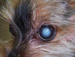
Cataracts in Diabetic Dogs

Most diabetic dogs will develop cataracts and go blind. This FAQ is designed to assist the owners of diabetic dogs in knowing what to expect and to make decisions regarding cataract surgery.

What Is a Cataract?

A cataract is an opacity in the lens of the eye. The entire lens may be involved or just a part of it. The patient will not be able to see through the opacity.

Why Do Diabetic Dogs Get Cataracts?

The lens of the eye is round, hard, and normally as clear as glass. Looking at the lens, it is hard to believe it is a piece of living tissue. The lens is suspended by fibers that can adjust its position so that one can focus vision. The lens is encased in a capsule and depends on the fluids of the eye for nutrients. The lens does not receive a direct blood supply.

Normally, the lens absorbs glucose from the eye fluids, using most of this for its own energy needs. Some of the excess is converted to another sugar called sorbitol. When there is excess sugar in the eye fluids, as in diabetes mellitus, there is excess sorbitol produced. Sorbitol pulls water into the lens, which in turn disrupts lens clarity and causes the cataract. Fructose is also produced from the excess glucose and also contributes to this water absorption.

Cataracts do not necessarily imply poor diabetic control. Even well-controlled dogs can still get cataracts.

How Long Does It Take To Go Blind?

Generally, the cataract has matured, and the dog is blind in a matter of weeks.

Until recently, blindness in a diabetic dog was basically a foregone conclusion, but there is a new product called Kinostat® that has changed that outlook. To review, the lens absorbs glucose from the fluids of the eye and uses this glucose as nutrition. Any extra glucose that is absorbed into the lens is converted to sorbitol by an enzyme called aldose reductase. Sorbitol pulls water into the lens to prevent the lens from becoming dehydrated. This is all well and good, but in the diabetic state, there is lots of excess glucose, and the excess glucose gets converted to excess sorbitol, which, in turn, pulls so much water into the lens that clarity and function are disrupted, and a cataract is formed. Kinostat is an aldose reductase inhibitor that curtails sorbitol production. Early use of Kinostat may significantly delay or even completely prevent the development of cataracts.

Kinostat is preventive only and will not reverse cataract formation that has already occurred. Kinostat is unfortunately not yet commercially available, though has been reportedly near coming to market for some time.

What Does It Mean To Say That a Cataract Is Mature?

A cataract’s maturity is determined by how much visual impairment the pet is believed to have. Since we cannot ask a dog to read an eye chart, we must determine this by visually inspecting the eye. A light is used to look into the eye and view the colorful area at the back of the eye called the tapetum.

(This is the area that flashes or appears colored in certain lighting.) When less than 10 percent of the tapetum is obstructed, the cataract is very young and does not significantly change vision. When 10-50 percent of the tapetum is obstructed, this cataract is called early immature. When 51-99 percent is obstructed the cataract is late immature. The mature cataract obstructs the entire tapetum. Ideally, a cataract is removed in the early immature stage for the lowest surgical complication rate.

When a cataract is hypermature, it starts to liquefy and dissolve. This can actually lead to restoring vision, which sounds like a positive turn of events; unfortunately, the dissolution process is quite inflammatory. The resulting inflammation can be painful and can damage the eye even more.

All cataracts do not progress all the way to hypermature and may stay static or progress at changing rates; however, diabetic cataracts are notorious for reaching hypermaturity and creating inflammation. 

What Is Uveitis?

Uveitis is inflammation of the eye’s uveal tract, which consists of the eye’s vascular tissues. In this situation, uveitis is the inflammation that results when the hypermature cataract begins to liquefy. Uveitis is painful and tends to cause the eye to become reddened and the pupil to constrict. When a pet has uveitis before surgery, success (pain-free vision) occurs in only 50 percent of cases 6 months after surgery as opposed to 95 percent of cases for whom there was no uveitis pre-operatively.

Can My Dog’s Vision Be Restored?

Maybe. It depends on the health of the eye, excluding the cataract. If the only thing wrong with the eye is the cataract and the patient’s diabetes mellitus is well-regulated, surgery can remove the cataract and place an artificial lens. If the eye is deeply inflamed or is otherwise damaged, the eye may be blind even without a cataract. A veterinary ophthalmologist can evaluate the eye to determine the feasibility of cataract surgery.

It is important to note that there are numerous eye drops advertised that claim to dissolve cataracts. None of these products actually work and wasting time with them may actually allow uveitis to set in and make for a much worse prognosis for vision.

Is it Cruel To Keep a Dog Blind?

Not at all. Dogs do not depend on vision the way humans do. A blind dog can get along very well as long as the furniture isn’t moved and the dog is properly supervised.

For tips on helping a blind dog adapt:

Blind Dog Support

There are many medical conditions that render a dog blind and as long as the condition is not painful, the dog can live a normal life as a successful and happy pet.

Cataract Surgery: What Is The First Step?

The first step is a consultation with your regular veterinarian. Your dog’s diabetes must be well-regulated before surgery is considered. If pre-operative lab tests show nothing to preclude anesthesia, your veterinarian may refer you to a veterinary ophthalmologist, as clinics do not usually have the specialized equipment necessary. A regular veterinarian is not qualified to perform cataract surgery.

What Happens At The Ophthalmologist’s?

It is necessary to determine if the eye is going to be visual after cataract surgery. There is, after all, no point in performing this surgery if the eye is going to be blind anyway. The most important test is called an ERG (an electroretinogram). This test checks the retina for electrical activity, which indicates the eye should be able to see after the cataract is removed.

In addition to the ERG, the ophthalmologist will check for uveitis. It should be treated before surgery to minimize the inevitable inflammation after surgery.

What Kind of Surgical Procedures Are Done?

There are two types of surgery: lens extraction and phacoemulsification. With lens extraction, the incision tends to be larger, the post-operative inflammation is greater, and the potential for leaving bits of the lens behind is also greater.

With phacoemulsification, an ultrasonic instrument is used to liquefy the lens, and a sort of vacuum cleaner sucks the lens away. This procedure is more difficult if the patient is older, and the lens is thus harder in consistency. This method is preferred for diabetic patients.

After either surgery, an artificial lens is usually placed for optimal post-operative vision.

What Kind of After Care Is Needed?

The patient will need to wear an Elizabethan collar after surgery to protect the eye. Cortisone eye drops are needed for probably several weeks. Oral anti-inflammatories will be needed for weeks to months after. Drops to keep the pupil dilated will also be used.

What Kind of Complications Are Possible?

Complications to consider are:

  • Long-term uveitis (probably of most concern for diabetic patients)
  • Opacification of the lens capsule (usually correctable with a laser)
  • Corneal clouding (can be managed with 5 percent saline eye drops 4 to 6 times daily)
  • Bleeding in the eye
  • Glaucoma
  • Retinal detachment (particularly if the cataract is hypermature)

Should Surgery Be Done On Both Eyes?

It is important to remember the old saying that the one-eyed man is king among the blind. A dog need only have one cataract removed to have vision restored. Doing both eyes is an option to discuss with the ophthalmologist, as some dogs need all the vision they can get.

Cataract surgery requires committed patient care both in the hospital and at home. Your veterinarian will be able to answer your questions or direct you to other appropriate resources. 

6725556

Bloat – The Mother of All Emergencies 

Many injuries and physical disorders represent life-threatening emergencies, but there is only one condition so drastic that it overshadows them all in terms of rapidity of consequences and effort in emergency treatment: the gastric dilatation and volvulus – the bloat.

What is it, and Why Is It So Serious?

The normal stomach sits high in the abdomen and contains a small amount of gas, some mucus, and any food being digested. It undergoes a normal rhythm of contraction, receiving food from the esophagus above, grinding the food, and moving the ground food out to the small intestine at its other end. Normally, this proceeds uneventfully except for the occasional burp.

In the bloated stomach, gas and/or food stretches the stomach many times its normal size, causing tremendous abdominal pain. For reasons we do not fully understand, this grossly distended stomach tends to rotate, thus twisting off not only its own blood supply but the only exit routes for the gas inside. The spleen, which normally nestles along the greater curvature of the stomach, can twist as well, cutting off its circulation. The distended stomach becomes so large that it compresses the large veins that run along the back, returning the body’s blood to the heart, creating a circulatory shock. Not only is this collection of disasters extremely painful, but it is also rapidly life-threatening. A dog with a bloated, twisted stomach (more scientifically called gastric dilatation and volvulus) will die in pain in a matter of hours unless drastic steps are taken.

What Are the Risk Factors for Developing Bloat?

Dogs weighing more than 99 pounds have an approximate 20 percent risk of bloat. The risk of bloating increases with age.

Classically, this condition affects dog breeds that are said to be deep-chested, meaning the length of their chest from backbone to sternum is relatively long while the chest width from right to left is narrow. Examples of deep-chested breeds would be the Great Dane, Greyhound, and the setter breeds. Still, any dog can bloat, even dachshunds and chihuahuas.

Classically, the bloated dog has recently eaten a large meal and exercised heavily shortly thereafter. Still, we usually do not know why a given dog bloats on an individual basis. No specific diet or dietary ingredient has been proven to be associated with bloat. Some factors found to increase and decrease the risk of bloat are listed below:

Summary of Factors Increasing the Risk of Bloat

  • Increasing age
  • Having closely related family members with a history of bloat
  • Eating rapidly
  • Feeding from an elevated bowl
  • Feeding a dry food with fat or oil listed in the first four ingredients.

Factors that May Decrease the Risk of Bloat

  • Adding table scraps, canned food, or non-kibble supplements to the dog’s kibble diet reduced the risk of bloat in some studies. More research is needed to fully understand the implications of this.
  • Happy or easy-going temperament
  • Feeding a dry food containing a calcium-rich meat meal (such as meat/lamb meal, fish meal, chicken by-product meal, meat meal, or bone meal) listed in the first four ingredients of the ingredient list.
  • Eating two or more meals per day

Contrary to popular belief, cereal ingredients such as soy, wheat, or corn in the first four ingredients of the ingredient list do not increase the risk of bloat.

In a study done by the Purdue University Research Group, headed by Dr. Lawrence T. Glickman: 

  • The Great Dane was the #1 breed at risk for bloat. (The incidence of bloat in this breed is reported to be 42%. Preventive gastropexy should be considered as described below.)
  • The St. Bernard was the #2 breed at risk for bloat.
  • The Weimaraner was the #3 breed at risk for bloat.

In the 1993 study from Germany (see below), the German shepherd dog and the boxer had the highest risk for bloat.

How to Tell if Your Dog Has Bloated

Classically, the dog is distressed and makes multiple attempts to vomit, and the upper abdomen is hard and distended from the gas within, though in a well-muscled or overweight dog, the distention may not be obvious. There are other potential emergencies (sudden abdominal bleeding from a ruptured tumor, for example) that might have a similar presentation, so radiographs may be needed to determine what has happened. The hallmark presentation of bloat is a sudden onset of abdominal distention, distress, anxiety, and pain (panting, guarding the belly, anguished facial expression), and multiple attempts at vomiting that are frequently unproductive. Not every dog will have a classic appearance, and some dogs will not have obvious abdominal distention because of their body configuration. If you are not sure, it is best to err on the side of caution and rush your dog to the veterinarian immediately.

What Has To Be Done

There are several steps to saving a bloated dog’s life. Part of the problem is that all steps should be done at the same time and as quickly as possible.

First: The Stomach Must be Decompressed


The huge stomach is by now pressing on the major blood vessels, carrying blood back to the heart. This stops normal circulation and sends the dog into shock. Making matters worse, the stomach tissue is dying because it is stretched too tightly to allow blood circulation through it. There can be no recovery until the stomach is untwisted and the gas is released. A stomach tube and stomach pump are generally used for this, but sometime surgery is needed to achieve stomach decompression.

Also First: Rapid IV Fluids Must be Given to Reverse the Shock


Intravenous catheters are placed, and life-giving fluid solutions are rushed in to replace the blood that cannot get past the bloated stomach to return to the heart. The intense pain associated with this disease causes the heart rate to race at such a high rate that heart failure will result. Medication to resolve the pain is needed if the patient’s heart rate is to slow down. Medication for shock, antibiotics, and electrolytes are all vital in stabilizing the patient.

Also First: The Heart Rhythm is Assessed and Stabilized


A special and very dangerous rhythm problem called a premature ventricular contraction, or “PVC,” is associated with bloat and it must be ruled out. If this is the case, intravenous medications are needed to stabilize the rhythm. Since this rhythm problem may not be evident until even the next day, continual EKG monitoring may be necessary. If the disturbed heart rhythm is noted at the very beginning of treatment, this is associated with a 38% mortality rate.

Getting the bloated dog’s stomach decompressed and reversing the shock is an adventure in itself, but the work is not yet half finished.

Surgery

All bloated dogs, once stable, should have surgery. Without surgery, the damage done inside cannot be assessed or repaired, plus bloat may recur at any point, even within the next few hours, and the above adventure must be repeated. If the stomach has not untwisted with decompression, the surgeon untwists it and determines what tissue is viable and what is not. If there is a section of dying tissue on the stomach wall, this must be discovered and removed, or the dog will die despite the heroics described above. Also, the spleen, which is located adjacent to the stomach, may twist with the stomach, necessitating removal of the spleen or part of the spleen as well. After the nonviable tissue is removed, a surgery called a gastropexy is done to tack the stomach into its normal position to prevent future twisting.

If the tissue damage is so bad that part of the stomach must be removed, the mortality rate jumps to 28 – 38 percent.

If the tissue damage is so bad that the spleen must be removed, the mortality rate is 32 – 38 percent.

After the expense and effort of the stomach decompression, it is tempting to forgo the further expense of surgery. However, consider that the next time your dog bloats, you may not be there to catch it in time and, according the study described below, without surgery, there is a 24 percent mortality rate and a 76 percent chance of re-bloating at some point. The best choice is to finish the treatment that has been started and have the abdomen explored. If the stomach can be surgically tacked into place, the recurrence rate drops to 6 percent.

Surgery will prevent the stomach from twisting in the future, but the stomach is still able to periodically distend with gas. This is uncomfortable but not life-threatening.

Results of a Statistical Study

In 1993, a study involving 134 dogs with gastric dilatation and volvulus was conducted by the School of Veterinary Medicine in Hanover, Germany.

Out of 134 dogs that came into the hospital with this condition:

  • 10% died or were euthanized prior to surgery (factors involved included expense of treatment, severity/advancement of disease, etc.)
  • 33 dogs were treated with decompression and no surgery. Of these dogs, eight (24%) died or were euthanized within the next 48 hours due to poor response to treatment. (Six of these eight had actually re-bloated.)
  • Of the dogs that did not have surgical treatment but did survive to go home, 76% had another episode of gastric dilatation and volvulus eventually.
  • 88 dogs were treated with both decompression and surgery. Of these dogs, 10% (nine dogs) died in surgery, 18% (16 dogs) died in the week after surgery, and 71.5% (63 dogs) went home in good condition. Of the dogs that went home in good condition, 6% (four dogs) had a second episode of bloat later in life.
  • In this study, 66.4% of the bloated dogs were male, and 33.6% were female. Most dogs were between ages seven and 12 years old. The German Shepherd dog and the Boxer appeared to have a greater risk for bloating than did other breeds.

(Meyer-Lindenberg A., Harder A., Fehr M., Luerssen D., Brunnberg L. Treatment of gastric dilatation-volvulus and a rapid method for prevention of relapse in dogs: 134 cases (1988-1991) Journal of the AVMA, Vol 23, No 9, Nov 1, 1993, 1301-1307.)

Another study published in December of 2006 looked at 166 dogs that received surgery for gastric dilatation and volvulus. The point of the study was to identify factors that led to poor prognosis.

  • A 16.2% mortality rate was observed. The mortality rate for dogs over the age 10 years was 21%.
  • Of the 166 going to surgery, 4.8% were euthanized during surgery, and the other 11.4% died during hospitalization (two dogs died during surgery). All dogs that survived to go home were still alive at the time of suture removal.
  • 34 out of 166 dogs had gastric necrosis (dead stomach tissue that had to be removed). Of these dogs, 26% died or were euthanized.
  • Post-operative complications of some sort occurred in 75.9% of patients. Approximately 50% of these dogs developed a cardiac arrhythmia.
  • Risk factors significantly associated with death prior to suture removal included clinical signs of bloating for greater than six hours before seeing the vet, partial stomach removal combined with spleen removal, need for blood transfusion, low blood pressure at any time during hospitalization, sepsis (blood infection, and peritonitis (infection of the abdominal membranes).

(Beck, J.J., Staatz, A.J., Pelsue, D.H., Kudnig, S.T., MacPhail, C.M., Seim H.B, and Monnet, E. Risk factors associated with short-term outcome and development of perioperative complications in dogs undergoing surgery because of gastric dilatation-volvulus: 166 cases (1992-2003). Journal of the AVMA, Vol 229, No 12, December 15, 2006, p 1934-1939.)

It is crucially important that the owners of big dogs be aware of this condition and prepared for it. Know where to take your dog during overnight or Sunday hours for emergency care. Avoid exercising your dog after a large meal. Know what to watch for. Enjoy the special friendship a large dog provides, but at the same time, be aware of the large dog’s special needs and concerns.

Prevention: Gastropexy Surgery

Preventive gastropexy is an elective surgery usually done at the time of spaying or neuter in a breed considered at risk. The gastropexy, as mentioned, tacks the stomach to the body wall, which drastically reduces the stomach’s ability to twist. The stomach may distend with gas in an attempt to bloat, but since twisting is not possible, this becomes a painful and uncomfortable situation but nothing more serious than that. That said, gastropexy is not an absolute guarantee against twisting but we are talking about a recurrence rate of 76% without gastropexy versus 6% recurrence with gastropexy.  

A study by Ward, Patonek, and Glickman reviewed the benefit of prophylactic surgery for bloat. The lifetime risk of death from bloat was calculated, along with estimated treatment for bloat versus the cost of prophylactic gastropexy. Prophylactic gastropexy was found to make sense for at-risk breeds, especially the Great Dane, which is at the highest risk for bloat.

7905375

Aggression Between Familiar Dogs

Aggression between familiar dogs in the same household is a common occurrence and is one of the most common reasons that dogs are surrendered, rehomed, or euthanized. This aggression can be secondary to high arousal or excitement levels. When a fight occurs, damage to the relationship between the dogs can be difficult to repair.

There are a variety of factors that need to be considered when determining the future relationship between these dogs. For instance, aggression between female dogs is reported to be the most common and is the most difficult to treat due to the severity of injuries and intensity of aggression that occurs between the dogs. Fights between male dogs are often due to resource guarding and are the easiest to treat. Other indicators for poor outcome include:

  • fights escalating in intensity.
  • fights occurring between two female dogs of similar size and age.
  • no noticeable early warning signs or triggers.
  • level three bite or above.

As with any behavioral concern, the first step is looking for underlying medical causes contributing to the behavior.

Treatment Plan

When aggression occurs between familiar dogs, the initial goal of the treatment plan should focus on improving the welfare for both dogs. Although it can be difficult, there should be complete separation of the dogs with no visual contact. Leashes, gates, and tethers can be useful.

Monitor body language of each dog. Since dogs are often very subtle with body language monitor closely for hard stares or stiff posture that is signaling to another dog that they are uncomfortable. It is important that both dogs be completely relaxed in close proximity to each other. Direct stares (longer than three seconds) need to be quickly redirected to reduce escalating tensions.

Basket muzzle training is very important for safety. All dogs should be conditioned to wear a muzzle. This is not a punishment but rather a predictor of good things. Go slow and always wait for each dog to put their own nose into the muzzle. We never force it on, and the dog must willingly place their nose in the muzzle. We never advance it on them.

The dogs should never be together until they are 100% comfortable wearing their muzzles. A muzzle with a buckle is ideal as plastic clasps can break easily. A Baskerville Ultra or Jafco Muzzle with the end square for treat dispensing would be best.

Once comfortable in muzzles you can start walking them next to each other, one adult per dog. This is a great way to rebuild their relationship.

Teaching alternative behaviors such as “touch” is a great behavior to teach your dogs. It can be used to redirect a dog when needed. It can also be used to teach new behaviors and even as an emergency recall.

Teach your dogs to relax on a mat. This can be used later if the dogs are to be reintroduced.

Establish a safe haven such as a crate or room for all dogs. Make sure good things happen in this area. Food dispensing, puzzle toys, classical music and pheromones, can be helpful. A tether or gate may be necessary. If using a crate, be sure to cover and place them out of sight from each other.

Practice Cue-Response-Reward interactions with each dog. A Cue-Response-Reward interaction system encourages predictability and consistency therefore reducing overall anxiety and instability in the relationship between the dogs.

Phase II

Once both dogs are comfortable wearing muzzles and relaxing on their mat.

  • Initially, bring them out for short periods together.
  • One adult per dog and sit on opposite sides of the room. A tether may be necessary.
  • Give them something to do, like a long-lasting treat or toy giving the dogs an opportunity to be together, stay calm and relearn to like each other.

Always separate when they cannot be directly supervised with your entire attention.

Medications

In many cases medications for one or both dogs may be needed to lower anxiety.

Grove Center Services

Adverse Reactions to Spot-on Flea and Tick Products  

Spot-on flea and tick products for dogs and cats have greatly improved the safety and convenience of controlling external parasites on our pets. Using pesticides that are many degrees less toxic and frequently more effective than the organophosphate or carbamate dips and sprays of the past, these spot-on products help keep our pets (and our homes) free of annoying pests that can also carry serious diseases.

When used according to the label directions, spot-on products are well tolerated by most pets. However, as with any product that is applied directly on the skin, there is the possibility that certain individuals will have adverse local reactions to one or more ingredients in the product. Some individuals will have similar reactions to many different spot-on products with different active ingredients, suggesting that their sensitivities may be to some of the inactive ingredients. These reactions are restricted to the area of skin that comes in direct contact with the product, so they do not reflect a systemic toxicosis but rather a local hypersensitivity. Skin reactions to spot-on products can vary from mild tingling sensations to actual chemical burns of the skin in especially sensitive individuals.

The mildest form of skin reaction to spot-on products is epidermal paresthesia, which is defined as an abnormal sensation such as an itch or prickling of the skin. Paresthesia occurs when the applied product “tickles” the nerve endings in the skin, causing the characteristic sensation. Although paresthesia may occur with any spot-on product, it is most commonly associated with products containing concentrated pyrethroids such as permethrin, cyphenothrin, and etofenprox. Pyrethroid paresthesia is a syndrome that has been well documented in both humans and animals. Human descriptions of pyrethroid paresthesia vary from “tingling” to “pins and needles” to “burning” sensations in the skin at the site of pyrethroid contact. These sensations begin within 30 minutes of application and may last 8 to 24 hours if untreated. Some pets appear to experience similar discomfort following application of spot-on products; the fact that the products are generally applied between the shoulder blades can make the sensation particularly annoying to the pets. Affected pets, cats in particular, may become hyperactive and agitated as they try to walk away from the sensation; others may become quiet, subdued and reluctant to move. Skin at the product application site will appear normal (if redness is noted, you’re dealing with contact dermatitis, see below).

Fortunately, treating epidermal paresthesia is fairly simple and entails bathing off the product with a mild dish soap (pet shampoos are too mild to remove all of the product). For pyrethroid paresthesia, applying vitamin E to the affected skin can provide quick relief— just use scissors to snip open a vitamin E capsule (the kind used as a vitamin supplement) and squeeze the oily contents onto the pet’s skin and rub it in.

A rare but more significant skin reaction (in terms of sensitivity) to spot-on products is contact dermatitis, in which an inflammatory reaction develops in response to the topically applied product. These responses – sometimes referred to as hypersensitivity reactions – can occur upon the first use of a product, but more commonly occur after several uneventful exposures to the product. The body sets up an inflammatory response to the site of application, and the skin will appear red and irritated. In more severe cases, wheals or blisters may develop and the skin may actually ulcerate. Unlike paresthesia, which tends to occur within 30 minutes of application, oftentimes the onset of contact dermatitis is delayed for several hours, with the full extent of the injury taking 12 to 24 hours to develop. Depending on the degree of the inflammatory reaction, the level of discomfort can range from mild to quite severe. After bathing off the spot-on product, pets who develop more than mild redness at the application site should be seen by their veterinarians for further treatment to reduce the inflammation and discomfort.

The good news is that skin reactions to spot-on flea/tick control products are quite uncommon, and most pets will have no problems when these products are used. Unfortunately, it is not possible to predict which individual will react to a given product until the reaction has occurred. For this reason, it is a good idea to observe your pet closely for at least an hour following the application of any spot-on product for any signs of discomfort.

Although not life-threatening, skin reactions can range from mildly annoying to very painful; for this reason, a pet that has had a reaction to a spot-on product should never have the same product applied in the future. If a spot-on product is still desired for flea and/or tick control, try a product with different active ingredients than the one that caused the reaction. Following application, watch the pet carefully and be ready to bathe at the first sign of problems. Unfortunately, there are rare individuals that cannot tolerate any of the topical spot-on products, and other flea control options such as sprays or dips may be needed.

More serious problems can result when spot-on flea/tick products designed for dogs are inappropriately applied to cats. Certain concentrated pyrethroids (e.g. permethrin) that are not harmful to dogs can cause life-threatening tremors and/or seizures if applied to cats—these are not really adverse events but are instead true poisonings since the product was misused.  Always read the label before using any topical flea/tick product on cats, and never apply products intended for dogs onto cats.   

Frontline (fipronil) is toxic to rabbits. 

If your pet has a reaction to a flea or tick control product of any kind, you should report the reaction. All spot-on product labels will have a phone number that you can use to report the reaction to the manufacturer. By law, the manufacturer is required to send monthly reports of adverse reactions to the appropriate federal regulating authority. Alternatively, you can go to the agencies’ websites to find out how to report reactions directly to them. Report reactions to flea/tick products containing heartworm preventatives, such as Revolution and Advantage Multi, to the Food and Drug Administration; report flea/tick products that do not contain heartworm preventatives to the Environmental Protection Agency.