Tag: Diagnosis

6665143

Bacterial Conjunctivitis in Cats

Conjunctivitis is inflammation of the conjunctiva, a tissue that makes up part of the eye and eyelid. In people, the conjunctiva is the white part. Conjunctivitis is common in cats and can be a result of infection or injury.

The primary cause of most conjunctival infections in cats is usually feline herpes virus. The treatment for herpes-based conjunctivitis involves a combination of antibiotics and antivirals, both in eye drops and pills. Even cats who have had the herpes vaccine can get conjunctivitis because the vaccination does not prevent herpes, but allows a milder case.  

However, some conjunctival infections that are primarily viral also have a bacterial infection at the same time.

The two bacteria species best known to cause conjunctivitis in cats are Chlamydia felis and Mycoplasma spp.

An infection caused by Chlamydia may cause redness, discharge, and excessive tears. Bacterial conjunctivitis caused by Chlamydia is treated with antibiotics. All cats in a household may need to be treated, as some cats who have it do not show any symptoms.

Mycoplasma is a natural inhabitant of every cat’s eye, so infection happens when there is an overgrowth of that bacteria. Treatment for the Mycoplasma infection is similar to that of Chlamydia: antibioticsThe goal in treating Mycoplasma conjunctivitis is to reduce the overgrown number of bacteria back down to a normal level because it is impossible to eliminate them entirely.

Who gets bacterial conjunctivitis?

Both cats and dogs can get conjunctivitis caused by bacteria or viruses. Conjunctivitis is the most common cat eye disease, and most cats will develop the infection once in their life. All breeds can develop infection, and juvenile and young adults are more likely to get it.

Clinical Signs of Either Form

  1. Red eyes
  2. Eyelid redness
  3. Eyelid swelling
  4. Excessive blinking
  5. Eye discharge
  6. Eye crusting
  7. Tearing
  8. Squinting
  9. Scratching the face to relieve irritation

 Diagnosis

A physical exam of the eye by your veterinarian is the first step, as this will rule out other causes of redness, such as trauma.  The easiest diagnosis is a cytology test, which means looking at scrapings of the irritated tissue under a microscope. If a lot of bacterial and inflammatory cells are seen, it is likely that the eye is infected. Checking scrapings is a quick test for infection that can be done at the clinic during your appointment. PCR is another test that can be used to detect the specific organism causing the infection, but typically needs to be sent out to a lab.

Treatment

Antibiotics are used to treat bacterial conjunctivitis, mainly doxycycline.  Mycoplasma and Chlamydia are the most common bacteria involved in eye infections, and doxycycline is effective against both.

Prognosis

Treatment should clear the signs and infection. However, if the eye still looks infected or just doesn’t look normal, contact your veterinarian. The ongoing bacterial infection may also be caused by an unknown factor that needs to be treated before the eye infection can be cured.

Prevention

There is a vaccine for Chlamydophila felis, but it is not required and is usually only suggested if your cat is at a higher risk for an infection or in a multi-cat household. There is no vaccine for a Mycoplasma spp. infection because it is normally in the eye anyway.

The key to preventing infection is separating sick cats away from the unaffected ones and bringing them to your veterinarian at the first sign of infection.

7941098

Arthritis in Dogs and Cats: What Can be Done?

Arthritis, also known as osteoarthritis or degenerative joint disease (DJD), is a degenerative, progressive, and irreversible condition of the joints. It is characterized by the progressive loss of joint cartilage, bony spurs/growths, and the thickening and scarring of connective tissue around the joint, usually as a result of injury.

Approximately 25 percent of dogs are diagnosed with arthritis in their life time, and as many as 60 percent of dogs have radiographic evidence of it.

Degenerative joint disorders are probably as common in cats as in dogs but are less likely to be associated with obvious clinical signs, such as lameness. In one study, 90 percent of cats over 12 years of age had radiographic signs of osteoarthritis.

Arthritis is classified as primary or secondary. Primary arthritis is associated with aging, due to years of wear and tear on the joints. Secondary arthritis is the result of an external event or force (e.g., trauma, poor joint alignment, etc.) that once damaged the joint cartilage.

Arthritis can affect any age, sex, and breed of dog and cat. Most predispositions to it relate to underlying causes, such as elbow dysplasia, hip dysplasia, osteochondrosis, and so forth.

Diagnosis

Diagnosis is based on what is found in the physical examination, X-rays and other diagnostic imaging, joint taps, cytology, force plate gain analysis, therapeutic drug trials, and other tests.

Physical Examination Findings in Dogs

Lameness is the most common sign. It may happen once in a while (episodic) progressive (gets worse over time), or be persistent. Stiffness is common after periods of rest. Stiffness and lameness may decrease when the dog warms up a bit with some activity. Lameness often gets worse after periods of overexertion. Pain, swelling, and decreased range of motion may be seen. Thickened joints, excess fluid in the joint space, and muscle weakening are likely to occur.

Physical Examination Findings in Cats

As opposed to the visible lameness seen commonly in dogs, many cats simply become less active, may hide, or develop behavioral changes, such as irritability, decreased grooming, or difficulty getting into position in the litterbox. Cats also may have joint swelling/thickening, too much fluid in the joint space, and decreased range of motion. There may or may not be pain when the cat’s affected joint is moved by you or your veterinarian.

Diagnostic Imaging

Radiographs (X-rays) and CT scans may show the excess fluid in the joints; the bony spurs; signs of an underlying disorder, such as elbow dysplasia, osteochondritis dissecans, hip dysplasia, or cruciate ligament rupture; and so forth.

Kinetic Tests

Force plate gait analysis in dogs can help find where the lameness is within the joint. It can also show the severity of lameness, both before and after therapy. A force plate is mounted to the floor, and the dog walks over it. Measurements are taken to see which areas use the least force (more affected) and vice versa. The gait analysis hasn’t been used as often in cats.

Therapeutic Trial

In some cases, a therapeutic trial of nonsteroidal anti-inflammatory drugs (NSAIDs) may help confirm the diagnosis if the lameness is less noticeable after taking them.

Treatment, Management, and Prevention

It is not possible to cure arthritis.

The goals are to alleviate your pet’s discomfort, to minimize further degenerative changes to the joint, and to restore the joint’s functionality. Multiple types of treatment are usually necessary to relieve pain, stiffness, and discomfort.

Managing your pet’s weight is important. Excess weight increases stress on the joints and muscles. If your pet is obese, your veterinarian will want your pet to lose weight. Daily, low-impact activities, such as walking and swimming, will not only help your pet with losing some pounds but can also improve joint mobility, muscle mass, and exercise tolerance.

Joint supplements known as chondroprotective agents will help support the cartilage and will have some anti-inflammatory effects. These agents will slow the breakdown of cartilage and/or provide the building blocks that can help build it. Some agents also increase joint fluid secretion and thus decrease inflammation.

The main components of chondroprotective agents are polysulfated glycosaminoglycan (PSGAG), glucosamine, and chondroitin sulfate. Oftentimes there is no improvement using chondroprotective therapies. The failure may be due to too little cartilage left in the affected joint as there’s bone on bone; unresponsive joint inflammation; variable bioavailability of the drug between different products; and the lack of analgesia.

Although the injectable PSGAGs are more expensive, they provide a faster and longer-lasting response than the oral forms. If there is no response after 4-6 weeks, your veterinarian may discontinue use of PSGAGs.

Diets containing omega-3 fatty acids may help reduce inflammation. Some studies have found that dietary supplementation with fish oil omega-3 fatty acids can improve the clinical signs of arthritis in dogs, and may allow the NSAID dose to be reduced.

NSAIDS are an important component of arthritis therapy in dogs. Side effects that you should be aware of include stomach upset, elevated liver enzymes, and potential worsening of chronic kidney disease. Few NSAIDs are licensed for use in cats; your veterinarian will advise you about what NSAID options are available for your cat.

Analgesics such as tramadol, gabapentin, and amantadine may provide pain relief in dogs.

Alternative therapies such as acupuncture, stem cell therapy, platelet-rich or conditioned plasma, physical therapy, rehabilitation therapy (e.g. radial shock wave therapy, pulsed signal therapy), green-lipped mussel supplements, vitamin E, and zoledronic acid may be beneficial in some canine patients. Studies to support their use are still being done.

Surgery 

Your veterinarian may consider surgical options if your pet’s response to medical treatment is low. In addition, your veterinarian may suggest surgery for certain underlying causes of arthritis, such as cranial cruciate ligament rupture, elbow dysplasia, osteochondritis dissecans, joint incongruity, intra-articular fractures, and joint instability. Reconstructive procedures can eliminate joint instability and correct the anatomic defects. If your pet has severe hip dysplasia, your veterinarian may suggest a total hip replacement and femoral head/neck ostectomy. If the arthritis is in the wrist or ankle (then joint fusion may be considered; this surgery is usually well tolerated and can result in reasonable functionality.

Monitoring and Prognosis

Your veterinarian may need to do periodic physical examinations every 1-4 months to monitor your pet’s response to therapy and the progression of the disease. In addition, if your pet is on an NSAID, blood tests including complete blood counts and biochemistry profiles, should be done every few months to ensure there are no side effects impacting the liver or kidneys.

With therapy and careful monitoring, arthritis can be managed in many dogs and cats, resulting in a good quality of life that you and your pet will appreciate. 

36322

Cleft Palate or Lip in Puppies and Kittens

Congenital clefts in the palate or lip are birth defects that puppies and kittens can get in the tissues connecting parts of the mouth. Human babies also get them. These cleft defects happen when the roof of the mouth does not close correctly before the baby is born.

Cleft lip is a defect of the lips, underlying bone, and/or front area of the roof of the mouth, also known as a hard palate. Oftentimes, part of the lip will be missing or misshapen. Sometimes, one or both nostrils appear to connect directly to the mouth. Cleft lip may also be called a harelip because the face appears similar to that of a hare’s. 

Cleft palate is a malformation or disconnection of the tissues that form the roof of the mouth. The soft palate towards the back of the mouth can be involved. A hole may be in the palate because the tissues never fully connected to each other while the fetus was developing. Severe palate defects result in holes from the palate that connect directly to the nasal passages. 

The big problem with a cleft lip or palate is that it is difficult, if not impossible, for the affected puppy or kitten to latch on properly to a nipple and therefore they cannot eat properly. Depending on the severity of the cleft, the infant may not be able to get sufficient nourishment at an age where lack of nutrition can completely compromise the infant’s health and ability to grow.

Causes

Many conditions and issues can cause cleft defects. Congenital clefts can be an inherited birth defect; genes may be passed from the mother or father to the developing fetus. Because of this, pets with a cleft lip or cleft palate should not be bred as they have a high likelihood of passing on the cleft genes to their offspring.

Occasionally there are other causes, such as trauma during fetal development. During pregnancy, too little folic acid or too much vitamin A given to the mother can result in cleft defects, although that is rare. Many medications, if given to a pregnant cat or dog, may also cause cleft birth defects. These include steroids such as prednisone, aspirin, anti-seizure drugs, and griseofulvin. Certain viral infections or illnesses can lead to congenital clefts in puppies and kittens if the mother is sick while pregnant. Luckily, most of these issues are rare.

Signs

Signs are not always obvious, especially if the defect is minor. An abnormal appearance to the lip may be visible, or you may not even see any problems because they are so far back in the mouth. Animals with severe defects where the mouth connects to the nose may have nasal discharge. Milk, saliva, and/or food may come out of the nose during or after feeding. If food material gets lodged in the nasal passage way, nasal discharge may be green or mucus-like.

In addition to signs of cleft, signs of complications can be seen at home. If the cleft connects with the nasal cavity, the puppy or kitten may cough when trying to eat or drink. Pneumonia can occur due to cleft palates or cleft lips: trouble chewing and properly swallowing food can lead to food or milk in the lower airways and lungs instead of the stomach. Pneumonia will also result in coughing as well as trouble breathing.

For those who appear to eat well enough on their own, poor growth, decreased activity level, and slow development may result from not getting enough nutrition.

Diagnosis

Your veterinarian will do a thorough physical examination. Sedation is sometimes needed for a good look in the mouth. If the pet has symptoms of pneumonia, X-rays and lab work may be needed. If surgery is planned, advanced imaging, such as computed tomography (CT) or magnetic resonance imaging (MRI) scans, may be necessary but that is rare. 

Treatment

Cleft lips that do not connect the mouth directly to the nose can be left alone if they do not cause any symptoms. However, for those with symptoms, surgery is usually needed. Without surgery, food can get stuck in the malformed areas, leading to serious infections. In severe cases, pets with cleft palates can’t eat or swallow properly, and will become ill from lack of nutrition. 

Unfortunately, surgery is costly, and multiple attempts may be needed to close the defect.

Most surgeons recommend waiting until the puppy or kitten is older and strong enough to handle anesthesia, if the pet can wait that long. If surgery is delayed, tube feeding (either through a tube inserted into the back of the mouth, through the nose, or directly into the stomach) may be necessary to help keep up with the growing baby’s nutritional needs. Treatment for associated respiratory diseases may require hospitalization where the infant can get IV fluids and IV or oral antibiotics.

Prognosis

If a pet has no signs, it may survive and live on to be a healthy adult.

Improvement from surgery is not guaranteed, and chances of survival are decreased if the pet is sick or if the symptoms are severe.

If you are a breeder, call your veterinarian for a checkup if you notice your puppies or kittens have a facial deformity, have trouble drinking milk, cough or spit up frequently, or do not thrive like the rest of the litter. Also call your veterinarian if you see similar signs in your new puppy or kitten. Early intervention can prevent some of the issues associated with clefts such as infections and may help improve your pet’s chances for a healthy outcome.

4127904

Chronic Bronchitis in Dogs is Neither Infectious nor Contagious

Definition and Causes

Chronic bronchitis is usually a non-infectious, inflammatory condition affecting the lining (mucosa) of the large airways (the trachea and bronchi) that results in a cough on most days for more than two months. In most cases, the specific cause of chronic bronchitis in dogs is not identified.  Chronic bronchitis is neither infectious nor contagious to other dogs; dogs will not pass it on to other dogs as they can in the acute form (see below).

Acute bronchitis, on the other hand, is most often caused by infectious agents, such as viruses (e.g. parainfluenza virus, adenovirus, canine influenza virus, distemper virus, or bacterial agents Bordetella bronchiseptica is the most common).  It is commonly termed kennel cough or canine infectious tracheobronchitis. Inflammation can also be caused by noxious irritants such as second hand smoke, foreign bodies, allergens (similar to hay fever in humans), foods allergens, and either migrating or primary lung parasites. In many of these cases, this acute bronchitis, occurring at the time of the infection or insult, will resolve with appropriate therapy from your veterinarian.  However, if the bronchitis persists long enough – generally, more than a few months – it is labeled chronic, and the cough itself can begin to cause and perpetuate airway inflammation.

Clinical Signs

Dogs with chronic bronchitis generally have a persistent hacking cough.  Some people describe it as sounding like a goose honking.  However, any trachea-bronchial inflammation/irritation can produce a similar sounding cough.  Often, the coughing occurs during the night or when the dog first starts to move around upon waking.  It also commonly occurs with excitement or exercise.

Diagnosis

Chronic bronchitis is a diagnosis of exclusion.  Other diseases can cause clinical signs similar to chronic bronchitis.  These include tracheal collapse, laryngeal paralysis, lung diseases, lung cancer, heart failure, heartworm disease and infectious causes of acute bronchitis (listed above).  Your veterinarian will likely attempt to rule out many of these causes with a history, physical examination, and additional testing.

Diagnostic tests that are used to establish a diagnosis of chronic bronchitis include chest x-rays (thoracic radiographs), airway endoscopy (passing a small camera into the larynx, trachea and bronchi), collecting samples from the lower airways for culture and analysis of the cells in the airways (broncho-alveolar lavage or a transtracheal washing), and sometimes even echocardiography (examining the heart using ultrasound).  A fecal analysis might be needed to rule out parasites.

Treatment

Once a diagnosis of chronic bronchitis is made, your veterinarian will generally treat this condition with a variety of medications.

There are several things to remember about treating chronic bronchitis:

  • The cough in chronic bronchitis is unlikely to completely disappear.  The aim is to reduce the coughing by more than 70-80%, and to reduce the severity of the coughing fits.
  • Coughing begets coughing – the more a dog coughs, the more they irritate their airways, which stimulates more coughing.  Therefore, the aim is to initially break the coughing cycle.

Treatment of chronic bronchitis generally relies on using a combination of medications:

  • Corticosteroids. These drugs are the main therapy for chronic bronchitis. They reduce and suppress the inflammation in the airways.  They can be given orally (pills) or with an inhaler.  Specific canine inhalers are required to properly administer steroids.
  • Certain antibiotics, such as doxycycline, minocycline, or azithromycin may be given to rule out common infectious agents of the airways.  These particular antibiotics also have an anti-inflammatory effect, and may decrease coughing even if there aren’t any infectious agents.
  • Cough suppressants.  These are usually narcotic-based preparations that require a prescription. They sometimes need to be given to the point of sedating the dog, especially early in treatment.
  • Bronchodilators.  These may help by relaxing the airways, allowing air to move more easily into and out of the lungs. They can be given orally, by nebulizer, or by inhalers.

Your veterinarian might prescribe a combination of these drugs, and alter the doses as the disease is controlled to minimize side effects while still maintaining good control of the coughing.

An important factor in successful therapy of chronic bronchitis is weight loss.  If your pet is overweight, a loss of even 5-10% of the body weight can dramatically improve the coughing.  When the dog is overweight, improving your dog’s condition is completely in your hands; therefore, your veterinarian might prescribe a weight-loss program for your pet in addition to the medications.

Your veterinarian might also ask you to keep a “coughing log” to keep track of the severity and frequency of coughing, as this can help determine how successful the treatment is.

Prognosis

The prognosis for treating chronic bronchitis is good.  However, because this condition generally occurs in older dogs, other causes of chronic coughing (listed above) might complicate the management.  If coughing persists or recurs despite appropriate therapy, your veterinarian might be required to pursue other causes, or refer you to a specialist for additional diagnostic tests.

7176337

Chagas Disease in Dogs  

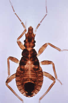
Chagas disease is an infectious disease caused by a microscopic parasite called Trypanosoma cruzi. This parasite cannot infect mammals on its own and instead is spread by an insect called the kissing bug, or, more formally, the triatomine. Kissing bugs spread the parasites through their feces. If the bug bites a dog and then defecates nearby, parasites can infect the dog through the open wound from the bite. Dogs can also be infected by eating kissing bugs, as the parasites enter the body through mucous membranes like the gums. 

Once the parasite infects an animal, if it is not treated promptly, it can reproduce and live in the body for the rest of the animal’s life. Chagas has been a serious problem in humans and dogs in South America for many years, but recently there have also been cases reported in the southern United States. 

Signs of Infection

Chagas has three stages after the animal has been infected: acute, latent, and chronic. 

Acute Phase

During the acute phase, the parasite enters the dog’s body and begins to infect macrophages, a type of white blood cell. Macrophages normally travel all through the body to fight infection and the parasite, hidden inside these cells, can move about undetected. 

When it reaches the heart, it begins to multiply and damage heart cells. The parasite can also infect many other body systems, including the nervous system, liver, kidneys, spleen, or lymph nodes. 

Most animals only have mild symptoms at this stage, such as slight fever or low energy, although dogs younger than six months are more likely to be severely affected. This stage generally lasts for several weeks and throughout this time, there are many parasites in the blood and tissues. Unfortunately, due to the lack of visible symptoms, acute phase infections of Chagas are often missed. As a patient’s immune system begins to fight the parasites, the number of parasites in the body goes down and the patient moves into the next phase of infection.

Latent Phase

During the latent phase, there are few parasites in the blood and the patient has no symptoms of illness. The immune system is generally not able to fully eliminate all the parasites in the body, but it keeps levels low enough that the patient doesn’t feel sick. A Chagas infection at this stage is difficult to identify through laboratory testing because there are so few parasites in the blood. Some dogs may remain in the latent phase for the rest of their lives and will thus continue on without any symptoms. 

Chronic Phase

Other dogs, however, will go on to the chronic phase of infection. We are not yet sure what triggers the chronic phase in some animals, but if the immune system becomes compromised through other diseases or injury, it can allow parasites to replicate again. As the immune system tries to fight against the growing number of parasites, it causes inflammation throughout the body, which can make the dog feel even sicker. Along with signs of systemic infection, like fever and lethargy, we also see symptoms of heart disease such as weakness, collapse, and, in severe cases, sudden death.

Diagnosis

Because Chagas disease is still relatively unknown in the United States, many cases are missed even after symptoms set in. Ask yourself the following questions to see if your pet is likely to have contracted Chagas:

  • Do they currently live or have they ever lived in areas where Chagas disease is common? Infections are widespread in Texas, Oklahoma, Louisiana, Georgia, and Tennessee, but have been reported all through the southern United States. 
  • Does your dog have symptoms of heart disease, such as lethargy, weakness, collapse, or labored breathing? Or do they have symptoms of systemic disease, like fever or loss of appetite?
  • Does your dog spend a lot of time outdoors, such as in a kennel, dog house, or as a working dog? Kissing bugs do not commonly live inside homes in the United States, but they can be found outside. 

If your dog fits any of the above criteria and your veterinarian suspects Chagas, they may take a blood sample to send to a laboratory for molecular tests or to look at under a microscope. They may also take X-rays or use ultrasound to look at your dog’s heart. 

Treatment

Drugs are available to fight the parasite infection and are quite effective during the acute phase. However, once the dog is in the chronic stage, it is no longer effective to only kill the parasites. Instead, the focus is on managing the symptoms of heart disease. Your veterinarian may recommend heart medications, prescription diets, or changes in exercise level.

Chagas Disease In Other Species 

Although Chagas is also a serious condition in humans, your risk of contracting it from your dog is extremely low. You cannot get Chagas by being bitten or licked by an infected dog. The main risk to humans from dogs is for those who are handling infected blood samples, such as veterinary or laboratory staff.

Chagas has been seen in both domestic cats and wild cats, such as bobcats. However, it appears to be less common in cats than it is in dogs. Research on the disease in cats is minimal, but if your cat is showing symptoms of heart disease, promptly inform your veterinarian.

Prevention

Research is being done on a canine vaccine for Chagas, but as of right now, it is not commercially available. Even if it were available, it only provides partial protection. If you live in an area where Chagas has been reported, you can take the following steps to protect your pets: 

  • Do not allow your pets to interact with or eat meat from other species that might be infected (raccoons, opossums, armadillos, rodents and skunks). Your dog can contract Chagas by ingesting kissing bugs that are on these other animals.  
  • Regularly spray outdoor kennels and spaces with pyrethrin/permethrin insecticides, which are effective against kissing bugs.
  • Before breeding female dogs, screen them for Chagas because it can be passed down to offspring. 

Note that pyrethrin insecticides are toxic to cats in high doses. Spray insecticides are 1-5% pyrethrin, while most cats have symptoms of poisoning at concentrations around 40%, so pyrethrin sprays intended to be used outside are generally safe even if you have cats. Be sure to check the concentration of pyrethrin in the product you are using, store the container where cats can’t access it and allow insecticides to dry fully before allowing cats to enter that space. Never apply a pyrethrin product directly to a cat. 

Being informed of the risk factors and symptoms of Chagas disease is the best way to ensure that your pets receive timely care. The earlier you catch the infection, the better your chances of reducing long-term complications. 

5666579

Bladder Stones in Dogs and Cats

Bladder stones are solid mineral deposits that form inside the bladder of dogs and cats and are quite common. Stones start out as crystals that form in the urine. Crystals form when a combination of events take place, such as urine pH change (pH is a measure of acidity and alkalinity in urine), increased urine concentration, and changes in the mineral makeup of the urine produced by the body. Over time, the crystals combine and layer together to form bladder stones. The process of forming bladder stones is called urolithiasis or cystolithiasis, 

The number of bladder stones produced can range from one or two stones to hundreds. Some bladder stones are small and grit-like, while others can grow to be larger than two inches in diameter. Kidney stones, more common in humans, occur much less often in dogs and cats. Only 2% of stones found in the urinary tract of our pets are found in the kidney.

The two most common types of bladder stones are those made of calcium oxalate and those made of struvite (also known as magnesium ammonium phosphate). Urate, cystine, xanthine, calcium phosphate, and silica are other mineral types that can form bladder stones but are rare.

Types of Bladder Stones 

Struvite bladder stones can occur with bladder infections. Certain bacteria will change the urine’s pH to reproduce faster. This pH change causes the urine to be more alkaline, causing struvite crystals to form. Struvite stones can also form without an infection, which is seen more commonly in cats than dogs.

Less is known about why struvite stones form without an infection. They often play a role in idiopathic cystitis in cats (also known as feline lower urinary tract disease), a condition associated with stress and straining to urinate.

Calcium oxalate stones tend to form with a more acidic pH and are rarely caused by bacteria. Other types of stones can occur with toxins, such as antifreeze poisoning, or can be breed-related, as occurs in Dalmatians.

Clinical Signs

Signs related to bladder stones include:

  •       straining to urinate
  •       urinating small amounts more frequently
  •       dribbling urine
  •       urinating in unusual places
  •       vocalizing or crying when urinating
  •       licking the vulva or penis frequently
  •       urine may have a strong odor or show mucus or blood

Diagnosis

To diagnose bladder stones, your veterinarian will perform a thorough physical examination. Sometimes large stones can be felt by your veterinarian during an exam when they feel the belly (also called abdominal palpation) to check for normal organs within the abdomen. Urine can be obtained to look for crystals, pH changes, and evidence of infection. Radiographs, also called X-rays, are almost always necessary to confirm bladder stones. Unfortunately, some stones do not show up well on X-rays, so your veterinarian will also take into account any signs present during a physical exam and examination of your pet’s urine.

Ultrasound is sometimes used to look for bladder stones, especially those invisible on X-rays. It can also be used to view any damage to the urinary tract from bladder stones.

Determining the type of mineral in a stone is difficult when looking at its shape or appearance. Urine pH can provide clues, but this is not very accurate. To determine the stone’s composition, it must be sent to a laboratory that does urolith analysis, and results can take several weeks.

Treatment Options

Any infection will need to be treated. Depending on the type of stone, removing bacteria that change the pH can prevent new stones from forming. Very small stones may dissolve with normalized pH. Unfortunately, large stones create a perfect environment for bacteria to live, so antibiotics may not be able to completely kill off all the bacteria. Some stones are made of different layers of various minerals, so adjusting the urine pH by getting rid of bacteria may not have a big impact on these “combination” bladder stones. Also of concern, giving antibiotics in situations where all bacteria cannot be killed can potentially cause antibiotic-resistant bacteria. That can make infection harder to control, even after bladder stones are gone.

In some cases, usually with small stones and in combination with antibiotics for bacteria, struvite stones can be dissolved by feeding a therapeutic diet recommended by your veterinarian. This diet acidifies urine pH and restricts certain minerals such as magnesium and phosphorus. Dissolving the stones can take between one week to two months, depending on many factors. Calcium oxalate crystals cannot be dissolved with diet, but specific therapeutic diets can change the urine environment such that enlargement or new stone formation is less likely.  

Sometimes, bladder stones migrate along the urinary tract and become lodged in the urethra, which is the tube that leads from the bladder to outside your pet’s body (to the hole from which they urinate). In such cases, your pet is at serious risk of a urethral blockage, which prevents your pet from urinating. When this occurs, a method called retropulsion (also called retrograde hydropulsion or hydropropulsion) may be attempted. A urinary catheter is inserted into the urethra, and sterile saline (and sometimes lubricant) is injected into the catheter to try to push the urethral stones into the bladder to allow for easier removal.

Sometimes veterinarians will use retropulsion to try to remove small stones from the bladder itself (in which case it may be called voiding hydropulsion or hydropropulsion). The increased pressure of additional fluid in the bladder allows for small stones to pass easily and quickly out of the bladder and through the urethra with the extra fluid once the catheter is removed.

If hydropulsion does not work and diet change is not an appropriate option for your pet, surgery is needed to remove the bladder stones. This type of surgery is called a cystotomy. Your veterinarian will surgically open the abdomen and bladder to physically remove the stones. Sutures, also called stitches, or staples are used to close surgical openings.

After surgery, recovery can take two to four weeks. Pets will often need pain medication for at least one week. Pets are usually given antibiotics after surgery if they had a urinary tract infection prior to surgery. Remember, more stones may form if the infection is not cured. Your pet will need to be on restricted activity (this means no exercise, no running in the house, going outside only on a leash, even to use the bathroom) for one to two weeks following the surgery so they don’t damage the surgical sites or break internal sutures. Urine may be blood-tinged for several days following surgery. Straining to urinate should improve by two to three weeks after the surgery.

Lithotripsy, a method to fragment stones into a smaller size so they can be passed or removed through the urinary tract, is extremely uncommon in veterinary medicine. The procedure is only available at a few referral institutions and veterinary schools.

Increasing water intake by providing more water or adding canned food can also help with bladder stones. Increased water allows for increased flushing of the bladder and dilution of minerals within the urine.

Consequences

If left untreated, bladder stones can grow to the point that urinating is difficult or impossible. This difficulty is especially problematic if stones become stuck in the urethra. The inability to urinate is a life-threatening situation. Other issues associated with bladder stones are chronic pain and an increased risk for urinary tract infections.

The inability to urinate is a life-threatening situation.

Prevention

Once your pet has had bladder stones, therapeutic diets selected for that specific stone can help prevent recurrence. It is crucial that only the therapeutic food be given to your pet. No additional treats (unless specific to the diet), bones, or flavored chew toys can be given. Even a small change in the diet can change the pH and mineral content of the urine and lead to the stones reforming. Increase water consumption as much as possible to help dilute the urine to further decrease the chances of bladder stone formation. 

4128968

Ataxia in Dogs and Cats

When your pet is stumbling and staggering around, almost as though drunk, the condition is called ataxia. It is an inability to make normal, coordinated, voluntary movements but is not caused by muscle weakness, involuntary spasms, or too little strength to move. Generally speaking, ataxia is a symptom caused by some type of central nervous system problem in which the brain cannot correctly tell the body what to do.

Ataxia affects a pet’s ability to coordinate their head, legs, and body. Perhaps the pet cannot place their foot appropriately while walking, resulting in the pet knuckling their feet and dragging their toes on the ground. Since the pet does not realize where their feet are, they cannot just flip the feet over to walk normally. Dragging knuckles on the ground can damage the tops of their feet. Some pets with ataxia do not knuckle their toes, but their foot placement is exaggerated.

Ataxia needs to be differentiated from lameness or weakness due to musculoskeletal disease or generalized illness.

Three types of ataxia can be seen in dogs and cats, depending on whether the inner ear, brain, and/or spine is involved: 

  • Cerebellar (brain)
  • Proprioceptive (brain or spine)
  • Vestibular (inner ear)

Cerebellar

The cerebellum is the part of the brain that coordinates voluntary movement, allowing your pet to reach toward the water bowl or you to move your arm appropriately when you want to pick something up. Common causes of cerebellar ataxia in pets are congenital defects and inflammatory diseases. Other causes include degenerative neurological diseases, brain tumors, and strokes.  

The pet’s head and torso sway and stagger (see video) and their feet step up high and wide in an exaggerated walk as though they are about to go up steps even though they’re not. They may have head tremors.

Proprioceptive

Proprioception is an awareness of where your body is positioned in space, strengthening coordination. It tells us how much force to use when pushing or pulling. Proprioception is how your pet knows without looking if their tail or foot is up or down, and you know if you are standing up straight or slanted. Proprioceptive ataxia is caused by a problem in the cerebral cortex of the brain or the spine. Common causes include brain tumors, infectious diseases of the brain or spinal cord, traumatic brain injuries, injured disks, and strokes. 

The pet’s unsteady body is seen swaying, knuckling, feet crossing over, and with a wide-based stance. You would see an inability to get the limbs into their normal positions. This form can affect one or more limbs and the torso.

Vestibular

Vestibular ataxia is caused by a problem in the vestibular system in the inner ear or within the balance center in the brain, both of which contribute to balance. The vestibular system helps your pet stay upright when walking forward and lets you walk or run on uneven ground without falling. The vestibular systems signal to the brain to let the eyes and extremities know what they are supposed to do. Here, the ataxia is characterized by leaning and falling to one side or, less commonly, both sides. It affects the head, trunk and limbs. It’s often the easiest form of ataxia to recognize and often is worse if the animal is removed from contact with the ground.

Pets with vestibular ataxia may have a head tilt, walk like they are going in a circle rather than straight ahead, and may have odd back-and-forth eye movements called nystagmus. Drooling, leaning, rolling, and falling are common. Nausea or vomiting may occur as a result of dizziness.

Diagnosis

Your veterinarian will start by taking a detailed history of what has been happening to identify likely causes and will do a neurological examination (neuro exam) to determine where the problem is located. A neuro exam is quite simple and has two objectives: (1) Confirm that the pet has a neurological condition, and (2) see where the problem is located. A neuro exam may sound like you need to see a specialist, but you do not as the exam does not require special knowledge or expensive equipment.  Your veterinarian will watch your pet walk, move, check reflexes, see what happens when they lift your pet’s leg, and other tests to see what your pet’s physical reaction is. The neuro exam checks on:

  • Mentation (level and quality of consciousness)
  • Posture of head, limbs, and body
  • Gait (how the pet walks)
  • Postural reactions (incoordination)
  • Spinal reflexes (normal, diminished, or increased)
  • Cranial nerves (e.g., vision, eye movements, facial sensation, etc.)
  • Palpation of the spine to look for swelling or atrophy
  • Nociception (ability to sense pain)

A neuro exam will suggest the location of the problem but cannot by itself determine which disease your pet has. The history you provide is the most important information in determining which diseases are likely causes of abnormal signs.

Treatment

Treatment depends on what is happening with your pet. Your veterinarian needs to figure out where the problem is located, and which diseases are most likely, to determine which tests and treatments to recommend. Hospitalization with some medication to help the pet’s specific signs, such as vomiting, and some IV fluids may be of most use, especially early in the game. Good nursing both at the clinic and at home can be helpful. 

The prognosis for pets with ataxia depends on where the lesions are located and what the specific disease is that is causing the abnormal signs. Some diseases are benign, and recovery only requires time and supportive care. Other diseases are serious and may be difficult to resolve.