Tag: Clinical Signs

4128376

Rocky Mountain Spotted Fever in Dogs

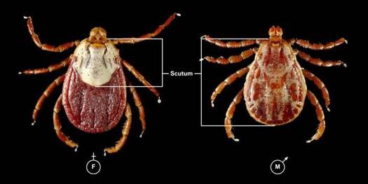
Rocky Mountain spotted fever (RMSF) is caused by Rickettsia rickettsii. This intracellular parasite is transmitted to dogs through the bite of an infected tick. The ticks that can transmit RMSF are the Rocky Mountain wood tick, the American dog tick, and the brown dog tick.

In the United States, RMSF is most common in the southern Atlantic states, western central states, and areas of the mid-Atlantic and southern New England coastal states. According to the Centers for Disease Control and Prevention (CDC), only about 1 to 3 percent of the tick population carries R. rickettsii, even in those geographic areas.

Prior research indicated that the infected tick must be attached for at least 2 hours in order to transmit disease. Research in Brazil demonstrated that unfed ticks had to be attached for more than 10 hours for transmission to occur, whereas fed ticks could transmit disease within as little as 10 minutes after attachment. These results may indicate that transmission across all tick species could occur earlier than once thought, depending on when the tick has last eaten. Transmission of the Rickettsia can then occur due to the bite or from exposure to the parasite while handling the tick. 

Clinical Signs

Clinical signs will show up 2 to 14 days after the bite occurred. The parasite creates an inflammation of the body’s small blood vessels, which results in damage to all the organs of the body.

Common signs include fever, lethargy, inappetence, pain, eye/nose discharge, nosebleed, cough, enlarged lymph nodes, lameness, skin necrosis/sloughing, hemorrhage, and peripheral swelling. Petechial hemorrhages (tiny hemorrhages in the skin) will occur in about 20% of affected dogs. Up to one third of the infected dogs will have central nervous system signs (lack of voluntary coordination of muscle movements, weakness, balance problems, cranial nerve abnormalities, seizures, stupor, spinal pain, etc.). Any organ in the body may be affected and the clinical signs may be mild or severe enough to result in death.

Diagnosis

Diagnostic tests for RMSF include blood tests looking for severely low platelet count, plus coagulation profiles, blood chemical analysis, and serology. (Paired titers, from blood samples taken 14 to 21 days apart, are often needed, but a single high titer in dogs that have clinical signs is consistent with an active infection.)  Response to antibiotic therapy is suggestive, but not diagnostic.

Treatment/Management

Specific treatment relies on the use of appropriate antibiotics. Response to the antibiotics usually is seen within 24 to 48 hours, although advanced cases may not respond at all to treatment. The most common antibiotics used are tetracycline, doxycycline, and minocycline. Chloramphenicol is usually reserved for pregnant bitches or young puppies. Fluoroquinolones, such as enrofloxacin, have shown efficacy, but their use is generally restricted to older animals. Side effects to any of the antibiotics may be seen. Your veterinarian will choose the antibiotic that best suits your pet’s age, pregnancy status, etc.

Blood transfusions to treat anemia and other supportive therapies may be needed.

Prognosis

Prognosis is excellent in dogs that are diagnosed and treated early and who have no complications. Lifelong immunity often occurs after the infection is cleared.

More severely affected patients are at higher risk for complications, such as kidney disease, neurological disease, vasculitis, and coagulopathies. Prognosis is guarded for patients that have complications.

Prevention

Limit your dog’s exposure to ticks and tick-infested areas.

Inspect your dog closely for ticks. The sooner you can remove a tick after it attaches, the better the chance that the organism will not have had time to become infective. Wear gloves when removing ticks, as the infective organism can get into your body through abrasions, cuts, etc. You can also use a tick remover tool. 
4477841

Nighttime Waking in Senior Dogs

Sleep is essential for dogs and people. Disturbances of the sleep-wake cycle occur in older pets and often disrupt the entire family. Interrupted sleep is a common problem in older pets and may occur for many reasons, including both medical and anxiety-related causes.

Diagnosis and Clinical Signs

There are many reasons an older dog may have difficulty sleeping at night. Dogs do get cognitive dysfunction, which is similar to dementia in people.  The hallmarks of these syndromes are progressive confusion, reversal of day-night wake-sleep patterns, and poor adaptability to new situations. Just like older people with early-stage dementia, the dog may function well in routine and familiar situations but be confused in new situations.

Waking at night may occur for reasons other than cognitive dysfunction, such as pain or discomfort. Dogs who are feeling discomfort may pace, pant, vocalize, or be restless at night but may not show any physical limitations during the day. The effects of pain or sore muscles may be more noticeable to the dog at night.

The urge to eliminate may wake many dogs. Senior pets are more likely to have medical conditions that would increase the pet’s urination or defecation frequency or decrease control of these bodily functions.

Many physical and medical conditions should be considered, including urinary tract infections, kidney disease, hormone imbalances, Cushing’s Syndrome, gastrointestinal (GI) upset, and neurological impairments.

Anxiety can cause nighttime waking. Dogs may be anxious because of weather-related noises such as thunderstorms or noises in the home from people or other animals.  The dog may also be anxious during the day but the severity is often increased at night since these noises are more pronounced and start when it is quiet. Furthermore, the dog’s reaction may be limited to restlessness or pacing, which is often not noticed during the day. Seizures may occur anytime but often seizures that occur at night are not seen by the family, so people are awakened by a dog in a confused state.

Thinking about the pattern of when the dog wakes can help figure out the cause and at least partially correct the problem. Some dogs have difficulty settling down or falling asleep while other dogs may go to sleep easily and then wake up in the middle of the night. Some dogs go to sleep, enjoy slumber much of the night, and then wake up early. A definitive diagnosis may be difficult because there are so many medical conditions to consider. Cognitive dysfunction is only diagnosed with certainty in the later stages, and there is no test for it.

Management

After evaluating possible medical causes, try to establish a predictable routine for nighttime slumber. The pet’s routine as a younger dog may provide some insight into his preferences but consider age-related changes. An older dog may require an orthopedic bed to ease pressure on bones and joints. Warmth may also be important as the dog may not be able to regulate body temperature and cold weather may affect sensitive joints.  Establish a bedtime routine that may include a late trip outside for elimination, a massage, and quiet time settling in a comfortable area. Most dogs prefer to be with family members but some prefer to rest alone. Quiet, soothing music may obscure sounds and a night light may help a dog with vision problems to walk in dark hallways with more ease.

Even though this condition may be frustrating, avoid punishing or scolding. After addressing your dog’s physical needs, try gently and calmly returning your dog to where he likes to sleep. Some dogs want to sleep in bed with people; this is a matter of personal preference and family tolerance. Unless it is unsafe due to aggression, it is acceptable for dogs to sleep with people. Be sure to consider if this pattern of slumber with canine companions will continue to be acceptable since once they have tried it, many dogs like to sleep on comfortable mattresses with comforters. Despite commonly perpetuated myths, this type of “spoiling” does not cause other behavior problems; it does, however, teach a dog it is nice to sleep in a person’s bed.

Nutraceutical and Pheromone Interventions

Cognitive dysfunction is a slow, degenerative, and progressive disorder. Supplements are available that may ease the severity and slow its course. Ask your veterinarian about diets designed for the unique needs of senior pets. Some of them provide antioxidants (vitamins C and E, beta carotene, selenium, flavonoids, and carotenoids) that may reduce the neurological damage caused by free radicals and fatty acids as well as dl-alpha-lipoic diet and l-carnitine. Others include botanic oils such as medium chain triglycerides (MCT’s) that provide an alternative energy source. These diets are shown to improve a senior dog’s performance on cognition tests.

Dietary supplements that contain phosphatidyl serine, ginkgo biloba, and grape extract (resveratrol) are likely to slow and minimize cognitive impairment. S-adenosyl-l-methionine, or SAMe, may reduce anxiety and increase awareness and activity in the senior dog. Furthermore, SAMe is safe or even beneficial for patients with compromised liver function. These supplements are most beneficial in the early stages of cognitive decline.

A dog-appeasing pheromone (such as Adaptil) may ease anxiety and promote a feeling of well-being. The diffuser may be especially useful to create an area and support the use of a desired area for resting. The collar is most useful for dogs who experience anxiety all the time.

Melatonin may help create a normal nighttime sleeping cycle. This supplement should be given at bedtime on a regular basis. Combined with a predictable nighttime routine, it can help create a normal sleep pattern.

Pharmacological Treatments

Sometimes medications may be helpful to induce sleep. Benzodiazepines (anti-anxiety medications) such as diazepam (Valium), alprazolam (Xanax), or oxazepam (Serax). Trazodone may also be helpful. These medications are relatively quick-acting but don’t last long so administration must be timed carefully. Medication should be combined with other recommendations described above.

Generalized anxiety may be reduced with medications such as SSRI’s or TCA’s but these medications require many weeks before positive effects are seen.  Cognitive dysfunction symptoms may be reduced with selegiline (Anipryl), which when given in the morning may help to keep the dog more alert and active during the daytime. Old age onset of anxiety may reflect the additive effect of lifelong learning or be a sign of progressive decline from cognitive dysfunction.

Expected Outcome

Nighttime waking may be caused by medical or behavioral changes common in senior dogs. Often these problems may be managed and good sleeping patterns can be re-established, though many of the underlying conditions may be persistent and worsen with age. Quick intervention is important since undesirable habits may form quickly and people may become frustrated when they’re sleep deprived.

4127238

Blastomycosis is a Systemic Fungal infection Affecting Dogs and Cats

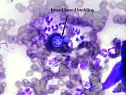
Blastomycosis, caused by Blastomyces dermatitidis, is a systemic fungal infection that affects dogs and cats. Blastomycosis is most common in certain geographic areas in North America, most often the Mississippi, Ohio, Missouri, Tennessee, and St. Lawrence River valleys, and in three provinces of Canada (Quebec, Ontario, Manitoba). It has also been reported in Africa, India, Europe, and Central America. (Fungal growth is supported by wet, sandy, acidic soils rich in organic matter, which is why it is found in valleys and is seen most frequently near water.)

Pathophysiology

Infection with Blastomyces occurs when a cat or dog inhales the fungal spores into the lungs. The incubation period is from 5 to 12 weeks. Some animals don’t show clinical signs for a long time after being infected, but those animals are not contagious to other animals and people. Blastomycosis organisms have a predilection for the respiratory tract, and pulmonary disease is the most common sign (88-94% of canine cases). Once the lung disease develops, yeast forms of the organism spread throughout the body. Organs typically affected include eyes, bones, skin, lymph nodes, subcutaneous tissues, brain, and testes. It can also be found in the nose, prostate, liver, mammary glands, and heart, but those locations are less common.

Dogs appear to be much more susceptible to infection than other species. Although the disease does occur in both people and cats, the incidence is much lower than in dogs. Dogs are ten times more likely to contract the disease than are people, and 100 times more likely than are cats. The incubation period in dogs is also shorter than in people. The reason dogs are more susceptible is unknown, but immune-deficiency may play a role. Annual prevalence in dogs in endemic areas is estimated at 1-2%. Many infected animals live within 0.25 mile of water. An increased number of cases can occur after periods of unusually heavy rainfall. Historically, young (i.e. 1-5 years), male, large-breed (e.g. hounds, pointers) dogs have had the highest risk of infection. (This is probably because these animals would tend to have more contact, due to hunting activities, with the organism’s geographical area.) However, any age, breed, or sex of dog can be infected.

Clinical Signs

The signs of illness will depend on what organs are infected. Some dogs will have eye problems, and some will have neurological signs (seizures, head tilt, etc.). Lameness may occur, if the infection is in the bones. Owners of dogs in the specific geographic areas should watch for coughing, difficulty breathing, eye inflammation, enlarged testicles, fever, swollen lymph nodes, ulcerated/draining skin lesions, bloody urine, difficulty urinating, nasal cavity signs (including bloody nasal discharge), and lack of appetite. (Weight loss occurs because of the decreased appetite.) Infections in the nasal passages may result in skull damage, and lead to infection of the brain. Large skin abscesses and neurologic signs are more common in cats than in dogs, while bone lesions are more common in dogs than in cats. Hypercalcemia can occur in dogs, although it’s rare in cats and non-domestic feline species.

Diagnosis

Diagnosis involves physical exam, blood tests, imaging (radiography, ultrasonography, CT, etc.), urinalysis to look for Blastomyces yeast, cultures, serology, and biopsies of affected organs. Fungal serology, to look for antibodies, is not always accurate and has been known to produce false negatives. The enzyme immunoassay (EIA) for B. dermatitidis galactomannan antigen appears to have a high sensitivity in urine (93.5%) and serum (87%). The EIA assay is commercially available; cross reactions with Histoplasma capsulatumCoccidioides immitis, and Penicillium marneffei can occur with this assay. The definitive diagnosis is finding the fungus in the tissues.

Treatment

Treatment involves various antifungal medications, including itraconazole, ketoconazole, fluconazole, amphotericin B, and some combination therapies. These medications usually need to be taken for a long period of time; how long depends on the specific case. Treatment usually can be done at home, unless the disease is severe. Pets with respiratory involvement should be on restricted activity. In severely ill dogs, intravenous fluids, oxygen, antibiotics for secondary infections, and pain medication may be necessary. Skin lesions may require wound cleaning and debridement.

Eyes that are severely affected may not respond well to the treatment because the medication does not penetrate eyes very well. Ocular blastomycosis cases may need systemic and topical corticosteroid therapy, topical anti-glaucoma medications, etc. Significantly affected eyes may require enucleation (removal of the eyeball).

Pets with severe lung disease may get worse at the beginning of treatment because the fungal organisms are dying; the mass death of organisms can cause severe respiratory problems.

Patients may not appear to improve for one to two weeks after the start of treatment. Close monitoring should be done for the first two weeks, and then rechecks are usually scheduled on a monthly basis. Rechecks may involve blood tests, biochemistry profiles, and imaging, if appropriate.

Prognosis

The prognosis for many pets is good, especially if owners can afford to treat the pet for long enough. Approximately 50% to 75% of affected dogs recover when treated with itraconazole, fluconazole, or an amphotericin-ketoconazole combination. Prognosis is poor for dogs with severely affected lungs, but if the dog survives the first 7 to 10 days of therapy, the prognosis improves. However, mortality rates in dogs with Blastomyces dermatitidis can be as high as 41%.

The prognosis for the retention of vision, in animals that have eye disease, is variable.

Dogs with brain involvement often die. Seizures are common upon death of the organism and may be uncontrollable.

Prognosis is also poor when at least 3 body systems are involved.

Relapses are most likely to occur in dogs that had a severe case at onset, or dogs that were not treated long enough. Relapses are most common within the first 6 months after treatment. Dogs that recover from the disease are probably not immune to getting it again.

After discontinuing therapy, animals may be rechecked at 1, 3, and 6 months for evidence of relapse. In one study, relapse rates for itraconazole and fluconazole were 18% and 22%, respectively. Relapses are treated like a new infection.

There is no way to prevent your pets’ exposure to Blastomyces other than by keeping them away from affected geographic areas.

Zoonotic Potential

Blastomycosis is not considered to be a zoonotic disease. It is acquired by humans via inhalation or direct contact with infective conidia/spores. Risk of infection is higher for excavation workers, and for people working or playing in wooded areas with waterways. Blastomycosis cannot be spread between dogs and other animals, or between dogs and people. However, immunocompromised people should limit their contact with infected pets and should wear gloves when cleaning and treating draining lesions.

4127904

Chronic Bronchitis in Dogs is Neither Infectious nor Contagious

Definition and Causes

Chronic bronchitis is usually a non-infectious, inflammatory condition affecting the lining (mucosa) of the large airways (the trachea and bronchi) that results in a cough on most days for more than two months. In most cases, the specific cause of chronic bronchitis in dogs is not identified.  Chronic bronchitis is neither infectious nor contagious to other dogs; dogs will not pass it on to other dogs as they can in the acute form (see below).

Acute bronchitis, on the other hand, is most often caused by infectious agents, such as viruses (e.g. parainfluenza virus, adenovirus, canine influenza virus, distemper virus, or bacterial agents Bordetella bronchiseptica is the most common).  It is commonly termed kennel cough or canine infectious tracheobronchitis. Inflammation can also be caused by noxious irritants such as second hand smoke, foreign bodies, allergens (similar to hay fever in humans), foods allergens, and either migrating or primary lung parasites. In many of these cases, this acute bronchitis, occurring at the time of the infection or insult, will resolve with appropriate therapy from your veterinarian.  However, if the bronchitis persists long enough – generally, more than a few months – it is labeled chronic, and the cough itself can begin to cause and perpetuate airway inflammation.

Clinical Signs

Dogs with chronic bronchitis generally have a persistent hacking cough.  Some people describe it as sounding like a goose honking.  However, any trachea-bronchial inflammation/irritation can produce a similar sounding cough.  Often, the coughing occurs during the night or when the dog first starts to move around upon waking.  It also commonly occurs with excitement or exercise.

Diagnosis

Chronic bronchitis is a diagnosis of exclusion.  Other diseases can cause clinical signs similar to chronic bronchitis.  These include tracheal collapse, laryngeal paralysis, lung diseases, lung cancer, heart failure, heartworm disease and infectious causes of acute bronchitis (listed above).  Your veterinarian will likely attempt to rule out many of these causes with a history, physical examination, and additional testing.

Diagnostic tests that are used to establish a diagnosis of chronic bronchitis include chest x-rays (thoracic radiographs), airway endoscopy (passing a small camera into the larynx, trachea and bronchi), collecting samples from the lower airways for culture and analysis of the cells in the airways (broncho-alveolar lavage or a transtracheal washing), and sometimes even echocardiography (examining the heart using ultrasound).  A fecal analysis might be needed to rule out parasites.

Treatment

Once a diagnosis of chronic bronchitis is made, your veterinarian will generally treat this condition with a variety of medications.

There are several things to remember about treating chronic bronchitis:

  • The cough in chronic bronchitis is unlikely to completely disappear.  The aim is to reduce the coughing by more than 70-80%, and to reduce the severity of the coughing fits.
  • Coughing begets coughing – the more a dog coughs, the more they irritate their airways, which stimulates more coughing.  Therefore, the aim is to initially break the coughing cycle.

Treatment of chronic bronchitis generally relies on using a combination of medications:

  • Corticosteroids. These drugs are the main therapy for chronic bronchitis. They reduce and suppress the inflammation in the airways.  They can be given orally (pills) or with an inhaler.  Specific canine inhalers are required to properly administer steroids.
  • Certain antibiotics, such as doxycycline, minocycline, or azithromycin may be given to rule out common infectious agents of the airways.  These particular antibiotics also have an anti-inflammatory effect, and may decrease coughing even if there aren’t any infectious agents.
  • Cough suppressants.  These are usually narcotic-based preparations that require a prescription. They sometimes need to be given to the point of sedating the dog, especially early in treatment.
  • Bronchodilators.  These may help by relaxing the airways, allowing air to move more easily into and out of the lungs. They can be given orally, by nebulizer, or by inhalers.

Your veterinarian might prescribe a combination of these drugs, and alter the doses as the disease is controlled to minimize side effects while still maintaining good control of the coughing.

An important factor in successful therapy of chronic bronchitis is weight loss.  If your pet is overweight, a loss of even 5-10% of the body weight can dramatically improve the coughing.  When the dog is overweight, improving your dog’s condition is completely in your hands; therefore, your veterinarian might prescribe a weight-loss program for your pet in addition to the medications.

Your veterinarian might also ask you to keep a “coughing log” to keep track of the severity and frequency of coughing, as this can help determine how successful the treatment is.

Prognosis

The prognosis for treating chronic bronchitis is good.  However, because this condition generally occurs in older dogs, other causes of chronic coughing (listed above) might complicate the management.  If coughing persists or recurs despite appropriate therapy, your veterinarian might be required to pursue other causes, or refer you to a specialist for additional diagnostic tests.

5770367

Alcohol Poisoning

Dogs and cats can get more than just drunk when they drink ethanol, isopropanol, or methanol — they can get a trip to the emergency room. Pets can die from ingesting alcohol. 

What are ethanol, isopropanol, and methanol?

Ethanol (ethyl alcohol) is most commonly formed from the fermentation of sugars. Ethanol is found in alcoholic beverages, some liquid medications and mouthwash bases, rotting/fermenting fruits, and rising (raw) yeast-containing bread dough. The “proof” of ethanol is twice the percentage of alcohol concentration (i.e. 80 proof = 40% ethanol). Although ethanol is also in some household inks, cleaners, and solvents, the levels are low enough to generally be of no clinical significance if these products are ingested by pets.

Isopropanol (isopropyl alcohol) is in rubbing alcohol (70%), some anti-freezes, some detergents, glass/window cleaners, perfumes/colognes, and alcohol-based topical sprays, including some pet flea-control and grooming products. Isopropyl alcohol is more than twice as potent as ethanol or methanol.

Methanol (methyl alcohol, wood alcohol) is most commonly found in automotive windshield washer fluids (20-80%) but is also in some gasoline additives, “canned heat” fuels (e.g., Sterno®), and some household solvents (e.g., paint removers).

Although the strength of these three alcohols varies, the clinical signs associated with their ingestion by dogs and cats are quite similar.

All poisoning problems boil down to the amount of alcohol ingested compared to weight, just like a dosage of medicine. Thus, when pets drink an alcoholic beverage that was left within reach or that was given to them intentionally by someone, it can cause a significant toxicity problem. In addition, significant absorption can occur through the skin or by inhalation.

Dogs are highly susceptible to the effects of alcohol. Signs of mild inebriation may occur with even minor exposure. Within 15 to 30 minutes after the pet has drunk the alcohol on an empty stomach (or within 1 to 2 hours on a full stomach), central nervous system (CNS) signs, such as staggering, excitement, or decreased reflexes, can begin. Behavioral changes can be seen, as can an increased need to urinate. As the problem gets worse, the pet may become depressed, have a slow respiratory rate, or go into cardiac arrest. Puppies and kittens are at particular risk because of their small size and immature organ systems. 

Alcohols are depressants, so many of the clinical signs associated with them are due to their effect on the CNS. Alcohols irritate the gastrointestinal tract. They also act as diuretics, and the alcohol and its metabolites are eliminated by the kidneys. Liver damage may occur after exposure, although it is much more common in animals that have repeated or chronic exposure. 

Clinical Signs

Clinical signs of intoxication can occur within 15-30 minutes of ingestion of alcohol. Signs include nausea, vomiting, thirst, urination, dehydration, lethargy, incoordination, disorientation, becoming cold (hypothermia), low blood pressure, and alcoholic bad breath. In severe cases, blindness, tremors, tetraplegia, respiratory depression, coma, or seizures may develop. Death is uncommon but may occur, especially if the pet has severe respiratory and cardiovascular depression, low blood pressure, and/or hypoglycemia. 

Diagnosis

Diagnosis is based on a history of exposure, combined with appropriate clinical signs and laboratory tests.  

Treatment/Management/Prevention

The treatments your veterinarian may use can include assisted ventilation, intravenous fluids (to improve elimination of the alcohol/metabolites, to provide cardiovascular support, and to correct electrolyte abnormalities), seizure control, and other supportive therapies. In addition, if the alcohol toxicosis is due to the ingestion of bread dough, your veterinarian may need to wash out your pet’s stomach with cold water to inhibit further alcohol production and to break up the dough mass for removal. In addition, your pet will be confined to prevent accidental self-injury.

Prognosis

Most dogs with alcohol intoxication can recover with adequate symptomatic and supportive care. Recovery may take 8-12 hours and tends to take longer if it was a severe intoxication or if isopropanol was involved.

Preventing Alcohol Toxicosis

All alcoholic beverages and alcohol-containing fluids should be kept out of reach of your dogs and cats. Consult your veterinarian before giving any ethanol-containing liquid medications.

Do not feed raw bread dough to your pets or leave bread dough out to rise in areas that can be reached by them. Always dispose of discarded bread dough carefully. 

The ASPCA National Animal Poison Control is available 24 hours a day at 888-426-4435. Expect an initial consultation fee of around $100.00 and additional follow-up is at no charge. You will be assigned a case number your veterinarian can use to communicate with a toxicology specialist before beginning treatment.