Tag: cats & dogs

5425457

Calcium Phosphorus Balance in Dogs and Cats

In renal insufficiency, phosphorus is not anyone’s friend. The same phosphorus that has so many helpful roles in the body (from transferring biochemical energy to combining with calcium to form bone), turns against us in a condition called renal secondary hyperparathyroidism.

The short version is that the failing kidney is no longer good at getting rid of excess phosphorus and phosphorus levels in the blood begin to rise. The rise in phosphorus upsets the delicate balance between calcium and phosphorus and activates a regulatory hormonal cascade that attempts to re-establish control. Without healthy kidney tissue to play its role in this balance, the body is fighting a losing battle. Calcium is mobilized from bone to balance the phosphorus but in the end this only serves to demineralize and weaken the bones and cause calcium phosphate deposits to form in soft tissues. These mineral deposits are inflammatory and damaging.

The hormones that play roles in the regulation of calcium and phosphorus are parathyroid hormone and calcitriol (which most of us know as Vitamin D). The story co-stars a substance called Fibroblast Growth Factor 23. These three materials interact to maintain a normal and effective blood calcium level without allowing phosphorus levels to run away.

We’ll review the story of how this works.

Keeping Calcium Under Control

While our discussion of renal disease largely revolves around phosphorus, the importance of calcium cannot be underestimated. The movement of calcium ions is what allows our muscle fibers to contract, not just in our arms and legs but also in our hearts, and involuntary intestinal and blood vessel muscles as well. Calcium combined with phosphorus makes up bone; in fact, bone can be considered a storage depot for calcium when we need some in a pinch. The blood level of calcium is tightly regulated by hormones within a narrow range as too much calcium is as dangerous as too little.

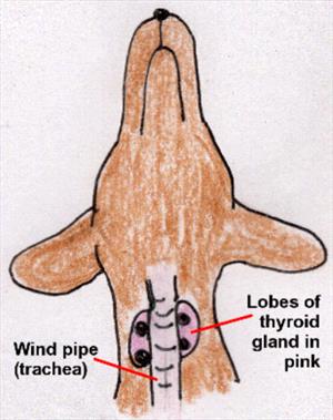
There are four tiny parathyroid glands around the thyroid gland in the throat area. These glands produce a biochemical called parathyroid hormone (often abbreviated PTH). PTH basically raises calcium and activates vitamin D, which further raises calcium. It sounds like calcium would just continue to rise out of control but there is a safeguard. When active vitamin D levels get to a certain point, they shut off PTH production. Calcium levels then drop down and between these two hormones, blood calcium is regulated within a healthy range.

Keeping Phosphorus Under Control

When it comes to phosphorus, PTH and calcitriol no longer work together. PTH signals the kidney to dump phosphorus while calcitriol signals the kidney to save it. In the normal body, the coordination of these hormones works out to keep calcium and phosphorus levels in balance, no matter what is happening with the blood calcium.

All this goes along swimmingly until the kidney is too damaged to respond to its hormonal signals or properly activate calcitriol.

Parathyroid hormone causes the kidney to dump phosphorus while vitamin D causes the kidney to save phosphorus.

What Happens in Kidney Failure?

In early kidney failure, the kidney becomes unable to get rid of phosphorus normally, and as a result phosphorus levels begin to rise. This activates a substance called Fibroblast Growth Factor 23 (affectionately known as FGF-23) from the bones. FGF-23 encourages the kidney to dump more phosphorus and dampen the activation of vitamin D (remember that vitamin D tells the kidney to save phosphorus). As part of its mission to dampen vitamin D, FGF-23 also instructs the parathyroid glands to cut back on PTH. All this vitamin D suppression is all well and good and helps keep the phosphorus levels reasonable, but pretty soon FGF-23 has dampened both PTH and vitamin D so well that calcium levels begin to drop.

And this is where calcium-phosphorus Hell breaks loose.

At this point, kidney failure is no longer early. Dropping calcium levels supersede any FGF-23-mediated suppression of PTH and the parathyroid glands crank up PTH production to bring calcium levels back up. To meet the body’s demand for circulating calcium, the bones liberate their structural calcium to save the day, but unfortunately doing this also liberates more phosphorus. This leads to an even higher phosphorus level. Circulating phosphorus binds the circulating calcium creating crystals that deposit in the body’s soft tissues and generate inflammation. Bones are weakened and replaced with fibrous tissue. Calcium is depositing uselessly all over the place and the parathyroid glands must crank even harder to keep calcium levels livable. A metabolic disaster has occurred at this point.

Making matters worse are other effects of excess parathyroid levels. In high amounts, nerves cannot conduct electrical impulses properly. Patients become dazed and poorly responsive.

The goal is to keep the phosphorus level from getting out of control in the first place. If this is not possible, the goal is to get the phosphorus level back under control and keep it there. 

Treatment

How do we Control Phosphorus Levels?

The goal phosphorus level defined by the International Renal Interest Society is between 2.5 and 4.6 mg/dl for earlier renal insufficiency stages and to keep phosphorus levels under 6 mg/dl for Stage Four (more advanced) patients.

Fluid Therapy

Often simply giving fluids under the skin at home provides enough extra circulation through the kidneys for the extra phosphorus to be excreted normally. Further treatment may not be needed. Hydrating a damaged kidney effectively maximizes the kidney’s remaining function, and this may be enough to control phosphorus.

Therapeutic Diet

It is important to remember that ultimately phosphorus balance is a matter of balancing phosphorus entering the body with phosphorus leaving the body. If the kidney is no longer effective at removing phosphorus, we can perhaps work on limiting the amount of phosphorus entering the body. The first step in doing this is with a therapeutic renal diet. These diets are designed to give the kidney less work to do, and that includes limiting phosphorus entering the GI tract. After one to two months on a phosphorus-restricted diet, blood tests will indicate whether or not additional phosphorus treatment is needed.

Phosphate Binders

These products are given with food to bind phosphorus in the food. The complex of phosphorus and phosphate binder cannot be absorbed into the body with the result being reduction of incoming phosphorus. If the pet will not eat food containing the binder, the binder can be given just before or just after the meal but the binder must be in the GI tract at the same time as the food or it will not work. There is no point in giving a binder to an animal that is not eating. Giving the binder before the meal can induce enough stress so that the pet will not eat so it is better to give the binder after the meal if the binder is not accepted mixed in the food.

There are a number of phosphate binders available and which one is selected will depend in part on the concurrent calcium situation. This means that the ionized calcium level (not the total calcium level) must be measured. Some binders also supplement calcium while others drop or have no influence on calcium. Some patients require more than one binder to get their phosphorus value low enough.

Calcitriol


You might think that calcitriol would not be helpful in this situation since it leads the kidney to retain phosphorus. The good news is that when small enough doses are given, calcitriol can still act as the off switch for parathyroid hormone without causing the kidney to retain phosphorus. The amounts needed for this beneficial effect are so small (they are measured in nanograms) that a compounding pharmacy is needed to custom-make the product at the proper dose.

  • Calcitriol cannot be used in patients with elevated blood calcium levels.
  • Calcitriol cannot be used in patients with phosphorus levels that are already abnormal. This is a preventive measure more than a treatment.

Renal secondary hyperparathyroidism and its associated high blood phosphate levels are one of the most common uremic toxin issues in treating kidney disease. Dedication is required to get phosphate binders into the pet along with food when the pet’s appetite is weak. Phosphate binders come in powders, liquids, capsules, compounded flavored chews, and other formats. Consider what approach is likely to work best for your pet but, remember, the binder only works with food.

4128675

Cherry Eye in Dogs and Cats

Prolapse of the Tear Gland of the Third Eyelid

Unlike humans (who only have eyelid, technically called the nictitans or nictitating membrane, arises from the inner corner of the eye and covers the eye diagonally as shown. The eye is lubricated by tear film, which consists of water, oil, and mucus. The oil comes from glands lining the outer eyelids, the mucus comes from glands in the conjunctiva (the pink part inside the eyelids), and the water comes from tear (or lacrimal) glands.

Each eye has two tear glands: one just above the eye and one located in the third eyelid. The gland in the third eyelid is believed to produce a full 30 percent of the total tear film water, so it is important to maintain the function of this gland.

The tear gland of the third eyelid is held in place by tissue fibers but some individuals have weaker fibers than they should so the gland protrudes. This protrusion is called a cherry eye.  In the smaller breeds – especially Boston terriers, cocker spaniels, bulldogs, and beagles – the gland of the third eyelid is not strongly held in place for genetic reasons. The gland prolapses (drops down) out to where the owner notices it as a reddened mass. Out of its normal position, the gland does not circulate blood properly, may swell, and may not produce tears normally. 

Treatment: Replacing the Gland in its Proper Location

By far the best treatment for cherry eye is replacing the gland back into its proper location. There are two techniques for doing this. The traditional tucking method (also called tacking) is probably the most commonly performed. Here, a single stitch is permanently placed, drawing the gland back where it belongs. Complications are uncommon but be aware of the following possibilities:

  • If the stitch unties, the surface of the eye could become scratched by the suture. If this occurs, the eye will become suddenly painful and the suture thread may be visible. The suture can be removed and the problem solved.
  • The tuck may not be anchored well enough to hold permanently. In fact, this surgery is notorious for this type of failure, and frequently a second or even third tuck is needed. If more than a couple of tucks have led to failure, it may be better to try the imbrication technique as described below. Some cases are repaired using both tuck and imbrication together.
  • Sometimes cherry eye is accompanied by other eyelid problems that make the repair more difficult or less likely to succeed. In these cases, again, if the simple surgery is not adequate, ask your veterinarian if a referral to a veterinary ophthalmologist for the second surgery to maximize the chances of a permanent resolution is in the best interest of you and your pet.

In a newer surgical technique called imbrication, or pocketing, a wedge of tissue is removed from directly over the actual gland. This technique is more challenging as it is not easy to determine how much tissue to remove. Tiny stitches that will eventually dissolve are used to close the gap so that the tightening of the incision margins pushes the gland back in place. 

Complications may include:

  • Inflammation or swelling as the stitches dissolve.
  • Inadequate tightening of the tissue gap may lead to recurrence of the cherry eye.
  • Failure of the stitches to hold and associated discomfort. Loose stitches could injure the eye depending on the type of suture used.

Sometimes both surgical techniques are used in the same eye to achieve a good replacement. Harmful complications from cherry eye surgery are unusual but recurrence of the cherry eye can happen. If it recurs, it is important to let your veterinarian know so that a second surgery, either with your veterinarian or an ophthalmologist, can be planned.

Expect some postoperative swelling after cherry eye repair but this should resolve and the eye should be comfortable and normal in appearance after about a week. If the eye appears suddenly painful or unusual in appearance, have it rechecked as soon as possible.

Treatment: Removing the Gland

Historically, the prolapsed gland was treated like a small tumor; it was simply removed. This was before the full significance of the gland was realized.

If the third eyelid’s tear gland is removed, it cannot be put back in place. If the other tear gland (the one above the eye) cannot supply adequate tears, not an uncommon phenomenon in older small breed dogs, then the eye becomes dry and uncomfortable. A thick yellow discharge results and the eye develops a blinding pigment covering for protection. This condition is called simply dry eye or more scientifically keratoconjunctivitis sicca and daily medical treatment is required to keep the eye both comfortable and visual. Not only is dry eye uncomfortable, but its treatment is often frustrating and time-consuming and there is expense involved. If left untreated, the eye can become blind. We would like the dog to maintain the greatest amount of tear-producing tissue possible, thus removing the gland for cosmetic reasons is not an acceptable treatment method.

4128893

Constipation and Megacolon in Dogs and Cats

Simple Constipation

An occasional episode of constipation is not cause for alarm. When a pet is constipated, stools seem unusually hard and there is unproductive straining. Hard stools might be found in unusual locations. Veterinary assistance may or may not be needed. Here is what you need to know if you think your pet is constipated.

  • One of the purposes of the colon (large intestine) is to store stool. Many pet owners become alarmed if their pet has not passed a stool in a few days, especially in a post-anesthetic situation. In fact, the colon can easily store several weeks’ worth of stool so if only a few days have passed it may be worthwhile to wait a little longer. If your pet seems to be uncomfortable or straining unproductively, then it is probably time for intervention.
  • Why do pets get constipated? Straining unproductively can be a symptom of either constipation or large intestinal diarrhea. In either case, small amounts of mucous, gooey, or even blood-tinged stool can be passed and there is a lot of pushing involved. Difficulty urinating can also appear as straining. The point is that if all you have noticed is straining, it may not be constipation. Straining to urinate is often an emergency situation so if there is any question about the pet’s ability to urinate, see the veterinarian right away.
  • It may be tempting to buy a commercially prepared enema at the drug store and attempt to relieve the pet’s problem at home. Some commercially prepared products are toxic to pets so it is important that human constipation products, be they enemas or laxatives, not be used in pets without specific veterinary instruction.
  • In fact, the colon can easily store several weeks worth of stool so if only a few days have passed it may be worthwhile to wait a little longer. If your pet seems to be uncomfortable or straining unproductively, then it is probably time for intervention and you should call your veterinarian.

Simple constipation can be caused by any number of reasons. Some animals excessively groom themselves (especially if they are itchy) and find themselves passing stools containing large amounts of hair. This is not an uncommon cause of constipation and often treatment for the excessive grooming is helpful.

Some animals, especially dogs, get in the habit of eating gravel, stones, dirt, bones, or plants. This does not usually indicate a dietary deficiency as many owners suspect, though we do not have a good explanation for this unusual dietary behavior. The stools produced can be sharp or painful to pass, often leading to straining and discomfort.

Some medications can have constipation as a side effect (sucralfate).

An important potential cause of constipation to check for is an electrolyte imbalance, especially in an older pet. This may be the only noticeable sign of an important metabolic problem such as insufficient kidney function. If constipation has been a recurring problem, then this kind of lab work database becomes especially important.

An internal obstruction may be causing the problem. For example, animals hit by cars often suffer a fractured pelvis. These usually heal without surgery but can heal such that the pelvic canal through which stool must pass is narrowed. Constipation may not result for years after the initial trauma. An old fracture is generally obvious with a radiograph of the abdomen.

Alternatively, an enlarged prostate gland is a common feature of older male dogs. The gland sits just below the colon and can press on the colon serving to narrow it. Neutering usually solves this problem, though sometimes the problem is more serious, such as a prostate tumor. An enlarged prostate is often palpable rectally though the size of the prostate is better assessed with a radiograph.

Treatment for Simple Constipation

Simply isolated episodes of constipation are easily treated with a DSS, soap and water, or K-Y jelly-based enema. It is important to appreciate that pets do not take kindly to enemas and this kind of procedure should not be attempted at home. It invites bites and scratches, especially if the patient is uncomfortable, to begin with, plus it is a messy undertaking. Enemas are best left to experienced professional staff.

A short course of medication may be prescribed. This might be stool softeners such as laxatone, lactulose, polyethylene glycol 3350 (Miralax®), or DSS or it might be a medication to increase the normal motility (contractile strength) of the large intestine such as cisapride or bisacodyl (Dulcolax®).

An old-fashioned remedy has been mineral oil taken orally (by mouth). It is best to avoid this temptation because mineral oil is a petroleum oil without flavor and it is easily inhaled into the respiratory tract accidentally when it gets in the mouth. Since it is a mineral-based compound, it cannot ever be removed by the body and the immune system will forever attempt to wall it off with inflammatory granulomas.

For a single episode of constipation, a diet change may or may not be recommended. There are two approaches that are commonly employed in this regard. The first is the addition of fiber to the diet. Fiber is not absorbed by the patient’s intestinal tract and passes to the colon where it contributes to the stool volume. The result is a larger, more bulky stool which, when passed, provides stronger sensory stimulation to the colon than a regular stool. This increased stimulation may result in better colon motility. This type of diet change is achieved most easily by switching to one of the prescription high-fiber diets formulated for this exact use; most manufacturers of therapeutic pet foods include such diets in their offerings. If this is not acceptable to the pet, fiber may be added to the regular diet in the form of:

  • Canned pumpkin
  • Bran cereal
  • Metamucil, Konsyl, Siblin or similar product

Your veterinarian can instruct you as to how much you should add.

The second theory of dietary management is that the colon would perform better with a smaller stool. In this case, a low-residue, high-digestibility diet is used. With such a diet, a greater amount of nutrients is absorbed by the patient and there is less undigested material passing to the colon to contribute to the fecal mass. One of the problems with constipation is that stool becomes dry when water is absorbed by the colon, making the stool harder to pass. The aforementioned high-fiber diets tend to absorb water and potentially make constipation worse whereas low-residue diets help preserve fecal water and create a softer stool.

Recurring Constipation

In recurring constipation, the same treatment methods as listed above are employed but on a more long-term basis. Enemas may have to be used more frequently and medications/diet changes may represent permanent management methods. The lab work database and the abdominal radiographs become especially important.

Some additional comments regarding the long-term use of the above methods:

  • Lactulose is an especially thick and overly sweet liquid and many animals find its taste objectionable (though cats are reportedly unable to taste the sweetness component). The objectionable taste frequently leads to drooling out the medicine into the fur of the chin and ruff. This may become a grooming or cosmetic problem.
  • Bisacodyl works by stimulating the pelvic nerves to increase the motility of the colon. It has been suggested that this kind of nerve stimulation should not be done indefinitely; thus this medication is typically recommended in a finite course.
  • Polyethylene glycol 3350 (Miralax powder) acts by pulling water from the body into the intestinal tract during stool formation, leading to a wetter, more pliant stool. The product is typically given twice daily in food and is generally well tolerated by pets though some trial and error is often needed to find an acceptable dose.
  • Cisapride has a broad dose range depending on individual response. Often a more conservative dose is selected to start. If this does not appear to be effective, there is a good chance that there is room to increase the dose. Do not change a drug dose on your own! If you think the cisapride dose you have been using is ineffective, notify your veterinarian and ask if a higher dose might be a good idea.
  • While neutering usually resolves most enlarged prostate problems, an old pelvic fracture may or may not necessarily be helped by surgically repairing the bones. If the above medical management is not effective, the subtotal colectomy (see below) may become needed.

Constipation vs Obstipation

When constipation becomes a more permanent and continuous problem, it is more correct to use the term obstipation. Here, patients are unable to effectively or completely empty the colon on their own (70% of affected cats are male, 30% female). The obstipated colon is dilated and packed with an enormous, rock-hard burden of feces. The patient is usually quite uncomfortable, with more frequent unproductive straining, lethargy, appetite loss, and even vomiting entering the picture.

Small hard bits of the stool is often found around the house as well as in the litter box. Sometimes liquid fecal secretions are passed around the hard fecal mass, leading the owner to incorrectly think the pet has diarrhea.

Usually, the only way to relieve this is through a more complete de-obstipation process, which frequently necessitates general anesthesia. The patient is hydrated, usually using fluids given under the skin and some enemas are given while the patient is awake. This helps moisten the hard fecal mass and sometimes helps with fecal evacuation. After this, the patient is anesthetized and the fecal mass is milked from the colon by hand. If the colon is severely backed up, often a single procedure is incompletely effective as some of the stool that is higher up may not be accessible at the time of the procedure.

There is no way to predict the frequency with which this procedure must be performed in a given individual; you must simply judge the patient’s discomfort to determine this.

At some point, repeated use of anesthesia may represent an undesirable expense or risk. At this point, home enemas may be reconsidered. As previously mentioned, this is a messy procedure that pets do not appreciate in the least. Your vet will need to show you the equipment and supplies as there are many different products available. The procedure will need to be done in an area that can be mopped or hosed off afterward (an outdoor area, for example). Often the patient will require a bath afterwards. For many pet owners, this is simply not something they want to do but for some people, this can be a valuable management procedure that can save a great deal of veterinary expense.

High-fiber diets are not appropriate after a patient has progressed from simple constipation to obstipation.

Subtotal Colectomy – A Permanent Solution for Cats

For cats, a permanent resolution of this problem can be achieved by surgically removing the diseased colon though this procedure is not nearly as effective in dogs. This generally eliminates the need for any stool softeners, pills, enemas etc. and the patient can resume a low-maintenance lifestyle. The constipation is replaced by a looser consistency stool and, though sometimes this firms up into a more normal consistency stool after a couple of months, it is important for an owner expect this change to be permanent. Patients appear much more comfortable with this new arrangement and most owners are so satisfied with results as to wish they had pursued surgical treatment sooner.

Still, it is important to realize that subtotal colectomy is a major surgery and there are special problems to be concerned about:

  • The colon’s bacterial population is enormous (some ten times higher than the bacterial population of the small intestine). This introduces special concern for any leaking from the intestinal incision. An infection could easily progress to peritonitis and become a lethal complication. This is by no means a common problem but it is important to report any post-operative lethargy or appetite issues immediately, especially if the patient had appeared to be stabilizing and the change is sudden. Leaking after intestinal surgery generally occurs around the third day after surgery if it is going to occur. Fever is a sign of infection but it is best not to attempt to take a rectal temperature at home given the proximity of the colon incision. Your cat will probably be going home on antibiotics.
  • Specific diets are often recommended during the recovery period, usually something highly digestible to minimize the amount of stool produced. The 20% of cats who have persistent loose stool problems generally must stay on this diet permanently.
  • It is not unusual for a patient to refuse food for several days after surgery. The cat must not be allowed to go without food for more than five days without some kind of nutritional support. This may require some form of assistance, such as force-feeding through a special feeding tube. 
  • Scarring of the surgery site (stricture) may lead to narrowing of the bowel and recurrence of the obstipation. If this occurs a second surgery would be necessary to remove the narrowed area.

Most cats do not experience complications with this surgery beyond the initial loose stool mentioned. Results are described as good to excellent.

In Summary: 

Some causes of constipation:

  • Side effects of some medications
  • Excessive grooming causes feces to contain hair
  • Eating gravel, rocks, bones, plants
  • An electrolyte imbalance, especially in an older pet
  • An internal obstruction, perhaps from being hit by a car even years ago

Simple constipation can be caused by any number of reasons. The colon can easily store several weeks’ worth of stool so if only a few days have passed, waiting a bit is reasonable if the pet is not straining without going.

Treatment of one episode:

  • Can be treated with veterinary enema, and possibly stool softeners or laxatives recommended by your veterinarian
  • Add fiber to the diet
  • Use a low-residue, high-digestibility diet

Some commercially prepared enemas for people are toxic to pets; do not give one without discussing it with your veterinarian. Enemas for pets whose colons are filled and cannot pass it are painful and first need sedation.
Recurring constipation uses the same treatments but on a long-term, possibly permanent, basis.

Some medical options for treating recurring constipation:

  • Lactulose, although it is a thick sweet liquid, and many animals find its taste objectionable
  • Dulcolax stimulates the pelvic nerves to increase movement in the colon
  • MiraLAX powder pulls water from the body into the intestinal tract, leading to a wetter, more pliant stool.
  • Cisapride provides improved movement of contents from the stomach to the colon.

Obstipation occurs when constipation becomes an ongoing problem, and they are unable to empty their colon. 

  • The obstipated colon is extended and packed with an enormous, rock-hard burden of feces.
  • High-fiber diets are not appropriate after a patient has progressed from simple constipation to obstipation.
  • Usually, the only way to relieve obstipation is through a more complete de-obstipation process, which frequently necessitates general anesthesia. The patient is hydrated and might be given an enema while awake because it moistens the hard fecal mass. After this, the patient is anesthetized, and the fecal mass is milked from the colon by hand. Often a single procedure doesn’t finish the job as some of the stool that is higher up may not be accessible at the time of the procedure.
  • At some point, repeated use of anesthesia may become too expensive or risky. At this point, home enemas may be reconsidered for obstipated pets.

Megacolon is obstipation in which the colon’s muscles have stretched so much that the colon becomes three or four times larger than normal and stays stretched that way. The colon muscles are unable to push feces into the rectum, so a lot of feces remain in the enlarged colon. The combination of the enlarged colon and obstipation is called megacolon.

For cats, a permanent solution called subtotal colectomy is surgically removing the stretched-out colon, though it is not nearly as effective in dogs. This procedure generally eliminates the need for any stool softeners, pills, enemas, etc. and the patient can resume a low-maintenance lifestyle. The subtotal colectomy is a major surgery with specific concerns:

  • The colon’s bacterial population is enormous, and the incision can leak. Infection can progress to peritonitis and become a lethal complication.
  • Specific recovery diets may need to be used permanently.
  • Cats may refuse food for several days afterward but must not be allowed to go without food for more than 5 days without nutritional support.
  • Scarring may lead to a narrowed bowel, causing obstipation again. A second surgery would be necessary to remove that narrowed area.
  • Results are usually described as good to excellent.
4128650

Colitis-Related Diarrhea in Dogs and Cats

What is Colitis?

In brief, colitis is the term for inflammation of the colon. The chief feature of colitis is a gooey, frequently splattery diarrhea featuring mucus, fresh blood, or both. The stool may start normal then finish soft or may seem gooey throughout. 

There is often accompanying cramping, gas, and a sense of immediate urgency (the sudden need to run for a bathroom). Vomiting can be a feature of this condition though the characteristic diarrhea is the hallmark. Colitis may be acute (lasting only a few days) or chronic (lasting weeks or months on end). Even in chronic cases, weight loss is usually not a feature of this condition.

What and Where is the Colon?

The colon is another term for the large or lower intestine. For those who do not know the lower intestine from the upper intestine, the colon/lower intestine is basically the last segment of the digestive tract. Before reaching the colon, food has been extensively processed. It has already been chewed up in the mouth, acid-treated in the stomach, and enzyme-treated in the small intestine. Most of the nutrients have already been absorbed by the time the undigestible leftovers have passed down to this last part of the GI tract.

But the digestive tract is not through with the food remnants quite yet and this is the where the business of the colon comes in. The colon has three functions: absorbing water, storing stool, and further digesting unabsorbed nutrients. The bacterial population in the colon is about 10 times more dense than that of the small intestine. These bacteria, often referred to as  good or helpful bacteria, take fibers that were undigestible to the host and actually process them into biochemicals that provide nourishment to the colon cells. The bacteria that live in the colon not only feed the cells of the colon, but they also control the pH of the colon environment so that excreted toxins are trapped in the stool and will be pooped away rather than reabsorbed back into the body. They also produce flatulent gases and pigments that lead to the ultimate color of stool. The relationship we have with our colon bacteria is mutually beneficial and is an excellent example of symbiosis.

Diarrhea Can Come from Either the Small Intestine or the Large Intestine and the Approach is Different in Either Case

In classifying diarrhea, it is important to determine whether the problem originates from the small intestine, the colon, or possibly both.  Small intestinal diarrheas tend to be more serious as they involve fundamental problems with obtaining nutrients from food. Diarrheas of the colon are less debilitating but still uncomfortable.

The following are characteristics of large intestinal diarrhea:

  • Large intestinal diarrheas are generally not associated with weight loss, and patients generally have normal energy levels and normal appetites.
  • Large intestinal diarrhea is associated with straining to defecate. Often, this straining is unproductive, leading to the erroneous conclusion that the patient is constipated.
  • There is cramping, flatulence, and a sudden sense of urgency. The pet may not be able to get to an appropriate area before the diarrhea erupts.
  • There is often mucus or slime in the stool
  • There is often fresh blood in the stool.
  • The stool may begin looking normal and formed but finish as a puddle.

A diagnosis of colitis is generally straightforward given the above classic findings, though how to proceed depends on the signs. Is the problem acute (i.e., suddenly there) chronic (has been happening for several weeks regularly), or episodic (happens then goes away, then happens again)?

Colitis Suddenly (Acute Colitis)

A pet that has sudden symptoms of colitis probably has stress-related colitis (common after boarding, moving, severe weather, or other change) or dietary indiscretion-related colitis (usually involves treats or raiding the garbage). These episodes are generally minor and can be cleared with a short course of medication such as metronidazole or sulfasalazine and/or dietary therapy. Parasites, especially Giardia and whipworms, can also cause colitis and the pet may be tested for those to rule them out or be dewormed. In general, a few days of medication and a bland diet should resolve the problem and the pet will be back to normal quickly. During recovery, it is common for the pet to have no stool at all for a couple of days. This is normal and not a sign of constipation. If, however, the pet’s diarrhea is not clearly improved in two to three days, contact the veterinarian to see if further testing is needed.

Diagnosing the Cause of Colitis

If the symptoms of colitis have been going on for a month or more or if they keep recurring and resolving over and over, then a medical workup is needed. It is important to make sure simple causes of colitis have been ruled out, so parasite testing becomes especially important.

A good fecal examination for worms and coccidia plus additional testing for giardia should be performed. If any of these tests are positive, then obviously the parasite in question can be addressed; it is always best to identify the cause of the colitis if it is possible to do so. That said, even if these tests are negative, it is still a good idea to include a broad spectrum de-worming and coccidia treatment, which should probably be given as these treatments are safe and inexpensive. Whipworms, in particular, are difficult to detect and commonly cause colitis symptoms. We want to be sure we have ruled out the simple causes of colitis before more advanced diagnostics begin. Similarly, a week or so of metronidazole, sulfasalazine, or tylosin may effectively treat a toxin-producing Clostridium perfringens infection and potentially solve the entire problem. Of course, a basic blood panel and urinalysis are in order, as they are with any chronic disease, to assess the patient’s general health.

After all the preliminaries above are covered, the patient is assessed for results. If it doesn’t look like one-time treatment of deworming, antibiotics, or diarrhea medication is going to solve the problem, then diagnostics continue to the next level. Most reference labs now have the capability to do PCR (DNA) testing for more obscure organisms such as Tritrichomonas in cats, Cryptosporidium in dogs and cats, and more. This is testing uses a fecal sample and can be used to non-invasively rule out unique infections.

The last step in colitis diagnostics is a colonoscopy with biopsies with the idea of examining colon tissue under a microscope to classify the inflammation. Depending on the type of cells infiltrating the colon lining, the colitis can be classified as lymphocytic/plasmacytic (a form of inflammatory bowel disease), histiocytic (which tends to stem from a type of E. coli infection,) or not truly colitis at all and the entire problem may be the psychosomatic condition known as irritable bowel syndrome. All these conditions have different treatments.

A colonoscopy requires a period of fasting (usually a couple of days) and enemas or some other kind of fluid to clear the colon of residual stool so the naked tissue of the colon can be viewed and sampled. Referral to a specialty hospital is likely needed. Colonoscopy is performed under general anesthesia and not every patient is a candidate for that, plus expense may be a concern. These disadvantages must be weighed against the quality and quantity of information that can be obtained by evaluating a tissue sample from the colon.

Depending on your pet’s specific case and the cause of colitis, your veterinarian may or may not recommend antibiotics. 

Management Tips for Colitis

Colitis is best managed when its cause is known and specific therapy can be instituted. When this is not possible, symptomatic management is often attempted. The following are therapeutic medications and strategies that can be helpful in the treatment of colitis.

Metronidazole and Tylosin

These medications are antibiotics with anti-inflammatory properties in the large intestine and the ability to kill harmful organisms.

Sulfasalazine


This medication consists of a sulfa antibiotic bound to a salicylate anti-inflammatory. The sulfa bond protects the anti-inflammatory medication until it gets to the large intestine, thus saving the anti-inflammatory effect for the disease of the large intestine. This is an effective medication but is typically given three times a day, which is an inconvenience. Cats are sensitive to salicylates, thus this medication is primarily used in dogs.

Dietary Fiber

The role of fiber in colitis is confusing as there is an assortment of fiber preparations (soluble fibers, insoluble fibers, and mixtures). In general, colitis is felt to be a fiber-responsive disease, but there are so many combinations of fiber types that it is hard to know what the patient may be responding to. Insoluble fibers, like cellulose, bulk up the stool and are stimulating to the colon lining. This may not be what is in order if the colon is already irritated although giving some structure to diarrhea may be a good thing. Soluble fibers, like psyllium, are fermented by the colon bacteria into nutrients for the colon cells, which helps them heal. Prescription high fiber diets often have a mixture of soluble and insoluble fibers that have been scientifically determined to help colitis patients. Alternatively, a low residue diet (one of high digestibility) could be used and soluble fiber added to it. The idea with this strategy is to have maximum intestinal absorption of nutrients in the small intestine so less material enters the colon, but once the material gets there the soluble fibers added help the colon cells to heal.

Prebiotics such as Fructooligosaccharides (FOS)

Prebiotics are basically food for beneficial colon bacteria. Feeding a diet rich in prebiotics promotes a healthy colon bacterial population, which in turn helps resolve diarrhea.

Fructooligosaccharides are carbohydrates involving fructose (fruit sugar) units attached to glucose (starch sugar) units. Regular dietary carbohydrates are digested by the bacteria of the small intestine, leaving only the undigested fibers and other dregs for the teeming masses of the large intestine. FOSs are not fibers but they are digested in the large intestine (not the small intestine) in the same way that fibers are, yielding the same biochemicals that fibers do. Why is this good? Tests in healthy animals indicate that this will help remove pathogenic bacteria from the large intestine and promote growing helpful bacteria. Think of it as an anti-crime program in the New York City of bacteria. Diets that contain FOSs or other prebiotics may be helpful in managing colitis. Several such diets are available.

Probiotics

A probiotic is a protected culture of live helpful bacteria that can colonize the patient’s intestine. The bacteria must be protected from the acid of the stomach so as to survive to the lower intestine. Once there, the bacteria make a home and make by-products that are nourishing to the intestinal and local immune system cells. There are numerous products on the market for both humans and animals; the problem has been that since these products are not regulated as drugs by the FDA, they are required only to be safe, not necessarily effective. In fact, a recent study found that most such products do not actually contain the live cultures they are advertised to contain. If you want to add a probiotic to a pet’s regimen, we recommend sticking to well-established veterinary companies. That said, probiotics are mainly made to colonize the small intestine, not the large intestine, so their benefit in colitis is still somewhat speculative.

Elimination Diet

Colitis can result from a food intolerance (an example would be lactose intolerance from which numerous people suffer). Intolerances can result from dyes, preservatives, contaminants or even natural proteins in the food. Similarly, colitis can result from an actual food allergy. The solution for these intolerances is the feeding a “pure” diet, ideally a home-cooked food made with carbohydrates and proteins that are novel or new to the patient. An 8 to 10 week diet course is typically needed and no other chews or treats can be eaten during the trial. Food allergy cannot be diagnosed by blood test or skin test. At this time, response to an elimination diet is the only test for food allergy or intolerance.  Most people are not in a position to home cook an appropriate food for their pet. Fortunately, several novel protein diets and hydrolyzed protein diets have been developed. For more details on using an elimination diet, see our food allergy page.

Prednisone is the cornerstone of treatment for inflammatory bowel disease, which must be diagnosed by biopsy. Sometimes a trial course of this medication is suggested for colitis but it is important to keep in mind that such trials can interfere with future diagnostics and can create some degree of immune suppression. Prednisolone could be disastrous in the event of an E. coli-related histolytic colitis, for example. Many patients with inflammatory bowel disease are never able to fully discontinue prednisolone, so be sure to discuss the pros and cons of attempting this therapy without a biopsy.

Histiocytic Ulcerative Colitis: A Specific Form of Colitis

This condition is also called Boxer colitis because the Boxer breed seems predisposed. This form of colitis is particularly ulcerative and involves infiltration of the tender colon lining with cells called histiocytes. These cells are the cells that are normally called into the scene of inflammation relatively late so that they can absorb the dead cells and debris that have been created by the inflammatory event.

Dogs with this condition typically show symptoms at an early age (less than two years) and become more debilitated than typical colitis patients. It is currently believed that this condition results from an inappropriate immune response against the common bacteria of the colon, especially E. coli. Dogs with this form of colitis do not respond well to the usual remedies listed above but instead seem to show an excellent response to the antibiotic enrofloxacin. This antibiotic is particularly effective against gram negative bacteria (so classified because their cell wall’s staining properties when tested). It has thus been inferred that these bacteria are at the root of the problem.

Histiocytic ulcerative colitis can be confirmed by biopsy, although signs of colitis in a young Boxer are highly suggestive of this condition.

In Summary:

  •   The colon is another term for the large or lower intestine.
  •   The colon has three functions: absorbing water, storing stool, and further digesting unabsorbed nutrients. 
  •   Inflammation of the colon is called colitis. Diarrhea is a characteristic sign; the condition may be accompanied by cramping, gas, and vomiting. Weight loss is usually not a feature.
  •   Colitis may be acute (lasting only a few days) or chronic (lasting weeks or months).
  •   There are “good’, or “helpful” bacteria that live in the colon and small intestine.
  •   It is important to find whether diarrhea originates from the small intestine, the colon, or possibly both.
  •   Small intestinal diarrhea tends to be more serious as it prevents food nutrients from being absorbed.
  •   Diarrhea of the colon are less debilitating but are still uncomfortable.
  •   Acute (or sudden) colitis can be stress-related or dietary, is usually mild, and can be cleared up with a short course of medication or a change in diet.
  •   Chronic, or recurrent, episodes of colitis mean the signs have been going on for a month or more, and this means a medical work up will probably be recommended by your veterinarian.
  •   Colitis is best managed when the cause is known and specific therapy can be instituted.
  •   Many different parasites can cause colitis, including coccidia and giardia.
  •   Food intolerance from dyes, preservatives, contaminants, or even natural proteins in the food can cause colitis.
  •   If other reasons have not been found, the last step in colitis diagnostics is a colonoscopy with biopsies to exam colon tissue under the microscope to classify the inflammation.
  •   Antibiotics that have anti-inflammatory properties in the large intestine as well as the ability to kill harmful organisms are often prescribed.
  •   An elimination diet to narrow down foods that may not be tolerated by your pet can be helpful.  
  •   Dietary fibers and pro- and pre-biotics may be suggested, as soluble fibers ferment by colon bacteria into nutrients, which help the colon heal. Prescription high-fiber diets often have a mixture of soluble and insoluble fibers that have been scientifically determined to help colitis patients.
  •   Histiocytic Ulcerative Colitis, also called Boxer colitis, is a condition in which the Boxer breed seems predisposed. This form of colitis is particularly ulcerative and involves infiltration of the tender colon lining with cells called histiocytes.
4503724

Chylothorax is more Common in Cats than Dogs  

First, the Basics

Let us consider the chest cavity for a moment. The chest can also be called the thorax and the chest cavity is sometimes called the thoracic cavity. The thorax houses the lungs and heart as well as some other structures such as nerves, large blood vessels, and other conduits (such as the esophagus transporting food through the chest and into the stomach on the abdominal side of the diaphragm) but the main residents of the chest are the heart and lungs.

The lungs are the focus of this topic. The diaphragm and rib muscles extend and contract to draw breath into the lungs and expel it again. In many ways, the lungs are like sponges filled with air. Life depends on the ability of their small chambers to expand with new air and contract to expel used air.  There is not a lot of extra room inside the chest cavity, so when something (such as fluid) starts taking up space in the chest, the lungs do not have room to inflate to their natural capacity. Breathing becomes hard work, and the patient’s focus becomes expanding the lung against limited space, sort of like trying to blow up a balloon that is too stiff. The fluid inside the chest cavity is called “pleural effusion.”  This fluid is not actually inside the lungs and generally does not lead to coughing or sputtering; it instead surrounds the lungs, making lung expansion difficult, leading to shallow rapid breaths and recruitment of the abdominal muscles to lend strength to the act of breathing.

Types of Fluids

There are many types of fluids that can accumulate in the chest cavity. When the fluid is blood, the problem is called hemothorax. When the fluid is pus, the problem is called pyothorax. When the fluid is actually air, the problem is pneumothorax. When the fluid is lymph, the problem is called chylothorax. With chylothorax, the fluid is milky when it is drained from the chest, and its whiteness is from fat. Chylothorax represents a specific problem and requires specific therapy.

Initial Tests

The effort the patient is making to breathe will probably be the first sign of trouble, and upon seeing this, your veterinarian is likely to recommend radiographs. On those, fluid in the chest will be obvious. From there, the chest will need to be tapped with a needle and the fluid drained. This will create some relief for the patient as the lung will once again be able to expand. The fluid will most likely need to be sent to the lab for testing to determine the type. If the patient is too distressed for radiographs, the chest may be immediately tapped to see if there is fluid pressure that can be relieved. After the fluid has been removed from the chest, new radiographs are often taken to see if there are any structures (such as masses or heart chamber enlargements) that have become visible now that the overlying fluid is gone. 

What is Lymph?

Part of the circulatory path of lymphatic fluid involves certain lymph vessels of the GI tract called lacteals, which are involved in the absorption of dietary fat. Because of this influx of fat, lymphatic fluid is milky white. Lymphatic fluid/chyle contains fat, water, and lymphocytes (cells of the immune system). We all know what blood is. We all know that blood circulates in veins and arteries and is moved by the pumping action of the heart. In fact, there is another circulatory system in the body: the lymphatic system. Lymphatic fluid, also called “lymph” or “chyle,” represents extra fluid draining between the cells of the body, gradually channeling into lymph vessels. The fluid is moved in these vessels by the natural movement of the body’s muscles, and on its way, it picks up assorted cellular debris and carries it along its route like driftwood. Lymph fluid (and its cellular driftwood) circulates through the lymph nodes, where cells of the immune system are exposed to the driftwood. In this way, the immune system sees the remnants of infection, tumor cells, foreign organisms, etc., and can react appropriately. Some immune cells circulate in the lymphatic fluid, facilitating the immune reaction and participating in the body’s defense.

When something goes wrong with the circulation of the lymph fluid and back pressure is created, lymph fluid can leak out and accumulate in the chest.

When the Diagnosis is Chylothorax

If there is a reason for the chyle build-up, it is important to find that reason. Often (especially in cats), the reason is heart disease. Heart disease generally interferes with lymphatic drainage, and poor drainage leads to chyle build up, but there can be other reasons for chyle buildup besides heart disease. Any sort of mass or growth in the chest could also be responsible. If a cause can be found, then it should be addressed if possible. If no cause can be found, then the condition is termed idiopathic and is simply managed either medically or surgically. An echocardiogram/ultrasound of the chest is almost always needed to rule out chest masses and assess the patient for heart disease. Most cases of chylothorax are idiopathic.

Breeds that seem predisposed include Siamese and Himalayan cats, and Afghan hound and Shiba Inu dogs.

Cats are diagnosed with chylothorax approximately four times as often as dogs.

When the Diagnosis is Idiopathic Chylothorax

There are several options for treatment, though they have pros and cons. The most conservative method is medical management. This means that whenever the patient seems to be having some distress, the chest fluid is drained (see video of a fluid drain). How often this is necessary is highly individual, but every few weeks is a common interval. In time, after many taps, scarring can build up to cause the fluid to loculate, which means that small pockets of fluid form rather than one drainable area. This makes tapping more difficult over time. Other problems with periodic tapping are the potential to introduce an infection with the needle stick and the fact that chyle is an inflammatory fluid that can, over a long time, create some problematic scarring between the chest wall and the lung. This is called restrictive or fibrosing pleuritis and is definitely something to avoid (see below).

A supplement called rutin may help. Rutin is available in vitamin stores and acts to stimulate cells called macrophages to carry away some of the fat in the chyle. In some individuals, this supplement is helpful in reducing the amount of chyle build-up.

Another treatment involves somatostatin, a chemical normally produced in the brain to regulate intestinal hormone and enzyme secretion. One of its effects is to reduce chyle flow through the thoracic duct. A commercial product can be used in pets, but its use should be considered somewhat experimental.

A low-fat diet (approximately 6 percent fat on a dry matter basis) is generally also used in conjunction with the above. Medical management such as this is often recommended before surgery, as some cases of chylothorax will spontaneously resolve.

Treatment

In many cases, the cause of chylothorax cannot be defined. In this situation, chylothorax is said to be idiopathic, and it must be treated without the benefit of treating its underlying cause. There are several options for treatment, though they have pros and cons. The most conservative method is medical management. This means that whenever the patient seems to be having some distress, the chest fluid is drained.  How often this is necessary is highly individual, but every few weeks is a common interval. In time, after many taps, scarring can build up to cause the fluid to loculate, which means that small pockets of fluid form rather than one drainable area. This makes tapping more difficult over time.

A supplement that may help is called rutin. Rutin is available in vitamin stores and acts to stimulate cells called macrophages to carry away some of the fat in the chyle. In some individuals, this supplement is helpful in reducing the amount of chyle build-up.

A low-fat diet (approximately 6% fat on a dry matter basis) is generally also used in conjunction with the above.  Medical management such as this is often recommended before surgery, as some cases of chylothorax will spontaneously resolve.

More permanent solutions require surgery. 

Thoracic Duct Ligation and Pericardiectomy

The thoracic duct is the largest lymph vessel in the body, and it runs alongside the aorta (the largest artery in the body) through the chest. Lymph fluid flows through it on the way to the subclavian artery, where it dumps into the bloodstream. When the thoracic duct is tied off, lymph fluid must find other channels for circulation, and the flow of lymph through the chest is greatly reduced. Ligation (tying off) of the thoracic duct resolves the chylothorax in approximately 50% of dogs and less than 40% of cats. See a video of thoracic duct ligation in a cat.

Success is hugely increased by stripping the pericardium from around the heart.

The pericardium is the fibrous sac containing the heart. When it is bathed in chyle, it becomes thickened and may slightly constrict the low-pressure right side of the heart. This causes increased pressure on the right side of the heart, which in turn causes increased pressure in the lymphatics. Stripping the pericardium relieves this pressure, and when thoracic duct ligation is combined with pericardiectomy, chylothorax resolved in 100% (10 out of 10) dogs studied and in 80% of cats studied. This is now the surgery of choice for chylothorax in cats and dogs.

Cisterna Chyle Ablation

The cisterna chyle is a structure where lymph collects just before it flows into the thoracic duct. The removal or destruction of the cisterna chyle further diverts lymph flow away from the chest. This procedure is generally reserved for patients who did not find success with thoracic duct ligation/pericardiectomy.

Complications

It can take up to 50 days to realize the benefits of surgery for chylothorax. Some animals simply will not achieve adequate resolution, and some (as many as 30% of dogs) will resolve their chylothorax only to develop some other type of chest fluid. The simple use of prednisone as an anti-inflammatory measure can resolve such fluid in as many as 60% of dogs, though it takes four to six weeks. If this is ineffective or a more rapid solution is needed, a port can be surgically placed under the skin to allow for easy fluid drainage, or a pump can similarly be placed to allow the owner to pump fluid from the chest into the abdomen.

Beware of Fibrosing Pleuritis

Chyle in the chest is irritating to the local tissue; the lungs can develop scarring from being in contact with chyle. Scarring prevents the lungs from expanding normally even after the chyle is removed. The only treatment is to surgically remove the scar tissue using a procedure called decortication, a process fraught with complications if the lungs are diffusely affected. If both lungs must be decorticated, it is common for life-threatening pulmonary edema to occur as the lungs try to re-expand. Sometimes, air leaks out of the lungs and fills the chest with air (pneumothorax). Before opting for any surgical treatment of idiopathic chylothorax, this potential complicating factor should be discussed with the surgeon.

Surgical treatment of chylothorax is something that not all veterinarians are comfortable performing. Discuss with your veterinarian whether referral to a specialist would be best for you and your pet.

7532954

Cutaneous Vasculitis in Dogs and Cats

Vasculitis is an inflammatory process of the blood vessels (arteries, veins, or capillaries). Cutaneous means the skin is affected. When skin blood vessels are inflamed, this is called cutaneous vasculitis. Weakened or narrowed vessels, and sometimes blood clots, decrease the flow of oxygen and nutrients to the skin. Cutaneous vasculitis is uncommon in dogs and cats.

What Does Cutaneous Vasculitis Look Like?

Signs of cutaneous vasculitis depend on the size and location of the affected blood vessels. Early cases may have only hair loss. Other signs of cutaneous vasculitis include redness, swelling, sores, and bleeding. A dog or cat with cutaneous vasculitis may only have skin changes or may also have other signs such as decreased appetite, lethargy, or fever.

Cutaneous vasculitis can cause skin changes in one place or over most of the body. Ears, tail tips, and paw pads are common sites. When a dog or cat’s ear flap (pinna) is involved, this is called pinnal vasculitis. One or both ears may have hair loss, crusts, and bleeding. When vasculitis affects a dog’s tail, tail wagging can knock scabs from the tail tip and cause bleeding. Severe cases of cutaneous vasculitis on the paw pads can lead to bleeding wounds (ulcers) in the center of the paw pads, making it painful for the dog or cat to walk. Because blood vessels in the ears and tail are small, pinnal, and tail tip vasculitis can lead to a severe block in blood flow. Parts of the ear and tail can then die off (necrosis) and appear black.

How is Cutaneous Vasculitis Diagnosed?

Many other skin issues look like cutaneous vasculitis. The veterinarian may take samples for skin tests to look for other causes of hair loss and crusty skin such as bacteria, yeasts, and mites. Because blood vessels are throughout the body, the veterinarian will also examine your dog and cat for other signs of vasculitis.

Several tests are then used to diagnose and determine the type of vasculitis. Different types of inflammation lead to different forms of cutaneous vasculitis.

Cutaneous vasculitis can also be a sign of another disease that is damaging blood vessels elsewhere in the body. 

For this reason, the veterinarian may also perform one or more of the following tests:

  • Blood tests
  • Urine tests
  • X-rays
  • Ultrasound, a noninvasive test that uses sound waves to take pictures of the body
  • Skin biopsy, a test in which a small piece of skin is removed through minor skin surgery, and the blood vessels are examined under a microscope by a veterinary pathologist.

Your pet may also be referred to a veterinary dermatologist, a veterinarian who specializes in ear and skin diseases, to determine if a biopsy is necessary and for additional care.

What Causes Cutaneous Vasculitis?

Although we don’t know why vasculitis happens, sometimes vasculitis follows an infection. It may also be the result of medications, vaccinations, or food. Although frostbite can occur on the ear flaps of dogs and cats, frostbite is a different condition. Cold weather is not a common cause of cutaneous vasculitis. Sometimes, a specific cause of vasculitis cannot be determined (idiopathic).

How is Cutaneous Vasculitis Treated?

The main goals of treatment are to stop inflammation and prevent more damage to the body while avoiding medication side effects where possible. Some cases respond quickly to one medication and then the medication can be stopped. Other cases require multiple medications before getting a good response. No matter which treatment is chosen, follow the instructions from your pet’s veterinarian.

What is the Long-term Prognosis for Cutaneous Vasculitis?

This depends on the type of cutaneous vasculitis and, sometimes, how quickly the vasculitis was diagnosed and treated. Lesions may come and go, especially as medications are adjusted. If your dog or cat continues to develop new signs of cutaneous vasculitis when medications are decreased, you may need to treat them for the rest of their lives.

The skin on the ears that has already died off (necrosis) will not grow back. Instead, there will be a scar, and part of the ear will remain missing. However, many medications can be used to manage vasculitis and prevent more damage to the skin. Diagnosing and treating the cause of the cutaneous vasculitis with your pet’s veterinarian can also make this disease easier to manage.

7425535

Behavioral Euthanasia and Quality of Life Decisions

Deciding to euthanize your pet who is sick, debilitated, or injured is difficult and heartbreaking. Sometimes the decision is a logical one if your pet is severely injured or has been diagnosed with an untreatable disease and is clearly suffering. In these cases, the quality of life is poor and will not improve with time, medical care, or love. 

When the issue in question is behavioral, such as after injuring a family member, another pet, or a stranger, the decision for euthanasia may not be clear. Your pet may panic because of noises, people, other animals, or being left home alone.

Perhaps they engage in abnormal repetitive behaviors and cannot stop. It can be hard to determine if your pet is suffering because the behavior has not improved.

In human medicine, these types of disorders are referred to as psychological or mental health disorders. There has been a history of discounting psychological disorders, but medical professionals now know that these are diseases, like cancer or diabetes. Sometimes the pet has a true emotional disorder that is not man-made through mistreatment or lack of training. These concerns cannot be solved “with love”, through “putting the animal in its place”, or by taking obedience lessons.

Psychological or behavioral diseases are diseases of the brain, just as cancer is a disease of the cell. Some cancers respond to treatment, some will not. Some will appear to respond initially, then relapse. Cancer which resolves quickly in one individual may be terminal in another. Some cancers generally considered treatable are fatal for others. Some patients may not be able to tolerate the treatment that is designed to save their life. Although veterinary behavioral medicine has progressed so many behavior disorders can be managed and treated, there will be variable responses among animals. 

If you are considering euthanasia for behavioral reasons, there may be alternative options to consider first.

Have You Taken Your Pet to the Veterinarian?

Many behavioral problems have underlying medical conditions that contribute to behavior. Conditions such as ear infections, dental disease, arthritis, and even skin allergies can cause pain and irritability, increasing your pet’s anxiety levels, or resulting in a bite when stressed. Scheduling a check-up with your veterinarian should be first on your list with any noted behavioral change, especially if this change is recent or your pet is older.

Have You Sought Out Professional Help? 

A board-certified veterinary behaviorist is a licensed veterinarian with advanced training in animal behavior for all types of behaviors. Applied Animal Behaviorists also have advanced academic training although these professionals do not need to be licensed veterinarians. Medications may be helpful in treating your pet’s aggression or anxiety problems, but only a licensed veterinarian can prescribe medications for your pet.

A qualified dog trainer can help you implement a management, training, and behavior modification plan often prescribed by your veterinarian or veterinary behaviorist. Unfortunately, experience levels, education, and training methods are not standardized among dog training professionals; therefore, it is important for you to do your homework before hiring a trainer. Understanding that some training methods can do more harm than good is important.

Have You Considered Rehoming? 

This may not be the solution for all pets, but some behavior problems can be managed well in a different environment. Fighting dogs and cats may do better when separated and away from the common stressors in their life. The same for dogs with a bite history towards kids. Sometimes a home without kids is just what your pet needs to feel safe. Not all pets benefit from, nor should be, rehomed.  In general, finding a new and safe home for your pet can be difficult. Animals with behavior problems are often at greater risk for abuse and neglect in new environments because, in these situations, they may not have as close of a relationship with the new owners as they did with you.

What Are Some Criteria To Consider When Making the Decision To Euthanize Your Pet?

If there are young children or elderly relatives in the home, they are often at a higher risk for injuries related to bites and scratches. Also, behavior modification can be difficult or impossible for some depending on the living situation and commitment made to the pet. Liability is always a concern along with safety for you, your family, and other pets in the home.

Rehoming is not an option. Some animals are not safe in any home. Most shelters will not adopt out animals with a bite history or a history of aggression towards other animals. 

Cats that eliminate outside the litterbox can also be harder to place. Transparency in shelter adoptions is important from both a legal and ethical standpoint.

Emotional well-being and mental suffering may not be as visible to us as physical pain and disease but can significantly affect your pet’s quality of life and, therefore, yours. When making euthanasia decisions, it is important to consider your pet’s overall emotional state and well-being.  

Remember, behavioral euthanasia is a hard decision, so have all the information before making that phone call. There is never a wrong answer but if you make this decision, it is important to know you are not alone and will always be supported.

Working with your veterinarian to explore medical options, you may decide that euthanasia is the best choice for your pet, your family, or the public. Regardless of the psychological cause, euthanasia may be a valid treatment plan and may be the only option. Behavioral euthanasia is hard for both the family and the veterinary team. Mutual respect and open communication without preconceived opinions or biases are important for everyone involved.

4127238

Blastomycosis is a Systemic Fungal infection Affecting Dogs and Cats

Blastomycosis, caused by Blastomyces dermatitidis, is a systemic fungal infection that affects dogs and cats. Blastomycosis is most common in certain geographic areas in North America, most often the Mississippi, Ohio, Missouri, Tennessee, and St. Lawrence River valleys, and in three provinces of Canada (Quebec, Ontario, Manitoba). It has also been reported in Africa, India, Europe, and Central America. (Fungal growth is supported by wet, sandy, acidic soils rich in organic matter, which is why it is found in valleys and is seen most frequently near water.)

Pathophysiology

Infection with Blastomyces occurs when a cat or dog inhales the fungal spores into the lungs. The incubation period is from 5 to 12 weeks. Some animals don’t show clinical signs for a long time after being infected, but those animals are not contagious to other animals and people. Blastomycosis organisms have a predilection for the respiratory tract, and pulmonary disease is the most common sign (88-94% of canine cases). Once the lung disease develops, yeast forms of the organism spread throughout the body. Organs typically affected include eyes, bones, skin, lymph nodes, subcutaneous tissues, brain, and testes. It can also be found in the nose, prostate, liver, mammary glands, and heart, but those locations are less common.

Dogs appear to be much more susceptible to infection than other species. Although the disease does occur in both people and cats, the incidence is much lower than in dogs. Dogs are ten times more likely to contract the disease than are people, and 100 times more likely than are cats. The incubation period in dogs is also shorter than in people. The reason dogs are more susceptible is unknown, but immune-deficiency may play a role. Annual prevalence in dogs in endemic areas is estimated at 1-2%. Many infected animals live within 0.25 mile of water. An increased number of cases can occur after periods of unusually heavy rainfall. Historically, young (i.e. 1-5 years), male, large-breed (e.g. hounds, pointers) dogs have had the highest risk of infection. (This is probably because these animals would tend to have more contact, due to hunting activities, with the organism’s geographical area.) However, any age, breed, or sex of dog can be infected.

Clinical Signs

The signs of illness will depend on what organs are infected. Some dogs will have eye problems, and some will have neurological signs (seizures, head tilt, etc.). Lameness may occur, if the infection is in the bones. Owners of dogs in the specific geographic areas should watch for coughing, difficulty breathing, eye inflammation, enlarged testicles, fever, swollen lymph nodes, ulcerated/draining skin lesions, bloody urine, difficulty urinating, nasal cavity signs (including bloody nasal discharge), and lack of appetite. (Weight loss occurs because of the decreased appetite.) Infections in the nasal passages may result in skull damage, and lead to infection of the brain. Large skin abscesses and neurologic signs are more common in cats than in dogs, while bone lesions are more common in dogs than in cats. Hypercalcemia can occur in dogs, although it’s rare in cats and non-domestic feline species.

Diagnosis

Diagnosis involves physical exam, blood tests, imaging (radiography, ultrasonography, CT, etc.), urinalysis to look for Blastomyces yeast, cultures, serology, and biopsies of affected organs. Fungal serology, to look for antibodies, is not always accurate and has been known to produce false negatives. The enzyme immunoassay (EIA) for B. dermatitidis galactomannan antigen appears to have a high sensitivity in urine (93.5%) and serum (87%). The EIA assay is commercially available; cross reactions with Histoplasma capsulatumCoccidioides immitis, and Penicillium marneffei can occur with this assay. The definitive diagnosis is finding the fungus in the tissues.

Treatment

Treatment involves various antifungal medications, including itraconazole, ketoconazole, fluconazole, amphotericin B, and some combination therapies. These medications usually need to be taken for a long period of time; how long depends on the specific case. Treatment usually can be done at home, unless the disease is severe. Pets with respiratory involvement should be on restricted activity. In severely ill dogs, intravenous fluids, oxygen, antibiotics for secondary infections, and pain medication may be necessary. Skin lesions may require wound cleaning and debridement.

Eyes that are severely affected may not respond well to the treatment because the medication does not penetrate eyes very well. Ocular blastomycosis cases may need systemic and topical corticosteroid therapy, topical anti-glaucoma medications, etc. Significantly affected eyes may require enucleation (removal of the eyeball).

Pets with severe lung disease may get worse at the beginning of treatment because the fungal organisms are dying; the mass death of organisms can cause severe respiratory problems.

Patients may not appear to improve for one to two weeks after the start of treatment. Close monitoring should be done for the first two weeks, and then rechecks are usually scheduled on a monthly basis. Rechecks may involve blood tests, biochemistry profiles, and imaging, if appropriate.

Prognosis

The prognosis for many pets is good, especially if owners can afford to treat the pet for long enough. Approximately 50% to 75% of affected dogs recover when treated with itraconazole, fluconazole, or an amphotericin-ketoconazole combination. Prognosis is poor for dogs with severely affected lungs, but if the dog survives the first 7 to 10 days of therapy, the prognosis improves. However, mortality rates in dogs with Blastomyces dermatitidis can be as high as 41%.

The prognosis for the retention of vision, in animals that have eye disease, is variable.

Dogs with brain involvement often die. Seizures are common upon death of the organism and may be uncontrollable.

Prognosis is also poor when at least 3 body systems are involved.

Relapses are most likely to occur in dogs that had a severe case at onset, or dogs that were not treated long enough. Relapses are most common within the first 6 months after treatment. Dogs that recover from the disease are probably not immune to getting it again.

After discontinuing therapy, animals may be rechecked at 1, 3, and 6 months for evidence of relapse. In one study, relapse rates for itraconazole and fluconazole were 18% and 22%, respectively. Relapses are treated like a new infection.

There is no way to prevent your pets’ exposure to Blastomyces other than by keeping them away from affected geographic areas.

Zoonotic Potential

Blastomycosis is not considered to be a zoonotic disease. It is acquired by humans via inhalation or direct contact with infective conidia/spores. Risk of infection is higher for excavation workers, and for people working or playing in wooded areas with waterways. Blastomycosis cannot be spread between dogs and other animals, or between dogs and people. However, immunocompromised people should limit their contact with infected pets and should wear gloves when cleaning and treating draining lesions.

4478882

Breast Cancer Happens in Companion Animals

Print an 8.5″x11″ pdf of this poster. 

Everyone reading this is a mammal (unless you are a particularly sentient anemone, coelacanth or axolotl), and one of the things that pegs us as mammals and unites us all is…well, boobs. Yes, even the men and boys. Boob-bearers, all of us.

Let me explain. 

But it still seems to shock people to learn that dogs, cats, rabbits, rats and a host of other domestic species can get breast cancer, too. Since the aim of Breast Cancer Awareness Month is to raise awareness, our aim here is to wave the flag for those who can’t speak for themselves and raise awareness of mammary cancer in companion animals. We also want people to know that breast cancer and mammary gland cancer are the same disease; it’s just called by a different name in other species. Check out the poster we created for use in veterinary hospitals that we hope will help pet owners understand that they’re the same.As a pre-amble ‒ and the way this is going it could be a “pre-ramble” ‒ October is Breast Cancer Awareness Month.

I think people are fairly keyed in to the fact that humans can get breast cancer, or mammary cancer, as it is called in companion animals; its gnarled and potentially deadly hand has touched the lives of many of you, I presume. My own mother had surgery to have a tumor removed that was, luckily, low grade.

Some numbers may help to illustrate the risks to dogs and cats. For humans, breast cancer happens more frequently in more developed countries, and accounts for about 10 percent of all deaths in women between 20 and 59 (source: World Health Organization).

Data for dogs and cats and other domestic species are harder to come by as there is no World Doggy Health Organization database to be accessed as easily as WHO data, but estimates are that 1 in 4 dogs who are not spayed and experience more than one heat cycle will develop mammary tumors. Those stakes are high enough that Vegas would avoid this bet like the plague.

Happily, for dogs or cats who are spayed before they are sexually mature (the usual timeframe for a spay is before 6 months of age) mammary cancer is virtually unheard of. Learn more about the specifics of the disease and treatment in dogs and cats.

The terminology surrounding lumps, tumors and cancer can be a little daunting sometimes, and is often a source of confusion. One thing to keep in mind is that the word tumor does not always mean cancer. Cancer implies malignancy, or a predilection to grow rapidly and spread to other parts of the body: it’s a process known as metastasis, in which a tumor says, in essence, I’m outta here and decides to infiltrate new and exciting regions for growth. A tumor can be benign or malignant, but cancer is always malignant.

So how do we know which lump is bad, and which is not likely to be a problem for our pets? Sadly, there’s no easy way. Any lump found on a pet’s mammary gland has the potential to be cancerous, so having your veterinarian remove the lump (or a piece of it, in what is known as an incisional biopsy) and submit it for analysis is an important part of keeping your pets safe from this disease. 

A biopsy is a piece of tissue that can be preserved and looked at under a microscope by a pathologist to determine what the lump is (mammary tissue vs. some other tissue that just decided on a whim to form a lump) and whether the lump is cancerous, or as doctors call it, malignant. You can’t tell if tissue is cancerous just by the way it looks, how it feels or where it is.

There is one intermediate step that can offer information to your veterinarian on how extensive or radical surgery to remove the lump should be. This is known as a fine needle aspirate, or FNA. With an FNA, you don’t get a piece of the mass, you just suck some cells up into the needle and place them onto a slide. It is much less invasive and doesn’t usually require any sedation or anesthesia, but the trade-off is that it is a less accurate test and less likely than a surgical biopsy to tell you the badness level of the tumor. An FNA is actually notoriously unreliable for mammary masses as far as predicting malignancy but can help make sure the lump is from mammary tissue and not something else that set up shop in the same area.

Medicine is often like that – the invasive stuff is the most accurate, and the easiest stuff gives you the least useable information. It’s like food where the good stuff tastes bad and the stuff that tastes great kills you.  The universe can seem like a vast, cruel joke some days.

I know this sounds complex, intimidating and potentially invasive – not to mention expensive. The good news is that the take-home on this is pretty simple: spay your pet before she goes into heat, unless you have a rock-solid breeding plan for your purebred pet, and have your veterinarian check out any suspicious lumps. That’s about it. Luckily, your trusted family veterinarian has the skills and knowledge to guide you and your pet through this maze to ensure the highest degree of success.

canine_Brachial Plexus_brachial_plexus_closeup_2

Brachial Plexus Avulsion in Dogs and Cats 

Brachial Plexus avulsion or brachial plexus injury can occur when an animal suffers an injury involving the front limbs or shoulders.

Caused by trauma such as an automobile accident, a bite or gunshot wound, a surgical complication, or a fall, brachial plexus avulsion or injury can happen to any breed or age of dog or cat.

Like people, dogs and cats have a network of nerves that arise from the cervical spinal cord in the region of the neck, cross each other in the armpit of the dog, and extend to supply stimulation to the muscles of the neck, shoulders, and front limbs. These nerves convey sensation from this region to the brain via the spinal cord. The brachial plexus is the term for the bundle of nerves in the armpit.

In the case of injury, damaged nerves can affect the function of a front leg. Usually, the damage is on only one side. 

Brachial plexus injuries usually involve more than one nerve. Nerve roots which arise from the spinal cord and contribute fibers to one or more nerves are more apt to be damaged than the nerve itself because the roots are less elastic.

When the nerve roots in the neck region are torn away from the spinal cord, the injury is called a brachial plexus avulsion. When the nerves located in the armpit are stretched or torn, the injury is called a brachial plexus injury.

Depending on the severity of the injury and which nerves are involved, your dog may be unable to move the injured shoulder or leg, may not be able to flex the elbow or toes, or may not put weight on the injured leg. They may lose feeling in the leg or paw. The seriousness of this type of injury depends on what kind of nerve damage your pet has.

How is it Diagnosed?

After hearing your pet’s history, a neurological examination will help your veterinarian determine if your pet has suffered a brachial plexus avulsion or brachial plexus injury and, if so, which type and how severe it is.

canine_Brachial Plexus_brachial_plexus_closeup_2

X-rays, ultrasound, and advanced imaging tests such as computed tomography (CT) and MRI may be recommended.

Treatment

Your veterinarian will treat any life-threatening injuries resulting from the accident or trauma first. If one or more nerves have been stretched rather than torn away from the spinal cord, recovery may occur given time. Treatment involves mainly supportive care. This care should be started early. Your veterinarian may prescribe analgesic drugs for pain as needed. Range-of-motion exercises to do when your pet is ready may be discussed and demonstrated for you. 

Monitoring

Watch your pet closely after this type of injury. An abrasion to the front of the paw may occur if the pet drags the limb. You may find a wound that was not visible at first. Your pet may fuss at or lick a wound or the affected leg. E-collars or other protective measures can be used if your pet starts to self-mutilate.

Your veterinarian will continue to check your pet’s progress and evaluate pain perception and the use of the injured leg.

Prognosis

How well your pet does will depend on the type and location of the injury and how severe it was. A mild injury may show signs of improvement within 1-2 weeks after the trauma happened. In severe cases, the affected leg may not improve even after several months. If there has been avulsion of multiple nerve roots, loss of sensation to the limb, or no progress is seen after several months, and lack of normal limb function is resulting in poor quality of life for the pet, amputation may be recommended.

Prevention is best: do not let your pets have free access to traffic or other hazards, and don’t let your dog ride in open truck beds.