Tag: cats & dogs

4127282

Whipworm Infection in Dogs and Cats

This worm is one of the “big four” intestinal parasites with which our canine friends must often contend: roundworms, tapeworms, hookworms, and whipworms. The whipworm that affects dogs (Trichuris vulpis) is substantially smaller than the other worms (a mere 30-50 mm in length, about two inches maximum) and is rarely seen as it lives in the cecum (the part of the large intestine where the small and large intestine meet). The head (or, more accurately, the digestive end of the worm) is skinny versus its stout tail (or reproductive end), which gives the worm a whip shape, hence the name.

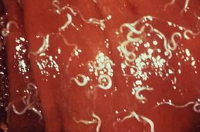
In the host’s digestive tract, food passes from the mouth to the esophagus to the stomach to the small intestine to the large intestine to the rectum, and then to the outside world. This means the large intestine is one of the last stops for nutrients, and by this point in the journey, nutrients have largely been broken down and absorbed. The large intestine is the home of the whipworm. The adult worms bite the tissue of the intestine, actually embedding their heads inside, and suck blood there.

Eggs are laid inside the large intestine and pass with the stool. Once in the outside world, the eggs require about two-to-four weeks to form embryos and become capable of infecting a new host. (This means that contaminated soil is the source of infection, not fresh feces.)

The new host is infected by consuming the egg, usually during grooming. After the host eats it, the egg hatches in the host’s small intestine releasing a larva.

The larva dives into the local glandular tissue and, after about a week, emerges into the small intestine and is carried downstream into the large intestine with the digested food. Once in the cecum or large intestine, its permanent home, it embeds in the tissue there, and after a total of 74 to 87 days from the time the egg was swallowed, the young whipworm is ready to mate.

Signs of Infection

A few whipworms generally do not pose a problem for the host, but if large numbers of worms are embedding themselves in the large intestine tissue, tremendous inflammation can result, leading to bloody, gooey diarrhea. Usually, there is not enough blood loss to be dangerous, but diarrhea readily becomes chronic and hard to control. A second syndrome of infection has emerged but is not well understood: signs mimicking those of Addison’s disease (hypoadrenocorticism). Here, waxing and waning weakness with the inability to conserve salt ultimately creates a dehydration crisis. The syndrome mimics Addison’s disease in every way except that testing for Addison’s disease will be negative and deworming yields a complete recovery.

Because female whipworms only periodically lay eggs (whereas other female worms lay eggs continuously), a fecal sample tested may easily be negative for eggs. This makes confirmation of a whipworm infection a challenge. It is common to deworm whipworms if the symptoms are suggestive of their presence, even if the fecal test is negative. Most common deworming agents do not work on whipworms, so something special must be selected. The most common products are fenbendazole (Panacur®), and febantel (Drontal Plus®).

Thanks to the long maturation cycle of young worms, a second deworming some 75 days or so after the first deworming is needed to fully clear the infection (easy to forget). Often another deworming in between these doses is recommended to further control the whipworm numbers.

Products Effective against Whipworms

More recently, regular heartworm prevention products have been developed to remove and control whipworms: Milbemycin-based heartworm preventives (such as Sentinel®, Interceptor®, and Trifexis®) will cover whipworms, and their regular use covers the second deworming as well. Moxidectin based heartworm preventive, such as Advantage Multi® and similar products, will also cover whipworms. 

Soil contaminated by whipworm eggs is contaminated for years. It is virtually impossible to remove the eggs from the soil or kill them. This potential for environmental contamination is concerning, but while there are scattered reports in the medical literature supporting human infection, the Companion Animal Parasite Council does not consider the canine whipworm to be transmissible to humans until there is stronger evidence.

Feline Whipworm Infection

There are species of whipworms that can infect cats: Trichuris serrata in North America and Trichuris campanula in Europe. Cats are clean animals and fastidious around feces, and they rarely get infected. When they do, worm numbers are so small that symptoms hardly ever occur. Whipworms are more of an interesting incidental finding in cats when whipworm eggs happen to come up on a routine fecal check. In other words, feline whipworm infection is generally not considered to be much of a problem.

team-single-fazenda

Workups for Itchy Dogs and Cats

Pruritus, or itching, in dogs and cats, can involve scratching and licking, chewing, or biting the skin. Pruritus is the most common symptom of skin disease in veterinary medicine. Many conditions can cause pruritus, including allergies, fleas, and other skin parasites. Some skin problems are more common in younger animals, while others are diagnosed more often in older pets. 

Medications are available to manage pruritus, but unless the underlying cause of the pruritus is diagnosed and addressed, it will return once the medication is stopped. Some anti-pruritic medications, such as steroids, can result in other health issues. Providing your dog or cat’s medical history can sometimes help your veterinarian determine the cause of the itching. 

A lack of response to some medications can help your pet’s veterinarian narrow down the possible causes. Additionally, some medications can actually cause itching.

When Did the Pruritus Start?

It can be helpful to know when the itching started. If allergies cause itching, it is beneficial to know if the problem is worse at different times of the year.  

Other Health Problems 

Sometimes, skin conditions develop secondary to another health problem. Your veterinarian will ask questions about other symptoms that could point to a specific cause of pruritus. For example, pets with food allergies may have frequent bowel movements or soft stools.

Location of Skin Lesions

Some pruritic skin conditions cause skin lesions more commonly in one body region than another.

Type of Skin Lesions

Although most pruritic skin conditions look the same to pet owners, there are actually many types of skin lesions, and some skin lesions occur more commonly with specific skin conditions. However, if the pruritus has become chronic, many pruritic skin conditions look similar to each other. Your veterinarian may need to run some tests to help determine the cause of the pruritus.

What Tests are Used to Diagnose the Cause of Pruritus?

Below is a list of some common tests for diagnosing the cause. Your veterinarian will select tests based on your pet’s medical history and examination findings. 

Skin Scrapings

To look for skin parasites, a scalpel blade is used to gently scrape the skin layers to collect cells and any skin mites. The sample is then examined under the microscope.

Skin Cytology

To look for bacteria and yeast organisms, skin cells and debris samples are collected and placed on a glass slide for examination under the microscope. This test can also help determine what types of cells are in the skin.

Parasite Therapy Trial

For some parasites, the only way to determine if they are the cause of the itching is to treat the parasites. A decrease in pruritus after the anti-parasite therapy would be consistent with the parasite causing the pruritus.

Ringworm Testing

To determine if a dog or cat’s skin is infected with ringworm (a disease caused by a fungus called a dermatophyte, not actually a worm), your veterinarian may start by examining your pet under a special ultraviolet lamp called a Wood’s Lamp. If the hairs glow (fluoresce), this may mean your pet has ringworm  Those hairs are collected for further testing. In a fungal culture, hair and skin samples are placed on a fungal culture plate. If a dermatophyte grows, the dog or cat is diagnosed with ringworm. In a dermatophyte PCR test, (a test that looks for viral diseases) the hair and skin samples are checked for dermatophyte DNA. 

Food Trial

A prescription or home-cooked diet is fed for several weeks with no other foods or treats to diagnose food allergies. Unfortunately, there is no reliable skin or blood test in dogs and cats to diagnose food allergies.

Allergy Testing

Skin or blood allergy testing is used in dogs and cats that have had other causes of pruritus ruled out through other tests. It is mainly used to select allergens for a type of allergy treatment called allergen-specific immunotherapy. There are no reliable hair or salivary tests to diagnose allergies.

Skin Biopsy

A sample of skin can be obtained through minor skin surgery. Skin biopsy is especially helpful for diagnosing some less common causes of pruritus, such as skin cancer. However, a skin biopsy cannot be used as an allergy test and cannot diagnose the cause of a pet’s allergies.

4128550

Dry Eye (Keratoconjunctivitis Sicca) in Dogs and Cats  

(Dry eye is formally known as keratoconjunctivitis sicca or KCS)

Why Tears are Good

We can all imagine the discomfort of dry, irritated eyes and the soothing that is provided by lubricating eye drops. Tears are essential to the comfort of our eyes but they do more than just provide lubrication. Tears contain anti-bacterial proteins and salts and serve to flush away the irritants and infectious agents that are constantly getting in our eyes. In addition, since the outer portions of the eye do not have a blood supply to remove metabolic waste, it is up to the tears to provide this service as well. 

Diagram shows the two lacrimal (tear-producing) glands of the canine eye. Graphic by marvistavet.com

Tears consist mostly of water, but also of oil and mucus secreted by their respective eyelid glands. The water portion of tears is secreted by two lacrimal glands in dogs and cats: one just above the eye and another in the third eyelid (or so-called nictitating membrane).

Dry eye with the classical ropey discharge.

(Photo courtesy of Dr. Michael Zigler)

Without tears, eyes become irritated, the conjunctival tissues around the eyes get red, the cornea itself in time will turn brown in an effort to protect the eye, and a gooey, yellow discharge predominates. Blindness can result.

Keratoconjunctivitis sicca is a fancy way of saying the eye is dry. “Kerato” refers to the cornea or clear covering of the eye that faces the outside world.

“Conjunctivae” are the moist pink membranes of the eye socket. “Itis” means inflammation and “sicca” means dry. Keratoconjunctivitis sicca, abbreviated KCS, means there is an inflamed, dry cornea and conjunctiva. It occurs when there is a deficiency in the water portion of the tear film, which normally accounts for 95% of the tear volume. Without water, one is left with oil and mucus; hence, the gooey yellow eye discharge characteristic of this condition.

Why do Eyes Become this Dry?

There are many causes of dry eye. Some are:

  • Canine distemper infection attacks all body interfaces with the environment including the eyes. Dry eye is part of the constellation of symptoms that can occur with canine distemper infection. 
  • In cats, herpes upper respiratory infection can lead to chronic dry eye (see more on herpes conjunctivitis).
  • There could be a congenital lack of tear-producing gland tissue (as described in certain lines of Yorkshire terriers).
  • Exposure to sulfa-containing antibiotics, such as trimethoprim-sulfa combinations, can lead to dry eye. It can be either temporary or permanent and occurs unpredictably.
  • Anesthesia will reduce tear function temporarily (thus eyes are lubricated with ointment by the attending nurse).
  • During surgery for cherry eye, removal of the third eyelid tear-producing gland, instead of replacing the gland in its proper location, can lead to KCS. So can too much damage to the gland prior to proper gland replacement.
  • A knock on the head in the area of one of the tear-producing glands can lead to KCS.
  • The most common cause of KCS appears to be immune-mediated destruction of the tear-producing gland tissue. We do not know what causes this type of inflammatory reaction but certain breeds are predisposed: the American Cocker Spaniel, the Miniature Schnauzer, and the West Highland white terrier.

How We Make the KCS Diagnosis

When KCS is in an advanced state, the situation is pretty obvious but early on in the case, it may look like a simple case of conjunctivitis. You may also notice a dry nose or nasal philtrum (area at the bottom of the nose). In either case, it is important to measure the tear production to determine how dry the eyes are.

The test that accomplishes this is called the Schirmer Tear Test.

To perform the test, a strip of specific paper is put just inside the lower eyelid in the outer corner of the eye and left for 60 seconds.

The moisture of the eye will wet the paper. At the end of the 60-second period, the length of the moistened area on the paper is measured. A length of 15mm or more is normal. A length 11 to 14mm is a borderline result. A height of less than 10mm is dry. A height less than 5mm is severely dry.

How do we Treat this Condition?

Not that long ago, all we had to treat this condition was tear replacement formulas and mucus-dissolving agents. These are still helpful but require an impractical frequency of administration. A breakthrough came with the discovery of cyclosporine topical therapy to control immune-mediated gland destruction.

Cyclosporine is an immunomodulating drug used to prevent organ transplant rejection in people and treat certain immune diseases in dogs and cats. When applied as an eye drop or ointment, it suppresses the immune destruction that is the most common cause of KCS, and tear production is restored.

The success of this treatment plus its convenient dosing interval (1 -3 times daily) has made this medication the primary treatment for KCS.

Animal hospitals used to make their own cyclosporine eyedrops out of oral cyclosporine and vegetable oil, but this largely ended when Optimmune® eye ointment (containing 0.2% cyclosporine) came out. Occasional patients simply do not show a good response to cyclosporine ointment but will respond when the concentration is increased. Higher-concentration products can easily be formulated by compounding pharmacies or one of the alternative medications listed below can be used. Treatment is almost always required for the lifetime of the pet.

After beginning cyclosporine eye drops or ointment, a recheck in three to four weeks is a good idea to check for improvement. If the Schirmer tear test is still showing poor results, the dosing frequency can be increased to three times a day; similarly, if excellent results are seen, the medication can be dropped to once a day.  Periodic rechecks are needed for dose adjustment and some dogs take as long as three to four months to show a response. Dogs with Schirmer tear tests as low as 2 mm still have an 80 percent chance of responding to cyclosporine. This medication has been a miraculous breakthrough in the treatment of KCS.

Tacrolimus is another medication that is an immune modulator. No commercial products are available for use in the eye, so they must be obtained from a compounding pharmacy. It is often tried in cases that are unresponsive or poorly responsive to cyclosporine. It is used in a manner similar to cyclosporine and is generally similar in cost.

Pilocarpine is a cholinergic drug, which means it works on the autonomic nervous system (the part that controls automatic functions such as glandular secretion). This medication can be given for the particular form of dry eye known as neurogenic KCS. In these cases, neurogenic stimulation of the tear gland is absent, so the pilocarpine is given in an attempt to stimulate the gland. Although the drug comes as an eye drop, for KCS it is actually given orally at an increasing dose until side effects are seen (diarrhea, drooling, vomiting). If side effects are encountered, the dose is reduced to that which the animal tolerates. It is continued indefinitely or until the neurogenic KCS subsides, usually twice daily. Neurogenic KCS typically affects only one eye.

Artificial tear solutions, gels, and ointments can be purchased in most drug stores. These can be combined with other therapies and are soothing. Their use is particularly important early in therapy until cyclosporine or tacrolimus takes effect and in eyes that do not respond to these latter medications. Over-the-counter products may be recommended two-12 times daily, depending upon their formulation and the severity of the KCS.

Topical antibiotics are often needed, especially when starting treatment for KCS because secondary infections are common with inadequate tears. These products do not increase tear production but help relieve the thick discharge.

Topical steroids may be beneficial in decreasing the inflammation associated with KCS. Typically they are combined with topical antibiotics in the same solution or ointment, especially when given to dogs.

Surgical Solutions

Parotid duct transposition is a surgical solution to unresponsive, severe KCS, although it is a delicate procedure, usually done by a veterinary ophthalmologist. The parotid duct is the salivary gland on either side of the face/cheek. It produces saliva that is carried to the mouth via a long duct. This duct can be carefully dissected out and moved to the outer corner of the inside of the lower eyelid in order to deliver saliva to the eye. Saliva is a reasonable substitute for tears, although in time some mineral deposits may form on the eye surface. Mineralization of the cornea can range from mild to severe and there is no good way to predict whether mineralization will be a problem prior to the transposition of the duct. The eye may water uncontrollably when the dog is fed and facial wetting may be objectionable in some cases.

4128639

Diabetic Ketoacidosis in Dogs and Cats

Ketoacidosis is one of the most extreme complications of diabetes mellitus that can be experienced. Unfortunately, most cases of ketoacidosis are in patients that were not previously known to be diabetic so the owner (and pet) must deal with two serious diagnoses: one acutely life-threatening and expensive and the other requiring ongoing commitment and daily treatment.

The diabetic patient has an insulin deficiency. To recap, there is a huge amount of glucose in the bloodstream but without insulin, none of it can get inside the cells that need it. It just circulates around uselessly. The tissues, some of which require glucose as their only food, are starving. Add to this some sort of second stressful condition such as infection or pancreatitis, and the tissue demand for food/fuel increases. The tissues, including the brain, become especially desperate and the body begins to frantically break down fat in order to liberate the small amount of carbohydrate (which can convert to glucose) contained therein.

Fat is able to convert to a biochemical called a ketone body. Ketone bodies can be used as an alternative fuel source by tissues that require glucose (brain, red blood cells, etc.). This will save the day in the short term but the problem is that ketone bodies do not “burn clean.” When ketone bodies are burned, pH and electrolyte imbalances ensue. These imbalances create dehydration, which in turn creates more pH and electrolyte imbalances. A metabolic disaster results if this state of glucose desperation persists.

Most patients in diabetic ketoacidosis are lethargic, depressed, and will not eat. They are dehydrated and frequently nauseated. Blood glucoses are extremely high and ketones can be detected in the urine. The goal is to gradually correct all the imbalances, get the patient out of the extreme fat-burning state, and establish some sort of initial regulation of the diabetes. 

Prognosis is generally fair as long as the complicating disease (the disease that is happening on top of the diabetes mellitus) can be resolved. That said, round-the-clock monitoring of electrolytes and blood sugars is needed to safely guide the patient through the crisis, and this kind of care has significant expense. Ketoacidosis involves potential disasters in potassium, phosphorus, pH, blood sugar, and sodium. All of these parameters must be controlled. The goal is to convert the complicated diabetic patient into an uncomplicated diabetic patient, but the patient will still be diabetic at the end of treatment.

What to Expect in the Hospital

The sooner the crisis is recognized, the faster treatment can be started. Because electrolytes can change moment by moment, blood testing is necessary throughout the day to keep track and keep the imbalances corrected. A facility that offers 24-hour care is ideal. Aside from the monitoring required to manage the ketoacidosis, testing to determine the precipitating stress is necessary as well.

In dogs, the most common precipitating/concurrent conditions are pancreatitis, urinary tract infection, and Cushing’s disease. In cats, precipitating/concurrent conditions include hepatic lipidosis, kidney infection, pancreatitis, and cholangiohepatitis.

Intravenous Fluids

Fluid therapy is the key to treatment. The patient is invariably dehydrated from the high circulating blood sugar levels, which cause excess fluid loss in urine, as well as from vomiting or diarrhea, which are common in ketoacidosis. Aside from simply providing fluids, the IV fluid provides a vehicle by which other metabolic derangements can be repaired.

Insulin

Blood sugar must be controlled if treatment is to be successful but to prevent brain damage, blood sugar levels must be dropped slowly. To achieve this, “regular insulin” (typically Humulin R®) is used, given either as multiple intramuscular injections or as a continuous drip. This type of insulin is short-acting and wears off quickly, which allows it to provide small adjustments. It is not until the patient is eating and nausea has been controlled that maintenance insulins can be started.

Potassium

Patients in ketoacidosis are greatly depleted in potassium. While insulin is needed to control blood sugar, insulin makes the problem worse by driving potassium into the body’s cells and out of the bloodstream. Typically, high amounts of potassium must be supplemented in the intravenous fluid solution.

Phosphorus

Low levels of phosphorus also accompany diabetic ketoacidosis and if levels drop too low, the patient’s red blood cells will begin to burst and be unable to maintain integrity. Phosphorus is also supplemented through the intravenous fluid solution.

Blood pH

The term ketoacidosis implies that the blood pH is overly acidic. If the situation is severe enough, sodium bicarbonate must be added to the intravenous therapy.

All these aspects require regular monitoring, which means lab work perhaps four times daily or more. Patients in diabetic ketoacidosis require close monitoring and intensive care.

When urine dipsticks no longer test positive for ketones and the patient is eating well and in good spirits, he or she is able to go home and be managed as a regular diabetic. Diet, monitoring, insulin etc. will be on-going concerns. Ideally, Ketostix, obtainable from any drug store, will be used at home to monitor for ketones to head off problems before they become extreme in the future.

4128004

Diabetes Mellitus: Introduction

ake-Homes for Pet Diabetes:

  1. Diabetes mellitus is caused by a deficiency of insulin. You will probably have to give insulin injections to correct the deficiency. (Don’t worry. It’s easier than you think.)
  2. The main symptoms of diabetes mellitus are excessive urination, excessive thirst, excessive appetite, and weight loss. Treatment should control these symptoms. Watching for these symptoms is the best way to know how your pet is doing.
  3. The starting insulin dose is going to be based on averages and will be tweaked based on trial and error, depending on both test results and control of the symptoms.
  4. You can save a lot of money if you get your own glucose meter and learn to do blood sampling at home (it’s easier than you think).
  5. Using too little insulin is a problem in the long term, but too much insulin is potentially an emergency in the short term. Be sure you know how to recognize hypoglycemia and what to do about it. See Summary, below.

What is Diabetes Mellitus?

In order to understand the problems involved in diabetes mellitus it is necessary to understand something of the normal body’s sugar metabolism.

The cells of the body require fuel in the form of fat or sugar to conduct their daily activities.  Some tissues can use either sugar or fat depending on circumstances, and some tissues (such as the brain and nervous system) depend almost exclusively on sugar as fuel. Diabetes mellitus mostly involves the metabolism of sugar (in particular, a sugar known as glucose), so we will focus on the sugar part of the situation. Glucose comes from the diet in the form of starches and sugars that we eat.

Tissues cannot absorb glucose without a hormone known as insulin. Insulin is produced by the pancreas as part of the body’s natural blood sugar regulation. Insulin can be considered to be a key that unlocks the door, allowing sugar in the bloodstream to enter the body’s cells. Once inside the tissues, glucose can be burned for fuel or stored, but without insulin, the sugar stays in the bloodstream and cannot be used by the body.

Ketones in urine for three days or more in a row warrant a visit to the veterinarian. 

In a Diabetic Animal, There Isn’t Enough Insulin

In fact, there may be no insulin at all. Remember that insulin was the key to unlocking the cell so that glucose could be brought inside. With no insulin, glucose cannot get in. Not only is glucose not being taken in and stored, but it is left floating around in the bloodstream in extremely high amounts. 

The body’s tissues are starving and the bloodstream has plenty of glucose to feed them, but without insulin, the glucose is unavailable.

So What Symptoms Result from this?

Because there is no way to remove glucose from the bloodstream, blood sugar levels are astronomically high. Normally, the kidney is able to conserve the bloodstream’s glucose but its mechanisms are overwhelmed and glucose spills into the urine in high amounts. If we continue our food delivery analogy, all the undeliverable food must be dumped. In the body, glucose dumping happens in the kidney with extra glucose dumping in the urine. This process involves the use of a lot of water with the ultimate result being a lot of urine being produced to process all that glucose to dump. This translates into excessive thirst and urination.

The tissues of the body are unable to access any of the glucose they need for fuel and are basically starving. Fat is mobilized and muscle is broken down to help feed the tissues but it does not do much good without insulin to bring fuel inside the cells. The patient shows excessive appetite because his body is in a state of starvation. Because the body is rapidly breaking itself down, weight loss is also a classic sign.

All the sugar in the urine provides a desirable growth medium for bacteria, and urinary tract infection is a common finding in diabetes mellitus. 

In diabetic dogs (but not cats), a specific type of cataract rapidly develops in the eye when high amounts of glucose enter the lens. Glucose normally feeds the lens, but the amounts of glucose coming into a lens in the diabetic state are much higher. Excess glucose is converted to another sugar called sorbitol, which in turn attracts water. The excess water disrupts the clarity of the lens, creating a diabetic cataract, which leads to blindness in almost all diabetic dogs. 

The main symptoms of diabetes mellitus are:

  • Excessive Thirst
  • Excessive Urination
  • Excessive Appetite
  • Weight Loss

Is it like Human Disease? Will We Need to Give Insulin Shots?

Dogs: Diabetes is most likely permanent and yes on the insulin shots.

Cats: Diabetes might not be permanent. Yes on the insulin shots if you want a chance at remission.

Diabetes mellitus is a classical disease in humans and most of us have heard some of the terms used to describe it. In humans, diabetes is broken down into two forms: Type I and Type II. These are also referred to as juvenile-onset and adult-onset diabetes or insulin-dependent and non-insulin-dependent diabetes. In short, Type I is the type where the pancreas produces no insulin at all, and Type II is the type where the pancreas produces some but not enough. Many pet owners wonder if dogs and cats have similar categories for their diseases.

Virtually all dogs have insulin-dependent diabetes and must be treated with insulin. There is no way around it. Their condition is similar to the Type I diabetic human in many ways.

Most cats have “non-insulin-dependent diabetes” at least to start. This suggests that some cats can get away without insulin injections and, indeed, some cats may qualify for oral options or temporary treatment with insulin. For cats, diabetes can resolve if we can re-activate the pancreas promptly.

Not all diabetic cats are similar to humans with Type II diabetes. Some diabetic cats, perhaps as many as 25 percent, have more severe hormone issues, such as acromegaly or Cushing’s disease, that make them not only insulin dependent but difficult to regulate.

Treatment: Giving Insulin by Injection

Since deficiency of insulin is the problem, it’s not surprising that giving insulin is the solution. You will need to learn to give injections, which is daunting to some owners at first, but almost everyone quickly becomes an expert. 

First, an insulin type and dose need to be selected. There are several types of insulins and it is not possible to know exactly how much insulin your individual pet will require; trial and error is needed. Your veterinarian will make a guess based on what works for other cats and dogs and what has been reported in the literature. Most pets require injections twice a day, approximately 12 hours apart, in conjunction with a meal. Because an overdose of insulin is potentially an emergency, it may be prudent to start with once-a-day insulin, just in case. If you like, discuss the pros and cons of each approach with your veterinarian. 

Insulin has traditionally been given by syringe in a shot, but insulin pens are proving more and more popular. The syringe method involves buying a box of syringes and a bottle of insulin, drawing up a measured amount of insulin, and giving a shot. The pen involves applying a needle tip to the pen, dialing a dose on the pen, sticking the tip into the pet’s skin, and pressing a button on the pen. Most people feel the pen method is much easier to perform but it may be difficult to find needle tips that are long enough for pet use as pet skin is much thicker than human skin.  In some situations, the pen is used as a dispenser for the syringe as the pen cartridges may be more cost-effective than bottles of insulin. Without video or first-hand experience with insulin pens or syringes, it may be hard to visualize the difference. See our section on insulin administration for more details. Not all pet insulins are available in a pen format. Your veterinarian will instruct you regarding options. 

Some insulins are available from the neighborhood pharmacy and some are available only through veterinary offices and veterinary pharmacies. Your veterinarian will either provide you with supplies or will give you the necessary prescriptions. If you are using syringes (instead of a pen) be sure the syringes and insulin concentration match. Insulin syringes are marked in insulin units (either U-100 syringes for 100 unit/cc insulins or U-40 syringes for 40 unit/cc insulins). Whenever you receive more supplies, always double-check these numbers.  

Never alter the insulin dose recommended by your doctor. To determine whether dose adjustments (or even a different type of insulin would be more appropriate), the pet will need a “glucose curve” where sugar levels are tracked over 10-24 hours. This can be done either by testing glucose levels with a glucose meter every 2 hours or so or by using a continuous monitor such as a FreeStyle Libre®. If a meter is used, it is best to test blood glucose in the pet’s own home and send the values to the vet but the curve can certainly be performed in the vet’s office. Note that stress and anxiety will alter blood glucose levels which means that values obtained at home will be more representative of the pet’s situation. 

Treatment: Oral Agents (Cats only)

Some cats will qualify for oral treatment of their diabetes. There are three medications that might be employed for this: bexagliflozin, velagliflozin, and glipizide.  

Bexagliflozin, an oral tablet medication, and velagliflozin, an oral solution (liquid), are options for some cats with diabetes mellitus where insulin injections are not practical.  Both of these drugs increase the kidneys’ ability to remove extra glucose from the bloodstream, keeping blood sugar levels in check. Lower blood sugar levels allow the pancreas to recover some and may alleviate many symptoms of diabetes. On the flip side, only certain cats will qualify for use, and there is potential for some very serious side effects. This includes diabetic ketoacidosis, a condition where insulin becomes so low that blood sugar cannot be used well in the body’s cells.  Following your veterinarian’s recommendations on proper monitoring is crucial. 

Finally, glipizide is a human medication used to reduce glucose levels by causing the pancreas’ beta cells to actually produce insulin.  In order for glipizide to have an effect, healthy beta cells need to be present. Glipizide is not as effective as bexagliflozin or velagliflozin in lowering blood glucose levels, so it does not include the intense monitoring or the same risk for complications as the others do. It is typically used for borderline patients or patients where insulin injections are simply not possible. Approximately 25% of cats will respond to this medication.

The various insulin formulations have different storage requirements. Check with your veterinarian for the proper storage of your pet’s prescribed insulin.

Diet and Feeding

Regulation can generally be worked out on whatever diet the pet is eating but there are some diets and feeding strategies that are helpful. For dogs, high-fiber/low-fat diets are preferred as they slow the absorption of sugars in the diet and help maintain a more regulated blood sugar level. Fiber also seems to make the body’s tissues more sensitive to insulin which also helps with regulation. Rewards are often employed to facilitate the insulin injection experience but these calories can add up and interfere with regulation so be sure your veterinarian is aware of all food items.  Diabetic dogs are best fed in two meals, approximately 12 hours apart. After they have been seen to eat their food, their insulin dose can be given.

For cats the strategy is different. First, cats seem to do best fed in multiple small meals daily so they should be allowed access to food at all times. Second, the high protein/low carbohydrate diets seem to be the most conducive to regulation. There are specific prescription diets, both canned and dry, for diabetic cats.

Learn more details on handling diets and feeding for cats and dogs.

What about Home Glucose Testing? 

Home glucose monitoring has many benefits: glucoses are monitored in the comfort of the pet’s own home without alterations from vet visit anxiety, a lot of money can be saved by monitoring at home, and questions about glucose status can be quickly answered and addressed. There are two ways to monitor glucoses at home: Continuous monitoring and a glucose meter.

Continuous Monitoring 

Continuous monitors have changed the way diabetic pets are monitored. A sensor with a tiny needle is implanted in the pet and paired wirelessly to either a phone or a reader. It reads tissue glucose levels constantly and reports its data to the device of your choice as well as to an internet portal where your veterinarian can access it. The sensor setup lasts up to 2 weeks and typically costs less than an in-house glucose curve run in the hospital. This is a fantastic option when it is time to get get a glucose curve. If one simply wants to spot-check a glucose level, one will need a glucometer and you will need to be able to take a blood sample from your pet.

Glucometer/Glucose Monitoring

 Many pet owners like to check glucose levels prior to insulin injections on a daily basis or simply want the ability to check a glucose level if there is a concern for hypoglycemia. For these situations, a glucometer is needed.  Human meters can be obtained at any drugstore, but we recommend getting a veterinary meter as the calibration is different. AlphaTrak is a popular brand, but there are presently several other brands available. 

The meter kit will contain measuring strips, solutions for calibration, and a spring-loaded lancet device for taking blood samples (usually from the ear margin). For details, please visit the Diabetes Monitoring section of this library. Taking single-drop blood samples turns out to be easier than you might think, and video links are provided in the monitoring section. Still, not every pet is amenable to blood sampling, and we do not want anyone getting bitten or scratched.

If you choose to use a glucometer at home, be sure to keep a log of when your pet was fed, when insulin was given, and what the glucose levels were that you found. (There are many phone apps to assist with this). Bring this log to your veterinarian when you come for checkups. Glucose levels obtained prior to the first insulin administration of the day are particularly useful. Your veterinarian will also be particularly interested in signs associated with poor regulation: excessive thirst, excessive urine production, excessive appetite, and weight loss

See the insulin administration guide for cats.

Hypoglycemia and Other Things to Watch for

The most serious problem to watch for is hypoglycemia (low blood sugar). This results from a mismatch in food consumption and insulin dose. If the dose is too high, you can get hypoglycemia. If the pet doesn’t eat, you can get hypoglycemia.  Your pet may look simply tired, weak, or sleepy. If she is roused, she will seem drunk or may not be able to fully come to alertness. This can be an emergency and can progress to seizures, so it is good to know what to do at home to prevent disaster.

If your pet appears wobbly or drunken, the blood sugar level may have dropped too low. This occurs after an insulin overdose. First, try to get your pet to eat. If the pet will not eat, administer light Karo syrup, honey, or even sugar water at a dose of one tablespoon per 5 pounds. If no improvement occurs, immediately see your veterinarian for emergency treatment. When your pet is more stable, a glucose curve will be needed to determine why this happened and what a more appropriate insulin dose might be.

It is best to make sure that your pet has recently eaten before giving the scheduled insulin dose.

Other Reasons to Return to the Vet

Bring your pet in for a re-check exam and testing if you note any of the following: 

  • the pet seems to feel ill.
  • the pet is losing weight.
  • the pet has a ravenous appetite or loses its appetite.
  • the pet seems to be drinking or urinating excessively. 
  • the pet becomes disoriented or groggy.

It is important for diabetic pets to have their teeth cleaned annually. Dental tartar seeds the body with bacteria and when blood sugar levels run high, infections in important organs can take root. The kidneys and heart are particularly vulnerable.

Some Pets are Difficult to Regulate

Your pet will probably require re-regulation at some point. There may be an underlying reason to sort out. If your pet seems to fit into this category, some reasons could be:

  • Improper administration of insulin. If possible, have your doctor observe you giving the insulin to your pet. Another possibility is that your insulin may be out of date.
     
  • Rapid insulin metabolism. Insulin wears off quickly in some animals. Your pet may require a different type of insulin or a second injection during the day, or even additional injections during the day.
     
  • Insulin overdose may actually lead to elevated glucose levels (and clinical signs of diabetes mellitus). In this situation, too much insulin brings the blood glucose too low, and other hormones respond to bring it back up (and generally overdo it).  
  • Steroid administration (such as prednisone, prednisolone, etc.) will interfere with insulin. 
      
  • Progesterone, a female hormone, also interferes with insulin. Unspayed female diabetics should be spayed once they are sufficiently regulated.

For more details on trouble with regulation, read about hard-to-regulate cats.

Additional Information on Feeding a Diabetic Pet

Regulation is achieved via a balance of diet, exercise, and insulin. Realizing that therapeutic diets are not always attractive to pets, there are some ideal foods that should at least be offered.

The most up-to-date choice for cats is a low-carbohydrate, high-protein diet. These diets promote weight loss in obese diabetics and are available in both canned and dry formulations. For dogs, high-fiber diets are still in favor as fiber seems to help sensitize the pet to insulin. Talk to your veterinarian to select an appropriate choice for your pet.

Avoid soft-moist diets as sugars are used as preservatives. Avoid bread and sweet treats. If it is not possible to change the pet’s diet, then regulation will just have to be worked out around whatever the pet will eat.

In Summary:

  1. Diabetes is caused by a deficiency of insulin. Diabetes mostly involves metabolizing sugar, particularly a sugar called glucose. You will probably have to give insulin injections to correct the deficiency. It’s easier than you think.
  2. A body requires fuel in the form of fat or sugar. Glucose comes from a diet of starches and sugars. Tissues cannot absorb glucose without insulin, so the level of insulin affects the glucose level.
  3. The main symptoms are excessive urination, thirst, appetite, and weight loss. Watching for these symptoms is the best way to know how your pet is doing.
  4. Tissues are unable to access any of the glucose they need for fuel and are basically starving. Rather than going into tissues, unmetabolized glucose goes into the bloodstream but cannot be removed from there, so glucose levels become astronomically high. The kidney compensates by dumping glucose straight into the urine. The kidney needs much water to do that, so a larger volume of urine is made just for dumping, causing a cycle of excessive thirst and urination.
  5. Your veterinarian will select the first dose of insulin based on glucose averages and will tweak it thereafter. You can use either a syringe or an insulin pen.
  6. While using too little insulin (high blood sugar or hyperglycemia) can lead to problems, using too much insulin (low blood sugar or hypoglycemia) is potentially life-threatening in the short term. Low blood sugar creates listlessness and potential unconsciousness; the fix is to offer food immediately.
    • Extremely low blood sugar can cause tremors, seizures, weakness, collapse, and incoordination, in addition to other signs.
    • Extremely high blood sugar can cause seizures, circling, pacing, weakness, coma, and incoordination, among others.
    • Both are emergency situations, and you should see your veterinarian or an emergency hospital immediately.
  7. Never alter the insulin dose without discussing it first with your veterinarian.
  8. It’s easiest for your pet if you get a glucose meter, install a continuous monitor, and do blood sampling at home. That way, you can find out their glucose status right away and do something about it.
  9. Some pets are difficult to regulate. Yours will probably require re-regulation at some point. Regulation needs a consistent balance of diet, exercise, and insulin.
  10. The current choice of food for diabetic cats is a low-carbohydrate, high-protein diet. High-fiber diets are best for dogs. 
  11. Dogs: Diabetes is most likely permanent, and they need insulin shots.
  12. Cats: Diabetes might not be permanent. To have a chance of remission, they need insulin shots. Some cats might be able to take pills rather than shots.
7779998

Diarrhea and Vomiting: First Aid

Diarrhea is the frequent evacuation of watery stools. Vomiting is the forceful expulsion of stomach contents through the mouth. 

What to Do

  1. Remove all food and water. 
  2. Check for signs of dehydration. 
  3. If diarrhea and/or vomiting continues or the pet acts ill, seek veterinary attention. 
  4. If no vomiting occurs for 6 to 8 hours, begin to frequently give small amounts of clear liquids (water, Gatorade, Pedialyte, or other electrolyte solution).
    • A rule of thumb is to give 1 teaspoon per pound of body weight every 2 or 3 hours throughout the day and night. 
    • If your pet does not vomit the fluid, the following day offer small frequent meals of boiled hamburger and rice or boiled chicken and rice.
  5. If your pet does not want to eat, starts to vomit, or continues to have diarrhea, see a veterinarian right away.
  6. Isolate the sick pet from other pets.

Diarrhea and vomiting can quickly lead to serious fluid loss and electrolyte imbalance, especially in very young or old animals, and those with certain health issues.  Severely dehydrated animals may need to be hospitalized and receive IV fluids to recover.

What NOT to Do

  • Do not administer any over-the-counter or prescription medications to your pet without talking to a veterinarian first.  
  • Do not allow the pet to eat or drink anything until there has been no vomiting for 6 to 8 hours.

Vomiting and diarrhea are associated with a host of problems that are referred to collectively as gastroenteritis. Some cases are quite severe (e.g., poisoning), and some are not (e.g., dietary indiscretion). If fever is present, infection may be a cause. 

Most infections that cause diarrhea and vomiting are contagious, so it is wise to assume that other pets might be vulnerable if they are exposed. 

If your pet is not feeling well and has vomiting and/or diarrhea, see a veterinarian.

4128884

Dilated Cardiomyopathy in Dogs and Cats

Dilated cardiomyopathy (DCM) is one of the more common acquired heart diseases in dogs. DCM is a primary disease of the heart muscle (cardio = heart; myo = muscle; pathy = disease) in which the heart muscle (myocardium) of the lower pumping chambers (ventricles) becomes weak and so loses its ability to contract normally. DCM most commonly affects the left side of the heart (the side that receives blood from the lungs and pumps it to the body), specifically the left ventricle.

Graphic by Tamara Rees of VIN

When the myocardium cannot pump blood out of the left heart effectively, the kidneys retain sodium and water to increase the amount of blood returning to the heart. This leads to an enlargement of the ventricles in order to compensate for the ineffective pumping. This is helpful for years, but ultimately becomes detrimental when it causes the blood pressure in the heart to back up into the lungs, thereby causing fluid accumulation within the lungs (pulmonary edema). This is called heart failure or congestive heart failure (CHF).

Although less common, DCM affecting the right ventricle can also occur. Blood backs up on the right side, which receives blood from the body and pumps it to the lungs, resulting in right-sided CHF, where fluid accumulates in the abdomen (ascites) and chest (pleural effusion). DCM affecting the right ventricle is almost always accompanied by DCM of the left ventricle.

What Breeds get DCM?

There are several breeds that are predisposed to DCM. These include Doberman Pinschers, Great Danes, Irish Wolfhounds, Boxers, Newfoundlands, Portuguese Water Dogs, Dalmatians and Cocker Spaniels.  DCM is not just limited to specific breeds. Large and giant breeds are most commonly affected, but it also occurs in smaller breed dogs and cats as well.

The causes of DCM in these breeds vary, as explained below.

What Causes DCM?

Because of the strong breed association, DCM almost certainly is inherited in many breeds. Genetic mutations that are associated with DCM have been identified in Doberman Pinschers, Boxers and Standard Schnauzers. Genetic testing for these mutations can be done for each.

Boxers get a specific type of cardiomyopathy called arrhythmogenic right ventricular cardiomyopathy (ARVC). Approximately 90% of these dogs in the USA have a ventricular arrhythmia and an otherwise normally functioning heart. They are prone to fainting and sudden death. The fainting most commonly happens when they develop a very fast heart rate (greater than 400 beats/min). Sudden death usually occurs when this fast rate degenerates into ventricular fibrillation (cardiac arrest). Approximately 10 percent also get DCM as a part of their disease. The disease is associated with a genetic mutation in a gene called striatin.

In some dogs, DCM is due to a nutritional deficiency. Taurine is an amino acid required for the development and function of the myocardium. Consequently, pets may develop DCM on taurine-deficient diets, such as vegetarian diets, and may benefit from appropriate supplementation. Some breeds, such as American Cocker Spaniels and Golden Retrievers, may have a predisposition to taurine-deficiency, possibly through defects in metabolizing taurine. Many, but not all, cases that are supplemented with taurine will improve. Some also need carnitine supplementation. If your pet is diagnosed with DCM, testing for a taurine deficiency may be warranted. Breeds such as Doberman Pinschers and Great Danes do not have taurine-deficient cardiomyopathy. Some cats may develop taurine-deficient DCM, although this has become rare as taurine is now added to virtually all quality cat foods (see Feline Cardiomyopathy).

L-carnitine is another amino acid that has rarely been implicated in the development of DCM in people. L-carnitine is required for the myocardial cells to produce energy and thus contract. There is some evidence that a deficiency in this molecule will contribute to myocardial dysfunction in Boxers (one small study only). Some American cocker spaniels need to be supplemented with it, along with taurine, to produce a beneficial response. However, the role of carnitine in most DCM cases is very limited.

In 2018, grain free diets were implicated in causing DCM in dogs, especially in breeds that do not typically get DCM.  Read more details about this potential cause.

Occasionally, toxins can cause DCM. The most common toxin is doxorubicin (Adriamycin), an anti-cancer drug used to treat various cancers in dogs. In some cases, dogs receiving doxorubicin  will develop DCM.

Infectious causes of DCM are rare. Puppies infected with parvovirus at two to four weeks of age can develop DCM. These days, vaccinating the mother protects the puppies against parvovirus during this susceptible period, so this cause of DCM is rarely seen. Chagas disease (Trypanosomiasis) can cause DCM in geographic areas where it is found (Texas, Mexico).

What are the Signs of DCM?

Signs of DCM vary depending on the breed of dog and stage of the disease. Loss of appetite, pale gums, increased heart rate, coughing, difficulty breathing, periods of weakness, and fainting are signs commonly seen. Since blood is backed up into the lungs, respiratory signs (CHF) due to pulmonary edema are most common. Blood returning to the right side of the heart from the body may also back up leading to fluid accumulation in the abdomen (ascites) or in the chest cavity (pleural effusion). Weakness or collapse may be caused by abnormal heart rhythms (arrhythmias) and occasionally, decreased blood flow to the body (depressed cardiac output).

In some breeds, sudden death or fainting can occur well before any signs of CHF.

How is DCM Diagnosed?

There are two different methods used to diagnose pets: (A) during a screening exam of an apparently normal dog (e.g. as part of a breeding program), and (B) during examination of a dog with clinical signs of heart disease.

Screening Exams for DCM


Many conscientious breeders and owners of dogs that are predisposed to DCM screen their pets for heart disease to try to minimize the risk of transmitting the disease to offspring. Screening for DCM in dogs can be expensive and complex. The screening test of choice depends on the breed of the dog and the stage of the disease.

The first step is a good physical examination. In most cases, the physical examination is completely normal. Occasionally, the veterinarian may detect an arrhythmia (abnormal heart rhythm). In Doberman Pinschers and Boxers, a 24-hour ECG recording using a 24-hour ambulatory ECG monitor (Holter monitor) is often the best way to screen dogs for early signs of DCM since an abnormal rhythm often occurs before any detectable changes in myocardial contractility. An echocardiogram (an ultrasound scan of the heart) is also used to identify dogs with DCM before they develop clinical signs, but many dogs with mild disease have equivocal findings. This examination is best performed by a board certified veterinary cardiologist.

Genetic testing should be done by breeders of the specific breeds where a mutation or mutations have been identified.

Diagnosis in Dogs with Clinical Signs


A thorough physical examination by your veterinarian, coupled with your pet’s clinical signs and specific breed, may help make the presumptive diagnosis of DCM. Tests that help support the diagnosis are an ECG (electrocardiogram) and x-rays (radiographs) of the chest. The ECG may show an arrhythmia and/or an elevated heart rate. The chest radiographs may show an enlarged heart and/or fluid in the lung tissue or chest cavity. Some dogs may have normal chest radiographs, but have arrhythmias on their ECG. These pets may be in the early stages of DCM (see above).

In dogs with clinical signs of heart failure, an echocardiogram is necessary to confirm the diagnosis of DCM. With an echocardiogram, a cardiologist can visualize the heart and assess its function. A decrease in heart pumping function (contraction) means that the patient has DCM. Your veterinarian may also perform blood tests to look for any underlying nutritional or infectious conditions if the specific case warrants such investigation.

How is DCM Treated?

Treatment of heart failure is based each individual patient. Drugs commonly used are diuretics (most commonly furosemide), ACE inhibitors, and pimobendan. The diuretic forces the kidneys to excrete more sodium and water. It is used to eliminate pulmonary edema (fluid in the lungs) and so improve breathing and/or effusion (fluid accumulation in the chest or abdominal cavities). Pimobendan increases the force of contraction of the ventricles and dilates blood vessels. Both furosemide and pimobendan are effective treatments that prolong survival and improve quality of life.

Pimobendan might increase the time until onset of heart failure in Doberman Pinschers with DCM when given prior to the onset of heart failure.

Management of arrhythmias is often an important part of managing DCM. Dogs with atrial fibrillation are most commonly treated with a combination of digoxin and diltiazem in order to reduce the heart rate. Sotalol alone or in combination with other antiarrhythmic drugs is used in Boxers and in some Doberman Pinschers to suppress ventricular premature complexes and tachycardia in order to stop the dog from fainting and from dying suddenly. Mexiletine is also commonly used in Doberman Pinschers.

In nutritional DCM, specific supplements will be prescribed. Patients with right-sided heart failure will also have fluid physically removed from the abdomen and/or chest cavity by the veterinarian to make the patient more comfortable.

In humans, DCM patients usually get heart transplants. However, this option does not exist for veterinary patients. Other surgical procedures have been evaluated, but currently none are being offered for patient care.

What is the Prognosis of a Pet Diagnosed with DCM?

Unfortunately, in most cases DCM is a progressive, irreversible, and ultimately fatal disease. Survival depends on the stage of disease, the breed of the patient, the specific type of DCM that patient has, and patient/owner treatment compliance. In taurine-deficient DCM, correcting the deficiency in cats results in complete cure. In dogs, correcting the deficiency may result in at least partial reversal of the disease and prolonged survival; however, some cases relapse after several years.

DCM is a slowly progressive disease. If it is diagnosed in the early stages, the patient may live several years before developing clinical signs. In some breeds, such as Doberman Pinschers, sudden death accounts for 30 percent of the deaths from DCM, well before these dogs ever develop CHF.

In other breeds with DCM, such as Doberman Pinschers and Great Danes, in dogs showing clinical signs of CHF medical therapy can help prolong survival. Historical average survival for Doberman Pinschers with clinical DCM was two to three months. However, with pimobendan, recent studies have seen extended survival for this breed to one year. Less is known about outcomes of other breeds with DCM. Once the diagnosis of DCM is made, ask your veterinarian to discuss your pet’s prognosis on an individual basis.

Can I do Anything to Prevent DCM or Slow its Progression?

Currently, the primary intervention that has been shown to alter the course of DCM is nutritional supplementation in dogs with a nutritional deficiency (i.e., taurine deficiency). Since the majority of cases are thought to be genetic, breeding from lines unaffected by the disease helps reduce the chance of inheriting DCM. Genetic tests, when they are available, are of value in determining breeding strategies. In Doberman Pinschers, pimobendan has been reported to prolong a composite survival endpoint although it did not prolong the time until the onset of heart failure and did not prolong the time until sudden death.

What about Other Supplements?

Multivitamin supplements, nutritional supplements, Co-enzyme Q10, and non-Western herbal supplements have all been used for DCM, but none have been examined critically to determine if they hurt or help patients. Use of these supplements is best discussed with your veterinarian.

4126835

Dust Mites: Minimizing Exposure in Dogs and Cats

House dust mites (Dermatophagoides farinae, Dermatophagoides pteronyssinus) are bugs that are  in every home, and some pets are allergic to them. Because these mites are very small, they cannot be seen without a microscope. House dust mites eat the skin scales and dander shed by humans and animals. They are most commonly found in the sleeping areas of people and pets. House dust mites also like to live in homes with high humidity.

A house dust mite allergy is NOT a sign of a dirty house. Homes with carpeting and areas with high humidity will always have some dust mites.  

The following are some ideas that can be used to reduce the amount of house dust mites in your home.

  1. House dust mites are most numerous in mattresses and beds. If possible, please keep the pet off of your bed. If this is not possible, use plastic mattress covers or some impermeable barrier for your pet’s sleeping area on the bed and wash and heat-dry bed linens weekly. Replace bedspreads, pillows and mattress covers regularly.
  2. Feather pillows are full of house dust mites and should not be used around your allergic pet.
  3. Pet beds should be covered in plastic, filled with cedar, or treated with insecticides inside the cover. Covers should be washed weekly and the pet beds should be aired out or put in the dryer. You should replace all pet beds every six months unless the whole bed, including the stuffing, can be laundered weekly.
  4. When possible, choose décor and furnishings that either do not retain dust or can be easily cleaned such as: closed bookshelves instead of open shelves, washable curtains instead of blinds and heavy draperies, furniture with simple designs instead of ornately curved pieces, wooden or plastic furniture instead of upholstery, and easily cleaned decorations instead of dried flowers or straw.
  5. Any upholstered furniture that is used by your pet should be covered with a plastic throw cloth (anti-bed-wetting mattress pads can be useful).
  6. Stuffed pet toys should be replaced with new ones that can be laundered and dried weekly.
  7. Vacuum and dust your pet’s environment frequently. Try to clean when the dust-sensitive pet is not at home as vacuuming and dusting stir up the allergens and increase exposure to them.
  8. Use air conditioning or central heat to keep household humidity low. Change or clean filters on air conditioning or heating systems on a regular basis. Do not confine your pet to the laundry room, bathroom, utility room, basement or other high humidity parts of the home.
  9. Regular use of pet flea control products is associated with lower levels of house dust mites in the home. If your pet has house dust mite allergies, we recommend that all pets in your household be kept on year-round monthly flea control.
  10. Large amounts of house dust mites can live in carpeting. If possible, remove the carpeting in your home or keep your pet out of carpeted rooms. If your home must contain carpeting:
    • Area rugs that can be thrown into the washing machine and heat-dried once a week are preferable.
    • Sprays that breakdown the house dust mite particles that cause allergies are available but it is uncertain how effective these sprays are.
smallanimalpuppykittencare_img

Diskospondylitis (Intervertebral Disk Infection) in Dogs and Cats  

Diskospondylitis is a bacterial or fungal infection of the disks between the vertebrae (intervertebral disks). This infection can occur in any area of the spinal column, and it can occur at multiple sites. Diskospondylitis is also known as spondylitis, intervertebral disk infection, and vertebral osteomyelitis.

Diskospondylitis occurs much more often in dogs than in cats.

The bacterial/fungal infection can reach the intervertebral disks several ways.

  • Through the bloodstream, which is the most common method. (Chronic infections of the skin, urinary tract, prostate, etc. can result in bacteria entering the bloodstream and travelling to the disk area.)
  • Via direct contamination from punctures or bite wounds near the spine, or from procedures or surgery near the spine.
  • From the migration of foreign bodies through the area near the spine.

Signs

Neurological signs have a gradual onset and are progressive. At the beginning, spinal pain, stiffness, unsteady walking, and other nervous system problems may occur. Spinal pain is the most consistent clinical sign. Impaired movement (paresis) is usually mild, unless the infection gets into the spinal canal. However, if the pet doesn’t get appropriate treatment, signs can progress to paralysis, often caused by fractures of the vertebrae. The muscles alongside the spine may atrophy (waste away); this is most likely due to local nerve damage or associated myositis (muscle inflammation).

Spinal cord compression can be caused by granulation tissue, bony changes, or fractures or dislocations (luxations) due to the infection. The vertebral fractures or dislocations may require surgery to stabilize them.

Occasionally, the infection may result in meningitis, meningomyelitis, or an abscess in the spinal canal.

Diagnosis

Diagnosis may require radiography, advanced imaging modalities (CT scan, MRI, bone scintigraphy), urinalysis, bacterial cultures, serology, and cerebrospinal fluid analysis.

Radiography

Radiographs (X-rays) of the spine will typically show damage to the vertebrae on either side of the affected disks.  In chronic cases, bone changes and deformities of the spine may be seen. Changes may not show up on the radiographs for 3-6 weeks after clinical signs start, so if the first radiographs are normal but the clinical signs are progressing, your veterinarian will likely repeat the radiographs.

If a typical lesion is detected, radiographs of the entire spine are recommended because some pets have multiple lesions. Infection causes disk degeneration, and destroys the ends of the adjacent vertebrae. These degenerative changes may weaken the vertebral column and make it susceptible to pathologic fractures, which stem from infection rather than trauma. The body may try to bridge the damaged area with bone, which can encroach on the spinal cord and nerve roots, causing more problems.

Advanced Imaging

Bone scintigraphy, computed tomography (CT), and magnetic resonance imaging (MRI) are more sensitive than plain radiography, particularly for early lesions. Myelography and/or MRI/CT are helpful for pets that have neurological problems, and these imaging methods are considered important if spinal surgery is going to be done.

Urinalysis

Pets may have pus and/or bacteria in their urine.

Bacterial Cultures

Culturing the urine, blood, spinal fluid, or other infected tissues may help identify the organism(s) that caused the infection. However, culture of the infected disk space will yield the most accurate results. Fluoroscopic-guided fine needle aspiration may be necessary to get the best sample. If spinal surgery is necessary, samples of the infected areas should be obtained and cultured.

Serology

Brucellosis serology (checking the blood for current and previous infections) should be performed. Brucellosis is a possible cause of diskospondylitis, and can be transmitted to people.

Cerebrospinal Fluid (CSF) Analysis

CSF analysis is called for in animals with neurologic signs. In addition to culturing the CSF, routine cytology, cell count, and protein determination are done.

Causes

Bacteria

Bacteria such as Staphylococcus, Streptococcus, Escherichia coli, and Brucella spp. are commonly isolated organisms. Pseudomonas, Proteus, Pasteurella, Corynebacterium, and Actinomyces spp. are sometimes found. Reproductive problems may occur with Brucella infections. There are reports of pets infected with Bordetella spp., Erysipelothrix rhusiopathiae, and Salmonella spp.

Fungi

Fungal organisms that can cause diskospondylitis include Aspergillus, Blastomyces, Histoplasma, Coccidioides spp., and occasionally Paceilomyces. In Europe, infection with Rasamsonia argillacea has been reported. A single case of infection with Westerdykella spp. has also been reported in a German shepherd dog. Fungal infections can be more difficult to diagnose than bacterial infections, so your veterinarian will also look for enlarged lymph nodes, lung or eye lesions, and fungal organisms in the urine.

Other Agents

Protothecosis, a disease caused by a type of green alga, has been associated with diskospondylitis.

Treatment/Management/Prevention

Antibiotic Therapy

Treatment involves a long course of antibiotics that are specific for the infective organism. Ideally, the antibiotic selection will be based on culture and sensitivity results from the infected disk. If disk culture is not possible, then antibiotic selection will be based upon the culture results of urine, blood, or other infected areas.

Antibiotic treatment may be needed for many months. Recurrence of the disease or persistence of signs can be a problem if antibiotic therapy is stopped too soon, if the antibiotic being used is ineffective, or if corticosteroids or other immunosuppressive drugs are used. If clinical signs do not improve while the pet is taking antibiotics, your veterinarian may decide to repeat the disk culture.

As for Brucella infections, treatment is only undertaken with the understanding that a cure cannot be expected, long-term antimicrobials will be required, and the return of the infection is common. Infected dogs should be neutered to reduce the risk of transmission to other dogs and people, to remove potential reservoirs of infection, and to resolve other clinical problems (infection of the testicles, for example). For all practical purposes, Brucella diskospondylitis should be considered incurable.

Antifungal Therapy

Antifungal drugs, particularly itraconazole, may be effective for treating some cases of fungal diskospondylitis.

Decompression

Your veterinarian may advise surgical exploration of a lesion that does not respond to treatment or that has persistent draining tracts that suggest foreign body migration is occurring. Decompressive surgery and/or stabilization may be considered if evidence of spinal cord compression is found on myelography or MRI, or if severe, progressive neurologic problems occur. Surgical stabilization of affected sites may be useful in cases in which spinal instability is a significant complication.

Supportive Therapy

If antibiotics are effective, pain medications may be needed for only a few days.

Monitoring and Prognosis

After your pet has had at least six weeks of therapy, your veterinarian may take more radiographs to see if the lesions have improved. Improvement of clinical signs often occurs before improvement can be seen on the radiographs. Prognosis for recovery is guarded and depends, to some degree, on the cause of the diskospondylitis.

4128893

Cryptorchidism (Retained Testicles) in Dogs and Cats

Cryptorchidism is a condition in which a male’s testicles have not descended (dropped) into the scrotum. At birth, a male puppy’s or kitten’s testicles are located near the inguinal ring. 

By 8 weeks of age, testes are palpable in the puppy’s scrotum. However, scrotal and suprascrotal testes can be difficult to palpate if the testes are small or the puppy is obese. Testes may also freely move between the scrotum and inguinal area in young puppies.

By 8 to 16 weeks, they’re palpable in the kitten’s scrotum. Some retained testicles can be palpated. However, testicles retained in the abdomen are typically not palpable on physical exam.

In the fetus, a structure called the gubernaculum connects the testicle (located next to the kidney during development) to the scrotum. If this structure fails to develop properly, the testicle will not end up in the scrotum but will end up in the abdomen, the inguinal canal, etc.

Cryptorchidism can be unilateral (only occurring on one side) or bilateral (occurring on both sides). Unilateral cryptorchidism usually involves the right testicle. Bilaterally cryptorchid animals are usually sterile because the higher body temperature inside the abdomen is enough to prevent sperm production. (The animals will, however, still exhibit male behaviors.)

Cryptorchidism is a fairly common defect in dogs. Dog breeds most likely to be affected include Yorkshire terrier, Pomeranian, French poodle, Siberian husky, miniature schnauzer, Shetland sheepdog, Chihuahua, German shepherd, dachshund, and brachycephalic breeds. Still, it can happen in any breed or mixed breed.

Since cryptorchidism is considered to have a genetic basis, animals with this condition should not be used for breeding.

Dogs with cryptorchid testicles are prone to testicular torsion (twisting) and testicular cancer, so these dogs should be neutered to prevent problems later.

Cryptorchidism in cats is uncommon. The most common breed associated with cryptorchidism is the Persian. Congenital abnormalities that have been known to occur simultaneously with cryptorchidism are patellar luxation, shortened tail, kinked tail, tetralogy of Fallot, tarsal deformity, microphthalmia, and upper eyelid agenesis. Unlike dogs, it may be possible to visually differentiate between a castrated cat and one with retained testicles, because cryptorchid cats have barbs on the penis.

Laboratory Diagnosis

A testosterone assay can be used to differentiate between a castrated male and a cryptorchid male.

Treatment

Surgical removal is the only treatment for cryptorchidism. Even if the animal is a unilateral cryptorchid, both testicles should still be surgically removed. (The cryptorchid testicle should be removed to prevent testicular torsion and testicular cancer, and the normal testicle should be removed to prevent cryptorchid offspring.) This surgery is more complicated than the usual neuter surgery because the cryptorchid testicle can be difficult to locate. Depending on the case, some pets will be able to go home on the day of the surgery, and some may have to stay in the hospital overnight. A 2-week recuperation (reduced activity) is advised because this surgery usually involves opening the abdomen, and the surgical site has to have time to heal before the pet resumes normal activities.

Many males will need to wear a protective Elizabethan collar during recuperation to prevent them from licking or chewing at the incision. Owners should check the incision regularly for redness and swelling, which could indicate a post-operative infection or self-trauma. If non-dissolvable skin sutures are used, they will need to be removed by your veterinarian about 10 to 14 days after surgery.

Prognosis

Cryptorchid animals that have had both testicles removed, and have no other defects, will generally live a normal lifespan for the breed.