Tag: cats & dogs

7750217

No Bones About It – Chewing Bones is Bad for Dogs’ Teeth

The Food and Drug Administration warned pet owners about bones and bone treats to include not only harmful bacteria (E.coli and Salmonella) that dogs can get and pass on to humans but the actual damage caused by the bone’s trauma to a dog’s teeth.

Other issues can occur with bones, such as blocked intestines, choking, wounds in the mouth, vomiting/diarrhea, and rectal bleeding (some of which could be fatal if not treated promptly).

Are Any Bones Safe for My Dog’s Teeth? 

No. Steak bones are too hard for teeth. Antlers are worse than bones because they’re even harder. Poultry bones – chicken, turkey, and duck – are awful because they are full of air and thus splinter easily. The pieces can get stuck in your dog’s palate (roof of the mouth) and cause infection or get stuck in the esophagus or intestines, most of which requires a surgical fix.

What Happens if My Dog’s Teeth Are Injured by Bones? How Will I Know if There is a Problem?

For many dogs, broken teeth do not show signs of discomfort even though they experience pain like humans. Some show apparent signs and symptoms of disease, including an open tract below the eye or under the chin that may be draining.

When the nerves die, the pain decreases until infection sets in. Signs relating to dental disease from a broken tooth include:

  1.  chewing on one side;
  2.  dropping food from the mouth when eating;
  3.  excessive drooling;
  4.  grinding teeth;
  5.  pawing at the mouth;
  6.  facial swelling;
  7.  fistulous tract below the eye or under the lower jaw;
  8.  regional lymph node enlargement;
  9.  shying away when the face is petted;
  10.  refusing to eat hard food;
  11.  refusing to chew on hard treats or toys.

Treatment for Broken Teeth

When presented with a tooth that is fractured and has pulp exposure (complicated tooth fracture), your veterinarian essentially has two choices:

  1.   Extract the tooth.
  2.   Refer to a veterinary dental specialist to perform treatment, which usually allows the tooth to be saved and returned to function. Specific treatment depends on the severity of damage to the tooth structure and if any other disease is affecting the tooth, and the functional significance of the tooth.

Types of Tooth Damage

  • Enamel fracture:  A fracture with loss of crown substance confined to the enamel 
  • Complicated crown fracture:  A fracture of the crown that exposes the pulp
  • Uncomplicated crown-root fracture:  A fracture of the crown and root that does not expose the pulp 
  • Complicated crown-root fracture:  A fracture of the crown and root that exposes the pulp 
  • Root fracture:  A fracture involving the root 

How Severe is the Damage to the Tooth?

Some fractures are limited to the enamel and require little or no therapy; others involve dentin and might not require endodontic care; others expose enamel, dentin, and pulp and require root canal care or extraction. The goal of endodontic care is to return the tooth to function if possible, and if not, extract the tooth to prevent further pain.

How Important is the Tooth?

The upper and lower canines (eye teeth) are the most common teeth broken, followed by the incisors and the upper cheek teeth.

Although endodontic care can be performed on any tooth, the canines and maxillary fourth premolars generally are the only teeth where endodontic therapy would be considered due to the importance of the tooth and ease of pulp chamber access.

Age of the Patient

The age of the patient is also essential when choosing endodontic therapy options. Canine teeth of patients younger than twelve months of age may have open root apices (the tip of the tooth’s root). Lower molar teeth generally have closed apices by seven months of age. Standard root canal therapy is not performed on teeth with open root apices because appropriate sealing of the apex cannot be assured.

Treatment options for teeth with open root apices include:

  1. Vital pulp therapy (partial coronal pulpectomy, direct pulp capping, and restoration) to promote the preservation of vital pulp tissue or
  2. A procedure called apexification is used to stimulate root development if the pulp is dead. Teeth with pulp exposure and closed root apices can be treated with standard root canal therapy.

Age of the Fracture

The age of the fracture affects endodontic treatment. Inflammation occurs less than two mm from the exposure site shortly after pulp exposure. In acute (sudden) fractures, the pulp appears pink or red at the fracture surface. The pulp of a long-standing fracture will appear brown or black. Healthy pulp tissue can be found several millimeters deeper within the pulp, which might respond to vital pulp procedures (i.e., vital pulp therapy).

 In the mature animal that has an acutely fractured tooth with a closed apex, standard root canal therapy results in a more predictable outcome compared to vital pulp therapy.

Aftercare Support

Tooth support is critical to the long-term success of endodontic treatment. If obvious periodontal disease is present before therapy, victory will be unlikely unless epic measures are taken, and strict home care is provided. 

Dogs love to chew. To find out what products are safe and effective in decreasing the accumulation of plaque and tartar, discuss with your veterinarian.

4127260

Kidney Transplants for Cats and Dogs

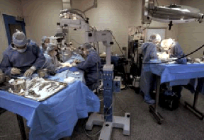
Kidney transplants are something everyone has heard of for people, but may not have considered possible for pets. In fact, while transplants are confined to specialized facilities with experienced surgeons, successful kidney transplants in cats have been going on since the mid-1980s. Canine programs have been less successful but are also available in limited facilities.

To the uninitiated, the impression may be that once someone receives a new kidney, life is renewed and all the kidney problems are solved. In fact, this is hardly the case. There are immune-suppressive medications needed to prevent the new organ from being rejected, not to mention potential for infection, on-going screening tests, and other issues.  A kidney transplant is a very big deal regardless of the species, and not every patient is even a candidate, so let’s walk through the main considerations.

Where Do the Donors Come from and What Happens to Them Afterward?

This question is important ethically for everyone involved in the transplantation program. No one wishes to harm the donor animal who cannot voluntarily become an organ donor in the way a human can, nor is there a mechanism to harvest organs from comatose brain-damaged patients as might be done for humans.

Feline kidney donors usually come from research facilities

These cats are classified as “specific pathogen-free,” which means they are free from infectious diseases. They are matched by blood typing and potentially by tissue typing, depending on the transplant program. A research cat is selected, and a kidney is removed for the recipient. After the procedure, the owner of the recipient cat must adopt the donor.

Some transplantation programs allow the owner of the recipient cat to provide the donor. Donors must be young adults, generally at least 10 pounds in size, be free of infectious diseases such as feline immunodeficiency virus, feline leukemia virus, and toxoplasmosis, and must have excellent kidney function. Basic blood testing and urine cultures must be normal.

You enter the program with one cat and leave with two cats.

For dogs, the recipient’s owner is generally responsible for finding the donor. The University of California transplant program has been on hold since 2007, but as an example, its former donor guidelines were that the donor must be less than six years of age, of similar size as the recipient (and preferably of the same breed), and tissue matching is needed if the donor is not related to the recipient. 

Is Your Cat a Candidate?

Kidney transplantation is not a procedure that is left until all medical therapies have been exhausted. The best candidate is free from other medical problems besides kidney disease.

Typical screening includes:

  1. Basic blood panel (including thyroid level) and urinalysis
  2. Urine culture (elimination of latent infection is crucial, and often a trial of immune-suppressive drugs is used after an infection has been cleared to ensure that the infection does not come back)
  3. Feline leukemia virus and feline immunodeficiency virus screening
  4. Ultrasound examination of the heart (heart diseases that involve high blood pressure development can damage the new kidney)
  5. Screening for toxoplasmosis (the immune suppressive drugs needed to maintain the new kidney could reactivate a latent infection with this parasite). A positive test for Toxoplasma does not preclude the transplant but a positive donor must be selected. Recipient cats testing positive for Toxoplasma are generally kept on clindamycin for life. Recipient cats testing negative will be periodically screened for Toxoplasma for the rest of their lives.
  6. Blood typing
  7. Blood pressure monitoring
  8. Urine protein to creatinine ratio to evaluate glomerular disease/renal protein loss.
  9. Teeth cleaning (performed under anesthesia)

Different programs may have additional required screening tests such as kidney biopsy, intestinal biopsy to rule out inflammatory bowel disease, test doses of immune-suppressive medications, etc. Obviously the details would be provided by the specific program being considered.

If the cause of the kidney failure is felt to be something that would lead the new kidney to fail as well, this may disqualify the patient from the program. Such conditions would include renal lymphoma or other cancer, amyloidosis (a malignant protein deposition), and pyelonephritis (a deep kidney infection, although if the infection is truly felt to have been eliminated, the patient might still qualify). Again, each program will indicate what conditions might serve to automatically disqualify a recipient.

An additional problem for dogs worth mentioning is intestinal intussusception. Imagine a telescope. Unfolded, it is basically a cylinder, but folded, and one segment collapses inside an outer segment. Now, imagine this happening to a piece of the intestine. A life-threatening obstruction results. A procedure called enteroplication is given in conjunction with the transplant. In this procedure, the intestines are tacked down with sutures into a formation that precludes any telescoping motions. This procedure, along with proper pain medication post-surgically, seems to prevent intussusception.

Cats with relatively early kidney failure are not yet candidates for transplantation. Cats with advanced kidney failure are not good candidates either, although dialysis (available at advanced critical care facilities such as those that perform kidney transplants) may improve the values.

The best candidates are those with an acute cause of kidney failure (such as a poisoning), cats who do not respond well to the usual medical management, or cats with a creatinine greater than 4.0 mg/dl. The cat should have a decent appetite and be as strong as possible prior to this major surgery.  Again, each transplant center will have its own criteria.

Prior to surgery, the recipient cat should be made as stable as possible. Usually, a blood transfusion is needed to correct the renal failure-associated anemia. Sometimes dialysis is also needed.

Is your Dog a Candidate?

Recipient screening is similar to that for the feline patient though heartworm testing is needed. Blood clotting tests are also needed for dogs. Similar conditions will also rule a patient out as a candidate (no cancer, heart disease, amyloidosis, or inflammatory bowel disease.) The adrenal hormone excess known as Cushing’s disease also precludes getting a kidney transplant. Kidney transplants for dogs are generally not as successful as for cats, so be sure you understand what you are getting into and the likelihood of obtaining the outcome you are hoping for.

What Kind of Home Care Will the Recipient Require?

The recipient is going to require suppression of his immune system for the rest of his life. This not only requires a substantial financial commitment for the medication but also the ability to give the cat oral medication at least twice a day for the rest of his life. The heart of this therapy is a medication called cyclosporine, a medication that has revolutionized organ transplantation for humans. Prednisolone, a commonly used cortisone derivative, is typically used as well, at least to start.

Cyclosporine is typically given twice a day with the lowest blood level of the day being approximately 500 ng/ml around the time of surgery and lowering to 250 ng/ml after a month or so of recovery after surgery. (Rejection of the new kidney occurs when levels dip below 200 ng/ml.)

Cyclosporine has some disadvantages that include:

  • Expense
    Cyclosporine is expensive. Concurrent administration of ketoconazole, a medication normally used to treat fungal infection, has the added benefit of “potentiating” cyclosporine. This means that less cyclosporine is needed to achieve the desired effect. As long as ketoconazole is well tolerated, it may become possible to dose the patient only once a day rather than twice. Approximately 30% of transplant patients will not be able to utilize this protocol due to the development of excessive cyclosporine levels or liver enzyme elevations from the ketoconazole.
      
  • Expenses for Monitoring
    Periodic blood level monitoring is needed to check that the right dosage is being used. For most drugs, “what you swallow is what your body gets.” For other drugs, there are individual variations in how the medication is absorbed, and cyclosporine is one. When two patients take the same amount of cyclosporine, they may not achieve the same serum levels; some individual fine-tuning is needed.
       
  • Long-term use of cyclosporine increases the risk for the development of cancer, specifically lymphoma. At the University of Wisconsin Renal Transplant Center, a 14% incidence of malignant tumor development is reported for cats with post-transplant time (and thus cyclosporine use time) of greater than one year.

In dogs, the chemotherapy agent azathioprine is also regularly used for its immuno-suppressive properties. It’s usually given every other day long term. Medication costs for dogs can vary from $150 per month to $2000 per month depending on the dog’s size.

What are Potential Complications for the Recipient?

  • Infection from immune-suppressive therapy is the second most common cause of transplant-associated death (rejection being the first). In cats, Toxoplasma infection is of particular concern, and while preoperative testing may have already been negative, some cats turn positive after the immune suppression begins. Also, in cats, old viral upper respiratory infections can be expected to recur, and they can be severe. In dogs, life-threatening infections can emerge in any organ system.   
  • Stricture (narrow scarring) of the ureter, which is the tiny tube that carries urine from the new kidney to the urinary bladder. If this occurs, another surgery is needed to trim the scarred area and re-attach the ureter to the urinary bladder. The original kidneys are generally not removed unless there is a reason to do so. The new kidney is simply an extra one. There is an approximately 21% incidence of this complication in the first 62 days post-operatively in cats. Mostly, males are affected.
  • The risk for developing diabetes mellitus increases by approximately five times after a kidney transplant.

What Kind of Survival Time Can You Expect?

In a recent study of feline kidney transplants, 59% of renal transplant patients were still alive 6 months after surgery and 41% were still alive 3 years afterwards. Apparently the first 6 months is a somewhat crucial time in determining long-term survival.

The University of Wisconsin Renal Transplant Center reports 70% survival at 6 months for cats and 50% survival at 3 years. Of the cats that survived to be discharged from the hospital (i.e. they did not succumb to problems directly related to the surgery), 96% survived to 6 months.

The picture is not nearly as bright in dogs. The University of California at Davis program had a success rate of about 40%. 

Kidney transplantation is an expensive undertaking. The University of California at Davis program, for example, required a deposit of $11,000 for cats and $13,000 for dogs. Transplantation involves the adoption of a donor and long-term medication and blood testing for the recipient. If this is something you are seriously considering, be sure to discuss the procedure with the transplant center closest to you as well as with your regular veterinarian.

7341109

Horner’s Syndrome in Cats and Dogs

A syndrome is a collection of symptoms that have significance when they go together. It is important to realize that having a syndrome is not the same as having a diagnosis. A syndrome, however, often has a limited number of causes such that recognizing a specific syndrome brings one substantially closer to a diagnosis.

What is Horner’s Syndrome?

Horner’s syndrome consists of five signs:

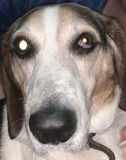
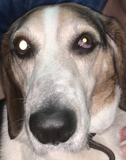
  • Constricted pupil
  • Elevated third eyelid
  • Retraction of the eyeball into the head
  • Slight drooping of the eyelid
  • Increased pink color and warmth of the ear and nose on the affected side (very hard to detect in small animals)

These signs appear on the side of the face (and eye) with damaged sympathetic nerves.

What is the Sympathetic Nervous System?

Our bodies have numerous functions that are controlled by our nervous systems, yet we are completely unaware of them. Our heart and respiratory rates, the amount of sweat and other secretions we produce, circulation to different body areas, pupil dilation (enlargement), and constriction (shrinking) are all regulated by our nervous systems automatically and without our knowledge or control. The part of our nervous system dedicated to these automatic systems is called the autonomic nervous system.

Sympathetic vs Parasympathetic

The autonomic nervous system is divided into the sympathetic nervous system and the parasympathetic nervous system. The parasympathetic system maintains a status quo, a normal business-as-usual state; the sympathetic system prepares the body for a fight-or-flight situation.

For example, the sympathetic nervous system kicks in with anxiety or fear leading to increased sweating, pupil dilation, increased heart and respiration rates, and increased blood flow to the muscles. The body is preparing to defend its life either by running or fighting. When danger passes, the parasympathetic nervous system kicks in to return everything to normal.  Both systems coexist to provide balance in a healthy body.

In the eye, the sympathetic nerve fibers dilate the pupil, widen the eyelids, drop the third eyelid, and keep the eye in a forward position in the socket. The parasympathetic nerves constrict the pupil, raise the third eyelid, and retract the eye for protection. Both systems are working at the same time, one system slightly dominating the other depending on what is happening.

When the sympathetic nerves controlling one of the eyes are damaged, only the parasympathetic nerves work and Horner’s syndrome is created.

How can the Sympathetic Damage Occur?

The nerve carrying the tiny nerve fibers that provide sympathetic control to the eye has a long path, and damage may occur anywhere along it. Also, some types of injuries are more likely to occur in certain areas along the path. 

This path begins in the brain’s hypothalamus. A group of nerves exit the hypothalamus and travel down the brainstem, continuing down the spinal cord, down the length of the neck, and just into the chest. This segment is called the central segment and is shown in blue in the illustration to the right. This pathway extends from the brain and brain stem down to the level of the second thoracic vertebra, all within the spinal cord.

From here, the nerves form right and left bundles and exit the spinal cord to make a U-Turn and travel back towards the eyes. The bundles are called the right and left sympathetic trunks (or the “pre-ganglionic segments”) and they extend from the top of the chest back to the area of the middle ear. They are shown in red in the illustration.

From there, the nerves connect to the last segment of nerves (the postganglionic segments – one on the right and one on the left), as shown in yellow. This segment starts just below the ear and travels to the eye.

The damage can occur in the neck or spinal cord area, the ear area, or the eye area. Damage can occur in the form of trauma, tumor involvement, infarction (abnormal blood clot), middle ear infection, or diseases of the eye itself. Each segment of the nerve pathway is vulnerable to different types of damage, so knowing which segment is involved gives us a good idea of what caused the damage.

Locating the Damage

As mentioned, localizing which area of the sympathetic nervous system is affected goes a long way in determining the nature of the damage, as different areas of the system are prone to different types of injury. Eye drops can be used to stimulate different areas of the above pathway and determine which area is damaged. Damage is described as being first order, second order, or third order. Most lesions turn out to be third order.

First Order Lesions (involving the blue segment)

Diseases that hit nerve fibers in the brain, brainstem, or spinal cord include tumors of the brain, vascular accidents (such as stroke) in the nerve tissue, fibrocartilaginous embolism in the spinal cord where disk material sprays into the spinal cord, or even a herniated intervertebral disk in the area of the neck. Horner’s syndrome stemming from any injury such as one of these might prompt a search for other neurologic issues. Advanced imaging such as an MRI might be a good idea.

Second Order Lesions (involving the preganglionic red segment)

Diseases that strike the sympathetic trunk include foreleg injuries especially if the foreleg is pulled and the nerves that exit the spinal cord in the armpit area become over-stretched. Sometimes a mass in the chest, such as a tumor or fungal granuloma, will damage the sympathetic trunk. Neck trauma such as pulling very hard on a leash could be severe enough to cause a second order lesion. If there is no obvious history to suggest injury, it might be a good idea to radiograph the chest to see if there are masses in the lung that might be involved in a second order lesion.

Third Order Lesions (involving the postganglionic yellow segment) 


These are the most common causes of Horner’s syndrome because ear infections are so common for small animals. Inflammation in the middle ear can easily lead to Horner’s syndrome. Third order lesions are associated with vestibular disease, the imbalance and dizziness of the middle ear infection, in many cases. When Horner’s syndrome localizes as third order, the ears should be thoroughly investigated as the source.

Treatment

It is not necessary to treat Horner’s syndrome. The syndrome is not painful and does not interfere with vision. The significance of the syndrome is that it indicates nerve damage that must be recognized. If you wish to treat the syndrome for cosmetic reasons, phenylephrine eye drops can be prescribed to relieve clinical signs. The most important thing is to determine what caused the Horner’s syndrome. Horner’s syndrome itself probably does not need treatment, but its underlying cause very well might.

4128244

Hot Spots (Pyotraumatic Dermatitis) in Dogs and Cats

What are Hot Spots?

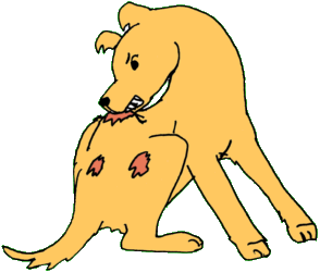
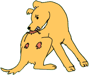
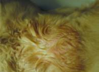
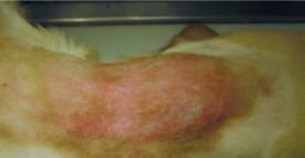
When the weather gets warm, we start seeing more dogs with hot spots. These wounds are weepy, wet, red, and sometimes bloody when they are fresh, and they are dry and scabby when they are resolving. They can cover large expanses of the pet’s skin or they can appear as smaller solitary lesions (sometimes in multiple locations). Hot spots generally have very clear margins defining them and separating them from normal skin.

They are caused by over-zealous self-licking, chewing, scratching, and rubbing (the pet creates them him/herself), and they can arise especially quickly (10 minutes of chewing can create a big one). The good news is that they almost always look worse than they are. The infection is quite superficial and often will resolve with topical treatment alone. The bad news is that about 30 percent of the pets who come to my hospital for hot spots actually have other kinds of skin diseases, such as deeper skin infections, bite wounds or other trauma, or even immune-mediated ulceration. If you aren’t entirely sure about what a hot spot looks like, it is probably best to have the veterinarian look at it.

The dog causes the hot spot by self-licking, chewing, scratching, and rubbing. What causes the pet to self-chew and lick is another story.

The pet may have an allergy, may have come in contact with an irritating substance, may have irritation from a grooming clipper, or may have some pain in the area from the underlying tissues. In many cases, the pet simply has fleas and is allergic to their bites. Anything that makes the dog itch will make the dog lick and chew, and if the licking and chewing is obsessive enough, a hot spot will soon follow.

First Aid

Treating a hot spot may or may not be a do-it-yourself project. Smaller hot spots can be treated at home with topical products made for this use. The important thing is to be aware that these areas are tender and so the pet may bite if you use something on the area that stings. Also, be careful about using human topical products as these may be toxic to pets when licked. Zinc oxide, for example, can be toxic when licked and is common in many human skin ointments.

Initial treatment usually involves removing the surrounding hair so that the hot spot can be disinfected. Once the superficial infection is properly cleansed, topical products can be used to relieve the associated inflammation. The lesion dries and scabs while it is healing. If the pet is really itchy or there are multiple hot spots, pills or injectable medication become necessary. Hot spots just under the ear/on the facial cheek, for example, are notorious for covering up a deeper skin infection below and often require more extensive treatment, especially in Golden Retrievers. More extensive treatments might include oral corticosteroids and/or oral antibiotics in addition to topical antiseptics and anti-inflammatories.

Good flea control is important for any itchy pet and is the foundation of itch prevention for most dogs and cats. Always be sure you have flea control secured in the approach to managing itchy skin in pets.

6721539

Hydrometra, Mucometra, and Pyometra in Dogs and Cats

Uterine disease in dogs and cats is often influenced by the hormone progesterone, which prepares a female for pregnancy and also helps the mother maintain a pregnancy. However, sometimes things don’t go as planned. One such event is when the uterus, or one of its horns, becomes filled one of several different types of fluid, and the effects can be relatively simple or substantial enough for an emergency hysterectomy to prevent death.

After a pet is no longer in heat, high levels of the hormone progesterone remain for nearly two months, and that high level of progesterone causes the uterine lining to thicken so as to be ready for pregnancy. Progesterone also suppresses the uterus’s immune function. If there is no pregnancy after several cycles, the uterine lining keeps getting thicker until eventually some cysts form inside the lining, a syndrome called cystic endometrial hyperplasia. Those cysts in the thickened lining leak fluid into the uterus.

The first stage of that fluid buildup is hydrometra. Unfortunately, two of the three stages do not have any significant clinical signs, so the pet owner is unaware that there is a medical problem.

A fluid-filled uterus has three stages called hydrometra, mucometra, or pyometra:

  • Hydrometra – watery fluid
  • Mucometra – fluid with mucous
  • Pyometra – pus (white blood cells and bacteria)

In hydrometra and mucometra, the fluid is sterile and there is no infection. Only pyometra is an infection, which occurs when bacteria crawl up the vagina through the cervix and into the uterus, causing a risk of developing endotoxemia (toxins in the bloodstream) and sepsis (in which the body’s response to an infection damages its own tissues) that are generally introduced by uterine disease. In pyometra, the uterus sometimes ruptures, which releases large amounts of pus and dead tissue into the abdomen.

Discovering hydrometra and mucometra is important: the conditions lead to decreased fertility and likely increase the risk of developing more severe uterine disease.

These conditions are more common in countries or areas where it is not typical practice to spay or neuter healthy dogs. They are not uncommon in sexually intact animals, especially in older females.

Radiography (X-rays) may detect uterine enlargement, but an ultrasound can tell if there is fluid in the uterus and may also indicate the type of fluid as well as rule out pregnancy. A dog with pyometra may be  quite sick, have a bloated belly, vaginal discharge, a poor appetite, and may be vomiting or drinking far too much water. Blood tests can detect infection and other underlying organ problems.

Treatment

Medical management sometimes involves using a drug called dinoprost (Lutalyse) to clean out the uterine contents; it is used to induce labor and the same contractions as occur in labor help eject the excess fluid. Sometimes vaginal infusion of warm saline can help empty the uterus.

While any stage of the first two conditions can be treated medically, they will recur. They will not resolve on their own and are a risk. Spaying is the only permanent fix.

Pyometra generally occurs in middle-aged to older female dogs in the six weeks following heat. The uterus fills with pus, bacteria, dying tissue, and toxins; at some point the uterus dies, releasing all of that dangerous material into the abdomen. It is a life-threatening emergency. A dog or cat with pyometra must be surgically spayed immediately (after stabilization) or she will die.

Oddly enough, the easiest one to fix is pyometra: treatment is a single surgery, despite being a bit more complex than a regular spay, and some antibiotics. Given that it’s otherwise fatal, it’s a good thing that the pet will have signs so that you know something is going on. Hydrometra and mucometra can be slow to respond to the dinoprost that lowers progesterone – sometimes taking a few months – and thus take much longer to treat than a surgical fix.

Pyometra is one of the main reasons for preventive spays. Removing the uterus is the only way to prevent hydrometra, mucometra, and pyometra.

4128334 (1)

Flatulence in Dogs

Imagine everyone is gathered together for a relaxing evening of snacks and television when gradually there is no way to avoid or ignore the odors being emitted by the otherwise happy family dog. Jokes abound but really a solution to this problem is wanted, especially if it is ongoing.

The Science of Flatulence

Flatulence comes from an excess of gases in the intestinal tract. These gases may represent air that has been swallowed (especially in short-faced/brachycephalic breeds), gas produced in the biochemical process of digestion, gas diffusion from the bloodstream, or gases produced by the bacteria that populate the intestinal tract.

Over 99% of the gases that pass from the intestinal tract are odorless; the gases with objectionable odors are typically those containing hydrogen sulfide.

Flatulence is a normal biological function. A surprising amount of air is swallowed with the simple act of eating and if this is not burped out, it must exit through the other end. The amount of air swallowed tends to be increased when dogs feel they must eat quickly or in the brachycephalic breeds who tend to breathe more by mouth rather than by nose. Swallowed air tends not to have objectionable odor.

The really stinky gases are produced by colon (large intestine) bacteria. Dietary fiber in pet food is not readily digestible by the pet’s own enzyme systems but is readily digested by the gas-producing bacteria of the colon. As these fibers are broken down, gases are produced. A diet heavy in fibers tends to favor these gas-producing organisms. The more supportive the intestinal environment, the more bacteria there will be and ultimately more gas will be produced.

What To Do About It

The following are easy changes that can be made in your management of the pet:

  • Feed smaller meals several times daily instead of one larger daily meal.
  • Feed a mixture of dry and canned foods.
  • Discourage rapid eating by placing an over-turned small bowl inside the pet’s regular food bowl. This prevents the pet from taking such a large mouthful. Commercial puzzle feeding bowls are also available for this same purpose.
  • Avoid soy and peas in the diet.
  • Avoid any treats containing milk, cheese or other forms of lactose.
  • Avoid fruit-based treats.
  • Change to a high digestibility/low residue diet. There are therapeutic diets sold at most veterinary offices that would be perfect. Ideally, rice would be the diet’s carbohydrate source.
  • If possible, walk the dog within 30 minutes of eating so as to encourage passing gases outside.
  • Avoid canned foods containing the texturing ingredient carrageenan.
  • Encourage regular exercise as this keeps intestinal gases moving and prevents intestinal gasses from pooling/building up.

Changing to a low residue diet means that most of the nutrients of the food are digested and absorbed by the pet before they reach the colon where the gas-forming bacteria are. This means there will be less food for the gas-forming organisms, which will ultimately mean fewer gas-forming organisms and less gas formed. Sometimes just going through a case and/or bag of a low residue diet solves the problem and the pet can return to a regular food afterwards. If necessary, the therapeutic diet can become the pet’s regular food. Low-residue diets are available through your veterinarian.

Prescription Low Residue Diets

Changing to a low residue diet means that most of the nutrients of the food are digested and absorbed by the pet before they reach the colon where the gas-forming bacteria are. This means there will be less food for the gas-forming organisms, which will ultimately mean fewer gas-forming organisms and less gas formed. Sometimes just going through a case and/or bag of such a low residue diet solves the problem and the pet can return to a regular food afterwards. If necessary, the therapeutic diet can become the pet’s regular food.

Sometimes Medication is Needed

A carminative is a medication that reduces flatulence. There is an assortment of available products, but unfortunately most are not helpful or even labeled for animal use. Changing the diet and ruling out actual intestinal disease are of primary importance in addressing flatulence. If further therapy is needed, the following products have some basis to suspect they might work:

Yucca shidigera supplementation

Currently this extract is labeled as a flavoring agent for pet food but it is also available as an oral supplement. Several studies have shown that it helps decrease the odor in flatulence.

Zinc acetate supplementation


Zinc binds to sulfhydryl compounds in flatulence ultimately serving to deodorize the gas.

Non-absorbable antibiotic

Such antibiotics serve to kill the gas-forming bacteria of the colon and may be helpful as long as their use is not ongoing. This is not an ideal method, however, as beneficial bacteria may also be killed off.

Probiotics

There are many ineffective probiotics being marketed so it is important to use one that has been shown to actually contain live cultures and that its cultures actually withstand stomach digestion so as to populate the small intestine with beneficial bacteria. It is unknown if this type of product would really help in flatulence as it is asking a great deal for such bacteria to travel all the way to the colon and attempt to displace the gas-forming resident bacteria. That said, there are several veterinary products that are felt to be reputable: Prostora®  Proviable®, Visbiome®,  and Fortiflora®. Fortiflora has been shown to be effective in reducing flatulence.

Prebiotics 

Prebiotics are special nutrients included in the diet to support and nourish beneficial bacteria and promote a healthy microflora in the colon. Special fibers are usually involved. Use of prebiotics allows the patient to essentially grow their own probiotics right there in their own colon. There are many intestinal diets that include prebiotic mixtures for this purpose.

Questionable Products

Activated charcoal tablets

Charcoal works by binding material in the intestinal tract so the material in question is not absorbed into the body, or in this case, so that the material is sequestered away from gas-forming bacteria. The idea is to make nutrients unavailable to gas-forming bacteria. Unfortunately, activated charcoal tablets are not likely to be effective as the charcoal binding sites are filled on the journey from mouth to colon and by the time the tablet sees the gas-forming large bowel bacteria, it has essentially been used up and cannot adsorb any more material.

Simethicone

Simethicone may control the volume of gas produced but not the odor. It is an antifoaming agent that reduces gas bubbles.

Pancreatic enzyme supplementation

In the absence of exocrine pancreatic insufficiency, it is unlikely that a patient would be helped by extra digestive enzymes. Furthermore, this treatment is relatively expensive for something that only might be helpful.

4128333

Esophagostomy Tube (E-tube) Care

Many people are intimidated by the idea of feeding a pet through an artificial tube, but the fact is the esophagostomy, or E-tube, makes feeding the sickly pet easy and free of mess. If you have been dealing with oral syringe feeding, meatball feeding, or even nasal tubes, the E-tube should be a breeze. Feeding through the tube is comfortable for the pet, plus the tube in no way precludes natural eating should the pet wish to do so. As your pet begins to feel better, regular eating can readily take place without disturbing the E tube.

The E tube can be in place as a temporary support measure or can be used long-term as an indefinite support measure. As you get used to using the tube, feeding will come to be a simple process and part of your regular pet routine.

To Feed your Pet

Have everything ready before beginning. You will need:

  • A syringe full of the liquid food in the appropriate amount. The diet should be warmed but not hot. Do not microwave the diet, or you may get hot spots that are too hot. To warm the food, microwave a tall glass of water and insert the syringe of food in the warm water and let it sit until the food is at least room temperature, and ideally close to body temperature.
  • A small glass or cup of tepid (lukewarm) water.
  • Any medication you should be giving at this time.

Clear the tube by squirting 6 ccs of tepid water through it to be sure it is not clogged. Next, hook up the food syringe and slowly deliver the food to the patient. It is tempting to just blast it all in there but for patient comfort, try to take several minutes or so and take your time. Follow the food with a chaser of 6 ccs of tepid water to clear the tube. Liquid medicine can be given through the tube as long as the tube is cleared with 6ccs of tepid water before and after each time. Do not put pills in the tube as it could clog. Pills can be crushed and given through the tube only if they are well dissolved in liquid. Any caking of pill powder has the potential to clog the tube, so be sure to flush the tube with tepid water after use.

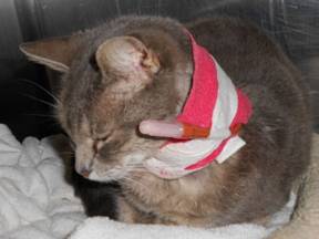
You will need to clean the stoma (the opening of the skin where the tube enters) daily with a baby wipe or moist tissue, otherwise discharge and/or crusting will accumulate there. Periodically the wraps will need changing. A special type of collar may be helpful in keeping the tube neat and comfortable.

Be sure to allow time for digestion between feedings. How much time depends on your pet’s feeding plan.
Be sure you understand the amount for each feeding and the feeding schedule provided by your veterinarian.

Having Problems?

Vomiting?

  • Are you giving the food too quickly? Rapid distension of the stomach is a stimulus to vomit. Try going slower.
  • Is the food too cold? Try warming the food to body temperature (around 100°F). Use a thermometer in the warming water bath to be sure the temperature is where you want it.
  • If these two solutions do not work, the tube may have slipped too low inside the esophagus. If the tube is dipping into the stomach, the patient may vomit. The doctor can take an x-ray to see if this is the case and then easily reposition the tube. If none of these things seem to be happening, the patient’s primary disease may be progressing. Your veterinarian will need to evaluate your pet more comprehensively.

Tube clogged?

A clogged tube can be a challenge. First, try to force 6 ccs or so of tepid water through the tube by pushing. If the clog does not give way, try hooking up a syringe of 6 ccs or so of water and alternately push and pull back, creating a “toilet plunger” effect. Continue fairly rapid push-pull action until the tube is cleared.

Note: some people feel that incubating 6 ccs of cola soda in the tube overnight is helpful in dissolving a clog. Whether or not this works is somewhat controversial, but it may be worth a try, as the tube will be useless if it cannot be unclogged.

Crusting or pus at the tube exit site?

The patient’s body does not like having a foreign body sticking out of it and frequently there is some inflammation at the exit hole. True infection is unusual, and most of the time simply cleaning the area with gauze or a moist tissue is sufficient to solve the problem.

Esophagostomy tubes can stay in place for months without needing to be replaced. Hopefully your pet’s condition will have resolved before that time. When the time comes, the tube’s anchoring sutures can be snipped and the tube pulled out. The hole left behind will simply heal on its own. If you have any questions about the tube or its care, your veterinarian will be happy to assist you.

4128431 (1)

Exocrine Pancreatic Insufficiency in Dogs and Cats

What the Pancreas Normally Does

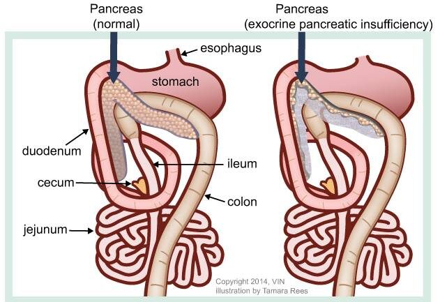
The pancreas is a small light pink glandular organ nestled under the stomach and alongside the upper small intestine. There are two types of pancreatic tissues to know about. The first tissue is the “endocrine” pancreas which is the part of the pancreas that secretes hormones involved in blood sugar regulation, such as insulin, (which you have probably heard about if you know anyone with diabetes mellitus). The “exocrine” pancreas produces enzymes we use to digest our food. These two parts of the pancreas are not in separate areas but instead these two different types of pancreatic tissues are all mixed together throughout the entire pancreas.

Digestive enzymes break down fats, starches and proteins into smaller units so that we can absorb them into our bodies. These enzymes are stored as inactive forms inside special granules in the exocrine pancreatic tissue (the “acinar cells”). The enzymes sit there harmlessly until a chemical signal tells the pancreas to secrete them down the pancreatic duct and into the intestine where the food is waiting.

Once nutrients are broken down into smaller molecules (i.e. they are digested), they can be absorbed down the entire length of the GI tract. Without an adequate amount of enzymes, we cannot break down/digest our food. If we cannot digest the food, we cannot absorb the food. We get skinny, have especially nasty, rather greasy diarrhea, or both. Often, pets will develop a dry, dandruffy coat from their inability to absorb dietary fats and can be anemic from a vitamin B12 deficiency. Difficulty in clotting blood (vitamin K-related coagulopathy) can result as well.

Exocrine Pancreatic Insufficiency (also called Maldigestion)

The most common cause of digestive enzyme deficiency in dogs is “pancreatic acinar atrophy,” where the pancreas simply becomes shriveled and useless. This condition seems to have a genetic basis but is not congenital and may develop at any age (though usually shows up before age 4 years).

The German Shepherd Dog and Rough-Coated Collie are particularly at risk and the mode of inheritance appears to be autosomal recessive (meaning genetic carriers will appear normal).

About 70% of dogs with exocrine pancreatic insufficiency are German Shepherd dogs and 20% are Rough Collies.

Recently a juvenile onset form of exocrine pancreatic insufficiency has been described in the Greyhound, however, because this breed is primarily used in racing, sickly puppies are commonly euthanized thus keeping the condition from being recognized in the pet population.

In cats, chronic pancreatitis is the usual cause of exocrine pancreatic insufficiency. There does not appear to be a genetic concern; though, rarely, cats that eat grasshoppers may get infected with a pancreatic fluke carried by grasshoppers (Eurytrema procyonis). This fluke, similar to a very small worm, can cause enough inflammation to cause pancreatitis or enough general pancreatic damage to cause exocrine insufficiency. Fluke infection is rare, as mentioned, but feline pancreatitis is actually very common especially in older cats. There is frequently a link to chronic intestinal disease. 

Diagnosis Requires Specific Tests

Trypsin-Like Immunoreactivity

The biggest breakthrough in the diagnosis of exocrine pancreatic insufficiency was developing the serum trypsin-like immunoreactivity test, a blood test. Prior to this, an assortment of inaccurate fecal tests were used. Trypsin is one of the digestive enzymes secreted by the exocrine pancreas. The TLI test looks for a normal level of trypsin-like enzymes in the bloodstream. This is harmless, normal, and measurable in a blood test.

A dog or cat with EPI will have almost no serum trypsin-like immunoreactivity in the bloodstream. The patient must be fasted for the test to be accurate, but only a single blood sample is needed to make the diagnosis. The feline version of this test often requires that the sample be sent to a university laboratory and generally a week or so is needed to get results but the canine test can be run in just a few days.

Other Tests

Another popular diagnostic is the fecal protease test, where a stool sample is tested for protein digesting enzymes. Fasting is not necessary and any fecal sample will do; however, three consecutive samples are needed to get a consistent result as there is tremendous variability in fecal enzyme activity over the day. Sometimes soybeans are given to dogs to help stimulate release of pancreatic protein digestion enzymes and get a more accurate test.

The fecal elastase test (elastase is another digestive enzyme) is the newest test and it is only available for dogs. A single fecal sample is needed but the problem is that sometimes normal dogs will test negative for elastase. This means that EPI can be ruled out when the elastase test is positive but not confirmed when the elastase test is negative.

Treatment

Dietary supplementation with digestive enzymes is an effective therapy for EPI even though most of the supplement given is digested in the stomach along with other dietary proteins. The little bit that survives the acid bath of the stomach and the patient’s own protein-digesting chemicals turns out to be enough to stop the diarrhea and enable the patient to actually gain some weight. Powdered enzymes (Viokase-V, Pancrezyme, PanaKare, and other brands) seem to work the best; tablets are available but do not seem to break down consistently. If the pet finds the taste of the enzymes objectionable, a compounding pharmacy can fill gel capsules with the powder. Again, enteric-coated tablets simply do not seem to work well.

In the past, it was suggested that incubating the enzymes in the patient’s food would help initiate the digestion process in the food bowl but this has not been found to be true; the enzymes can be fed immediately mixed with the patient’s regular pet food. Some patients respond best when an H2 blocker-type antacid (such as famotidine) is given concurrently with the enzymes.

It is important to thoroughly mix the enzyme powder into the food, because if it is sprinkled on top, it can be abrasive and lead to ulceration in the pet’s mouth. If ulceration has already occurred in this situation, incubating the food with the enzymes can help resolve the problem. A six-hour incubation at room temperature should suffice. If the food is to be refrigerated, a 24-incubation has been recommended. 

Raw beef or lamb pancreas can also be used as a possibly inexpensive form of enzyme replacement but the problems with raw foods include parasite and bacterial contamination. Cooking the pancreas relieves these concerns but inactivates the desired digestive enzymes. Raw pancreas can be stored frozen without losing digestive enzyme activity.

Generally a high-digestibility diet is the best choice for an EPI patient. These foods are low in fiber and may be especially helpful for patients with trouble gaining weight. Many animals simply use enzymes mixed with their regular food if prescription diets prove too expensive.

EPI patients commonly have an overgrowth of bacteria in their intestines, which means that the unabsorbed nutrients in the tract have fed the bacteria living there, instead of the patient, and an over-population of bacteria has occurred. This results in a vitamin B-12 (also called cobalamin) deficiency as the bacteria consume the vitamin instead of the patient getting his share. Periodic injections of vitamin B-12 and blood test monitoring have been recommended for patients with EPI. The B-12/cobalamin deficiency is a particularly big problem for cats with EPI, who typically require injections every couple of weeks for life. Folate deficiency is also common in cats and supplements are required for the first month of therapy. Regular cobalamin and folate blood levels important for cats with EPI. Supplementation with the fat soluble vitamins (especially vitamin K and vitamin E) are not usually needed.

Treatment is for life and without enzyme supplementation, all the unpleasant symptoms will recur. The good news is that a response to therapy is generally seen within a week of beginning it. Response can be excellent but approximately one dog in five will simply not respond well. Many do not ever regain a normal amount of weight. 

What if Results Are not as Rapid as Expected? 

Most pets respond in a matter of days to enzyme supplementation but if a week or so goes by and results are underwhelming, here are some tips to consider:

  1. Use more enzyme powder with each meal. 
  2. Check the powder’s expiration date. Consider a different brand of powder.
  3. Be sure to supplement vitamin B12 by injection. Many pets will not get better until this deficiency is corrected
  4. Treat the bacterial imbalance. Some patients will not get better without a course of antibiotics.
  5. Consider incubating the enzymes in the food. This is not supposed to matter but some pet owners feel that it does.
  6. Consider a prescription diet if one is not already in use.

If all of these factors are already controlled and diarrhea still has not cleared up, it may be time to consider that a second disease is afoot. Discuss this possibility with your veterinarian.
Want more support online? Join an Exocrine Pancreatic Insufficiency Forum for dog owners.

4127238

Blastomycosis is a Systemic Fungal infection Affecting Dogs and Cats

Blastomycosis, caused by Blastomyces dermatitidis, is a systemic fungal infection that affects dogs and cats. Blastomycosis is most common in certain geographic areas in North America, most often the Mississippi, Ohio, Missouri, Tennessee, and St. Lawrence River valleys, and in three provinces of Canada (Quebec, Ontario, Manitoba). It has also been reported in Africa, India, Europe, and Central America. (Fungal growth is supported by wet, sandy, acidic soils rich in organic matter, which is why it is found in valleys and is seen most frequently near water.)

Pathophysiology

Infection with Blastomyces occurs when a cat or dog inhales the fungal spores into the lungs. The incubation period is from 5 to 12 weeks. Some animals don’t show clinical signs for a long time after being infected, but those animals are not contagious to other animals and people. Blastomycosis organisms have a predilection for the respiratory tract, and pulmonary disease is the most common sign (88-94% of canine cases). Once the lung disease develops, yeast forms of the organism spread throughout the body. Organs typically affected include eyes, bones, skin, lymph nodes, subcutaneous tissues, brain, and testes. It can also be found in the nose, prostate, liver, mammary glands, and heart, but those locations are less common.

Dogs appear to be much more susceptible to infection than other species. Although the disease does occur in both people and cats, the incidence is much lower than in dogs. Dogs are ten times more likely to contract the disease than are people, and 100 times more likely than are cats. The incubation period in dogs is also shorter than in people. The reason dogs are more susceptible is unknown, but immune-deficiency may play a role. Annual prevalence in dogs in endemic areas is estimated at 1-2%. Many infected animals live within 0.25 mile of water. An increased number of cases can occur after periods of unusually heavy rainfall. Historically, young (i.e. 1-5 years), male, large-breed (e.g. hounds, pointers) dogs have had the highest risk of infection. (This is probably because these animals would tend to have more contact, due to hunting activities, with the organism’s geographical area.) However, any age, breed, or sex of dog can be infected.

Clinical Signs

The signs of illness will depend on what organs are infected. Some dogs will have eye problems, and some will have neurological signs (seizures, head tilt, etc.). Lameness may occur, if the infection is in the bones. Owners of dogs in the specific geographic areas should watch for coughing, difficulty breathing, eye inflammation, enlarged testicles, fever, swollen lymph nodes, ulcerated/draining skin lesions, bloody urine, difficulty urinating, nasal cavity signs (including bloody nasal discharge), and lack of appetite. (Weight loss occurs because of the decreased appetite.) Infections in the nasal passages may result in skull damage, and lead to infection of the brain. Large skin abscesses and neurologic signs are more common in cats than in dogs, while bone lesions are more common in dogs than in cats. Hypercalcemia can occur in dogs, although it’s rare in cats and non-domestic feline species.

Diagnosis

Diagnosis involves physical exam, blood tests, imaging (radiography, ultrasonography, CT, etc.), urinalysis to look for Blastomyces yeast, cultures, serology, and biopsies of affected organs. Fungal serology, to look for antibodies, is not always accurate and has been known to produce false negatives. The enzyme immunoassay (EIA) for B. dermatitidis galactomannan antigen appears to have a high sensitivity in urine (93.5%) and serum (87%). The EIA assay is commercially available; cross reactions with Histoplasma capsulatumCoccidioides immitis, and Penicillium marneffei can occur with this assay. The definitive diagnosis is finding the fungus in the tissues.

Treatment

Treatment involves various antifungal medications, including itraconazole, ketoconazole, fluconazole, amphotericin B, and some combination therapies. These medications usually need to be taken for a long period of time; how long depends on the specific case. Treatment usually can be done at home, unless the disease is severe. Pets with respiratory involvement should be on restricted activity. In severely ill dogs, intravenous fluids, oxygen, antibiotics for secondary infections, and pain medication may be necessary. Skin lesions may require wound cleaning and debridement.

Eyes that are severely affected may not respond well to the treatment because the medication does not penetrate eyes very well. Ocular blastomycosis cases may need systemic and topical corticosteroid therapy, topical anti-glaucoma medications, etc. Significantly affected eyes may require enucleation (removal of the eyeball).

Pets with severe lung disease may get worse at the beginning of treatment because the fungal organisms are dying; the mass death of organisms can cause severe respiratory problems.

Patients may not appear to improve for one to two weeks after the start of treatment. Close monitoring should be done for the first two weeks, and then rechecks are usually scheduled on a monthly basis. Rechecks may involve blood tests, biochemistry profiles, and imaging, if appropriate.

Prognosis

The prognosis for many pets is good, especially if owners can afford to treat the pet for long enough. Approximately 50% to 75% of affected dogs recover when treated with itraconazole, fluconazole, or an amphotericin-ketoconazole combination. Prognosis is poor for dogs with severely affected lungs, but if the dog survives the first 7 to 10 days of therapy, the prognosis improves. However, mortality rates in dogs with Blastomyces dermatitidis can be as high as 41%.

The prognosis for the retention of vision, in animals that have eye disease, is variable.

Dogs with brain involvement often die. Seizures are common upon death of the organism and may be uncontrollable.

Prognosis is also poor when at least 3 body systems are involved.

Relapses are most likely to occur in dogs that had a severe case at onset, or dogs that were not treated long enough. Relapses are most common within the first 6 months after treatment. Dogs that recover from the disease are probably not immune to getting it again.

After discontinuing therapy, animals may be rechecked at 1, 3, and 6 months for evidence of relapse. In one study, relapse rates for itraconazole and fluconazole were 18% and 22%, respectively. Relapses are treated like a new infection.

There is no way to prevent your pets’ exposure to Blastomyces other than by keeping them away from affected geographic areas.

Zoonotic Potential

Blastomycosis is not considered to be a zoonotic disease. It is acquired by humans via inhalation or direct contact with infective conidia/spores. Risk of infection is higher for excavation workers, and for people working or playing in wooded areas with waterways. Blastomycosis cannot be spread between dogs and other animals, or between dogs and people. However, immunocompromised people should limit their contact with infected pets and should wear gloves when cleaning and treating draining lesions.

7941886

Winter Holiday Hazards for Pets


The holiday season is upon us, and many people plan to include their furry companions in the festivities. As you gear up for the holidays, it is important to try to keep your pet’s eating and exercise habits as close to their normal routine as possible. Also, please be sure to steer pets clear of the following unhealthy treats, toxic plants, and dangerous decorations.

Be Careful with Seasonal Plants and Decorations

  • Oh, Christmas Tree: Securely anchor your Christmas tree so it doesn’t tip and fall, causing possible injury to your pet. This will also prevent the tree water—which may contain fertilizers that can cause stomach upset—from spilling. Stagnant tree water is a breeding ground for bacteria, and your pet could end up with nausea or diarrhea should he imbibe.
  • Avoid Mistletoe & Holly: Holly, when ingested, can cause pets to suffer nausea, vomiting, and diarrhea. Mistletoe can cause gastrointestinal upset and cardiovascular problems. And many varieties of lilies can cause kidney failure in cats if ingested. Opt for just-as-jolly artificial plants made from silk or plastic, or choose a pet-safe bouquet.
  • Tinsel-less Town: Kitties love this sparkly, light-catching “toy” that’s easy to bat around and carry in their mouths. But a nibble can lead to a swallow, which can lead to an obstructed digestive tract, severe vomiting, dehydration, and possible surgery. It’s best to brighten your boughs with something other than tinsel.
  • That Holiday Glow: Don’t leave lighted candles unattended. Pets may burn themselves or cause a fire if they knock candles over. Be sure to use appropriate candle holders, placed on a stable surface. And if you leave the room, put the candle out!
  • Wired Up: Keep wires, batteries, and glass or plastic ornaments out of paws’ reach. A wire can deliver a potentially lethal electrical shock and a punctured battery can cause burns to the mouth and esophagus, while shards of breakable ornaments can damage your pet’s mouth and digestive tract.

Avoid Holiday Food Dangers

  • Skip the Sweets: By now you know not to feed your pets chocolate and anything sweetened with xylitol, but do you know the lengths to which an enterprising pet will go to chomp on something yummy? Make sure to keep your pets away from the table and unattended plates of food, and be sure to secure the lids on garbage cans.
  • Leave the Leftovers: Fatty, spicy, and no-no human foods, as well as bones, should not be fed to your furry friends. Pets can join the festivities in other fun ways that won’t lead to costly medical bills.
  • Careful with Cocktails: If your celebration includes adult holiday beverages, be sure to place your unattended alcoholic drinks where pets cannot get to them. If ingested, your pet could become weak, ill, and may even go into a coma, possibly resulting in death from respiratory failure.
  • Selecting Special Treats: Looking to stuff your pet’s stockings? Stick with chew toys that are basically indestructible, Kongs that can be stuffed with healthy foods, or chew treats that are designed to be safely digestible. Long, stringy things are a feline’s dream, but the most risky toys for cats involve ribbon, yarn, and loose little parts that can get stuck in the intestines, often necessitating surgery. Surprise kitty with a new ball that’s too big to swallow, a stuffed catnip toy, or the interactive cat dancer.

Plan a Pet-Safe Holiday Gathering

  • House Rules: If your animal-loving guests would like to give your pets a little extra attention and exercise while you’re busy tending to the party, ask them to feel free to start a nice play or petting session.
  • Put the Meds Away: Make sure all of your medications are locked behind secure doors, and be sure to tell your guests to keep their meds zipped up and packed away, too.
  • A Room of Their Own: Give your pet his own quiet space to retreat to—complete with fresh water and a place to snuggle. Shy pups and cats might want to hide out under a piece of furniture, in their carrying case, or in a separate room away from the hubbub.
  • New Year’s Noise: As you count down to the new year, please keep in mind that strings of thrown confetti can get lodged in a cat’s intestines if ingested, perhaps necessitating surgery. Noisy poppers can terrify pets and cause possible damage to sensitive ears. And remember that many pets are also scared of fireworks, so be sure to secure them in a safe, escape-proof area as midnight approaches.