Tag: cat and dogs

4126859

Bloody Nose (Epistaxis) in Dogs and Cats

Some blood-tinged droplets sneezed on the floor might be the only sign, or there might be a steady, inexorable bloody drip from one or both nostrils. These findings are alarming as well as messy in the home and we want to identify the cause and take care of it promptly if it is possible to do so. The problem is that there are many causes, and not all of them are localized to the nose, and many are very serious diseases. The following is a review of tests typically necessary to get to the bottom of the bloody nose as well as the conditions that might be responsible.

First Aid

So, you are at home with your pet and a bloody nose starts and does not seem to be stopping. Here are some tips to get the bleeding controlled in the time prior to your vet appointment:

  • Keep yourself calm. If your pet sees you getting frantic, they will, too. Excitement = higher blood pressure = more bleeding.
  • Get an ice pack and apply it to the bridge of the nose (obviously, be sure your pet can breathe around the ice pack). The cold will constrict small blood vessels, which will slow the bleeding.
  • Do not insert absorbent material or cotton swabs in the pet’s nose, as this will generate sneezing, which will make the bleeding worse. A dose of an oxymetazoline nasal spray such as Afrin may help constrict blood vessels and lead to relief.
  • If the pet has a condition that involves recurring nose bleeds, consider the oral use of the Chinese herb Yunnan Baiyo, which promotes blood clotting tendency. Ask your veterinarian for details.

If these steps do not stop the bleeding or the pet is having difficulty breathing, go to your vet’s office or local emergency clinic at once.

Don’t forget that a pet with a bloody nose will likely swallow a great deal of the draining blood. This may lead to an especially black stool or even vomit with blood clots in it.

After a bloody nose, such findings are usually just a reflection of the bloody nose and do not necessarily indicate bleeding in the GI tract.

Information Your Veterinarian Will Need

You can help your veterinarian tremendously by taking some time to think about the following information and bringing up anything pertinent.

  • Does your pet take medication? Non-steroidal anti-inflammatory medications (aspirin in particular) will inactivate blood clotting factors. Do not assume your vet knows all the medications your pet is taking; list them for your vet.
  • Do you have any rat poison or has your pet been consuming any dead rodents that might have been poisoned? Most rat poisons act by disabling the ability to clot blood.
  • Look closely at your pet’s face. Is there any deformity or asymmetry? Is the bridge of the nose swollen? Are either of the third eyelids elevated? Does one eye seem to protrude more. does one eye tear more? Does the nose leather (textured tip) look normal?
  • Could there have been any trauma to the nose? Does your pet play roughly with another animal?
  • Is your pet exposed to foxtails or other grass awns that could become lodged in the nose?
  • Has your pet been sneezing? Has the pet been rubbing at the nose?
  • Open your pet’s mouth if possible. Look at the gums under the lips. Is there blood in the mouth? Do the gums seem pale? If they are, this suggests a serious loss of blood and you may have an emergency on your hands.
  • Is there any evidence of bleeding anywhere besides the nose? You may see a black tarry stool with Intestinal bleeding may present with a black tarry stool. Any unusual bruising should be reported. Any unexplained swelling that might be bleeding under the skin should also be noted.
  • Is this the first nosebleed or have there been others?
  • Is the blood coming from both nostrils or only one?

Where to Start

After the veterinarian performs a general examination of your pet, some more specific tests are needed with the idea of prioritizing the most likely conditions and least invasive forms of testing.

Blood Tests First

A basic blood panel and urinalysis will probably be needed as a database for the animal’s health as well as to assess the degree of blood loss. This information also serves as a pre-anesthetic evaluation should rhinoscopy or nasal imaging become necessary. A platelet (a blood cell involved in blood clotting) count will be needed as will coagulation tests (common tests are the PT or prothrombin time; the PTT or partial thromboplastin time; the ACT or activated clotting time; and the buccal bleeding or symplate time.) These tests evaluate a complicated biochemical cascade responsible for clotting blood. The pattern of abnormalities found in these tests will sort out blood clotting disorders.

Other blood tests that may be helpful involve titers for fungal infections, a classic cause of the nosebleed. Fungi are inhaled and if the patient is immune-compromised or excessively exposed, the fungus can take root and begin to grow in the nasal cavity.

In cats, the most common nasal fungal infection is caused by Cryptococcus neoformans. The good news here is that a blood test for fungal antigen is very accurate. Any positive number is significant and warrants treatment.

In dogs, fungal infections are not so simple. The most common organisms are Aspergillus fumigatus and Penicillium species. Blood tests are not as accurate especially since there are other species of Aspergillus besides fumigatus and each requires its own blood test. Complicating matters is the fact that nasal tumors predispose a dog to fungal infections so a dog can easily have both problems in the same nose. Blood tests for fungal infections may be included in the initial battery of tests. A negative Aspergillus test does not rule out Aspergillus infection.

Blastomyces dermatitidis is another fungus that can get into a dog’s nose. Urine antigen testing is accurate for diagnosis and blood testing is also available if results are ambiguous. As with other fungi, treatment is long term and challenging.

Another condition worth mentioning is hyperviscosity syndrome. In this situation, an extremely high blood protein level makes the blood so thick that blood vessels break from the pressure. Certain types of cancer (multiple myeloma, lymphoma, and certain types of leukemia) as well as infection with Ehrlichia canis, a blood parasite can cause this syndrome.

A routine blood panel should show the unusual globulin levels that typify hyperviscosity syndrome.

Another relatively simple parameter to measure is blood pressure. High blood pressure can occur as a complication of numerous diseases. When blood pressure rises, small blood vessels begin to burst and bleed, not just in the nose but often in the eyes or nervous system as well. Do not be surprised if your veterinarian checks for retinal hemorrhage.

Tick-borne infections (Ehrlichia, Babesia, and others) commonly involve low platelet counts. Platelets are blood cells involved in clotting and when they become infected with blood parasites, they do not work properly in the clotting cascade. Tick panels are blood panels that screen for infection with numerous tick-borne parasites, most of which can be managed or eradicated with antibiotics.

The bottom line is that there are many causes of nose bleeds but many can be ruled out with non-invasive testing and it is the non-invasive tests that we want to perform first.

Blood Clotting Disorders of Pets

  • Rat poisoning
  • Von Willebrand’s disease
  • Hemophilia
  • Liver failure
  • Disseminated intravascular coagulation

Diseases Causing a Low Platelet Count

  • Immune-mediated thrombocytopenia
  • Anaplasma infection
  • Bone marrow disease
  • Drug reactions (methimazole, chemotherapy drugs, excess estrogens, sulfa class antibiotics) 
  • Feline leukemia virus infection
  • Feline immunodeficiency virus (FIV)
  • Ehrlichia infection (dogs) 
  • Rocky Mountain spotted fever
  • Hemangiosarcoma
  • Other cancers
  • Babesia

Cruising Towards Anesthesia

If the basic blood tests and clotting parameters are normal, then the chances are that the problem is localized to the nose but there are a few more tests that are required before the patient is anesthetized for a nasal examination.

  • Radiographs of the chest should be performed to rule out obvious cancer spread or obvious disseminated fungal disease.
  • An oral examination should be performed as best as possible. Dental disease can be bad enough to create nasal bleeding, given that the roots of larger teeth connect with the nasal cavity. Oral tumors that have eroded into the nasal cavity may be evident if one can get a good look in the mouth. Many patients will not allow much oral exam and certainly probing the gums and getting a thorough inspection will require anesthesia but it is absolutely worth looking for obvious lesions if it is possible to do so.

Diagnostics Requiring General Anesthesia
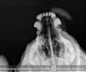
If nothing has been revealed by the preceding tests, it is now time for radiographs of the nose, superficial rhinoscopy, and a dental inspection all of which require general anesthesia. Radiographs generally start the procedure as the other procedures might alter the radiographic appearance of tissues. The radiographs help evaluate the tooth roots and sinuses. Nasal tumors are common causes of nosebleeds in elderly dogs and the bone destruction they cause is evident on radiographs. Referral for more advanced imaging such as CT scanning or MRI, may be needed to determine the extent of bone destruction or to clarify radiography findings.

An otoscope (the same gadget used to look in your pet’s ears) can be used to look inside the nasal cavity superficially to remove foreign bodies lodged there. Deeper peeking requires an actual endoscope which may not be readily available in general practice.

The teeth can be cleaned under anesthesia with specific attention to the tooth roots (remember, an abscessed upper tooth root penetrates into the nasal sinus above.

If it seems appropriate to do so, some nasal discharge can be flushed through the nose and into a gauze sponge packing the throat. This may be helpful in identifying infectious organisms but may initiate more bleeding so some judgment is required on whether the benefit is worth the risk.

Then What?

If the simple tools of general practice do not reveal adequate information, referral for endoscopy may be needed. Deeper visualization of the nasal tissues is possible with this equipment, plus biopsy specimens can be taken although bleeding is the chief risk. Taking a biopsy is particularly difficult in the nose, not just because of the hemorrhage but because nasal tumors are surrounded by so much inflammation it is difficult to get a representative sample. Often it is not possible to see the area being biopsied directly, especially if prior sampling has led to bleeding.

If radiographs are diagnostic for cancer and aggressive therapy is contemplated, prognosis is highly dependent on the type of tumor so biopsy becomes especially important in this situation.

And Then What?

At the end of all these procedures, sometimes the area of bleeding is simply not accessible without surgery. This would be the final and most invasive procedure in retrieving a difficult foreign body or tissue sample. Extensive bleeding is expected and this is generally the last resort after a long road of diagnostics.

In a study by Bissett et al published in the December 15, 2007, issue of the Journal of the American Veterinary Medical Association, 176 cases of dogs with bloody noses were reviewed to determine which underlying causes were most common. Of these 176 dogs, a definite underlying cause was found in 115 cases.

  • 30% had nasal tumors
  • 29% had trauma
  • 17% had nasal inflammation of unknown cause (idiopathic rhinitis)
  • 10% had low platelets
  • 3% had some other blood clotting abnormality
  • 2% had high blood pressure
  • 2% had tooth abscess

Conspicuously absent is the nasal fungal infection but since these are frequently regional in nature in dogs, the population studied may not have been in an area where fungi are common pathogens.

Other Relevant Studies

Evaluation of factors associated with survival in dogs with untreated nasal carcinoma: 139 cases (1993-2003) by Rassnick et al published in the Journal of the American Veterinary Medical Association  229:401-406, 2006.

This study reviewed the outcomes of 139 dogs diagnosed with nasal carcinoma. Since many dogs are euthanized at the time of this diagnosis, this study included only dogs that were alive seven days after the initial diagnosis was made. Dogs studied received only pain medication, anti-inflammatories and antibiotics (no surgery, chemotherapy or radiation therapy). The following statistical findings came out:

  • 80% of dogs were purebred. The median age was 11 years and the median body weight was 48 lbs.
  • 77% had nose bleeds. Median survival time for dogs with nosebleeds was 88 days vs. 224 days for dogs with carcinomas that did not have nose bleeds.
  • Approximately half of the patients studied were felt to have improvement with the supportive care described above.

Ultimately, the therapy for a recurring nosebleed depends on the cause, and the causes are many and varied. If your pet has a nose bleed that lasts more than 5 minutes, seek veterinary care right away. If nose bleeds are recurrent, your pet will need a medical checkup to sort out the possible causes, as reviewed above. Contact your veterinarian to assess your pet’s situation as early in the course of disease as possible.

4128320-1

Brushing your Dog’s Teeth

What would happen if you stopped brushing your own teeth? Even if you only ate hard food, as most dogs do, there still would be problems. You should be brushing your dog’s teeth twice daily if you can manage it; it’s not as difficult as you might imagine.

What are the benefits of brushing? Brushing removes daily accumulation of plaque from the teeth. Even though dogs do not commonly get cavities, they do suffer from periodontal disease. If untreated, gum disease can lead to bad breath, pain, and tooth loss.

How Do You Brush Your Dog’s Teeth?

Step one is to pick an appropriate pet toothbrush.

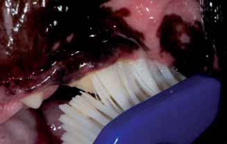
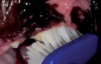
Save yourself time by not buying a child’s toothbrush, which is usually too hard for dogs. The ideal dog toothbrush will have a long handle, an angled head to better fit the mouth, and extra soft bristles. For dogs less than 30 pounds, a finger toothbrush that fits over the tip of your finger works well.

Step two is to select an appropriate toothpaste.

The best pet toothpastes contain enzymes that help control plaque. Try to avoid toothpastes with baking soda, detergents, or salt that are sometimes found in human pastes. Fluoride may be incorporated to help control bacteria. Place the toothpaste between the bristles as this allows the paste to spend the most time next to the teeth and gums.

Step three is to get the brush with paste into your dog’s mouth to get all the teeth brushed.

Most dogs accept brushing if they are approached in a gentle manner. If you can start when they are young, it’s quite easy, but even older pets will accept the process. Start slowly; you can use a damp washcloth or piece of gauze to wipe the teeth, front and back in the same manner you will eventually be using the toothbrush. Praise your dog during the process and offer teeth-healthy treats, such as dental chews, as a reward. Do this twice daily for two weeks, or until your dog is familiar with the approach. Then take the pet toothbrush, soak it in warm water and start brushing daily for several days. When your dog accepts this brushing, add the pet toothpaste.

Most attention should be given to the outside of the upper teeth. The toothbrush bristles should be placed at the gum margin where the teeth and gums meet at a 45-degree angle. The movement should be in an oval pattern. Be sure to gently place the bristle ends into the area around the base of the tooth as well as into the space between the teeth. Complete ten short back and forth motions, then move the brush to a new location. Cover three to four teeth at a time.

Dog home dental care should include daily brushing using an enzymatic pet toothpaste and a proper toothbrush.  Taking an active role in your dog’s dental care will help reduce dental disease, bad breath, and potentially life-threatening heart and kidney disease. Everyone wins.

4127773

Aural Hematoma in Dogs and Cats

Why is an Aural Hematoma a Problem?

A hematoma is swelling created by a broken blood vessel after bleeding has occurred inside a tissue. Hematomas in the earflaps (aural hematomas) occur when head shaking breaks a blood vessel within the earflap. The earflap may partially or completely swell with blood. The swelling may be so large that the opening of the ear canal is occluded. The extra weight of the earflap may be uncomfortable and may lead to a permanent change in the carriage of the ears. This condition is more common in dogs but can occur in cats as well. The earflap will feel fluctuant and fluid-filled, like a water balloon.

A small hematoma may not actually be a problem and may not require repair. There are several situations where the hematoma should be repaired.

  • The hematoma is so big that the ear canal is occluded (blocked). If this is the case, the ear cannot be evaluated for infection, nor can any infection be treated. In this situation, the hematoma must be relieved before the ear canal can be accessed.
  • The hematoma is in a location where natural healing will create scarring in such a way that the ear canal will be permanently narrowed. A permanently narrow ear canal can predispose the patient to a lifetime of ear infections. This is particularly a problem in cats.
  • The hematoma should be repaired if the owner feels the heavy ear flap is unacceptably uncomfortable for the pet.
  • To maximize the ability of the ear flap to stand up straight after hematoma resolution or to prevent excessive scarring in the ear flap for cosmetic reasons.

What do we do to Relieve it? 

There are probably as many ways of correcting ear hematomas as there are veterinarians. Some veterinarians have good results using medical therapy, treating with a corticosteroid. Some veterinarians will use surgery or low-level laser therapy in the treatment regimen for aural hematomas. You and your veterinarian need to discuss the best way to treat your pet. The following are some of the commonly performed procedures.

Aspiration

This procedure involves simply using a syringe to remove the fluid contents from the hematoma. The problem is that a space is left behind when the fluid is removed, and this space readily refills with more fluid leading to only temporary results. The benefits of the aspiration method are that it is inexpensive and relatively easy to perform, but the disadvantages are that it may introduce infection and may require multiple attempts. If the clot in the hematoma is already well organized and on its way to scarring, there may not be much fluid left to aspirate, and the technique may not work at all. Usually, other methods are utilized.

Pie-Crusting Sutures

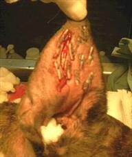
Here, an incision is made in the earflap surgically. The hematoma is drained of fluid and blood clots. To prevent the hematoma from refilling with fluid, multiple sutures are placed in the hematoma area either vertically or horizontally, either partly or completely through the earflap, with or without ear cartilage removal. Sometimes bandages are applied post-operatively, sometimes not. Sutures are generally left in place for three weeks to allow good scarring to take place so that refilling will not occur. The earflap is essentially quilted to close any space where fluid might refill.

Teat Cannula Placement

A teat cannula is a small device used in the treatment of udder inflammation in cows. It can be placed in the opening of the teat to allow drainage of milk or infected discharges. Teat cannulas can also be surgically placed in a dog’s aural hematoma if the earflap is large enough to accommodate the device. The hematoma is drained of fluids and allowed to heal over the next several weeks. This method is generally successful but does involve the dog tolerating a gadget inserted in its earflap for several weeks as well as accompanying fluid drainage.

What if there is a Concurrent Ear Infection?Usually, there is a reason why a dog has been shaking his head: an ear infection. This means that the ear infection must be treated along with the hematoma. The ear will need cleaning, a microscopic examination of the discharge, and medication. Sometimes ear shaking just happens and there is no underlying infection, but be prepared for the expense and trouble of treating an ear infection along with that of the hematoma.

What if We Leave it Alone?

If left alone, an ear hematoma will resolve by itself. The fluid will be re-absorbed back into the body and the earflap will no longer bulge. The problem is that a lot of scarring is associated with this process and the ear is often not cosmetically appealing afterward (i.e. it becomes a “cauliflower” ear). Resolution of a large hematoma can take several months during which it may be uncomfortable for the pet. If the patient is a poor anesthetic risk, it is certainly reasonable to forgo surgery. Corticosteroids, such as prednisone, can be used to reduce scarring.

Aural Hematoma in Cats

The situation in cats is somewhat more complicated than in dogs largely because the cartilage in the feline ear is more sensitive to inflammation and scarring is more severe. This makes the untreated hematoma more likely to form a permanently narrowed ear canal and long-term ear infection potential. Cats’ ear cartilage tends to experience more healing deformity than dogs’ ear cartilage and more curling and softening of the thinner pinnal areas is seen.

What this comes down to is that there is less leeway in letting the ear heal on its own in cats than dogs. Surgical repair is especially important as there is a greater tendency for a cat’s canal to narrow. That said, a more natural cosmetic appearance of the actual ear flap is harder to achieve in cat than dogs. It is more important to focus on the function.

As with dogs, a cat’s hematoma is generally brought about by ear infections and subsequent head-shaking. (In cats, most ear infections stem from ear mites but there are plenty of exceptions.) Bandaging is often used post-operatively as is the Elizabethan collar to protect the ear from being scratched. The cat will need confinement during a healing period of approximately 3 weeks.

6778455

Allergic Conjunctivitis in Dogs and Cats

Conjunctivitis is inflammation of the conjunctiva in the eye. The conjunctiva is a pinkish mucous-secreting membrane, similar to the lining of your mouth and nose, that covers the eyeball and lines the inner surface of the eyelids. The conjunctiva also covers the third eyelid, or nictitating membrane, that dogs and cats have but you do not. Inflammation, as indicated by the suffix ‘-it is’, is tissue that is red, warm, swollen, and usually painful as a result of disease or injury.  

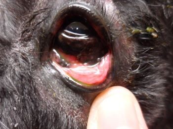
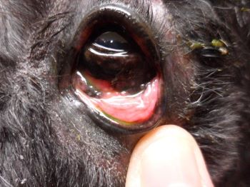
In conjunctivitis those membranes become red and swollen. Dogs and cats with conjunctivitis normally have cloudy, yellow, or greenish discharge from the eyes; a lot of blinking or squinting; and redness and swelling around the eye. Green or yellow discharge often indicates a bacterial infection; clear or whitish discharge is more likely to be caused by allergies or a bit of debris in the eye. Usually both eyes are affected, but not always. Sometimes other signs are seen, such as itching, hairlessness around the eye, discharge from the nose, sneezing, or coughing.

What we normally call “pink eye” in people is a type of conjunctivitis. The condition can be caused by several reasons, such as bacteria, herpes virus or allergies.

Any breed of dog or cat may develop allergic conjunctivitis. All breeds predisposed to atopic dermatitis, a hypersensitivity or over-reaction to a variety of commonplace and otherwise harmless substances like pollen, are also predisposed to allergic conjunctivitis. Allergic conjunctivitis is most common in young adults but can occur at any age.

In this allergic condition, the following are frequent contributors:

  • Allergies
  • Atopy (genetic tendency towards allergies)
  • Dust
  • Food allergy
  • House dust, molds

Treatment

Allergic conjunctivitis is most commonly treated with eyedrops or ointments containing corticosteroids, such as dexamethasone or hydrocortisone. Your veterinarian may check first for a scratch on the cornea (called a corneal ulcer) by instilling a yellow dye called fluorescein. If the eye has a scratch, medication without steroids is used, as steroids can delay healing of the scratch. Oral corticosteroids and/or antihistamines may also be helpful, especially in those pets with associated skin disease. Over-the-counter topical antihistamines may be beneficial, particularly in itchy dogs, but ask your veterinarian which one is best for your pet’s situation. However, topical mast cell stabilizers and antihistamines have variable clinical results.

A sterile saline eye wash can be used once or twice a day to clean the eyes and remove the accumulated discharge. Saline may also reduce surface irritation by flushing dirt, dust, debris, and pollens away from the eye.

Preventing damage to the eye is important as pets with allergic conjunctivitis may scratch at their eye with their paws or rub their face on furniture. This rubbing can lead to a corneal ulcer and associated pain and damage to the eye. Using an Elizabethan collar to prevent scratching may be part of the treatment plan from your veterinarian. This is especially important in short-faced breeds like pugs and shih-tzus, who are particularly prone to eye injuries. A recheck examination may be recommended by your veterinarian after treatment. If signs have not improved, your veterinarian may suggest sending your pet to a veterinary dermatologist or ophthalmologist for a work-up or recommending allergen testing to try and determine what your pet is allergic to. This step is usually reserved for dogs or cats with severe allergies.

Prevention

Allergic conjunctivitis is caused by allergies, so the best way to prevent it from happening again is to remove whatever your pet is allergic to from your home, if possible. Even if you can figure out what allergen they are reacting to and remove it or deal with it, you may not be able to eliminate it. If dust is found to be a cause, keep household dust to a minimum, and consider using an air purifier to filter out dust.

Allergic skin testing or food trials may be needed to find out the cause of the inflammation. In those cases, you may be able to determine if your dog or cat is reacting to something like dust or an ingredient in their food.

In many cases, allergic conjunctivitis can’t be cured, but with the help of your veterinarian (and perhaps a few steps taken around the house) it can be managed and you can make your pet more comfortable.