Tag: cat and dogs

5394716

Glomerulonephritis in Dogs and Cats

When a patient, human or non-human, is said to have kidney failure, renal insufficiency, or even chronic renal failure, what most people are talking about is a toxin build up when the kidney cannot adequately remove the body’s harmful wastes. This toxic state is called uremia and is associated with nausea, appetite loss, weight loss, listlessness, and other unpleasant issues.  It is also not the kind of kidney disease we are going discuss here. 

Glomerular disease is a completely different kind of kidney disease and may not involve any toxin build up at all. Glomerular disease is one of protein loss. 

What is a Glomerulus Anyway?

Consider for a moment what an important resource protein is to your body. Your blood, for example, is full of necessary circulating proteins handling clotting, fluid balance, transporting other chemicals etc. Your body went to a lot of trouble to build those proteins and you can’t afford to waste them. If you were to lose them, your body would have to break down muscle in order to recreate them because that is how important they are.

On the other hand, your blood carries an assortment of metabolic wastes that you need to get rid of. You need to filter out these bad materials without losing what is valuable. The millions of glomeruli you have are in charge of keeping your blood proteins where they belong — in the blood — while allowing for smaller wastes and extra fluid to filter out and be made into urine. There are other valuables in your blood besides cells and protein, but different areas of your kidney handle those.

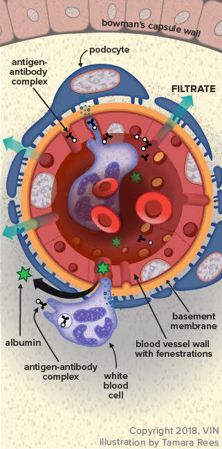
The illustration to the right shows the nephron, which is the functional unit of the kidney. There are millions of these making urine every moment of every day. Only about 30 percent of them must be working in order to maintain normal kidney function. The rest form a back up system so that we will have plenty of extra nephrons should some of them get plugged with debris, damaged by scarring or infection, or starved for oxygen during a traumatic event.

The glomerulus, which in a way looks like a little dandelion tuft, is where our interest lies today. Blood flows through an afferent arteriole into the glomerulus. Inside the glomerular tuft, the blood vessel narrows into a complicated spiral of tiny capillaries, so small that the blood cells pass through single file. The capillaries are gripped by cells called podocytes. Like hands, the podocytes have tiny fingers (ironically called foot processes) that encase the capillaries. Fluid and small molecules can flow in between the fingers while cells and large molecules like proteins cannot pass through.

The cells and large molecules/proteins exit the glomerulus through an efferent arteriole and return to normal circulation. This first step in filtration is driven both by blood pressure as well as by the protein content of the blood.

Now imagine what would happen if there were holes punched in that filtration system so that protein can pass through the fingers. This is what happens in glomerular disease.

How does the Glomerulus get Leaky?

Sources of chronic inflammation are believed to be the ultimate cause of the problem. The chronic inflammatory state leads to the circulation of antigen:antibody complexes in the blood and these complexes stick in delicate glomerular membranes like flies in fly paper. Once stuck there, they call in other inflammatory cells and soon a hole is eaten into the membrane by the ensuing reaction. The holes in the filtration membranes are big enough for proteins to traverse.

There are many possible sources of chronic inflammation that could be generating antigen:antibody complexes. Chronic ear or skin infections could be the cause. Long-standing dental disease could do it.

A latent, more internal infection might be the cause (such as heartworm, Lyme disease, prostate infection, or Ehrlichiosis). Even a tumor might generate enough of the immune system’s attention to lead to this sort of reaction. If it is at all possible to identify and resolve the underlying cause of inflammation, this should be done as other therapy is unlikely to fully resolve the protein loss.

How is the Diagnosis Made?

There are several common scenarios that might lead to the diagnosis but they all boil down to one or both of two findings: excess urine protein found on a routine urinalysis and/or low albumin found on a blood test.

Let’s start with excess urine protein found on a routine urinalysis.

A urinalysis examines a urine sample for some of its chemical contents and properties. Protein content is one of the parameters that is checked and reported as a small, medium or large amount. On a urinalysis report this will be designated as “+,” “++,” or “+++.”

This seems like it would be easy enough to interpret but unfortunately there is more to the story. A small amount of protein in a well-concentrated sample may be  normal while the same amount of protein in a dilute sample would be highly significant. How dilute or concentrated the urine is depends on the patient’s water consumption, and we need a method to examine urine protein that is independent of the water consumption. Further, we need to determine if any protein in the urine is truly coming from the kidneys; after all, a bladder infection or other bladder condition might generate urine protein. To help distinguish renal protein loss, the rest of the urinalysis will be helpful. When your veterinarian is confident that other issues with the urinary tract have been excluded, it is time for a urine protein:creatinine ratio (we will come back to this).

Low Blood Albumin Level found on a Blood Panel

Albumin is one of those proteins that the body really wants to conserve and here’s why. There are plenty of substances the body needs to circulate that simply are not water soluble. How do we circulate these things if they won’t dissolve in water? We bind them to a carrier protein that will circulate and carry them as if they were commuters on a city bus. The albumin molecule is that city bus, carrying important biochemicals around your body.

There’s more. Albumin also is important in keeping water in the bloodstream. This sounds odd but blood is basically a liquid and without enough water, it sludges and clots abnormally. Furthermore, if water is not held in the vasculature, it leaks into other body cavities such as the chest and abdomen, filling these cavities with liquid.

Your body prioritizes the maintenance of its albumin levels and will not allow them to drop. When the albumin levels are down, a serious protein loss is afoot. It could be intestinal or liver-related, but glomerular protein loss is going to be one of the first conditions to rule out. If there is no protein in the urine, the focus shifts to other organs but if there is protein in the urine, it must be quantified and that means there is a urine protein:creatinine ratio.

Interpretation of the Urine Protein: Creatinine ratio

The urine protein:creatinine ratio compares the amount of protein in the urine to the amount of creatinine, one of the metabolic wastes filtered by the kidneys. By using this ratio, it does not matter how dilute the urine is or how concentrated it is. The ratio allows for protein loss to be quantified and then we can tell how significant the protein loss actually is. If the urine protein: creatinine ratio is found to be abnormal, ideally it is repeated in 2 to 4 weeks to be sure that the protein loss is persistent, but this depends on how high the ratio is and whether or not there is a known inflammatory condition that would be expected to damage the glomeruli.

Determining how serious a patient’s protein loss is depends on overall kidney function as well. In other words, a protein-losing kidney that is effectively removing the daily load of toxins and wastes is in less trouble than a protein-losing kidney that is failing.

The International Renal Interest Society (IRIS) considers a urine protein:creatinine ratio of greater than 0.5 for dogs or 0.4 for cats to be abnormal, and if it is persistent, then further diagnostics and treatment are recommended.

Depending on how your pet responds to the therapies above, a biopsy may be recommended. Biopsy is most commonly recommended for patients with UPC ratios more than 3.5 or with significant proteinuria combined with low albumin levels or high blood pressure.

The goal of treatment is to reduce the UPC ratio to below 0.5 or to reduce it by at least 50%. Higher reductions are sought for cats (see later).

Ratios greater than 3.5 are particularly concerning and require more aggressive treatment and more extensive diagnostics. These patients have an increased risk of abnormal blood clotting and generally have more extensive kidney damage.

The urine protein: creatinine ratio varies by up to 30% above or below baseline as a matter of course. A significant change in the ratio caused by disease progression (up) or response to therapy (down) must be greater than 30%.

If Intervention is Recommended what Does that Mean?

Adding omega 3 fatty acids to the regimen appears to improve the protein loss situation and supplementation is recommended. Most renal diets are already fortified with these anti-inflammatory fats but additional use is felt to be beneficial.

Low Protein, Low Sodium Diet

Most commercial renal diets would fit in this category. It seems paradoxical that a disease that causes body protein to be lost would be treated with a protein-restricted diet but, in fact, supplementing protein causes albumin to drop faster.

ACE Inhibitor

These medications have been shown to reduce renal protein loss. Typically enalapril is recommended for dogs and benazepril is recommended for cats. These medications inherently reduce blood flow to the kidneys so care must be taken in patients with elevated creatinine ratios to be sure the uremia does not worsen. Lower doses are used and monitoring becomes more important.

Omega 3 Fatty Acid Supplementation

Most commercial renal diets are fortified with omega 3 fatty acids. These anti-inflammatory fats have been shown to improve survival of dogs with renal disease. It is still unclear how helpful they are for cats but studies are ongoing.

Angiotensin II Receptor Blockers (ARBs)

Angiotensin II receptor blockers are becoming more popular in human medicine and their use is trickling down to manage canine glomerular disease. These medications work with ACE inhibitors to further help reduce urinary protein loss though they can also be used alone. Like the ACE inhibitors, they not only reduce urine protein loss but also lower blood pressure as well and seem to have some effect on reducing the clotting tendency. They are new to veterinary medicine and protocols are still being worked out. The two commonly used medications are losartan and telmisartan.

Spironolactone

Aldosterone is the hormone that acts on the kidney to retain sodium and water and get rid of potassium. Spironolactone is an antagonist of this hormone, which means it increases urine production, retains potassium and removes sodium. In humans, it has been found to reduce urine protein loss by 34 percent, which makes it an attractive medication for this situation especially in patients with nephrotic syndrome (see below). In dogs it might be used when ACE inhibitors or ARBs have not controlled the proteinuria. It is not a medication for cats.

The goal in managing urine protein loss is a 50% reduction in urine protein:creatinine ratio for dogs and a 90% reduction in urine protein:creatinine ratio for cats. A combination of the above medications is likely to be prescribed, and urine and blood test monitoring will be periodically (at least quarterly) recommended in hope of achieving and finally maintaining these results.

Nephrotic Syndrome

In severe cases, a complication called nephrotic syndrome can result due to the extreme urinary protein loss. Nephrotic syndrome is defined as the combination of: 1) significant protein loss in urine; 2) low serum albumin; 3) edema or other abnormal fluid accumulation; or 4) elevated blood cholesterol level. This is a severe complication of glomerular disease and suggests a poor prognosis, especially if creatinine levels are elevated in the blood. High blood pressure is a common complication of nephrotic syndrome. Patients also tend to form inappropriate blood clots (embolism) that can lodge in small blood vessels, causing loss of circulation to entire organs or sections of organs. Nephrotic syndrome is an advanced state of urinary protein loss and must be treated aggressively.

Biopsy the Kidney?

There are pros and cons to this relatively invasive test. The kidney receives 25 percent of the blood supply at any given time, which means it is highly vascularized and can bleed in an extreme way. Blood transfusion is needed for 10 percent of dogs and 17 percent of cats having this procedure, and a three percent mortality rate has been reported. So why take the chance on this procedure? The main reason is to obtain information on prognosis. There are different types of glomerular disease and glomerular inflammation, all of which may have different associated expectations. There is a type of glomerular disease called amyloidosis that involves abnormal protein (called amyloid) infiltrating the kidneys and has a much more progressive and damaging course. Approximately 50 percent of glomerular disease patients have diseases that can benefit from immune-suppressive therapy but the only way to identify these patients is with a biopsy. 

Conclusion

When the kidney cannot retain blood proteins, the body loses its ability to carry out normal blood functions. In an attempt to replace these proteins, muscle is broken down and the patient becomes debilitated. Maintaining proper nutrition and using medication to reduce the protein loss are crucial to managing this form of kidney disease. It is important for the pet owner to keep up the monitoring schedule and to stay in contact with the veterinarian as to the pet’s progress and response to therapy. 

In Summary

  1. Glomerular disease is one broad type of kidney disease in which the primary problem is loss of renal proteinuria. Glomerulonephritis is one broad classification of kidney inflammation. It usually results in protein loss in urine. There are subtypes that can be determined by specialist pathologists based on renal biopsy.
  2. It is not typical Glomerular disease differs somewhat from “classic” kidney failure renal disease, although it glomerular disease can lead to chronic kidney disease if undetected.
  3. In the kidney, there are a million nephrons that make urine every minute of the day and send that urine to the bladder and out of the body through a filtration system. A body only needs approximately 30% of those nephrons working correctly for normal kidney function, at least as far as veterinarians could detect with typical lab tests. The rest are a backup system called the functional reserve.
  4. Inflammatory cells punch small holes along the filtration route. Those holes are big enough for proteins to pass through.
  5. Sources of chronic inflammation are believed to cause the problem, possibly stemming from issues such as chronic ear or skin infections, dental disease, heartworm disease, vector-borne diseases like Lyme disease, or feline immunodeficiency.
  6. Diagnosis is suspected by excess urine protein found in urine, and/or low albumin found on a blood test. Definitive diagnosis of glomerulonephritis and its specific type requires a kidney biopsy.
  7. When the kidney cannot retain blood proteins, the body cannot carry out normal blood functions. In an attempt to replace these proteins, muscle is broken down and the patient becomes debilitated.
  8. The urine protein:creatinine ratio (UPC) found in a urine test compares the amount of protein in the urine to the amount of creatinine, a metabolic waste filtered by the kidneys. The ratio tells what the magnitude of the protein loss actually is. A persisting, reproducible urine protein:creatinine ratio of greater than 0.5 for dogs or 0.4 for cats is too high. Mild elevations can be due to causes other than glomerulonephritis.
  9. Determining how serious the protein loss also depends on overall kidney function. A protein-losing kidney that is still effectively removing the daily load of toxins and wastes is in less trouble than one that is failing. Once the urine protein:creatinine reaches a certain ratio, a biopsy may be recommended. Ratios greater than 3.5 (typical US units) are particularly concerning.
  10. The best treatment is when a specific cause is found, such as a systemic infectious disease, and that disease can be successfully treated. However, sometimes despite extensive searching with imaging and blood tests, no underlying cause can be found.
  11. One goal is to reduce the urine protein:creatinine ratio to below 0.5 or to reduce it by at least 50% in dogs and 90% in cats.
  12. Potential interventions expected to help specifically with proteinuria include: omega-3 fatty acid supplementation; a controlled protein, low sodium diet; and RAAS inhibition (with drugs called ARBs or ACEIs). Accompanying hypertension, when present, may require additional treatment.
  13. In severe cases, a complication called nephrotic syndrome can result due to extreme urinary protein loss. The prognosis for this complication is comparatively poor, although it does depend on whether the problem developed acutely or chronically.
  14. A kidney biopsy can be informative about a prognosis and suggest whether additional treatments are expected to help. Since immunosuppressive treatments can also do harm, biopsy is usually recommended to justify trying that type of medication. Biopsy can be expensive and requires pre-planning to make it safe. For sample evaluation to be truly valuable, the specimens are analyzed by a specialist veterinary nephropathologist.
  15. It is important for you to keep up the monitoring schedule and stay in contact with your veterinarian.
5989537

Ectopic Ureters in Dogs and Cats

What is it?

An ectopic ureter is a birth defect in which one or both ureters in the kidneys connect to an abnormal location. This defect leads to the most common complaint: difficulty in house training and the pet leaking pee (urine). Ectopic ureters cause involuntary dribbling of pee, uncontrollable need to pee (urination), and urinary tract infections. Ongoing urinary tract infections can be fatal if they reach the kidneys, so it is important to diagnose and correct the ureter placement through surgery.

Normal Anatomy

Each animal has two kidneys, which make urine. Each kidney has a collection tube, called the ureter, that connects to the bladder at a location named the trigone. Urine is emptied from the bladder through a single tube called the urethra. Therefore, urine moves from the kidneys, through the ureters, and into the urinary bladder, where the urine is stored until the pet purposely pees. When urination occurs, urine empties through the urethra outside the body.

Two Types

There are two types of ectopic ureters based on where these collection tubes connect inside the pet’s body. Ectopic ureters do not connect and open at the bladder trigone, as they do in a normal animal.

Intramural ectopic ureter:

This type is the most common type in dogs. The collection tubes attach to the bladder at the trigone but then tunnel through the bladder wall, finally opening downstream from the bladder (i.e., the urethra).

Extramural ectopic ureter:

This type is rare in dogs but more common in cats. This is when the ureters pass the bladder and connect and open downstream from the bladder. This connection can be further along the urinary tract or even to the reproductive tract (i.e., urethra or vagina).

Who Gets it?

An ectopic ureter is an abnormality that occurs while the fetus is developing. It is a rare abnormality in dogs, and even less common in cats. It is most commonly seen in young pets because that is the age when signs are first noticed.

Cats

  • Sex: Both females and males equally
  • Breeds: Himalayan, Persian, Maine Coon
  • Age: Mostly young pets

Dogs

  • Sex: Females nine times more likely than males
  • Breeds: Labrador Retriever, Golden Retriever, Siberian Husky, West Highland White Terrier, Miniature Poodle, Newfoundland, and Soft Coated Wheaten Terriers
  • Age: Mostly young pets

What are the signs?

  • Urine leakage or dribbling (incontinence)
  • Hard to house train; lots of accidents
  • Urinary tract infection, more common in females
  • Licking of genital area
  • Rash on the genital area
  • Urine staining and genital area is persistently wet
  • Male dogs can have fewer signs and develop signs of urinary incontinence later in life.
  • Not all cats and dogs will show signs.

How is it Diagnosed?

A complete physical exam and basic diagnostic tests, such as a complete blood count, blood chemistry, and urine analysis and culture will be done first.

Diagnosis requires imaging. The method chosen depends on availability, training of the person doing the imaging, and your animal’s specific case. There is no one imaging method that is the best, and multiple methods may be needed.

Radiographs and ultrasound are two imaging methods that show the kidney shape and size.

Contrast CT and cystoscopy are two methods that are gaining popularity as they give a better visual of the ureters. Cystoscopy is a procedure where a small camera is placed at the pet’s urethral opening and goes into the body looking for the ureter openings. It gives the veterinarian a great visual of where the abnormality is located.

The type of ectopic ureter, location, and size have no effect on the prognosis and outcome of treatment. However, imaging is necessary to determine which surgical procedure to perform and the surgical approach the veterinarian will use.

  • Radiographs are usually readily available and cost-effective, but have limited ability to identify specifics.
  • Ultrasounds are non-invasive, visualize much of the urinary tract, and are relatively readily available. Your veterinarian may be able to diagnose ectopic ureters with ultrasounds, but it may be difficult to visualize the lower urinary tract
  • CT scans provide a good visual of the entire urinary tract but are expensive and not always available
  • Cystoscopy provide a good visual of the entire lower urinary tract and sometimes a repair can be done right then.  It is not always available.

What is the Surgery and Treatment?

The type of surgery used to correct the misplaced ureter depends on its location. Studies show that there is no significant increase in risk or complications between any of the surgeries. All the surgical options have similar long-term outcomes.

Ureteroneocystostomy

This procedure forms a new ureter and bladder opening. The surgery fixes the extramural type by creating a new bladder opening for the ureter in the correct spot. This surgery may cause swelling of the ureter during healing, but it will resolve in a few weeks.

Ureteronephrectomy

This procedure removes the ureter and kidney. The surgery is done on patients that also have a nonfunctional or chronically infected kidney attached to it. However, it can be difficult to tell if a questionable kidney is not functioning.

Laser Cystoscopy

This surgery is a new method specific to treating the intramural type. The laser cuts a new ureter opening into the bladder at the normal area. Surgery is less expensive because it can be done right after the initial cystoscopic diagnosis, while the patient is still under general anesthesia.

What is the Aftercare and Long-term Outcome?

Depending on the type of surgery, the aftercare and time of recovery may vary. The main post-surgical problems are continued uncontrolled peeing, leakage of pee into the body, swelling at the surgical site, and urinary tract infection. Studies show uncontrolled peeing resolves in cats that get a ureteronephrectomy. For dogs undergoing any of the surgeries for the birth defect, almost half will still have urinary leakage. If this occurs, additional medications will be needed for the rest of the pet’s life to help control urination.

It is not clear why some animals have continued urine leakage after surgery. Pets affected with ectopic ureters should not be bred.

6313300

Distichiasis Requires Permanent Eyelash Removal in Dogs

Distichiasis is quite common in dogs. Distichiasis is a condition in which extra hairs grow out of the eyelash area. It happens when there are two or more hairs growing out of a Meibomian gland opening. (Meibomian glands are located along the margin of the eyelid.) These hairs are not supposed to be there. In some cases, these extra hairs can be long and stiff and irritate the eye, resulting in a corneal ulcer. The severity of the problem depends on how stiff the hairs are, how long they are, where they’re located, and how many extra hairs there are.

Distichiasis is different from entropion. Distichiasis involves extra eyelashes; entropion is an inward roll of the eyelid that causes eye irritation from normal eyelashes or hair.

Untreated distichiasis can cause corneal ulcers, chronic eye and eyelid pain, and excessive tearing. It is quite uncomfortable and/or painful for the animal, depending on the amount irritation. If the excessive hair causes any clinical signs at all, the hair should be permanently removed.

Signs include increased blinking, lots of extra tears that often look like a tear streak, and squinting. Dogs don’t typically paw at the eye. The more severe cases are the most easily diagnosed, as the milder cases often involve small softer hairs that can easily be missed.

It’s seen most often in puppies or young adults and is typically diagnosed before a dog is three years old. Any dog can have it, but it’s considered to be one of the most commonly inherited diseases in dogs, and considered by some to be the most common congenital eye problem.

Breeds that seem to be predisposed to distichiasis include:

  • Cocker spaniel
  • Cocker Spaniel, American
  • English bulldog
  • Flat-coated Retriever
  • Golden retriever
  • Lhasa apso
  • Miniature Longhaired Dachshund
  • Miniature poodle
  • Pekingese
  • Poodle
  • Pug
  • Shetland sheepdog
  • Shih tzu
  • Toy poodle
  • Yorkshire terrier

Treatment

There are several treatment options and your veterinarian’s choice will generally depend on how many extra hairs are involved and what equipment the veterinary facility has. General anesthesia is usually needed. Occasionally a very cooperative dog might allow treatment with only a local anesthetic, but that would be extremely rare.

  1. Cryosurgery freezes the lid margin at the places where there are extra hairs.
  2. Surgery will remove the hairs permanently.
  3. Electrolysis will remove the hairs permanently.

These procedures destroy the hair follicles, preventing hair regrowth. However, no procedure can be guaranteed, so retreatment may be necessary in some cases. Several follow-up visits will be needed, to make sure that the hairs are not going to regrow.

Plucked hairs will just grow back, so plucking is not a permanent solution. Lid splitting and thermocautery can destroy the normal lid margins, leading to severe permanent scarring and entropion, so they are not good treatment options.

The eyelids will have some post-operative inflammation, which your veterinarian will also treat.

Prognosis

Once the hairs have been removed permanently, the prognosis is good. However, dogs with distichiasis should not be used for breeding, because of the hereditary aspects.

4128442

Disseminated Intravascular Coagulation (DIC) in Dogs and Cats

Disseminated intravascular coagulation (DIC) is an extreme complication of numerous already life-threatening conditions leading to the deregulation of the body’s natural mechanisms of blood clotting and blood clot dissolving. In other words, a really sick patient begins both bleeding and clotting abnormally at the same time, leading to disaster and frequently to death. DIC is a disease of the blood.

Setting the Scene for DIC

The patient must already have a serious problem before DIC sets in. Typical conditions that are associated with DIC include those involving dying internal tissue, widespread inflammation, red blood cell destruction, poor circulation, particulate matter in the bloodstream, or loss of blood vessel integrity.

The following conditions are associated with disseminated intravascular coagulation:

  • Pancreatitis
  • Rattlesnake bite
  • Immune mediated hemolytic anemia
  • Immune mediated platelet destruction (thrombocytopenia)
  • Hemangiosarcoma
  • Blood transfusion reaction
  • Severe liver failure
  • Overwhelming infection (sepsis)
  • Crushing injury
  • Heatstroke
  • Protein-losing enteropathy
  • Glomerular disease
  • Heartworm disease
  • Cushing’s disease

DIC is primarily a canine problem but can occur in cats. The usual underlying causes in feline patients are lymphoma, hepatic lipidosis, and feline infectious peritonitis.

In the normal body, there are small bleeds occurring regularly as we bump into things or cut ourselves. Blood clots patch these small blood vessel tears up and as healing takes place other blood mechanisms dissolve the old clots leaving scar tissue.

In DIC, the normal clotting mechanism is exaggerated. In the normal body, a substance called antithrombin (formerly called antithrombin III) is involved in dissolving old blood clots but in the event of exaggerated clotting, antithrombin becomes prematurely depleted. There are other anti-clotting proteins that are also depleted by the exaggerated clotting state. The result of this depletion is an excess of fibrin, the material of which blood clots are made.

Active proteins involved in the manufacture of fibrin stimulate the blood vessel cells to release inflammatory biochemicals (note: the patient likely started with a disease involving widespread inflammation). Ultimately, there is both inappropriate clotting and bleeding at the same time.

Recognizing DIC

The sooner DIC is recognized, the more likely the chance of a positive outcome. At first, there are no signs at all, just subtle blood test changes. It is important for the medical staff to watch for these lab changes in patients known to have diseases associated with DIC.

There are several factors that go into the diagnosis of DIC and a patient need not have them all:

Low platelet count

Platelets are the white blood cell fragments that are involved in normal blood clotting. In DIC, they are depleted.

Evidence of inappropriate bleeding

This could be bruising in the skin, excessive bleeding after a blood sample is taken, or spontaneous bleeding from the gums or from any orifice.

Increases in blood clotting times

Tests called the PT and PTT are run to assess how long different blood clotting proteins take to produce a blood clot. These times are compared to standardized normal times. Increased clotting times indicate a tendency to bleed inappropriately.

Clotting times well below the normal range can indicate a hypercoagulable state.

Presence of fibrin degradation products (sometimes called fibrin split products)
Fibrin is the material that clots are made of. When antithrombin and other biochemicals remove clots, fibrin fragments become detectable. These are fibrin degradation products. A fibrin degradation product of note is called the D-Dimer. It is notable because there are in-hospital test kits that can be used to detect it. The presence of D-dimer definitely indicates that a clot has been made and broken down (though, there are many reasons for such a thing to have occurred other than DIC). A negative D-dimer test rules out DIC with 95 percent confidence.

The absence of D-Dimer rules out DIC with 95% confidence in dogs but is less reliable in cats.

Reduced fibrinogen blood levels

Fibrinogen is a fibrin precursor and the absence of it suggests that it is depleted. The use of fibrinogen reduction as a marker for DIC has been questioned because there are numerous other factors that can reduce fibrinogen.

A Special Note on Thromboelastography (TEG Testing)

In this kind of testing, a gadget called a thromboelastograph hemostasis analyzer accepts a sample of blood, forms a clot with it, and measures the strength and elasticity of the clot. The test determines if there is an excessive or reduced tendency to clot. Dogs with reduced ability to clot on the TEG test have a much higher mortality rate. This test may help determine the prognosis with DIC, at least in dogs.

Treatment

Ultimately what all this clotting and bleeding comes down to is loss of blood flow to the tissues. Treatment centers on restoring normal circulation. This means intravenous fluid administration is crucial to restore tissue perfusion. Often plasma transfusions are used to replenish consumed blood clotting proteins. Plasma is incubated with an anticoagulant substance called heparin before it is administered. Heparin activates anti-thrombin, which as mentioned has been depleted in DIC.

The most significant factor in the treatment of DIC is removal of the original disease that predisposed the patient to DIC in the first place. If this can be achieved, it would be the best chance at resolving DIC.

4128712

Clostridium perfringens Causes Diarrhea in Dogs

When pets get chronic diarrhea, one of the tests that sooner or later comes up is for Clostridium perfringensThis bacterium produces an unpleasant toxin. The situation sounds simple: a pet gets infected with toxin-forming bacteria, gets diarrhea, and the diarrhea should at least improve when toxin-forming bacteria are removed. As with most things, the situation turns out to be more complicated.

What is Clostridium perfringens?

Clostridial diseases are classic in medicine, both veterinary and human. It is a Clostridium that causes tetanus and botulism, and clostridia are responsible for gangrene, “black leg,” lamb dysentery, and other famous maladies.

Clostridia have several features in common that make them especially nefarious but most importantly:

  • They produce toxins.
  • They are anaerobic (they grow in the absence of oxygen).
  • They form spores (essentially armor) so as to withstand environmental change, including disinfectants that would kill more vulnerable bacteria.

Clostridium perfringens is one of the brothers of the evil Clostridium family but is further classified into five biotypes (A, B, C, D and E) depending on what combination of four toxins it produces. Dogs are almost exclusively infected with biotype A.

The toxins of significance that it can produce are called enterotoxins. We are particularly concerned about the alpha, epsilon, and net E/F toxins. These toxins can be seen in normal dogs, but if they are in high enough amounts, they are associated with diarrhea.

How Would Clostridium perfringens Infection Happen?

In order to produce toxin(s), the organism must form a spore (sporulate). Before it can do that, it must make its way into the host’s intestine from the outside world. The organism enters the body orally: in food, grooming of fur, chewing up rotten dead things, etc.

When the organism arrives in the small intestine, it forms a spore and begins to produce its toxin(s). Alternatively, the organism may have been happily and innocuously living in the intestine for who knows how long when something causes it to sporulate and produce toxin.

The trigger may be dietary, may be related to infection with another organism, or may even be related to medications, especially antibiotics. The type of diarrhea produced is generally a colitis, meaning the large intestine is affected. Such types of diarrhea are mucousy, possibly bloody, and associated with straining. More watery diarrhea, as comes from the small intestine, is also possible. The severity of the diarrhea can range from mild to life-threatening, depending on how much toxin is produced. The important concept is that C. perfringens toxin can be the cause of chronic diarrhea, or it can be a complicating factor in diarrhea caused by something else.

Can we Test for Toxin-Producing Strains Of Clostridium perfringens?

The culture will not tell us if the strain present can produce the enterotoxin. Clearly, we need to know more than whether there are any C. perfringens. How about culturing for Clostridium perfringens? Again, cultures are not very helpful. Clostridium perfringens can be cultured from the feces of 80% of dogs, whether they have diarrhea or not. Some dogs seem to be unaffected.

This is where PCR testing comes in; it is a form of DNA testing whereby the C. perfringens are tested for the DNA needed to make the different enterotoxins. In this way, we can detect the genes that are capable of producing enterotoxin. Furthermore, the number of gene copies can be measured so that we can tell if there are large or small amounts of toxin genes. Large amounts of toxin genes are associated with disease, so in this way, we can tell if one of the Clostridium perfringens enterotoxins is likely to be contributing to the patient’s diarrhea. 

There is also a test for the toxin, but it is an ELISA format, which means it is either negative or positive and does not say how much or how little toxin is present. In a perfect world, this test is combined with the test for the toxin-producing genes so as to confirm that not only are there large amounts of toxin genes, but they are confirmed to be producing toxin.

The severity of the diarrhea can be mild all the way to life-threatening.

When Should We Treat for Clostridium perfringens?

Let’s begin with the obvious: a pet does not need to be treated for C. perfringens unless he has diarrhea. Since 80% of dogs harbor Clostridium perfringens whether they have diarrhea or not, culturing Clostridium perfringens from a fecal sample will not be adequate for diagnosis; we have to find the toxin or, at the very least, verify the presence of significant amounts of Clostridial genes capable of producing toxin. And all this must be found in a patient that actually has diarrhea.

The chances are that a fecal check for worms has been done, and a trial course of an anti-diarrheal medication has been tried. A possible next step would be a PCR panel that detects the DNA from an assortment of viruses and bacteria associated with diarrhea. Often this type of panel includes a test for Clostridial enterotoxin DNA. The laboratory will report a quantification of gene copies for CPA (the gene for the alpha toxin), CPE (the gene for the epsilon toxin), and CP net E/F (the gene for the net E/F toxin). If the number of toxin genes for any of these toxins is significant, treating with antibiotics against Clostridium is indicated.

Keep in mind that Clostridial diarrhea might be the entire problem and curative with the right antibiotic or it might be secondary to a bigger problem yet to be discovered. Treatment for significant Clostridial diarrhea thus might yield only partial results.

Treating the Toxin-Positive Dog with Diarrhea

Any number of antibiotics can be used to remove it. Some choices include ampicillin, amoxicillin, metronidazole, erythromycin, and tylosin. Tetracycline was formerly on the list, but it has developed too much resistance.  If C. perfringens is responsible for the diarrhea, a response to antibiotics is generally seen in a couple of days, but the full course of medication your veterinarian prescribed should still be completed.

5771095

Cuterebriasis is a Parasite Causing Skin Infections in Dogs and Cats

Cuterebriasis is caused by a fly called Cuterebra. The parasite infests small mammals that spend time outdoors, such as dogs, cats, rabbits, ferrets, squirrels, and rodents. Cuterebra are large non-feeding flies that lay eggs near animal burrows, nests, or vegetation. These opportunistic parasites do not seek out animals, but when an animal wanders by, in response to the host’s body heat the eggs attach and hatch. Infestation is most common in summer and fall. 

Infestations of the skin tend to occur around the head and neck because the animal has stuck its head near a burrow that contains the eggs. Once a larva hatches, it can be licked and swallowed during grooming, enter the body via the mouth or nostrils, or it may enter through an open wound. The larva causes a lesion in subcutaneous (below skin) tissue. [See special note below.] The host acts like an incubator. The larva opens a tiny breathing hole (a fistula) in the skin. It just lives there, as though renting space; it does not feed on the host. An advanced-stage larva is the size of the first joint of your thumb and is a light tan color. 

About a month after infestation, the Cuterebra larva crawls out of the skin to pupate and then emerge as an adult fly to begin the cycle. The length of time it stays in the ground depends on seasonal factors. 

What pet owners typically see is the swelling at the air hole and matted hair from the pet’s over-grooming. Cats often groom to the point of irritation. Sometimes the pet has pain at the site. Some sites become infected and pus can be seen. Your veterinarian can diagnose cuterebriasis just by visual inspection. 

Removing the larva is one of the simplest things in veterinary medicine; your veterinarian will probe and enlarge the breathing hole, grasp the parasite with forceps, and just pull the invader out. Squeezing the lesion with your fingers can rupture the larva and cause an infection. The larva should be removed as a whole piece, rather than in parts, in order to reduce the body’s reaction. Typically, no other treatment is required, although sometimes the wound will need to be flushed with saline, debrided (unhealthy tissue removed), or covered with antibiotic ointment. Because of the “housing” that was created around the subcutaneous parasite, it may take longer to heal than you might expect, but it will heal.

Special Note

The most common problem with Cuterebra is the subcutaneous site infestation. However, occasionally aberrant migration of Cuterebra spp. can occur in other tissues and can cause more significant disease. 

Ophthalmomyiasis externa occurs when larvae infect the conjunctiva (the mucous membrane that lines the inner surface of the eyelids and is continued over the forepart of the eyeball), the skin around the eyes, and the eyelids.

Ophthalmomyiasis interna anterior occurs when larvae are in the anterior segment of the eye’s globe.

Ophthalmomyiasis interna posterior occurs with larvae in the posterior segment.

Cuterebra spp. larvae have been found in the central nervous system (CNS) in some patients, leading to severe neurologic abnormalities. For example, cerebrospinal cuterebriasis has been associated with ischemic encephalopathy in cats.

Migration of larvae through the nose, trachea, pharynx, and larynx has also been reported. Other abnormalities, such as disseminated intravascular coagulation (DIC), systemic inflammatory response syndrome (SIRS), and protein losing nephropathy, have occurred in dogs.

7736303

Cellulitis in Dogs and Cats

Cellulitis is a painful inflammation of the tissue under the skin. In animals, it is generally caused by a bacterial infection that has entered the skin from a cut, open wound, or broken skin from an animal bite.

The most common bacteria causing cellulitis are Staphylococcus and Streptococcus. These bacteria are found in your pets’ environment (in the soil, water, and air), on humans and other animals, and are known opportunists. An open wound is a perfect environment for bacteria to thrive.

You may have heard infections caused by these bacteria referred to as “staph” or “strep.”

It is extremely important to seek your veterinarian’s help as quickly as possible if you suspect a wound has become infected. If left untreated, cellulitis can quickly become very serious, sometimes resulting in the tissues becoming necrotic (tissue death), and can be fatal to your pet in severe cases.

Symptoms

Cellulitis can occur on any part of your dog’s or cat’s body where there is a break in the skin, allowing bacteria to enter. For this reason, cellulitis is often the secondary health issue, the first being the wound that breaks the skin. A wound does not always need to be large for bacteria to enter and may go unnoticed until the symptoms of cellulitis appear.

In the affected area, your pet may have:

  • sensitivity and pain;
  • skin that feels hot to the touch, redness;
  • an abscess;
  • accumulated pus or green discharge;
  • swelling;
  • obsessive licking of the area.

The infection may cause your pet to have a fever, refuse to eat, act lethargic, or appear depressed. You may notice an unpleasant odor if there is an abscess or discharge. Often, this odor is the first clue that something is going on with your pet if you are unaware that they have been injured.

Treatment

Your veterinarian will take your pet’s history and examine them for wounds or abscesses. Blood tests and cultures may be needed to determine the type of bacteria in your pet’s body and their overall health.

If an abscess is found and hasn’t already burst, it will be opened, drained, and cleaned, usually under anesthesia or sedation. A piece of sterile tubing, cloth, or other material is often placed into the abscess to allow continued drainage. Stitches may be used to keep this drain in place, and drains are often covered with a bandage.

A lab test may be performed on the fluid from the abscess to identify further the culprit or culprits causing the infection. This is important to ensure that the correct antibiotics are prescribed. Culture results can take several days to return, and a change in antibiotics may be needed. Your pet may have other issues stemming from the original injury, which will be treated as needed.

Your veterinarian will send you home with antibiotics to tackle the infection, possibly anti-inflammatories, and pain medication as needed. Based on culture results, antibiotic therapy can be tailored to your pet’s specific needs, and multiple antibiotics are sometimes needed. Additionally, topical antibiotic ointments are available. You will likely be given a list of aftercare instructions, which may suggest the continued use of warm compresses on the abscess site to encourage drainage. Use a protective e-collar if your pet won’t leave the treated area alone. These cones are available at your veterinary clinic or a pet or feed store.

Be sure to finish the prescription medications as directed.

Sometimes, the drain is not enough, and the abscess fills up again despite your best efforts. Contact your veterinarian if this happens.

Should You Be Concerned About Catching This from Your Pet?

Always wash your hands well before and after treating your pet, and wear gloves if possible. If your immune system is compromised, let your veterinarian know, and pay special attention to hygiene when dealing with your pet’s wound.

Prognosis

Your veterinarian is your best source for information and treatment and for receiving a positive outcome when dealing with cellulitis.   

In minor cases and with proper and timely care, your pet should recover without problems.

4128726

Behavior Supplements in Dogs and Cats

Many colleagues and pet owners ask about the use of supplements in the treatment of behavioral issues. There is limited documented evidence on the use of these supplements, but to address this demand for information, here is what we know is being used and impressions from what experience and data are available.

There are many different products available if your pet shows unwanted behaviors such as anxiety, fear of strangers, aggression, or stress-related conditions. Always check with your veterinarian prior to giving your pet any supplement, even if it is available without a prescription.

Behavioral Supplements

Behavioral supplements are a combination of dietary supplements such as vitamins, minerals or herbal supplements, and diet. There are many common supplements available. The common ingredients of many of these supplements are listed below.

Reducing Fear and Anxiety

  • L-Theanine: This plant based amino acid is thought to help regulate behavior and mood by affecting the central nervous system. Studies have shown decreased anxiety in cats, decreased fear of strangers in dogs, and decreased noise related anxiety during thunderstorms.
  • Magnolia officinalis: This has been shown to have an anti-anxiety affect when studied in mice. Humans have also reported improved mood while taking this supplement.
  • Phellodendron amurense: This has been shown to have neuroprotective abilities, meaning it may help protect the brain from the effects of stress and prevent mood disorders.
  • Alpha-lactalbumin: This cow milk derivative contains high concentrations of amino acids, including tryptophan. It is believed that chronic anxiety depletes tryptophan in the brain.
  • Melatonin: This hormone is secreted during the evening and is known to facilitate sleep. Melatonin has been used for a variety of fear and anxiety-based behaviors, however scientific evidence indicating its effectiveness is lacking.
  • Tryptophan: Studies show that there may be a link between the metabolism of tryptophan and fear in dogs and cats. There is not a veterinary supplement that contains tryptophan alone, but this ingredient is found in some therapeutic diets.
  • Alpha-casozepine: This cow milk derivative has been shown to reduce anxiety without causing sedation or sleepiness. This supplement has also been shown to reduce fear of strangers and sensitivity to nail trims in dogs. Cats were also shown to have a reduction of feline lower urinary tract disease, which may be caused by stress.
  • Souroubea spp. containing betulinic acid and Platanus spp.: Extracts of flowering plants combined and formulated for use in dogs only have been used for stress-related behaviors caused by noise phobias.

Supporting Cognitive Functions

  • S-Adenosylmethionine (SAMe): This supplement plays a critical role in creating feel-good neurotransmitters and is sometimes used to treat fears and anxieties. Dogs and cats with cognitive decline given SAMe showed improved cognitive abilities and increased activity in senior dogs.
  • Phosphatidylserine, Pyridoxine, Vitamin E, Ginkgo biloba extract, and Resveratrol: These antioxidants are used to decrease the clinical signs of brain aging.
  • Omega 3 Fatty Acids
    • DHA (docosahexaenoic acid) is beneficial for the development of nervous and retinal tissue in puppies. It is thought to be essential for cognitive learning, memory, and trainability in puppies.
    • EPA (eicosapentaenoic acid) is an anti-inflammatory thought to reduce the signs of aging associated with cognitive decline, while improving learning.
  • Apoaequorin: Obtained from jellyfish, it is believed to have a direct benefit on learning and attention in dogs with cognitive decline.
4126859

Bloody Nose (Epistaxis) in Dogs and Cats

Some blood-tinged droplets sneezed on the floor might be the only sign, or there might be a steady, inexorable bloody drip from one or both nostrils. These findings are alarming as well as messy in the home and we want to identify the cause and take care of it promptly if it is possible to do so. The problem is that there are many causes, and not all of them are localized to the nose, and many are very serious diseases. The following is a review of tests typically necessary to get to the bottom of the bloody nose as well as the conditions that might be responsible.

First Aid

So, you are at home with your pet and a bloody nose starts and does not seem to be stopping. Here are some tips to get the bleeding controlled in the time prior to your vet appointment:

  • Keep yourself calm. If your pet sees you getting frantic, they will, too. Excitement = higher blood pressure = more bleeding.
  • Get an ice pack and apply it to the bridge of the nose (obviously, be sure your pet can breathe around the ice pack). The cold will constrict small blood vessels, which will slow the bleeding.
  • Do not insert absorbent material or cotton swabs in the pet’s nose, as this will generate sneezing, which will make the bleeding worse. A dose of an oxymetazoline nasal spray such as Afrin may help constrict blood vessels and lead to relief.
  • If the pet has a condition that involves recurring nose bleeds, consider the oral use of the Chinese herb Yunnan Baiyo, which promotes blood clotting tendency. Ask your veterinarian for details.

If these steps do not stop the bleeding or the pet is having difficulty breathing, go to your vet’s office or local emergency clinic at once.

Don’t forget that a pet with a bloody nose will likely swallow a great deal of the draining blood. This may lead to an especially black stool or even vomit with blood clots in it.

After a bloody nose, such findings are usually just a reflection of the bloody nose and do not necessarily indicate bleeding in the GI tract.

Information Your Veterinarian Will Need

You can help your veterinarian tremendously by taking some time to think about the following information and bringing up anything pertinent.

  • Does your pet take medication? Non-steroidal anti-inflammatory medications (aspirin in particular) will inactivate blood clotting factors. Do not assume your vet knows all the medications your pet is taking; list them for your vet.
  • Do you have any rat poison or has your pet been consuming any dead rodents that might have been poisoned? Most rat poisons act by disabling the ability to clot blood.
  • Look closely at your pet’s face. Is there any deformity or asymmetry? Is the bridge of the nose swollen? Are either of the third eyelids elevated? Does one eye seem to protrude more. does one eye tear more? Does the nose leather (textured tip) look normal?
  • Could there have been any trauma to the nose? Does your pet play roughly with another animal?
  • Is your pet exposed to foxtails or other grass awns that could become lodged in the nose?
  • Has your pet been sneezing? Has the pet been rubbing at the nose?
  • Open your pet’s mouth if possible. Look at the gums under the lips. Is there blood in the mouth? Do the gums seem pale? If they are, this suggests a serious loss of blood and you may have an emergency on your hands.
  • Is there any evidence of bleeding anywhere besides the nose? You may see a black tarry stool with Intestinal bleeding may present with a black tarry stool. Any unusual bruising should be reported. Any unexplained swelling that might be bleeding under the skin should also be noted.
  • Is this the first nosebleed or have there been others?
  • Is the blood coming from both nostrils or only one?

Where to Start

After the veterinarian performs a general examination of your pet, some more specific tests are needed with the idea of prioritizing the most likely conditions and least invasive forms of testing.

Blood Tests First

A basic blood panel and urinalysis will probably be needed as a database for the animal’s health as well as to assess the degree of blood loss. This information also serves as a pre-anesthetic evaluation should rhinoscopy or nasal imaging become necessary. A platelet (a blood cell involved in blood clotting) count will be needed as will coagulation tests (common tests are the PT or prothrombin time; the PTT or partial thromboplastin time; the ACT or activated clotting time; and the buccal bleeding or symplate time.) These tests evaluate a complicated biochemical cascade responsible for clotting blood. The pattern of abnormalities found in these tests will sort out blood clotting disorders.

Other blood tests that may be helpful involve titers for fungal infections, a classic cause of the nosebleed. Fungi are inhaled and if the patient is immune-compromised or excessively exposed, the fungus can take root and begin to grow in the nasal cavity.

In cats, the most common nasal fungal infection is caused by Cryptococcus neoformans. The good news here is that a blood test for fungal antigen is very accurate. Any positive number is significant and warrants treatment.

In dogs, fungal infections are not so simple. The most common organisms are Aspergillus fumigatus and Penicillium species. Blood tests are not as accurate especially since there are other species of Aspergillus besides fumigatus and each requires its own blood test. Complicating matters is the fact that nasal tumors predispose a dog to fungal infections so a dog can easily have both problems in the same nose. Blood tests for fungal infections may be included in the initial battery of tests. A negative Aspergillus test does not rule out Aspergillus infection.

Blastomyces dermatitidis is another fungus that can get into a dog’s nose. Urine antigen testing is accurate for diagnosis and blood testing is also available if results are ambiguous. As with other fungi, treatment is long term and challenging.

Another condition worth mentioning is hyperviscosity syndrome. In this situation, an extremely high blood protein level makes the blood so thick that blood vessels break from the pressure. Certain types of cancer (multiple myeloma, lymphoma, and certain types of leukemia) as well as infection with Ehrlichia canis, a blood parasite can cause this syndrome.

A routine blood panel should show the unusual globulin levels that typify hyperviscosity syndrome.

Another relatively simple parameter to measure is blood pressure. High blood pressure can occur as a complication of numerous diseases. When blood pressure rises, small blood vessels begin to burst and bleed, not just in the nose but often in the eyes or nervous system as well. Do not be surprised if your veterinarian checks for retinal hemorrhage.

Tick-borne infections (Ehrlichia, Babesia, and others) commonly involve low platelet counts. Platelets are blood cells involved in clotting and when they become infected with blood parasites, they do not work properly in the clotting cascade. Tick panels are blood panels that screen for infection with numerous tick-borne parasites, most of which can be managed or eradicated with antibiotics.

The bottom line is that there are many causes of nose bleeds but many can be ruled out with non-invasive testing and it is the non-invasive tests that we want to perform first.

Blood Clotting Disorders of Pets

  • Rat poisoning
  • Von Willebrand’s disease
  • Hemophilia
  • Liver failure
  • Disseminated intravascular coagulation

Diseases Causing a Low Platelet Count

  • Immune-mediated thrombocytopenia
  • Anaplasma infection
  • Bone marrow disease
  • Drug reactions (methimazole, chemotherapy drugs, excess estrogens, sulfa class antibiotics) 
  • Feline leukemia virus infection
  • Feline immunodeficiency virus (FIV)
  • Ehrlichia infection (dogs) 
  • Rocky Mountain spotted fever
  • Hemangiosarcoma
  • Other cancers
  • Babesia

Cruising Towards Anesthesia

If the basic blood tests and clotting parameters are normal, then the chances are that the problem is localized to the nose but there are a few more tests that are required before the patient is anesthetized for a nasal examination.

  • Radiographs of the chest should be performed to rule out obvious cancer spread or obvious disseminated fungal disease.
  • An oral examination should be performed as best as possible. Dental disease can be bad enough to create nasal bleeding, given that the roots of larger teeth connect with the nasal cavity. Oral tumors that have eroded into the nasal cavity may be evident if one can get a good look in the mouth. Many patients will not allow much oral exam and certainly probing the gums and getting a thorough inspection will require anesthesia but it is absolutely worth looking for obvious lesions if it is possible to do so.

Diagnostics Requiring General Anesthesia

If nothing has been revealed by the preceding tests, it is now time for radiographs of the nose, superficial rhinoscopy, and a dental inspection all of which require general anesthesia. Radiographs generally start the procedure as the other procedures might alter the radiographic appearance of tissues. The radiographs help evaluate the tooth roots and sinuses. Nasal tumors are common causes of nosebleeds in elderly dogs and the bone destruction they cause is evident on radiographs. Referral for more advanced imaging such as CT scanning or MRI, may be needed to determine the extent of bone destruction or to clarify radiography findings.

An otoscope (the same gadget used to look in your pet’s ears) can be used to look inside the nasal cavity superficially to remove foreign bodies lodged there. Deeper peeking requires an actual endoscope which may not be readily available in general practice.

The teeth can be cleaned under anesthesia with specific attention to the tooth roots (remember, an abscessed upper tooth root penetrates into the nasal sinus above.

If it seems appropriate to do so, some nasal discharge can be flushed through the nose and into a gauze sponge packing the throat. This may be helpful in identifying infectious organisms but may initiate more bleeding so some judgment is required on whether the benefit is worth the risk.

Then What?

If the simple tools of general practice do not reveal adequate information, referral for endoscopy may be needed. Deeper visualization of the nasal tissues is possible with this equipment, plus biopsy specimens can be taken although bleeding is the chief risk. Taking a biopsy is particularly difficult in the nose, not just because of the hemorrhage but because nasal tumors are surrounded by so much inflammation it is difficult to get a representative sample. Often it is not possible to see the area being biopsied directly, especially if prior sampling has led to bleeding.

If radiographs are diagnostic for cancer and aggressive therapy is contemplated, prognosis is highly dependent on the type of tumor so biopsy becomes especially important in this situation.

And Then What?

At the end of all these procedures, sometimes the area of bleeding is simply not accessible without surgery. This would be the final and most invasive procedure in retrieving a difficult foreign body or tissue sample. Extensive bleeding is expected and this is generally the last resort after a long road of diagnostics.

In a study by Bissett et al published in the December 15, 2007, issue of the Journal of the American Veterinary Medical Association, 176 cases of dogs with bloody noses were reviewed to determine which underlying causes were most common. Of these 176 dogs, a definite underlying cause was found in 115 cases.

  • 30% had nasal tumors
  • 29% had trauma
  • 17% had nasal inflammation of unknown cause (idiopathic rhinitis)
  • 10% had low platelets
  • 3% had some other blood clotting abnormality
  • 2% had high blood pressure
  • 2% had tooth abscess

Conspicuously absent is the nasal fungal infection but since these are frequently regional in nature in dogs, the population studied may not have been in an area where fungi are common pathogens.

Other Relevant Studies

Evaluation of factors associated with survival in dogs with untreated nasal carcinoma: 139 cases (1993-2003) by Rassnick et al published in the Journal of the American Veterinary Medical Association  229:401-406, 2006.

This study reviewed the outcomes of 139 dogs diagnosed with nasal carcinoma. Since many dogs are euthanized at the time of this diagnosis, this study included only dogs that were alive seven days after the initial diagnosis was made. Dogs studied received only pain medication, anti-inflammatories and antibiotics (no surgery, chemotherapy or radiation therapy). The following statistical findings came out:

  • 80% of dogs were purebred. The median age was 11 years and the median body weight was 48 lbs.
  • 77% had nose bleeds. Median survival time for dogs with nosebleeds was 88 days vs. 224 days for dogs with carcinomas that did not have nose bleeds.
  • Approximately half of the patients studied were felt to have improvement with the supportive care described above.

Ultimately, the therapy for a recurring nosebleed depends on the cause, and the causes are many and varied. If your pet has a nose bleed that lasts more than 5 minutes, seek veterinary care right away. If nose bleeds are recurrent, your pet will need a medical checkup to sort out the possible causes, as reviewed above. Contact your veterinarian to assess your pet’s situation as early in the course of disease as possible.

4128320-1

Brushing your Dog’s Teeth

What would happen if you stopped brushing your own teeth? Even if you only ate hard food, as most dogs do, there still would be problems. You should be brushing your dog’s teeth twice daily if you can manage it; it’s not as difficult as you might imagine.

What are the benefits of brushing? Brushing removes daily accumulation of plaque from the teeth. Even though dogs do not commonly get cavities, they do suffer from periodontal disease. If untreated, gum disease can lead to bad breath, pain, and tooth loss.

How Do You Brush Your Dog’s Teeth?

Step one is to pick an appropriate pet toothbrush.

Save yourself time by not buying a child’s toothbrush, which is usually too hard for dogs. The ideal dog toothbrush will have a long handle, an angled head to better fit the mouth, and extra soft bristles. For dogs less than 30 pounds, a finger toothbrush that fits over the tip of your finger works well.

Step two is to select an appropriate toothpaste.

The best pet toothpastes contain enzymes that help control plaque. Try to avoid toothpastes with baking soda, detergents, or salt that are sometimes found in human pastes. Fluoride may be incorporated to help control bacteria. Place the toothpaste between the bristles as this allows the paste to spend the most time next to the teeth and gums.

Step three is to get the brush with paste into your dog’s mouth to get all the teeth brushed.

Most dogs accept brushing if they are approached in a gentle manner. If you can start when they are young, it’s quite easy, but even older pets will accept the process. Start slowly; you can use a damp washcloth or piece of gauze to wipe the teeth, front and back in the same manner you will eventually be using the toothbrush. Praise your dog during the process and offer teeth-healthy treats, such as dental chews, as a reward. Do this twice daily for two weeks, or until your dog is familiar with the approach. Then take the pet toothbrush, soak it in warm water and start brushing daily for several days. When your dog accepts this brushing, add the pet toothpaste.

Most attention should be given to the outside of the upper teeth. The toothbrush bristles should be placed at the gum margin where the teeth and gums meet at a 45-degree angle. The movement should be in an oval pattern. Be sure to gently place the bristle ends into the area around the base of the tooth as well as into the space between the teeth. Complete ten short back and forth motions, then move the brush to a new location. Cover three to four teeth at a time.

Dog home dental care should include daily brushing using an enzymatic pet toothpaste and a proper toothbrush.  Taking an active role in your dog’s dental care will help reduce dental disease, bad breath, and potentially life-threatening heart and kidney disease. Everyone wins.