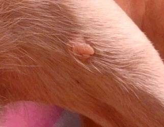
Benign Sebaceous Gland Tumors
We receive a fair amount of emails related to the Papilloma article from people with older dogs with numerous “warts” wondering if their dog’s warts will go away as viral warts usually do. The problem is that in older dogs, what looks like a viral wart is probably a sebaceous gland tumor, while there is a very good chance it is benign, it will not be going away any time soon.
It is not uncommon for an elderly dog to develop scores of “warts” which are not warts at all but are sebaceous growths. Most sebaceous growths are benign but one cannot say for sure simply by looking.

Here are some reasons to remove a sebaceous growth:
- when the growth has been bleeding.
- when the growth is itchy or is in a location where it is bothering the pet.
- when the growth is in a location where it interferes with normal grooming of the pet (i.e. the growth gets caught in the grooming clippers, etc.).
- when there is a question as to whether the growth actually IS a sebaceous tumor and biopsy is needed to settle the question.
- when you don’t want to take any chances that a sebaceous growth is malignant (as mentioned, you can’t tell by looking).
These growths are typically small (pea size or smaller) and originate from the skin’s sebaceous glands, the oil-producing glands of the skin. Because these growths are small, they are generally amenable to removal with local anesthetic. This is helpful since often patients are older and not good anesthesia candidates. It is usually not practical to remove a large number of sebaceous growths with local anesthesia at the same time but the most troublesome can be selected for removal.

Viral warts are different and occur primarily on the face of young adult and adolescent dogs. Sebaceous gland tumors occur in any location, often in large numbers, and usually in older dogs (and occasionally in older cats).
There are several types of sebaceous gland tumors:
Nodular Sebaceous Hyperplasia
About 50% of sebaceous growths are technically not tumors at all and are classified as excessive growth of the gland tissue. It is thought that growths of this group ultimately develop into actual benign sebaceous adenomas as described below. These lesions are round, cauliflower-like, and sometimes secrete material that forms a crust. Occasionally they even bleed. They are particularly common in Cocker spaniels, Beagles, Miniature Schnauzers, Poodles, and Dachshunds. This growth is technically not a tumor but is actually an area of excessive sebaceous cell division.

Sebaceous Epithelioma
Another 37% of sebaceous growths fit into this category. These look just the same as sebaceous hyperplasias to the naked eye but tend to occur in larger breeds and usually on the eyelids or head. They often pigment into a black color. They were formerly described as benign but it turns out they are able to spread in a malignant fashion if given enough time. Since it is not possible to distinguish a low-grade malignant epithelioma from hyperplasia, it is smart to remove any sebaceous growth and not take a chance.
Sebaceous Adenoma
These lesions also look the same as the others to the naked eye. These are also actual benign tumors that probably arose from areas of hyperplasia. As previously mentioned, if given enough time a sebaceous hyperplasia growth will develop into an adenoma. Both are benign.
Sebaceous Carcinoma
About 2% of sebaceous tumors are malignant and may be locally invasive but even malignant sebaceous tumors rarely spread. They have a greater tendency towards ulceration than benign growths. Cocker spaniels seem to be predisposed.
Again, in most cases, the removal of sebaceous gland tumors is straightforward and can frequently be done with a simple local anesthetic. In the event that further treatment is needed, your veterinarian will make recommendations or possibly refer you to a veterinary dermatologist.
