Tag: Antibiotics

716183

Corneal Ulcers and Erosions in Dogs and Cats

One of the classic eye problems veterinarians must address is a red eye. The red eye may or may not be obviously painful but when it is, the pet can be observed squinting or even rubbing at his/her face. The conjunctiva (the pink moist tissue lining the inner surfaces of the eyelids) becomes an angry red and can even swell or puff up around the eye (a condition called chemosis). In short, it is clear when the eye suddenly hurts, and that veterinary attention is needed.

The Corneal Erosion

There are several causes of acutely red and painful eyes and one of the most common causes is a wound or scrape to the surface of the eye. The clear surface of the eye is called the cornea; because it is the outermost layer of the eye, it is prone to scrapes and tears. Common causes of corneal erosions include:

  • Rough contact with plants, thorns, or bushes
  • Scratches from another animal (note: the cat scratch wound can be especially serious as the wound quickly heals over, sealing infection within the eye.)
  • Self-trauma (rubbing or scratching at a painful ear or even at the eye due to some other eye problem can lead to an inadvertent scratch to the eye.)
  • Chemical irritation (such as getting shampoo in the eye during a bath.)
  • Foreign body injury (plant material can get stuck under an eyelid and can scrape the cornea.)

A special fluorescent stain is used to confirm if there is an ulcer or erosion. Normally, water will run smoothly off the surface of the cornea, like rain washing off a windshield. If the cornea is damaged, the stain will stick to the damaged area and look bright green under a fluorescent lamp.

Treatment

Antibiotics

A routine corneal ulcer or erosion should heal easily. Since the damaged cornea is at risk of becoming infected (or may even already be infected, as demonstrated by a purulent discharge), a topical antibiotic is needed and ideally should be used four times a day or more. Since it is a rare pet owner that can accommodate any medication administration four times a day, we usually have to make do with three times a day but to properly sterilize the eye surface, the antibiotic should be used four times a day. Either drops or ointment can be used depending on the owner’s preference.

Pain Relief

The second part of treatment is pain relief: Atropine 1% drops or ointment. The atropine acts by temporarily paralyzing the pupil’s ability to constrict (the main source of pain is spasm in the pupil). Pupillary dilation is expected when this medication is used and the pet may be reluctant to experience bright sunlight while under the influence of this medication. Because the tear duct system is connected to the nose and mouth, the patient will also taste the above medications and atropine is famous for its bitter taste. Dogs do not seem to mind this unduly but cats will drool shortly after the medication is given in an effort to get the taste out of their mouths. This is a normal reaction to the Atropine as is the dilated pupil.

A special collar, called an Elizabethan Collar, may be needed to prevent self-trauma of the eye. If you think your pet will rub the eye, it is important to have the pet wear this special collar until the erosion is healed. Be sure to request one if you think your pet needs it and if you are given one be sure the pet wears it for the entire course of treatment.

Re-Check in One Week

It is important that the eye be stained again after one week of therapy. Most ulcers will have healed in this time but some will require an additional week. If the ulcer has not healed after two weeks, it is no longer considered routine.

Some other procedures, and possibly a visit to a veterinary ophthalmologist, may be necessary. 

 If the inflammation associated with the ulcer goes deeper into the eye, the situation becomes more serious; it is very important not to skip that one-week re-check. If there is any question about the eye’s healing progress, the eye should be re-checked sooner.

It is important that the owner observe the progression of healing at home. If the eye is doing well but suddenly becomes more painful, if a discharge develops, or if the eye simply does not look right, have your vet recheck the pet sooner than the planned one-week appointment.

Some Special Ulcer Conditions

Indolent Ulcer

Some ulcers form with a small lip on their edge.  Since the ulcer is trying to heal from the bottom up, the lip interferes and creates an ulcer that seems to never get any smaller. Several techniques can be used to remedy this situation: the lip can be rubbed away, hyaluronan or PSGAG eye drops can be used to strengthen the cornea, or surgery can be performed. A technique that has gained popularity over recent years is called the grid keratectomy where a needle is used to scratch a grid of lines on the cornea. The cornea is then able to heal in, grid by grid. Poodles and boxers are notorious for these ulcers but they can occur in any breed. 

Melting Ulcers

When infection is present, the cornea will develop a yellow or tan gooey appearance because the bacteria or fungi causing the infection elaborate enzymes that actually dissolve corneal collagen fibers. The cornea softens and appears to be melting and can actually perforate. Culture and cell sampling for analysis are very helpful in determining the right antimicrobial therapy. In addition to antibiotic drops, the eye will need some sort of medication to inactivate the aforementioned collagen-dissolving enzymes. Often, this involves taking a blood sample from the patient and actually delivering the patient’s own serum as an eye drop.

Descemetocele

(Pronounced des’ĕ-met’ō-sēl) Descemet’s membrane is the thin attachment of the cornea to the fluid of the eye below. A Descemetocoele is an ulcer that has penetrated through the cornea completely except for the last thin membrane. An eye with a descemetocoele is at high risk for rupture and measures must be taken to protect the eye. Usually, surgery is needed. The brachycephalic breeds (Pekingese, pug, etc.) are predisposed to this problem due to their prominent eyes.

Again, corneal ulcers and erosions usually heal routinely but stay alert for any changes that could indicate a more serious turn of events. Be sure self-trauma (rubbing the eye) is prevented and be sure to go to a recheck at the proper time. If anything seems not to be proceeding properly, be sure to contact your veterinarian.

4128677

Chronic Bronchitis in Dogs

What is Bronchitis?

Most of us have heard of bronchitis, and most of us have probably suffered from it at some point. The bronchi are the larger airways through which air is moved throughout the lungs. The trachea (windpipe) brings air from the throat down into the chest and branches into two mainstem bronchi, one for each lung. Each mainstem bronchus branches into smaller bronchi, which eventually become smaller structures called bronchioles that, in turn, branch, and re-branch, eventually ending with the alveoli, small chambers where oxygen is absorbed into our bodies from the air we inhale.

Listen to a dog cough. Coughing, reverse sneezing, and retching to vomit are commonly confused. The dog in this video is coughing.

Bronchi are basically tubes made of cartilage, forming the larger “pipes” in our lungs.  

When the bronchi are injured by irritants (cigarette smoke, air pollution, biochemical products of infection, etc.), they respond with a healing process that is inherently inflammatory. When the injury becomes ongoing, the inflammatory/healing process also becomes ongoing and the end result involves excessive mucus production in the airways. The mucus obstructs the smaller bronchi, which stimulates coughing, which in turn leads to irritation and then to inflammation, and then to more mucus. A vicious cycle results.

Once the vicious cycle of mucus production to coughing to inflammation to more mucus has become entrenched, it is unlikely to be cured and therapy is geared towards controlling the symptoms.

What is Chronic Bronchitis?

In order to fit the definition of “chronic bronchitis”, the coughing condition in question must (obviously) be chronic. Bronchitis can be temporary, as in short-term infectious diseases such as Kennel Cough, or it can be chronic, which basically means it just doesn’t seem to go away. To fit the definition of “chronic”, the cough must be present daily for at least two months. Chronic bronchitis is a lifelong condition once it starts; it never fully goes away but can be managed.

The second part of the definition of chronic bronchitis is that the chronic cough condition must include bronchial inflammation. (In other words, chronic bronchitis must be both chronic and bronchitis.) There are many causes of ongoing cough; bronchitis is certainly not the only one. For example, heart disease can cause chronic cough, as can tracheal collapse, and certain types of parasitism or even cancer can cause ongoing coughing. The character of the cough (wet, dry, “honking,” productive, etc.) is not particularly helpful in determining the cause of the cough, as coughing can sound similar regardless of its cause. Diagnostic tests are needed to get the right diagnosis, whether it is bronchitis or something else. It is very important to determine whether or not a reversible underlying cause is present.

Diagnosis and Testing

There are many conditions that make dogs cough chronically, including but not limited to:

  • Pneumonia
  • Tracheal collapse
  • Fungal infection in the lung 
  • Lungworms 
  • Cancer 
  • Heart failure
  • Heartworm infection

Note that asthma is conspicuously missing from this list. Asthma involves recurring episodes of airway inflammation and narrowing rather than continuous (non-episodic) airway inflammation. Asthma is not a natural clinical entity in dogs, though it is certainly a well-described condition in cats.

Chest radiograph of a dog with bronchitis. The heart is the oval structure in the center. The lacy white texture of the lungs comes from mucus and other inflammatory secretions clinging to (and visually highlighting) the airway walls. Image courtesy of MarVistaVet.

Radiographs (X-rays) of the lungs are crucial in ruling out conditions that have a specific treatment. In bronchitis, mucus in the airways tends to outline the shape of the airways in patterns that look like parallel lines (so-called tram lines) when looking at an airway’s side view or circles (so-called doughnuts) when looking at an airway to end on. In bronchitis, the airways may be dilated as well, especially if the case is advanced. Other conditions tend to have characteristic radiographic appearances of their own but there is definitely room for ambiguity.

In a perfect world, bronchoscopy can be performed, and samples of the airway secretions can be analyzed. In bronchoscopy, a small video camera is inserted down the airway system. The color and character of the airway walls can be examined, and a fluid wash can be used to aspirate out secretion samples. General anesthesia is required for this procedure. This YouTube video shows a bronchoscopy and sampling of the airway secretion.

Alternatively, a tracheal wash can be done to obtain samples using tubing and fluid washes. In this procedure, only samples are taken, and there is no camera involved and no visual inspection of the airways. Sedation is generally needed.

Sedation and anesthesia are not without risk in patients with lung compromise, so the benefit of obtaining diagnostic information must be weighed against patient risk. Often invasive procedures are skipped, and trial courses of medication are used.

Treatment

There are several aspects to the treatment of bronchitis. Often a weight loss program relieves so much chest restriction that medications become unnecessary. Physical therapy techniques can be used to clear secretions. Medications can be used to relieve inflammation, dilate airways, and reduce secretions, but some trial and error are often needed to get the right combination.  Newer inhalant therapies can spare the patient many potential drug side effects.

Trial Courses of Medication

Different individuals respond to different medications, and in bronchitis, there are several options to choose from and even combine. 

Corticosteroids

Corticosteroids such as prednisone relieve inflammation and cut secretions as no other medication can. Typically, a short course is used to control symptoms initially followed by a lower dose maintenance course. This helps dry up the mucus in the airways and stop the cough-irritation vicious cycle. The problem is that oral steroids have side effects potential with long-term use. Furthermore, because they are suppressive to the inflammatory process, it is important to rule out infectious causes of coughing that might be made worse with steroid use.

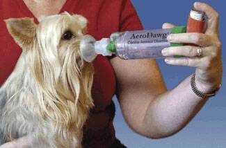
As an alternative to oral steroids, inhaled steroids can be used as is done with human asthmatics. A special facemask is used, and a spacer connects the mask to the metered dose inhaler. A couple of puffs from the inhaler doses the dog with inhaled steroids, and then it is possible to avoid the systemic side effects associated with oral steroids. Dogs typically need to use inhalers at least twice a day to control the symptoms of bronchitis. Airway dilators can also be given via inhalation in the same way as steroids.

Cough Suppressants

The cough itself is involved in the vicious cycle of irritation described above. Suppression of the cough reflex through medication may be helpful in establishing patient comfort. On the other hand, we want our patient to be able to clear the mucus from the inflamed airways if there is an excess of mucus. Common medications used are hydrocodone and butorphanol. Over-the-counter human products are often ineffective and may be combined with harmful additional medications such as decongestants or pain relievers.

Never medicate your pet without checking with your veterinarian first about what product to use and how to dose it.

Airway Dilators

Although airways technically do not constrict in chronic bronchitis, it may be helpful to relax them, allowing air to pass through bronchi that had previously been so small as to be blocked by mucus. Common medications include albuterol, terbutaline, and theophylline.  These are frequently used in combination with corticosteroids to reduce cough.

Mucolytics

An over-the-counter supplement called N-acetylcysteine may be helpful. This supplement works by breaking down sulfur bonds in the airway mucus, creating a more watery secretion. Thinner secretions may be easier to clear. The use of this supplement has not been formally studied for this use but it is an option that might be helpful.

Check with your veterinarian about what to use and how to use it.

Antibiotics?

Chronic bronchitis is not an infectious disease but all the inflammation and mucus in the lung certainly compromises the patient’s normal resistance to infection and ability to clear inhaled bacteria. Sometimes antibiotics are a helpful temporary addition to the therapies listed above.  More commonly, antibiotics are used early in the treatment course to rule out infectious causes of cough before the use of corticosteroids, which are immune suppressive.

Other Therapies

Nebulization and Coupage

It is important for the bronchitis patient to cough up inflammatory secretions as long as coughing does not become excessive and perpetuate the inflammation. It is not easy to draw the line as to when the cough is helpful and when it is excessive; only experience will tell. In patients for whom coughing is encouraged, it may be useful to moisten secretions with a vaporizer or even a nebulizer; the difference in these pieces of equipment is the size of the water droplet they generate. Inhalation of water vapor a couple of times daily will help keep secretions moist.

Coupage is a form of respiratory physical therapy that allows for moistened secretions to be moved into more central airways so they can be coughed up. See a YouTube video of how to do coupage.

Obesity Management

Pressure on the chest from body fat hampers the patient’s ability to take a deep breath. Many patients with chronic coughs do not require medication after excess weight has been lost simply because the chest can then expand properly.

Minimize Airway Irritants

It is especially important to minimize irritants circulating in the air. If possible, cigarette smoking should be limited to outdoor areas. If house painting, cleaning, or remodeling is planned, the pet should probably be boarded or stay elsewhere until any fumes have dissipated. It may be a good idea to get a good air filter for home use to minimize dust, pollens, or molds in the air.

Chronic bronchitis represents an ongoing airway change that generally cannot be cured; however, symptoms are manageable for most patients. Discuss the options for therapy with your veterinarian, expect some trial and error to be necessary, and it is likely the outcome will be a system that keeps your pet comfortable.

6021696

Anal Glands and Anal Gland Abscess in Dogs and Cats

What are anal glands?

Anal glands are two sac-like structures that release an unmistakable, foul-smelling fluid. It’s really unfortunate when veterinarians get that fluid on their clothing because then they smell like that all day, as does the exam room. Dogs and cats use this fluid to mark their territory when they poop. Anal glands are not important for your pet’s health and can be removed if medically necessary (i.e, constant infections, recurrent abscess formation), but not without risking incontinence.

What animals have anal glands?

  • Female and male cats, dogs, and many other animals
  • People do have them. They are like sweat glands and much smaller vs. cat or dog anal glands.

What is the anatomy of an anal gland?

Each animal has two glands located on the left and right side of the anus. Each gland connects to the end of the anal canal through a small connecting tube (duct). When your pet is defecating (pooping), the feces (poop) passes through the anal canal and squeezes the sacs, releasing the smelly fluid.

What is an anal gland abscess?

An anal gland abscess is a painful infection of the anal glands. During an infection, pus builds up in the sac. The infection prevents the foul-smelling anal gland fluid from leaving the sac. The anal gland swells with the fluids and may even burst. This abscess is extremely painful and should be treated immediately.

Can my pet get an anal gland abscess?

Animals with anal glands can have an anal gland abscess. There is no breed, gender, or age group that is more likely to have this happen. There are a variety of potential explanations of why the anal gland swells and even ruptures, such as underlying diseases or allergies. By working together, you and your veterinarian will create the best treatment plan to prevent abscesses from forming in the future.

What are the symptoms?

  • Scooting across the floor to put pressure on the anal gland and release the fluid
  • Trauma and biting of the tail and anal area
  • Matting of the hair at the anal area
  • Reluctance to sit and poop
  • Constipation and anal discharge
  • Crying, whimpering or signs of pain
  • Being withdrawn and lethargic, not wanting to eat
  • Swelling and redness of anal area

Should I treat the anal gland abscess?

Yes, and treat it immediately. Although this is not a medical emergency, make an appointment with your veterinarian as soon as possible. An abscess is painful for your pet and infection can spread to different parts of the anus. The veterinarian will clean the abscess and treat the infection. In some cases, anesthesia or sedation is needed to clean it. Treatment only stops after your veterinarian has rechecked your pet to determine if the abscess is healing properly. The goal of this treatment is to minimize the swelling and discomfort to your pet. Treatment may include:

  • Cleaning and unclogging of the anal duct to prevent fluid buildup
  • Anti-inflammatory medications to reduce swelling and pain
  • Antibiotics to treat infection
  • Recheck by your veterinarian is necessary

Is there any additional testing needed?

There may be additional testing depending on each case. Standard tests that may be needed include complete lab work, especially if anesthesia is needed. In complex or unusual cases, culture or biopsy may be needed.

Does my pet need surgery?

These sacs only produce scent markers and are not necessary for your pet’s health. If your pet continues to have infected and abscessed anal glands, a more permanent treatment may be needed. This involves removing the anal glands entirely. The surgery may affect sphincter strength, causing incontinence. This severe side effect makes surgery a last resort for chronic anal gland abscess offenders.

How can I prevent it?

If your pet continues to have anal gland swelling and infection, your veterinarian may suggest high-fiber diets, hypoallergenic diets, or surgical removal of the anal sacs. Knowing the cause of your pet’s continued anal gland abscesses is important for coming up with a prevention method. Your veterinarian will work with you to come up with the best plan to prevent discomfort and pain in your pet.