4128376

Rocky Mountain Spotted Fever in Dogs

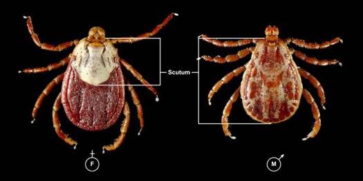
Rocky Mountain spotted fever (RMSF) is caused by Rickettsia rickettsii. This intracellular parasite is transmitted to dogs through the bite of an infected tick. The ticks that can transmit RMSF are the Rocky Mountain wood tick, the American dog tick, and the brown dog tick.

In the United States, RMSF is most common in the southern Atlantic states, western central states, and areas of the mid-Atlantic and southern New England coastal states. According to the Centers for Disease Control and Prevention (CDC), only about 1 to 3 percent of the tick population carries R. rickettsii, even in those geographic areas.

Prior research indicated that the infected tick must be attached for at least 2 hours in order to transmit disease. Research in Brazil demonstrated that unfed ticks had to be attached for more than 10 hours for transmission to occur, whereas fed ticks could transmit disease within as little as 10 minutes after attachment. These results may indicate that transmission across all tick species could occur earlier than once thought, depending on when the tick has last eaten. Transmission of the Rickettsia can then occur due to the bite or from exposure to the parasite while handling the tick. 

Clinical Signs

Clinical signs will show up 2 to 14 days after the bite occurred. The parasite creates an inflammation of the body’s small blood vessels, which results in damage to all the organs of the body.

Common signs include fever, lethargy, inappetence, pain, eye/nose discharge, nosebleed, cough, enlarged lymph nodes, lameness, skin necrosis/sloughing, hemorrhage, and peripheral swelling. Petechial hemorrhages (tiny hemorrhages in the skin) will occur in about 20% of affected dogs. Up to one third of the infected dogs will have central nervous system signs (lack of voluntary coordination of muscle movements, weakness, balance problems, cranial nerve abnormalities, seizures, stupor, spinal pain, etc.). Any organ in the body may be affected and the clinical signs may be mild or severe enough to result in death.

Diagnosis

Diagnostic tests for RMSF include blood tests looking for severely low platelet count, plus coagulation profiles, blood chemical analysis, and serology. (Paired titers, from blood samples taken 14 to 21 days apart, are often needed, but a single high titer in dogs that have clinical signs is consistent with an active infection.)  Response to antibiotic therapy is suggestive, but not diagnostic.

Treatment/Management

Specific treatment relies on the use of appropriate antibiotics. Response to the antibiotics usually is seen within 24 to 48 hours, although advanced cases may not respond at all to treatment. The most common antibiotics used are tetracycline, doxycycline, and minocycline. Chloramphenicol is usually reserved for pregnant bitches or young puppies. Fluoroquinolones, such as enrofloxacin, have shown efficacy, but their use is generally restricted to older animals. Side effects to any of the antibiotics may be seen. Your veterinarian will choose the antibiotic that best suits your pet’s age, pregnancy status, etc.

Blood transfusions to treat anemia and other supportive therapies may be needed.

Prognosis

Prognosis is excellent in dogs that are diagnosed and treated early and who have no complications. Lifelong immunity often occurs after the infection is cleared.

More severely affected patients are at higher risk for complications, such as kidney disease, neurological disease, vasculitis, and coagulopathies. Prognosis is guarded for patients that have complications.

Prevention

Limit your dog’s exposure to ticks and tick-infested areas.

Inspect your dog closely for ticks. The sooner you can remove a tick after it attaches, the better the chance that the organism will not have had time to become infective. Wear gloves when removing ticks, as the infective organism can get into your body through abrasions, cuts, etc. You can also use a tick remover tool. 

4128496

Ringworm Environmental Decontamination: How to Clean Your Home When Your Pet Has Ringworm

Ringworm Environmental Decontamination:

How to Clean Your Home When Your Pet Has Ringworm

Dermatophytosis, commonly called ringworm, is a fungal infection of the fur and skin of dogs and cats. When your pet has ringworm, it is really important to clean and disinfect your home to prevent the spread of ringworm to other pets and to people.

How to Clean and Disinfect the Home

Thoroughly cleaning and disinfecting the home when a pet has ringworm is called environmental decontamination and is a two-step process.

STEP 1: Remove pet hair

Wipe, sweep, and/or vacuum the surface then thoroughly wash the surface until visibly clean of pet hair. Remove any excess water before Step 2.

STEP 2: Disinfect the surface.

Apply disinfectant and let sit for 5 min.

Step 1 is the most important step because many surfaces can be rid of ringworm by removing pet hair. Additionally, many disinfectants do not work in the presence of debris.

Below is a guide to cleaning and disinfecting various materials. Information is summarized from publications by veterinary dermatologist Dr. Karen Moriello of the University of Wisconsin School of Veterinary Medicine.

MaterialCleaning StepDisinfection Step
Carpeting (small area rugs)Vacuum dailyLaunder. Do not overload the washing machine. Wash separately from other laundry.
Carpeting (wall to wall)Vacuum dailyProfessional steam cleaning
or
Chemical disinfection (spray with a disinfectant then rinse with a carpet shampooer after 10 minutes of contact time)
Upholstered furnitureVacuum daily
or
Lint rollers or duct tape daily
Professional steam cleaning
or
Chemical disinfection (spray with a disinfectant then rinse with a carpet shampooer after 10 minutes of contact time)
Hardwood floorsVacuum daily
or
Use disposable electrostatic wipes daily (Swiffer® pad)
Wash with a cleaning product safe for hardwood flooring (e.g., Murphy’s Oil Soap®)
Hospital towelsLaunder daily and use laundry detergent.
Wash twice on a long cycle (for ≥14 minutes). Use laundry detergent. Wash separately from other fabrics. Do not overload the washing machine.
Cat trees, pet clothing, fabric collars, etc.Discard all non-washable items. Wait until after your pet’s infection has been treated to buy new items. 
Pet food bowlsSoak dishes in hot, soapy water, scrub until clean, and rinse. Wear dishwashing gloves for protection against infection.

Laundry Tips for When Your Pet Has Ringworm

  • Wash towels, bedding, and clothing that have been in contact with your infected pet separately from other household laundry. Wear gloves when handling dirty laundry.
  • For any laundry, thoroughly washing is more important than the temperature setting for the washer or dryer. Use the longest wash cycle possible and do not overload the washing machine. This will help the washing machine remove infected pet hairs from the fabric.

Disinfection Tips for Ringworm

  • Newer research shows that if the surface is first thoroughly cleaned, many disinfectants can be used to clean household surfaces. Examples include ready-to-use bathroom cleaners that are effective against Trichophyton spp. For any product, follow the instructions on the label and pay close attention to contact time.
  • Household bleach (sodium hypochlorite) is an inexpensive option. Although dilutions as high as 1:10 have previously been reported, a lower dilution of 1:100 is also effective and less harsh on surfaces. Prepare fresh solutions each week and store in a dark container. Pay attention to the warnings on the bleach container and do not mix bleach with other cleaners. Bleach will damage some floor finishes, wood, and discolor fabrics.
  • Accelerated hydrogen peroxide is a proprietary compound of concentrated hydrogen peroxide combined with other cleaners. It is different from the bottles of dilute hydrogen peroxide available at drugstores and may be available from your pet’s veterinarian.
  • In households with less than 2 pets infected with ringworm, twice weekly cleaning and disinfection should be sufficient. However, any visible pet hair should be removed daily.

Tips for Making Cleaning and Disinfection Easier When Your Pet has Ringworm

  • Try to keep your pet in an easily cleanable room that does not have carpeting or clutter. Bathrooms and other rooms with hard surface flooring work great.
  • Change your pet’s bedding daily and use easy-to-launder fabrics.
  • After vacuuming, dispose of the vacuum bag at least weekly and wear gloves when handling the vacuum bag.
  • Because cleaning and disinfection need to continue until your pet’s ringworm infection has been cured, follow the treatment recommendations from your pet’s veterinarian. This will help your pet recover from the infection as soon as possible.
7840712

Rhinitis in Dogs and Cats

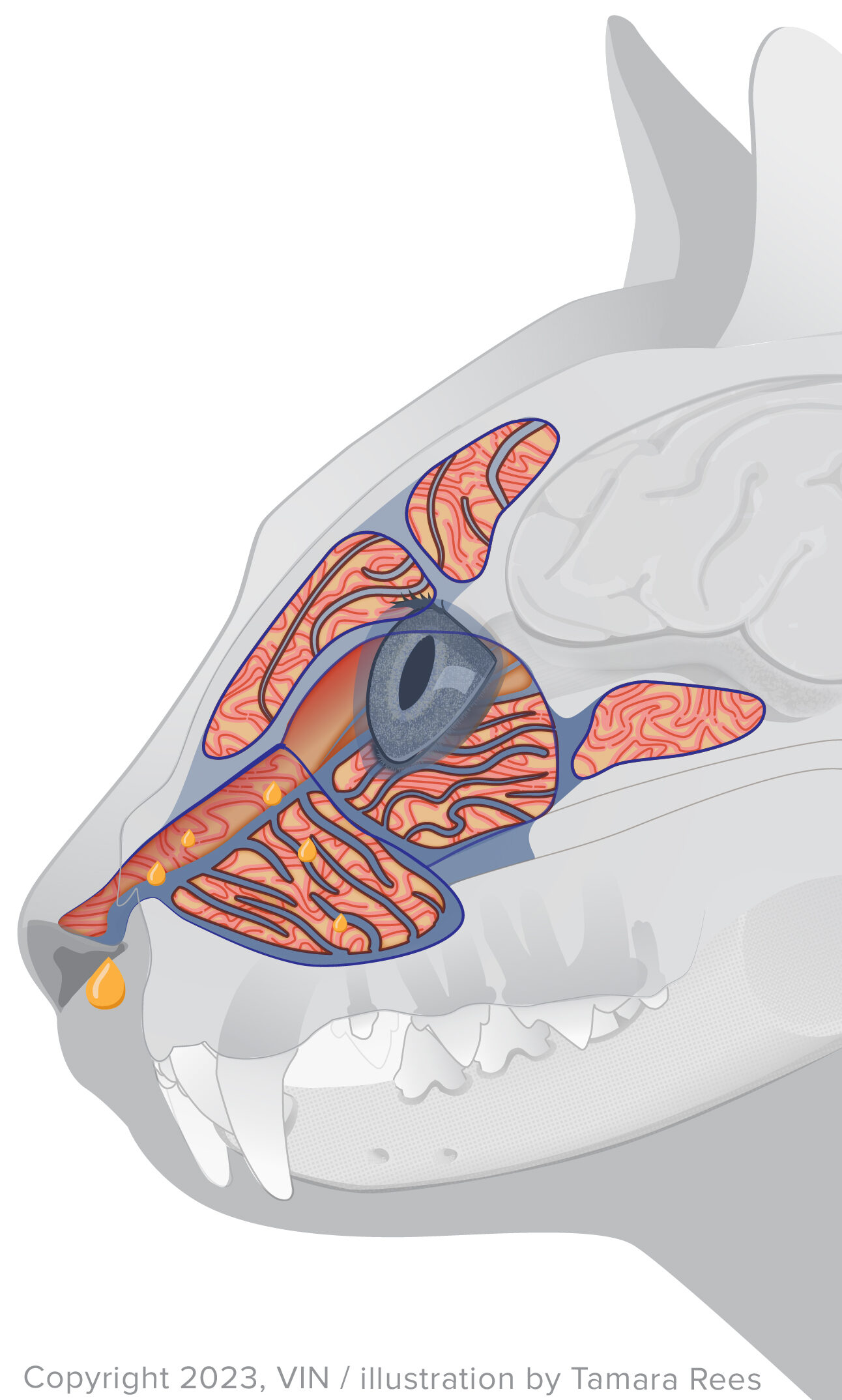
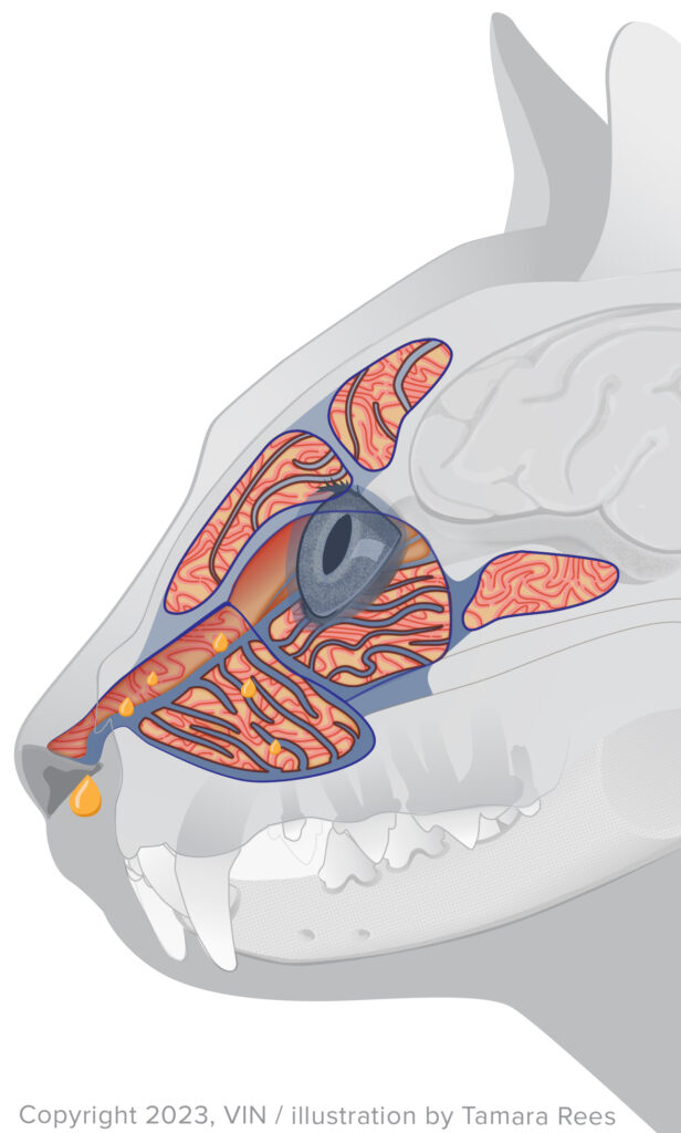
Rhinitis is inflammation of the mucous membranes in the nose. If the lining of the sinuses is inflamed, that is called sinusitis. If both the nose and sinuses are affected, it is rhino-sinusitis. Rhinitis can occur by itself or as part of an upper respiratory illness/infection. Rhinitis can be mild, with just a bit of clear nasal discharge seen, or it can be severe enough to have green/yellow/hemorrhagic (bloody) discharge.

Potential Causes of Rhinitis 

Many things can cause inflammation of the nose lining in dogs and cats. Some of them are:

  1. Tooth root abscess
  2. Viruses (feline herpesvirus, feline calici, and canine distemper, for example)
  3. Fungal infections
  4. Parasites (nasal mites, for example)
  5. Inflammatory polyp
  6. Birth defects (a cleft palate, for example)
  7. Facial/head trauma
  8. Foreign bodies (blades of grass, seeds, and grass awns, for example)
  9. Allergies
  10. Idiopathic lymphoplasmacytic rhinitis (chronic inflammatory rhinitis)
  11. Cancer
  12. Bacteria

Primary bacterial causes of rhinitis in cats are uncommon. The bacterial infection is usually secondary to another disease.

Aspergillus fumigatus is the most common cause of fungal rhinitis in dogs.

Cases of rhinitis can be chronic (ongoing) or acute (short-term). Chronic rhinitis has intermittent, recurrent episodes of sneezing and nasal discharge; sometimes there will be a partial response to antibiotics, but not enough to get rid of it forever. Chronic rhinitis/rhino-sinusitis cases in cats are often referred to as chronic snufflers.

Idiopathic lymphoplasmacytic rhinitis (chronic inflammatory rhinitis) is uncommon in the cat but may represent a hypersensitivity reaction to one or more environmental air allergens. In dogs, it was diagnosed in 20% of rhinitis cases, in one study. The German shepherd dog, Labrador retriever, dachshund, cocker spaniel, and Yorkshire terrier are the most commonly affected dog breeds. Median age of affected dogs is 8-9 years.

Clinical Signs

Signs may include sneezing, pawing at the face (which can indicate a foreign body), snoring, nasal obstruction, and/or a nasal discharge. When the nasal passages are narrowed because of inflammation, the pet will breathe with an open mouth and experience shortness of breath. Sneezing is seen most frequently in acute rhinitis as the pet attempts to clear discharge out of the upper airways. Sneezing tends to be intermittent with chronic rhinitis. Conjunctivitis (inflammation of the conjunctiva, or pink eye) and tearing may be seen in conjunction with inflammation of the upper respiratory passage.

Diagnosis is based on history and physical examination. To determine the underlying reason for the rhinitis, the diagnostic workup often requires anesthesia for a complete examination. The workup usually includes radiographs of the skull, a culture of the nasal passage and its discharge, nasal wash, a microscopic examination of the discharge, and endoscopy (a type of imaging in which a lighted tube is inserted into the nasal passages).

Treatment

Treatment depends on the underlying cause of the rhinitis. Medications such as antibiotics, antihistamines, steroids, or anti-fungal medications may be called for. Secondary bacterial nasal infections are common, and nasal discharge often will resolve with antibiotic treatment. For cases involving foreign objects or cancer, surgery may be needed.

Prognosis

The prognosis is dependent on the underlying cause of the rhinitis and how chronic the case is.

portfolio-single-swiper-3

Rectal Prolapse in Dogs and Cats

Rectal prolapse occurs when the rectum, which is the last part of the intestines, gets turned inside out (everted), and sticks out of the anus. It looks like a red or pink tubular structure sticking out of the area from which your pet normally poops. It can also be crusty, ooze, or be a dark color if it has been around for a while. Rectal prolapse is not the same thing as hemorrhoids, which are swollen veins from the rectum that bulge out of the anus.

Why a Prolapse Happens 

Prolapse can occur after a pet has been straining to poop for a while. Some of the most common reasons this can happen is if the pet has intestinal worms, is dehydrated, constipated, has diarrhea or loose stool, or has an intestinal blockage such as a tumor or has eaten an object that he is having trouble passing. Over time, straining leads to weakening of the structures that keep the rectum in place, causing the rectum to stick out through the anus. 

Other Symptoms 

If your pet has a rectal prolapse, you may also have noticed that he has been spending a lot of time trying to poop recently. An example of straining is standing in the classic, “hunched” posture used for emptying the bowels for longer than usual. It can also mean pooping more times than usual, even if he is able to finish quickly. You may also have seen an unusual change in the poop, such as being small and dry, looser than usual, or covered in intestinal worms. If you haven’t noticed any change in regular habits, either because keeping an eye on your pet’s bathroom habits is gross or because you have a sneaky pet that hides in order to go, it isn’t a big deal. Your veterinarian will know what to do. 

Diagnosing a Rectal Prolapse 

To officially diagnose a rectal prolapse, your veterinarian will give a pretty thorough physical examination, including a rectal exam in which they feel the prolapsed tissue. A fecal sample will likely be taken to check for worms and other intestinal issues. If a blockage is suspected, x-rays and/or an abdominal ultrasound may be necessary. 

Treatment 

To treat the prolapse, the vet will have to carefully clean the area before replacing it back into its original position. If it is swollen, additional medications or solutions may need to be applied to shrink the prolapse. Sometimes sedation or anesthesia is needed because a prolapse can be painful, especially if it becomes infected. 

If the prolapse can be replaced, the vet may tie a “purse-string” suture around the anus to keep the opening small and prevent the rectum from prolapsing again. This fix is temporary until your pet heals. The suture will need to be removed later (your vet will tell you when), but it is important that you don’t let your pet chew the area or the suture may break. Antibiotics and pain medications may be sent home, as well as an e-collar, to ensure the rectal tissue heals properly. 

Occasionally, a rectal prolapse cannot be replaced, or the tissue is too damaged. In these cases, surgery will be necessary to either remove the damage tissue (called a resection and anastomosis) or tack the rectum into the body wall from inside the abdomen (called a colopexy). 

In addition to treating the prolapse, your veterinarian will also want to treat the cause of the prolapse (e.g., treat for intestinal worms). Stool softeners may also be prescribed for a short period to keep your pet from straining. Most pets improve and do not continue to have issues once the rectal prolapse heals. If you have any questions or concerns about your pet’s diagnosis and treatment, give your vet a call so they can guide you further.

7003957

Recessed Vulva in Dogs

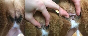
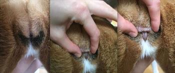
A recessed (or juvenile) vulva means a dog has excess skin folds that hang over her vulva, enough to potentially cause problems of one kind or another. The skin folds are a structural problem thought to possibly be a genetic issue, but the genetic connection has not been determined. The condition tends to be seen mostly in medium to large-sized breeds, most often in obese dogs.

Some dogs will never have one single problem related to having a recessed vulva, and it will be found during a physical exam or during a procedure for something unrelated. However, some dogs will have life-long problems because the vulvar area collects some moisture when the dog urinates. Combined with body heat in that area, the moisture creates a perfect breeding ground for bacteria and predisposes the dog to urinary tract infections or vaginitis. The vagina is normally not sterile. The bacteria can infect the skin folds, which is called perivulvar dermatitis, move into the vagina causing vaginitis, or move up into the urinary tract, which leads to a bladder infection.

Your veterinarian can diagnose a recessed, or hooded, vulva simply by looking at the genital area, and the vulva is not seen because it’s covered with skin folds.

Clinical signs tend to depend on how much inflammation and infection the dog has. Your dog may scoot on her behind, trying to relieve the itching in her vulva (it may look like a dog scooting because of inflamed anal glands), or licking her vulva excessively.

You may notice a bad odor from her vulva; that odor is usually from infected tissues. Some dogs might leak urine while sleeping or resting due to a condition called USMI, which is a common problem in spayed dogs and could also potentially contribute to a bladder infection (bacterial cystitis).

If she has a bladder infection, you may see blood in her urine, accidents in the house even if she is perfectly housetrained, see her straining to urinate, or notice a foul odor from her urine. It’s possible that her signs of a bladder infection are the only ones you will see if she has a recessed vulva.

Overall, bacterial cystitis is a common medical problem for female spayed dogs (in general) in that many of them may have a bout of bacterial cystitis during their lifetime. Repeated or recurrent bouts of bacterial cystitis are more problematic and it’s important to look for underlying drivers. A recessed vulva could be one of these predisposing causes.

Treatment

If the skin folds of the recessed vulva are affecting both your and your dog’s quality of life, your veterinarian can provide medical management or a surgical fix. Medicated shampoos or wipes may be enough to prevent skin infections. In overweight dogs, weight loss will likely help because there would be less tissue to fold over the vulva. If your dog has not gone through a heat cycle, the recessed vulva might improve after the first heat cycle.

The surgical procedure is called a vulvoplasty. Prior to surgical intervention, it’s important to investigate and look for co-morbid causes for recurrent bacterial cystitis. The evaluation typically includes a physical examination, complete urinalysis, urine culture, possibly a complete blood count, and a chemistry panel. Imaging with radiographs (X-rays) and a complete abdominal ultrasound are also likely to be done. Direct visualization of the lower urinary tract and vagina can be performed with cystoscopy, and this is often done prior to vulvoplasty surgery in order to look for problems such as malformations and other abnormalities.

During vulvoplasty, the surgeon removes a piece of tissue from around the vulva so that the skin has a more normal structure. The new ventilation helps the skin dry out and not be moist enough all the time for bacteria to grow. Additionally, it keeps those skin folds from rubbing up against each other and causing irritation.

You may be provided with analgesics for pain and possibly antibiotics to prevent an infection. Some dogs will need sutures; most types will need to be removed in about 10-14 days at the follow-up appointment.

Before the vulvoplasty occurs, any skin infection near the vulva must be cleared up as it prevents the incision from healing normally, usually by taking antibiotics. A skin culture can determine the most appropriate antibiotic.

Appropriate post-operative care is critical: for two weeks she must be kept quiet with restricted activity, as otherwise during running, jumping, and playing the incision can rip open. That can cause some serious complications. She should be taken outside only on a leash. An e-collar needs to stay on so she will not lick the incision, which also can cause infections.

Rarely, in dogs with significant skin folds, it is possible that not enough skin is removed during the procedure. In this case, the symptoms will likely return. A second surgery can correct it.

Generally, however, the prognosis after a vulvoplasty is excellent and most owners are quite pleased.

7750222

Oral Joint Health Supplements #2

On the last program of Texas Vet News I talked about the large number of oral joint health supplements that are on the market for horses.  Even though these products are widely used, many of them are of questionable quality because there are no regulations governing them.  Because of this problem, Dr. Stacey Oke has developed a system for horse owners to help them choose a product that may be of better quality than some others. 

First of all, choose a product from a company that has been in the animal health pharmaceutical business for years instead of months.  It is likely that companies with a good reputation are not going to stake their reputation on manufacturing a poor quality product for short-term sales.  Also, look for products that are backed by clinical research published in peer-reviewed veterinary journals.  Many companies report they have clinical trials but don’t have information available; even if they do have information, clinical trials that are not published in peer-reviewed journals are worthless. 

It is also important to look at the label and make sure all of the ingredients in the product are listed, including active, inactive, and filler ingredients.  Manufacturers that do not list all ingredients of their products likely use fillers and may not contain the labelled ingredients.  Products that make claims that seem too good to be true are probably not as good as they claim. 

Dosing instructions should be accurate and clear, and the amount of ingredient per scoop should be printed clearly on the label.  Also, look for products with clear contact information and check to see if the company you choose has veterinarians on staff to answer your questions. 

4128711

Orphan Puppy & Kitten Care

The Nest Box

Keep the babies together as long as they are about the same age; this will help socialize them to their own species and will help in keeping them warm. Try to assemble the following equipment.

  1. The nest box in which the babies live should have tall sides so that they cannot climb out by mistake and become chilled. A cardboard pet carrier is perfect as it is portable, dark inside, and closable. These are inexpensive and should be available from a pet store or from your veterinarian.
  1. Place towels in the bottom of the box and cover them with a disposable diaper so the babies rest directly on the diaper. Most diapers have elastic leg holes and may have to be trimmed so they will lie flat. Expect to change the diaper several times daily. Keeping the babies clean and dry is very important, and a diaper is perfect for wicking away wetness.
  2. Place a heating pad under the nest box so that only half of the nest box is warmed. This way, the babies may crawl off the warm side of the box if they feel too hot. Alternatively, a water bottle filled with warm water can be buried in the blankets as a heat source as long as the babies have room to move away from it if they are too hot.

If the baby’s temperature drops below 94⁰F, the heart rate drops and intestinal motion ceases. Death occurs if this is not corrected.

Initial warming when babies have gotten cold should take place over an hour or two to avoid shock.

The Diet

  1. There are several brands of orphan formula on the market. For kittens, KMR® by PetAg and Just Born® by Farnam Pet Products come in both a powder and a liquid. The powdered form seems less associated with diarrhea than the liquid plus with the powdered form, the water content can be increased in the event of dehydration. For puppies, PetAg makes a canine formula called Esbilac® plus there is a canine version of Just Born formula. Again, both powder and liquid forms are available.
  2. Mix up the powder according to the directions on the can. If you are using the liquid form, you may want to dilute it with one part water for every two parts of formula. As the babies get older, less water may be used whether you are mixing up the powder or the liquid. If diarrhea occurs at any time, you should add more water to the formula to make up for fluid lost as diarrhea.
  3. Store the can of powder in the freezer after opening. Do not mix up more than a day’s worth of formula. Use a blender to mix the formula several hours ahead to allow time for the bubbles to settle. 

Makeshift Formula Until Commercial Formula can be Obtained

Puppies   
1 cup whole milk
1 tsp. salad oil
1 drop multi-vitamins (if you have any)
2 egg yolks

Kittens
1/2 cup whole milk
1 egg yolk
1 drop multi-vitamins
3 crushed Tums (antacid)

For either one, then mix in a blender.

The Bottle

  1. Obtain a pet nurser bottle from a pet store or your veterinarian.
  2. Use fine scissors or a hot needle to make a hole in the nipple. The hole should be big enough that formula will slowly drip out if the bottle is held upside-down and gently squeezed. The nipple should not collapse when the baby is sucking.
  3. Warm the bottle in a cup of hot water. Always test the formula before giving it to the babies. Taste it to be sure it is not sour. Do not use a microwave oven to heat the bottle as it may not heat evenly – some areas of the bottle can be scalding hot.

How / When To Feed

  1. Expect to feed them every two to three hours during the day. If this is done, the babies should be able to sleep through the night.
  2. Do not wake the babies at feeding time. Let them sleep. When they wake up hungry, they will let you know.
  3. During feeding be sure to tip the bottle so that no air is swallowed. It is more natural to feed them right side up rather than on their backs like human infants.
  4. Play with/rub them after feeding to burp them.
  5. Occasionally small amounts of formula will come out of the nose. The baby is drinking too fast. If excessive amounts of formula appear to be coming out the nose or if you are concerned, call your veterinarian.

Tube feeding involves using a syringe and lubricated tube to introduce formula through the mouth and directly into the stomach. Your veterinarian will need to show you how to do this should it become necessary. A baby who rejects three meals in a row will need tube feeding.

Urination / Defecation

  1. Infant animals are unable to take care of these matters alone and must be given help. Normally their mother’s tongue does the job as she washes them. Use a cotton swab, tissue, or your finger to gently rub the baby’s genital area. Have a tissue ready to catch the urine.
  2. Rubbing the anal area as well may also be necessary if the babies do not seem to be defecating as much as expected. Watch for diarrhea. Normal infant stool is loose but should not be watery.
  3. It is normal for baby kittens and puppies to have round bellies. If you think the baby might be constipated, usually taking the temperature rectally with a normal rectal thermometer provides enough extra stimulation to get things back to normal. Only the bulb of the thermometer tip needs to be inserted rectally.

Expect the baby to need stimulation for urination/defecation for the first three weeks of life.

Bathing

  1. Using kitten or puppy shampoo and warm water, bathe the babies a couple of times daily. Urine will burn their tender skin and caked feces can lead to infection, so keeping the babies clean is important.
  2. Take care not to submerge the infant in water. Be careful that it cannot drown or choke on the water and be sure the water temperature is acceptable.
  3. Gently blow dry the babies when the bath is over. Do not allow chilling.

How to be Sure You Are on Track

Starting Solid Food

The best way to be sure everything is going well is to track weight gain in your new babies. A postal scale or food scale (ideally one that measures weight in grams) will be helpful. A puppy or kitten should gain 10% of the birth weight every day and should be drinking 22 to 26 cc of formula per 100 grams of body weight over the course of the day. Puppies are variable in growth expectancy depending on breed, but kittens are more predictable and should gain 50 to 100 grams weekly. Kittens weighing less than 90 grams (approx three ounces) at birth have a high (59%) mortality rate.

  • When the babies start biting and chewing at their bottle instead of sucking (three to four weeks of age), they may be started on some finely textured canned food. At first it may be necessary to mix solid food with a little formula and /or smear a little around their mouths gently with a finger. Prescription recovery diets have a very soft texture and are readily accepted but any pate style canned food should do well. Your vet’s office will most likely have a recovery formula diet available.
  • Between ages four and six weeks, they should begin readily accepting solid food. New homes may be found for them at age eight weeks.

A Note on Colostrum

Colostrum is the first milk produced by the mother shortly after giving birth. It is rich in antibodies which will protect the babies for the first several months of life. Colostrum is only produced for a day or two and the baby is only able to absorb its antibodies for a day or two.

Without colostrum (if their mother did not nurse the kittens during the first two days of life) the babies have a serious immunological disadvantage. Especially great care should be taken in cleanliness and the babies can be vaccinated as early as age two weeks depending on circumstances. They may require a plasma transfusion to make up for the colostrum. There is no substitute for a real mother.

See more information on raising orphan kittens.

Many foster parents have difficulty distinguishing male and female kittens at this early age. See some guidelines and (illustrated) instructions.

4126881

Omega Three Fatty Acids for our Pets

Did you know that skin diseases account for as much as 25% of the cases seen by small animal veterinarians?

Skin problems typically faced by pets and their owners include:

  1.   Itching
  2.   Dandruff
  3.   Blackheads
  4.   Odor
  5.   Crusting
  6.   Redness
  7.   Rashes
  8.   Oiliness

The nutritional aspect of skin disease is quite broad. There are true nutritional deficiencies that affect the skin and other skin diseases that can be made dramatically better by using supplements.

Just because a condition responds to a nutrient does not necessarily mean that there is a deficiency of it.

Everyone wants their pet to have a lustrous beautiful coat and would like to do what it nutritionally possible to ensure this. Recently essential fatty acids have received a great deal of press. A brief primer follows. 

What is a Fatty Acid?

Biochemically, a fatty acid is what we just call fat. When we talk about different types of fatty acids, we are talking about different types of fat. A fatty acid consists of a long carbon chain (say 20 or so carbons in length) with biochemical acids called a carboxyl group at one end and a methyl group on the other end.

Saturated Vs. Unsaturated

Each carbon has four binding sites. In the carbon chain, two sites will be taken up by other carbons (i.e., the two adjacent carbons on the chain). In a saturated fat, the other two sites are taken up by hydrogen atoms. Saturated fats are typically solid at room temperature, like lard and butter, and are usually made from animals. Saturated fats are generally burned as fuel by our bodies. An example of a saturated fatty acid is depicted in the above illustration.

Unsaturated fats have two adjacent carbons held together by a biochemical double bond. These fats are generally liquid at room temperature and come from plants, such as olive oil, corn oil, etc.

Unsaturated fats can be classified as omega three fatty acids or omega six fatty acids, depending on the location of the double bond relative to the methyl group at the end of the chain. These types of fatty acids are essential, meaning that our bodies cannot make them; instead, we must eat them in our diet. These fats are not burned for fuel but are used as structural components.

The omega six fatty acids are used as the main structural components in our cells. Omega three acids are used in the retina and central nervous system.

For healthy skin and coat, the diet must contain adequate omega six fatty acids as these make up the surface of the skin.

Examples of omega six fatty acids (also called n-6 fatty acids): Linoleic acid, gamma linolenic acid, and Arachidonic acid. Evening Primrose oil is an excellent example.

Examples of omega three fatty acids, also called n-3 fatty acids, include: Alpha linolenic acid (ALA), Eicosatetraenoic acid (EPA), docosahexaenoic acid (DHA).

Cold water fish oils are an excellent source of DHA and EPA. A terrific source of ALA would be flax seed oil. DHA has anti-inflammatory properties, which is why it is so frequently recommended in inflammatory conditions. Many people prefer to use flax seed oil as their omega 3 source because flax seed oil does not taste fishy and people readily convert ALA to DHA. This is great for people but it turns out that pets are only able to convert about 10% of ALA to DHA, so for them cold water fish oils are better. Fortunately, most dogs and cats like the fishy taste. 

Should we Supplement Essential Fatty Acids?

There is no question that a diet must contain adequate omega 6 fatty acids to maintain optimal skin and coat quality. A diet found to be “complete and balanced” will have an amount of omega 6 fatty acids that should be optimal for a normal animal.  

But there’s more.

Omega 6 Fatty Acids and Dandruff

Research has shown that dogs with oily, dandruffy skin (seborrhea) have insufficient omega 6 fatty acids in their skin despite eating a diet that should be optimal. When omega 6 fatty acids are supplemented, the seborrhea improves. This finding supports the old-time remedy of adding a spoonful of corn oil to the diet to ensure a glossy coat. Seborrhea is a complex condition but animals with it may need more omega 6 fatty acids.

Anti-Inflammatory Effects of Omega 3 Fatty Acids

Omega 6 fatty acids constitute our cell membranes. During some biochemical situations, it is necessary to produce hormone-like substances called prostaglandins and leukotrienes. These substances are made from omega 6 fatty acids and the resulting prostaglandins and leukotrienes are not necessarily good for us. In fact, these substances are responsible for itching and inflammation leading to the clinical skin problems listed above. One way to address this is to supplement omega 3 fatty acids, which become incorporated into cell membranes along with the omega 6s. After a couple of months of supplementation, omega 3 fatty acids infiltrate cell membranes significantly. When it comes time to make prostaglandins, the omega 3s are mobilized instead of the omega 6s only in this case, the prostaglandins that result are not inflammatory. When omega 3 fatty acids are supplemented, itching can be substantially reduced and even arthritis pain improved.

One problem with this is that no one really knows how much omega 3 fatty acid to supplement. There is some evidence that a ratio of omega 6 to omega 3 fatty acids in the supplement is crucial. If this is so, clinical research becomes hugely complicated as the diets of pets cannot be standardized easily for study. If pets in a study eat different diets, then it is impossible to tell what overall omega 6:omega 3 ratio each is receiving. Essential fatty acids are being pursued as treatment for diseases of virtually every organ system; watch for new research in this area.

Conditions that Have Been Shown to Benefit from Omega 3 Fatty Acid Supplementation

  1.   Renal (kidney) insufficiency
  2.   Heart failure
  3.   Lymphoma
  4.   Airborne allergies

Research is ongoing. We know that supplementing with omega 3 appears to be benign with the potential to do a great deal of good. 

4128011

Osteosarcoma in Dogs

What is Osteosarcoma?

Osteosarcoma is by far the most common bone tumor of dogs, usually striking the leg bones of larger breeds. Osteosarcoma usually occurs in middle aged or elderly dogs but can occur in a dog of any age; larger breeds tend to develop tumors at younger ages.

  • Osteosarcoma can develop in any bone but the limbs account for 75-85 percent of affected bones. Osteosarcoma of the limbs is called appendicular osteosarcoma.
  • It develops deep within the bone and becomes progressively more painful as it grows outward and the bone is destroyed from the inside out. The lameness goes from intermittent to constant over 1 to 3 months. Obvious swelling becomes evident as the tumor grows and normal bone is replaced by tumorous bone.
  • Tumorous bone is not as strong as normal bone and can break with minor injury. This type of broken bone is called a pathologic fracture and may be the finding that confirms the diagnosis of bone tumor. Pathologic fractures will not heal and there is no point in putting on casts or attempting surgical stabilization.

How do we Know my Dog Really has an Osteosarcoma?

Radiographs (x-rays)

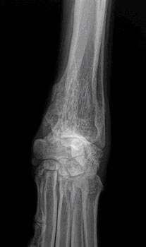
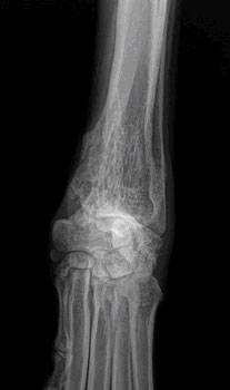
The lytic lesion looks like an area of bone has been eaten away. One of the first steps in evaluating a persistent lameness is radiography (x-rays). Bone tumors are tender so it is usually clear what part of the limb should be radiographed. The osteosarcoma creates some characteristic findings.

  1. The sunburst pattern – shows as a corona effect as the tumor grows outward and pushes the more normal outer bone up and away.
  2. A pathologic fracture may be seen through the abnormal bone.
  3. Osteosarcoma does not cross the joint space to affect other bones in the joint.

Radiography is almost completely diagnostic in most cases, but there are a few other far less common conditions that can mimic the appearance of a bone tumor, so a confirming test is going to be needed if one is to be complete. If a basic blood panel and urinalysis haven’t been done, this would also be a good time to do so as basic information about liver and kidney function will be needed for treatment regardless of whether this turns out to be a bone tumor or not; plus, a tissue sample from the bone is needed for confirmation (see later).

Tissue Sampling: Biopsy and Needle Aspirate

Radiographs are close to being confirmatory but still they are not definitive. Since life and death decisions are going to be made, it is best to obtain a tissue sample for confirmation. This can be done by either biopsy or by needle aspirate.

Biopsy

A small piece of bone can be harvested surgically. The bone is preserved, sectioned, and examined under the microscope to confirm the diagnosis of osteosarcoma. There are several problems associated with this diagnostic.  Sometimes a bone tumor is surrounded by an area of bone inflammation and it may be difficult to get a representative sample. The tiny hole that results when a core of bone is removed can create a weak spot and the bone can actually break. Even if the procedure goes well, often there is increased pain and lameness for the patient afterwards. With so many potential problems, most specialists have switched to needle aspirate for diagnosis.

Needle Aspirate

With needle aspirate, a large bore needle is inserted into the area of the tumor and cells are withdrawn for analysis. A full core of bone is not removed, just a sampling of cells. This is usually sufficient to confirm osteosarcoma. If there is ambiguity, certain stains can often settle any questions the pathologist may have.

With a Diagnosis Confirmed, Staging is the next Consideration

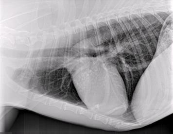
Osteosarcoma is an aggressive tumor and it can be assumed to have spread by the time it is first diagnosed; there is no possibility of preventing spread. That said, how well treatments can be expected to work depend on whether or not the tumor spread has progressed so as to be visible. Because osteosarcoma spreads to the lungs as one of its first stops, chest radiographs are important in checking for visible tumor spread. If there is already visible tumor spread at diagnosis, this changes what treatments are recommended.

Some specialists recommend nuclear imaging of the skeleton to identify any spread to other bones, which might also alter recommendations; however, this form of imaging is not readily available.

What if it isn’t Really an Osteosarcoma?

The location and radiographic appearance of the osteosarcoma in the limb are quite classic but there are a few outside possibilities that should at least be mentioned. Only a few other possible conditions cause similar lesions in bone: the chondrosarcoma, the squamous cell carcinoma, the synovial cell sarcoma, or fungal bone infection.

Chondrosarcoma

Chondrosarcoma is a cartilage tumor, possibly not as malignant as the osteosarcoma. The chondrosarcoma generally occurs on flat bones such as ribs or skull bones and is not usually found in the limbs. Still, should a chondrosarcoma occur in the limb, treatment recommendations still include amputation of the affected bone and many of the same treatments as for osteosarcoma.

Squamous Cell Carcinoma

The squamous cell carcinoma is a tumor of the external coating of the bone, called the periosteum. This is a destructive tumor locally but it tends to spread relatively slowly. Again, a bone suspected of malignant tumor should be amputated, and the tissue then analyzed and then treatment adjustments should be made thereafter. The squamous cell carcinoma tends not to arise in the same bone areas as the osteosarcoma; it tends to arise in the jaw bones or in the toe bones.

Synovial Cell Sarcoma This is a tumor of the joint capsule lining. Its hallmark is that it affects both bones of the joint. The osteosarcoma, no matter how large or destructive it becomes, will never cross over to an adjacent bone. Prognosis is much better with amputation with synovial cell sarcoma.

Fungal Bone Infection

Coccidiodes immitis is a fungus native to the lower Sonoran life zone of the southwestern United States. It is the infectious agent of the disease called San Joaquin Valley Fever or just Valley Fever. (More scientifically, the condition is called coccidioidomycosis.)  In most cases, infection is limited to a few calcified lymph nodes in the chest and possibly lung disease. In some rare cases, though, the fungus disseminated through the body and can cause a very proliferative bone infection.

The bone infection of coccidiodomycosis grows rapidly and lacks the lytic lesions that are typical of the osteosarcoma. Other fungi, such as Histoplasma capsulatum, also have potential for bone involvement. Pursuit of this possibility makes sense if you live in an area where these fungi are a concern. Disseminated fungal disease is serious and even though this diagnosis is not cancerous, amputating the limb is most likely going to be necessary.

Treatment of osteosarcoma involves two aspects: treating the pain and fighting the cancer’s spread.

How do we Treat the Pain?

Keep in mind that dogs are usually euthanized because of the pain in the affected bone. Treating the pain successfully will allow a dog to live comfortably and extend life expectancy by virtue of extending comfort.

There are two ways to address the pain: amputatating the limb and palliative radiotherapy (usually combined with periodic bisphosphonate infusion treatments).

For most patients, there is one tumor on a leg and no visible tumor spread in the lungs. These are the patients with the best potential results and they are good candidates for amputation. Patients with a lot of arthritis in the other legs or with tumor spread evident in the chest already are probably not candidates for amputation and it may be more appropriate to keep the leg and simply relieve the pain with radiotherapy.

Amputating the Limb

Since the tumor in the limb is the source of pain, it makes sense that amputating the limb would resolve the pain. In fact, this is true. Removing the affected limb resolves the pain in 100 percent of cases. Unfortunately, many people are reluctant to have this procedure performed because of misconceptions.

  • While losing a leg is handicapping to a human, losing one leg out of four does not restrict a dog’s activity level. Running and playing are not inhibited by amputation after recovery from surgery. That said, if the remaining legs are arthritic, the stress on them can pose a mobility issue.
  • While losing a limb is disfiguring to a human and has social ramifications, dogs are not self-conscious about their image. The dog will not feel disfigured by the surgery; it is the owner that will need to adjust to the dog’s new appearance.
  • Median survival time for dogs who do not receive chemotherapy for osteosarcoma is 3 to 5 months from the time of diagnosis regardless of whether or not they have amputation. Do you want your dog’s last 3 to 5 months to be painful or comfortable?

Read a letter about amputation from a veterinarian who sees too many owners who reject the option of amputation out of hand. His letter includes videos of two happy dogs without limbs.

Limb-sparing Surgery (removing the tumor but not the leg)
Limb-sparing techniques developed for humans have been adapted for dogs. To spare the limb and thus avoid amputation, the tumorous bone is removed and either replaced by a bone graft from a bone bank or the remaining bone can be re-grown via a new technique called bone transport osteogenesis. The joint nearest the tumor is fused (i.e., fixed in one position and cannot be flexed or extended.)

  1. Limb sparing cannot be done if more than 50% of the bone is involved by tumor or if neighboring muscle is involved.
  2. Limb sparing does not work well for hind legs or tumors of the humerus (arm bone.)
  3. Limb sparing works best for tumors of the distal radius (forearm bone).
  4. Complications of limb sparing can include: Bone infection, implant failure, tumor recurrence, and fracture.

While amputation can be viewed as a pain management strategy, limb-sparing is only performed in conjunction with chemotherapy. It is important to keep in mind that grafting of a new bone structure requires healing time and that a great deal of post-operative confinement time is needed (in a patient whose life expectancy is going to be measured in months). For the right patient, limb-sparing can be the best choice but be sure to understand all the details of post-operative care from the specialist.

Palliative Radiotherapy for Pain Control

Sometimes amputation is simply not the right choice and happily there is an effective alternative treatment. Radiation can be applied to the tumor in two, three or four doses, depending on the protocol. Improved limb function is usually evident within the first 3 weeks and typically lasts 2 to 4 months. When pain returns, radiation can be given again for further pain relief if deemed appropriate based on the stage of the cancer at that time.

There are a couple of caveats:

  • When pain is relieved in the tumorous limb, there is an increase in activity that can in turn lead to a pathologic fracture of the bone.
  •  Radiotherapy does not produce a helpful response in about 1/4 of patients. Remember, amputation controls pain in 100 percent of cases but if amputation is simply not an option, there is a 3 out of 4 chance that radiotherapy will control the pain.

Current standard treatment involves pairing palliative radiation with monthly infusions of medications called bisphosphonates. 

Bisphosphonates

This class of drug has become the standard of care in humans with bone tumors and have been found helpful in managing osteosarcoma pain in dogs as well. Bisphosphonates act by inhibiting bone destruction, which in turn helps control the pain and bone damage caused by the bone tumor. The most common bisphosphonate in use for dogs has been pamidronate, though a new drug zoledronate is taking its place gradually. Treatment is given as an IV drip over two hours in the hospital every 3 to 4 weeks. In humans, an assortment of potential side effects have emerged (fever, muscle pain, nausea all lasting 1 to 2 days in up to 25 percent of patients, renal disease in certain situations, low blood calcium levels, jaw bone cell death); these issues so far have not panned out as problems for dogs and cats. Bisphosphonates are important in managing bone tumor pain in patients that have no undergone amputation.

Analgesic Drugs

At this time there are numerous analgesic medications available for dogs with this tumor. No single medication, however, is a match for the pain involved in what amounts to a slowly exploding bone. A combination of medications is needed to be reasonably palliative and should be considered only as a last resort if amputation or radiation therapy will not be pursued. There are several types of drugs that can be combined.

  • Non-Steroidal Anti-Inflammatory Drugs (NSAIDs)
    These are anti-inflammatory pain relievers developed for dogs: carprofen, etodolac, deracoxib, meloxicam, firocoxib, and others. These are typically given once or twice daily in tablet form at home. The patient should have good liver and kidney function in order to take medications of this class.
  • Narcotic Pain Relievers
    While these drugs do not have anti-inflammatory properties, they are well-known analgesics and have been used in an assortment of forms for thousands of years. They are particularly useful in chronic pain because they do not interact negatively with other pain relievers. Drowsiness is a potential side effect.
  • Miscellaneous Supplemental Pain Relievers
    There are two drugs that have surfaced as adjunct pain relievers for animals with chronic pain: gabapentin and amantadine. Gabapentin works on neurologic pain and is rapidly surfacing in the treatment of arthritis, surgical pain, and other chronic pain states. Amantadine works by reducing what is called wind up, a phenomenon where nerves become sensitized to pain leading to the experience of pain from stimuli that normally do not cause pain.

These different drugs are often given together to create analgesia to the osteosarcoma patient when amputation and radiotherapy are not going to happen. It is important to realize that there is a limit to how much pain relief can be achieved against a bone tumor with only pills. It will not be long before the pain of this tumor, as evidenced by not using the leg, tenderness to the touch, etc., overpowers the effect of oral medications.

How do we Treat the Cancer?

 Osteosarcoma is unfortunately a fast-spreading tumor. By the time the tumor is found, it is considered to have already spread. Osteosarcoma spreads to the lung in a malignant process called metastasis. Prognosis is substantially worse if the tumor spread is visible on chest radiographs, so if you are contemplating chemotherapy, chest radiographs should be taken.

  1. Chemotherapy is the only meaningful way to alter the course of this cancer.
  2. Young dogs with osteosarcoma tend to have shorter survival times and more aggressive disease than older dogs with osteosarcoma.
  3. Elevations of alkaline phosphatase, one of the enzymes screened on a basic blood panel, bode poorly. These dogs have approximately 50% of the survival times quoted below for each protocol.
  4. A tumor in the lymph nodes local to the leg being amputated also bodes poorly. In the study by Hillers et. al published in the April 15th, 2005 issue of the Journal of the AVMA, median survival was significantly longer (318 days vs. 59 days) in dogs where the tumor was not evident in local lymph nodes at the time of amputation. 

Cisplatin (given by IV every 3 to 4 weeks for 3 treatments)

  1.   The median survival time with this therapy is 400 days.
  2.   Survival at 1 year: 30% to 60% 
  3.   Survival at 2 years: 7% to 21%
  4.   Giving less than 3 doses does not increase survival time (i.e., if one can only afford one or two treatments, it is not worth the expense of therapy)
  5.   Cisplatin can be toxic to the kidneys and should not be used in animals with pre-existing kidney disease.

Carboplatin (given by IV every 3 to 4 weeks for 4 treatments)

  •   Similar statistics to cisplatin but carboplatin is not toxic to the kidneys and can be used if the patient has pre-existing kidney disease.
  •   Carboplatin is substantially more expensive than cisplatin.

Doxorubicin (given IV every 2 weeks for 5 treatments)

  1.   The median survival time is 365 days.
  2.   10% still alive at 2 years.
  3.   Toxic to the heart. An ultrasound examination is needed prior to using this drug as it should not be given to patients with reduced heart contracting ability.

Doxorubicin and Cisplatin in Combination (both given IV together every 3 weeks for four treatments)

  •   48% survival at 1 year
  •   30% survival at 2 years
  •   16% survival at 3 years. 

What is Median Survival Time? 

There are a number of ways to statistically evaluate the central tendency of a group. The median is the value at which 50% of the group falls above and 50% of the group falls below. This is a little different from the average of the group, though more people are familiar with this term. When evaluating median survival times, you are looking at a 50% chance of surviving longer than the median and a 50% chance of surviving less than the median. 

What Does Chemotherapy Put my Dog Through?

Most people have an image of the chemotherapy patient either through experience or the media and this image typically includes lots of weakness, nausea, and hair loss. In fact, the animal experience in chemotherapy is not nearly as dramatic. After the pet has a treatment, expect 1 to 2 days of lethargy and nausea. This is often substantially helped with medications like Zofran, a strong anti-nausea drug commonly used in chemotherapy patients. These side effects are worse if a combination of drugs is used but the pet is typically back to normal by the third day after treatment. Effectively, you are trading 8 days of sickness for 6 to 12 months of quality life. Hair loss is not usually a feature of animal chemotherapy. In dogs, hair loss may occur in breeds that have continuously growing coats, such as poodles, Scottish terriers, and Westies. 

Axial Osteosarcoma

While osteosarcoma of the limbs is the classical form of this disease, osteosarcoma can develop anywhere there is bone. “Axial” osteosarcoma is the term for osteosarcoma originating in bones other than limb bones, with the most common affected bones being the jaws (both lower and upper). Victims of the axial form of osteosarcoma tend to be smaller, middle-aged, and females outnumber males two to one.

In the axial skeleton the tumor does not grow rapidly as do the appendicular tumors, thus leading to a more insidious course of disease. The tumor may be there for as long as two years before it is formally diagnosed. An exception is osteosarcoma of the rib, which tends to be more aggressive than other axial osteosarcomas.

Treatment for axial osteosarcoma is similar to that for the appendicular form: surgery followed by chemotherapy. There is one exception, that being osteosarcoma of the lower jaw. Because of the slower growth of the axial tumor and the ability to remove part or all of the jaw bone with little loss of function or cosmetic disfigurement, it has been reported that 71% of cases survived one year or longer with no chemotherapy at all.

Additional information can be found at Bone Cancer Dogs, Inc., a nonprofit corporation.

Not all veterinarians are comfortable treating osteosarcoma. Discuss with your veterinarian whether referral to a specialist would be best for you and your pet.

5217577

Obesity is Unhealthy in Dogs and Cats

Obesity has become an extremely important health problem in the Western world, not just for humans but for dogs and cats as well. Obesity in pets is associated with joint problems, diabetes mellitus, respiratory compromise, and decreased life span. Recent studies show approximately 60% of dogs and cats in the U.S. are classified as obese or overweight, making obesity an epidemic. Most pet owners underestimate their pet’s body condition, in part because overweight pets are so common that an overweight body condition now seems to be normal. 

 We can assure you that there is nothing normal about being overweight.

Why Obesity is Bad

A common justification for over-feeding treats is that a pet deserves a higher quality of life as a trade off for longevity. While this might on some level makes sense (after all, a pet munching on a treat is certainly getting a great deal of satisfaction from doing so), the other consequences do not make for higher life quality in the big picture. Here are some of problems that obese animals must contend with while they are not enjoying their treats and table scraps.

Arthritis


The over-weight animal has extra unneeded stress on joints, including the disks of the vertebrae. This extra stress leads to the progression of joint degeneration and creates more pain. Weight management alone decreases and can even eliminate the need for arthritis medications. The problem is compounded as joint pain leads to poorer mobility, which in turn leads to greater obesity.

Respiratory Compromise


The obese pet has a good inch or two of fat forming a constricting jacket around the chest. This makes the pet less able to take deep breaths as more work is required to move the respiratory muscles. Areas of the lung cannot fully inflate, so coughing results. The pet also overheats more easily. Many cases of tracheal collapse and chronic cough can be managed with only weight loss.

Diabetes Mellitus


Extra body fat leads to insulin resistance in cats just as it does in humans. In fact, obese cats have been found to have a 50% decrease in insulin sensitivity. Weight management is especially important in decreasing a cat’s risk for the development of diabetes mellitus.

Hepatic Lipidosis


When an overweight cat goes off food or partially off food because of illness or psychological stress, body fat is mobilized to provide calories. Unfortunately, the cat’s liver was not designed to process a large amount of body fat. The liver becomes infiltrated with fat and then fails. A stress that might have been relatively minor, such as a cold, becomes a life-threatening disaster.

Reduced Life Span


A study of age-matched Labrador retrievers found that dogs kept on the slender side of normal lived a median of 2.5 years longer than their overweight counterparts.

Unwillingness to Accept Therapeutic Diets


If the pet should develop a condition where a therapeutic diet is of great benefit, the pet that has been maintained primarily on a diet of table scraps may be unwilling to accept commercial pet food of any kind, much less a food modified to be beneficial for a specific disease process. This unwillingness will hamper treatment.

Increased Surgical/Anesthetic Risk


Obesity poses an extra anesthetic risk because drug dosing becomes less accurate. (It is hard to estimate a patient’s lean body mass for drug dosing if it is encased in a fat suit.) Furthermore, anesthesia is inherently suppressive to respiration and adding a constrictive jacket of fat only serves to make proper air exchange more challenging. And still further, surgery in the abdomen is hampered by the slippery nature of the extra fat as well as difficulty visualizing all the normal structures through the copious fat deposits. One never knows when a pet will require an emergency surgery (to say nothing of regular teeth cleanings).

So is the enjoyment of all those extra treats really worth it?

How Did My Pet Get So Fat Without Eating That Much?

You might think weight management might be easier for a pet than it is for a human. After all, the pet relies completely on someone else for feeding and exercise so it should follow that if the humans in control can regulate feeding and exercise, the pet should lose weight. It seems like this would be true but, as with humans, there is tremendous individuality with how different pets store the food they have eaten. Beyond this, sometimes it is hard to know what a pet is eating or the owner may not have a good sense for how much should be fed. Here are some factors involved.

A Cup of Food Depends on the Cup

When food packages refer to a certain number of cups of kibble being appropriate for a certain body weight, they are referring to an actual measuring cup. This may seem obvious but many mugs, coffee cups, and other scooping cups may not be equal to a cup measure.

If you do not have a cup measure, you can often get one from your veterinarian’s office as most manufacturers of reducing diets for pets provide free cup measures.

onsider measuring food with a gram scale. This a far more accurate way of measuring food amounts compared to cup measures.

The Package Guidelines are just Guidelines


Many packages of food include on their label some sort of feeding schedule that indicates how much food should be fed to a pet of a certain weight. This information is also available on most pet food web sites as well. The problem is that each pet is an individual and just as one person weighing 150 lbs can be obese and another person of the same weight may be skinny, the same is true of pets. These guidelines are meant as a starting point only. If your pet is too fat on the recommended feeding schedule, then you should reduce the amount of food or change to a diet that is higher in fiber so that a satisfying volume of food can still be eaten without adding calories.

Genetics


Some animals simply have the genes that predispose them to obesity. Dog breeds with genetic tendencies towards obesity include the: Golden Retriever, Cocker Spaniel, Dachshund, Beagle, Shetland Sheepdog, Boxer, Cairn Terrier, Basset Hound, and Labrador Retriever.

Children at Home


It is almost impossible to keep children from providing extra treats to their dog. This may include snacks spilled during play (pets have no “five-second rule”) or purposely feeding the pet unwanted food under the dining table. Similarly, pets that are allowed to roam (usually cats) often find food left out by neighbors, either to purposely feed their own pets or strays, or as unsecured trash. It is almost impossible to control the diet of an outdoor cat.

Low Metabolism


Some pets do not burn calories efficiently; they simply have a slow metabolism. This might be genetic as mentioned or it might be the result of a disease such as hypothyroidism or Cushing’s disease. Testing for health problems such as these is helpful to get the best treatment for resolution of the obesity. It seems like increasing exercise and eating a healthier diet would be easy to accomplish for a pet but it generally does not turn out that way.

Underestimating the Power of Treats


Many people express their affection for the pet by providing regular treats, and the pet happily obliges by begging or even performing cute behaviors. For some people, feeding treats to the pet constitutes a major part of the human-animal bond and they do not wish to give it up or reduce it. Pet treats are often high in calories, though, and four or five treats readily converts into an extra meal’s worth of added fat. Free feeding of dry food encourages the pet to snack as well; meal feeding represents better calorie control.

Treats should not exceed 10% of the pet’s feeding allotment.

Neutering


Sterilizing a pet is good for public health (fewer strays means fewer dog bites, less public resources needed for animal shelters etc.), good for a better house pet (less urine marking, tendency to fight or roam), no unwanted litters, reduced risk of many diseases, etc. The change in the hormonal picture, though, creates a tendency to form more fat cells (creating increased fat storage capacity – especially in female cats), and typically slows metabolism. Spay/neuter is a excellent time to switch from free feeding to meal feeding.

Evaluation by Body Score

Sometimes it is hard to recognize that your pet is overweight as the weight gain has come on gradually or it is hard to actually accept that your pet is more than just a little chubby and is now fully obese. To assist in this evaluation, body condition scoring has been developed and is fairly easy to accomplish. There are two scoring systems: a five-point system (where three out of five is considered optimal) and a nine-point system where four to five out of nine is considered optimal).  To evaluate your pet, feel for a small amount of padding over the ribs. It should be possible to feel the ribs and there should be a small tuck in the belly where the hind legs meet the body. 

See the body condition charts for dogs and cats.

What can be Done: Diet and Exercise

This sounds simple, but in fact when you simply try to cut back on food, it just does not seem to work. Begging for food can result plus simply reducing the amount of a regular diet (one not meant for weight loss), can lead to deficiency in vitamins or minerals.

As with humans, a more formal approach seems to work best. This means feeding a prescription diet made for weight loss (typically “lite” or “less active” diets are meant to prevent weight gain, not actually cause weight loss), feeding a measured amount, and coming in for regular weigh-ins at the vet’s office.

This means:

  • There must be control over what the obese pet eats. That’s easy enough if there is only one pet and roaming is not allowed, but trickier if there is more than one pet in the home. Use your ingenuity to feed the pets separately.
  • Feed in meals. Leaving food out encourages snacking. Feeding in meals makes it easier to feed multiple pets different foods or different amounts of food.
  • Commit to regular weigh-ins. Know what the goal weight is and how long it should take to reach this goal/or how to tell if the pet is on target. It is important not to try to go too fast. If the weight loss is not on track, sometimes it is necessary to feed more rather than less. Your veterinarian may need to be in contact with the clinical nutritionists at the pet food company so as to make the best recommendations.
  • Consider interactive toys that can be used when you are not home or where your own participation is minimal.

Be sure to rule out health issues that might specifically cause obesity as an initial step in obesity management.

Nutrigenomics

Nutrigenomics is the study of how food influences the expression of genes and how genes influence the disposition of nutrients. This field is still in its infancy but is rapidly developing such that one day nutritional programs can be designed based on one’s own individual genetic composition. We all have seen how different people metabolize the same food in completely different ways and how changing to a similar diet can have varying effects among individuals.

There is currently only one commercial diet on the market that uses principles of nutrigenomics to activate genes of fat-burning and create a fat burning metabolism and that only available through veterinary clinics. This food system employs dry food, canned food, and treats in any combination but in amounts determined by the pet’s initial body composition and calculated healthy weight. This approach has not been found to be more effective with weight loss over the more traditional high fiber diets but at the end of the program when the desired weight had been achieved, the animals on the nutrigenomic diet had been metabolically altered to reduce storage of consumed fat. Pets on traditional weight loss plans were not metabolically altered and were still “fat storers” and potentially ready to regain the fat stores they had worked so hard to lose. This food is for sale only through veterinarians so if you are interested in this concept, talk to your veterinarian.

Do it Yourself without a Therapeutic Diet

Therapeutic diets are available only through veterinary clinics and authorized diet delivery services. These types of foods are likely to cost more and/or be less convenient to obtain than foods available at the grocery store or pet supply store so a pet owner may be tempted towards do-it-yourself weight loss. As discussed, weight loss often turns out to be more complicated than you imagine. If the problem is simply cutting out treats, adding more exercise or in some way changing the human behavior that led to the pet’s weight condition, then you may have a good chance at getting results. If the problem is the pet’s metabolism, then it may be an uphill battle. Regular weigh-ins are still crucial and the pet should not lose more than two percent of body weight per week. High-fiber diets tend to work best for dogs while low-carbohydrate diets tend to work better for cats.

For more specific information, consult your veterinarian, and see the Association for Pet Obesity Prevention.