6683660

Resource Guarding in Dogs

What is resource guarding and why does it occur?

Resource guarding is a relatively common behavior problem in dogs. It is defined as a dog using avoidance, threatening, or aggressive behaviors to retain control of food or other items in front of a person or other animal.

 Sometimes, the signs of resource guarding are subtle. In these cases, your dog may show avoidance behavior or mild signs of aggression and anxiety such as stiff or crouched body posture, pinned back ears, lip licking, and physically blocking access to the resource. The aggression may escalate to more severe and overt signs of aggression such as growling, snapping, or biting. Food and food-related items are the most common resources that are guarded by dogs.

However, any resource that is valuable to the dog may be guarded. This includes but is not limited to toys, beds, furniture, bones, resting areas, and even certain people. Resource guarding may have a genetic component and occurs in males or females of any breed. This issue can develop at any age. Resource guarding may be an issue that appears and progresses over time, or it may begin suddenly after a change in environment or routine. 

 Resource guarding does not have anything to do with dominance. It is a common misconception that dogs show aggression or develop behavior problems because they do not understand their “place in the pack.” This misconception is problematic because it often leads to training using force and punishment to show the dog who is “top dog.” However, dominance in this sense has been scientifically debunked. Using force and punishment in a situation like resource guarding often makes the behavior problem worse and can cause your animal to fear you and damage your relationship. To successfully manage resource guarding, we must change the underlying motivation and emotion behind the behavior (anxiety, fear, frustration). Studies have shown that reward-based methods are more effective and humane when managing behavior problems such as resource guarding. 

 To a degree, resource guarding can be considered a normal canine behavior since obtaining resources is necessary for survival. However, just because it can be considered normal does not make it desirable, safe, or acceptable in a household. Unfortunately, over time, dogs may learn that their aggressive responses are effective at protecting their resources, so the behavior will persist or even worsen. Additionally, if a dog’s mild signs of aggression are ignored or punished, this can cause their behaviors to escalate to more severe forms of aggression.

For example, a dog may initially show mild signs such as freezing or hunching over their resource with a stiff body posture. If this behavior is punished or ignored the dog will likely realize subtle signs are not effective. In this situation, dogs are likely to escalate to more overt forms of aggression, such as growling, snapping, or biting. Then it becomes more challenging to manage and treat the issue.

How can my veterinarian help with Resource Guarding? 

Your veterinarian may be able to help you with this problem themselves, or they may refer you to a veterinary behaviorist. If you are experiencing this problem with your pet, it is important that you discuss it with your veterinarian.

Resource guarding may be caused by an underlying medical issue, especially if the behavior starts suddenly in an adult dog. Your veterinarian will complete a thorough physical exam and may request further testing based on their findings to determine if there is a medical issue. Sometimes medications can be helpful in the treatment of resource guarding especially if anxiety is a cause for the behavior.

Treatment and Management: What to do: 

  1. Safety is the utmost priority. You may need to use tools such as baby gates, crates, or exercise pens to separate the dog from people or other pets when the object is nearby.
    • Separate pets from each other when resources are nearby.
        
    • Attempt to avoid clutter in your home so that it is possible to have more control over things. You do not necessarily want to restrict access to the resources or take them away completely, but you do want to control the situation so your dog can get to their resources in a way that is safe for everyone.
          
  2. Identify all situations and resources that are guarded. Recording in a diary or on a calendar may be useful. You may want to record information such as:
  3. Learn to understand and respect what your dog is telling you. Learn to recognize dog body language and understand how it can escalate from more mild, subtle signs of aggression (such as lip licking, stiff body posture, ears pinned back) to the overt signs of aggression (growling, snapping, biting). If we respect dogs’ body language when they show mild signs of anxiety and aggression, we can likely avoid the behavior becoming more severe.
      
  4. Make sure your dog gets enough exercise and play every day.
       
  5. Behavior Modification
    • All household members must follow the same guidelines to manage this behavior. It must be a collaborative effort.
         
    • Here are some examples of behavioral modification techniques that may be used.
      • “Drop it”- Teach your dog to drop the item of value in exchange for a higher-value item or treat.
          
      • Relaxation exercises- this is especially helpful if resting places or certain people are guarded by the dog. With relaxation training, you can teach your dog to relax other places than the valued resting spot or away from the person that they guard. This can also include teaching a “place” behavior such as to a mat or dog bed.
           
      • Basket muzzle training – training your dog to be comfortable wearing a basket muzzle can be a useful tool to restrict access to certain resources for some situations.
           
    • Be realistic about what you can expect from your dog and what you can handle in your specific situation.
         
    • In rare and severe cases of resource guarding and aggression, euthanasia may be considered a humane option for both the dog’s welfare and human safety.
         

Treatment and Management: What NOT to do: 

  1. Do not use force or punishment to attempt to correct this behavior.
      
  2. Do not attempt to remove the resource of value from your dog as this could lead to injury.
       
  3. Your dog’s resource guarding behavior may never diminish completely resulting in long-term management To reduce the behavior.
       

Prevention and Avoidance

  1. Expose puppies to a wide range of toys and encourage them to share by calmly exchanging toys during play.
       
  2. Train verbal cues such as “Drop it”, “Trade”, and “Leave it” using positive reinforcement training before resource guarding develops.
        
  3. Provide your dog with a safe, comfortable space such as a crate or exercise pen that is only for your dog. It is important to not play with your dog’s food or put your hand in the bowl while eating. It is always important to add something like a special treat when humans are in close proximity to the bowl.
       

The treatment of resource guarding needs to be highly individualized for each dog and household. These dogs often need a combination of behavioral modification, training, and environmental and management changes.

5482526

Renal Failure Dietary Therapy

Many aspects of kidney failure require attention. The goal of renal therapy, dietary or otherwise, is to prevent or at least postpone advanced uremia (poisoning by toxins the kidneys could not adequately remove) and extend life expectancy and quality. Diet is an important part of achieving these goals and we are lucky to have commercially available diets made specifically for renal patients.

The idea behind using diet as therapy for kidney disease involves making alterations in the patient’s diet to correct or at least dampen the toxic metabolic state of kidney failure. A modified diet can thus be used to correct imbalances and slow progression of disease while maintaining a healthy muscle mass and body condition.

In one famous study where 38 dogs in kidney failure were tracked for 2 years, dietary therapy reduced the risk of dying by 69% over dogs allowed to continue eating regular dog food.

Another study in dogs showed that beginning the renal diet when the creatinine was between 2.0 and 3.1 delayed the onset of uremic crisis by 5 months.

A study of 50 cats with stable, naturally occurring renal failure were divided into two groups, one receiving a renal diet and the other receiving regular food. The cats on the renal diet survived over twice as long as the others.

There are more studies where these came from showing great survival and life quality benefit for renal patients who eat renal diets rather than regular maintenance diets.

What Makes a Renal Diet Different?

A renal diet takes into consideration reduction in uremic toxins, control of high blood pressure, calcium/phosphorus balance, maintenance of proper potassium levels and reduction in inflammation. Let’s review the features of a renal diet and why they are important:

Phosphorus Restriction

This is an important part of a renal diet since phosphorus balance is crucial. Phosphorus comes into the body through the diet and leaves it through the kidney. The problem is that when kidney function is poor, not as much phosphorus is removed as needs to be.

Obviously using less phosphorus in the diet may be enough to keep the blood phosphorus levels normal, thus balancing the intake with the output,

but sometimes addition of medication (i.e. a phosphate binder) is needed to further reduce intake. Restricting dietary phosphate has been shown to slow the progression of renal disease.

If the goal phosphorus level has not been achieved in 4 to 6 weeks after starting the renal diet, a phosphorus binder should be used.

Potassium Supplementation

In chronic kidney disease, potassium is not conserved properly and becomes depleted. When potassium depletion becomes excessive, the patient’s overall muscle strength, energy and general life quality are affected. Potassium supplements may be needed in the form of oral gels or powders but ideally, potassium is supplemented in the easiest possible way which generally means incorporating more potassium in the diet. If potassium levels can be maintained simply with food, this would mean less medication for the patient to have to take.

Omega 3 Fatty Acids

Studies suggest that kidney failure patients taking omega 3 fatty acids are likely to live longer than patients who do not take them. This has led to the supplementation of most renal diets with fish oils. The full import of fatty acid supplementation is still being worked out.

Other dietary features include B vitamin supplementation (since the damaged kidneys tend to lose excess B complex), which have non-acidifying features to help control acidosis.

Low Protein?

Since a number of renal toxins come from the break-down and processing of protein, one way to give the kidneys less work to do is to eat less protein. How much less protein depends on how serious the kidney disease is as there is a minimum protein requirement for maintaining body condition and a protein-restricted diet must not be restricted below that level.

Exactly how to restrict protein involves a lot of choices. Older animals tend to require a higher dietary protein level in general when compared to their younger counterparts. Protein also adds palatability to the food so that if we try to restrict protein too much we may end up with a pet who will not eat at all. Further, plant proteins tend to produce less difficulty with phosphorus balance but animal origin proteins may be needed for required amino acids.

It is no surprise that decades of research have gone into how protein selection is accomplished to create an effective but tasty diet.

  1. There is no protective value to restricting protein prior to the onset of kidney failure. It is not preventive or at all helpful for a healthy senior pet to be restricted in dietary protein.
  2. High-protein diets do not cause kidney failure (though they certainly make the patient worse after kidney failure has begun).
  3. Protein restriction is probably the least important dietary modification in early stages of kidney disease. There are special diets available for patients with less restrictive needs and others for patients in more advanced states.

At What Point Should a Special Diet be Started?

This question has been controversial for a long time. For many animals, changing diet to a less palatable food represents a definite reduction in life quality. There was some thinking that we are changing the diet too soon. On the other hand, if a pet is in a more advanced state of disease before the switch is made, the pet will be much less willing to change to a food of less palatability. The companies that make these foods have put a great deal of research into improving palatability over the years, which has helped tremendously.

Now the International Renal Interest Society finally has guidelines.

The IRIS Guidelines recommend considering a change to a renal diet by IRIS Stage II (creatinine test between 1.6 mg/ml and 2.8 mg/ml with an SDMA test between 18 & 25 in cats; creatinine test between 1.4 mg/ml and 2.8 mg/ml with SDMA test of 18 & 35 for dogs). The diet should have clear benefits at IRIS stage III which is later on, but the recommendation has been moved to a lower stage because animals in stage III are less likely to accept diet change. It was deemed that the change should be made earlier to have a better chance to be in place when it is more crucially needed.

These guidelines allow the patient to benefit the most from the preventive advantages of the diet. If the pet finds the diet palatable, then there should be no life quality issues with changing foods.

What if my Pet Will Not Eat the Renal Diet?

Animals with insufficient kidney function frequently do not feel well and will not be inclined to eat bland food. Here are some tips in increasing acceptance of renal diet.

Is the Pet Feeling Iill?

As mentioned, the pet that feels ill may be disinclined to eat a new food. Consider using other medical treatments until the pet is feeling better before changing diets.

Consider Feeding Access

If the pet feels ill, he or she may not feel like walking across the house to the feeding area. Be sure the food is accessible.

Offer a Choice of Renal Diets

At this point, there are many renal diets available in different textures and different flavors. There are kibbled foods, stews, morsels and gravy, pates, and loaf formats. Commonly a renal patient will eat one food for a while and then stop accepting it only to accept a new format, even if it was rejected on a prior location. Your vet can help you get a selection so that even a picky pet find something acceptable.

Appetite Stimulants/Medications

Medications such as capromorelin, mirtazapine or cyproheptadine can be used to enhance hunger and encourage eating. 

If the pet simply does not respond, it may not be possible to use a renal diet and other medications may be needed (as reviewed in other areas of our Kidney Failure Center) to provide therapy.

If the patient is simply too sick to eat at all, assisted feeding may be in order.

Home Cooking a Renal Diet

Home cooking an appropriate renal diet is a complicated task as might be surmised from the information above. Commercial diets have decades of research behind them as well as government regulation and professional quality control. They are also convenient to use. That said, some pet owners prefer to have more control over the ingredients they feed their pets and want more involvement with diet choices. Commercial diets tend to represent a “one size fits all” approach that may not be best.

For renal patients, it is important not to simply try to make up your own diet based on recipes from non-professional sources. Many recipes float around the internet and many pet owners may support them but this is not the same as getting guidance from a nutrition professional. Your regular veterinarian can guide you to an appropriate service for nutritional consultation. or you may wish to visit the American College of Veterinary Nutrition.

7485946

Reverse Sneezing in Dogs

What is Reverse Sneezing?

Reverse sneezing is a disconcerting event in which a dog makes unpleasant respiratory sounds that sound like it is dying — or will die in the next few minutes. This is called reverse sneezing because it sounds a bit like a dog inhaling sneezes. The sound the dog makes can be scary, and reverse sneezing sounds similar to the honking noise made by a dog with a collapsing trachea. However, the good news is that reverse sneezing is a far simpler condition that usually does not need any treatment. 

Because it can be difficult for an owner to recognize a reverse sneezing episode when it first happens, it is a good idea to check with the veterinarian to determine if it’s reverse sneezing or something more serious.

Causes of Reverse Sneezing

The most common cause of reverse sneezing is an irritation of the soft palate and throat that results in a spasm. During the spasm, the dog’s neck will extend and the chest will expand as the dog tries harder to inhale. The problem is that the trachea has narrowed, and it’s hard to get the normal amount of air into the lungs.

Anything that irritates the throat can cause this spasm and subsequent sneeze. Causes include excitement, eating or drinking, exercise intolerance, pulling on a leash, collar/tags pressing on and irritating the throat, mites, pollen, foreign bodies caught in the throat, perfumes, viruses, household chemicals, allergies, and post-nasal drip.

If an irritant in the house is the cause, taking the dog outside can help simply because the dog will no longer be inhaling the irritant. Brachycephalic dogs (those with flat faces, such as Pugs and Boxers) with elongated soft palates occasionally suck the elongated palate into the throat while inhaling, causing reverse sneezing. Small dogs are particularly prone to it, possibly because they have smaller throats.

Monitoring and Treatment

Reverse sneezing itself rarely requires treatment. If the sneezing stops, the spasm is over. Some veterinarians may recommend massaging the dog’s throat to stop the spasm; some may suggest covering the nostrils for a few seconds to make the dog swallow, which clears out whatever the irritation is and stops the sneezing.

Treatment of the underlying cause, if known, is useful. If mites are in the laryngeal area, your veterinarian may use drugs such as ivermectin to get rid of the mites. If allergies are the root of the problem, your veterinarian may prescribe antihistamines. If reverse sneezing becomes a chronic problem rather than an occasional occurrence, your veterinarian may need to look up the nasal passages (rhinoscopy), and may even need to take a biopsy to determine the cause of the problem. Sometimes, however, no cause can be identified.

Some dogs have these episodes their entire lives; some dogs develop the condition only as they age. In most dogs, however, the spasm is a temporary problem that goes away on its own, leaving the dog with no after-effects.

Cats are less likely to reverse sneeze than dogs are.  However, owners should always have the veterinarian examine the cat in case it’s feline asthma and not a reverse sneeze. Feline asthma requires more treatment than reverse sneezing does.

7974260

Respiratory Disease in Dogs Sweeping Across the US? Outbreak of Disease or Media Attention?

Many dog owners are currently asking, “What’s going on with this reported outbreak of respiratory disease in dogs in the US? What new disease is this?

I’m not sure there’s a new disease here. I’m not even sure if there’s a major outbreak (or any outbreak). Various groups are reporting stories of respiratory disease, which we refer to as canine infectious respiratory disease complex, or CIRDC, in dogs in various parts of the US.

CIRDC signs include:

  1.  A “honking” cough that may sound like retching
  2.  Sneezing, and discharge from the eyes and or nose
  3.  Lethargy, decreased appetite, and labored breathing
  4.  Not all dogs show all of these signs, and each case can be slightly different.

There’s always limited info about numbers due to a lack of funding, no real surveillance system, and testing that doesn’t necessarily change treatment for these dogs. Also, the disease description is usually vague…coughing dogs, some that get pneumonia, and unfortunately, a few that die.

The issue is, this largely describes our normal state. CIRDC is endemic (i.e. always present in dog populations), with various known bacterial and viral causes.  These include canine parainfluenza virus, Bordetella bronchiseptica, canine respiratory coronavirus, canine pneumovirus, canine influenza virus, Streptococcus zooepidemicus… roughly in that order of occurrence, and maybe the mysterious bacteria, Mycoplasma. There are also presumably a range of viruses that have been present for a long time but that we don’t diagnose.

I get lots of emails every week asking whether there’s more or more severe CIRDC activity at the moment. The thing is, I’ve been getting those for years, from across North America. To me, that reflects the fact that there’s always circulation of CIRDC and that we notice it more at times, either because of local clusters or, increasingly, local raises in awareness.

We see CIRDC all the time, everywhere. There’s a background level of disease that usually flies under the radar, alongside periodic clusters. Media and social media can drive outbreak concerns. They can be great to get the word out and help sort out issues, but often, they lead to false alarms.

For example, we might have 100 dogs with CIRDC every week in Guelph (a complete guess since we have no way to track this). Usually, few people hear about it. The dogs typically get better, and life goes on. However, if someone starts talking about it on social media, we might hear about 50 of those 100 cases. All of a sudden, we have an ‘outbreak of a disease affecting dozens of dogs’ when in reality, we might just have our normal background level of disease that people are actually noticing.

The same thing happens more broadly. There are thousands of coughing dogs in the US every day since there are millions of dogs. Once people start talking about it, some of these comments go from, “Oh, my dog is coughing. I guess he picked up something at the park.” to comments like OMG, my dog has this new disease that’s sweeping the nation.” 

With the first approach, no one but the owner usually knows or is concerned.  When people are very worried about their beloved pets, panic, and post on social media, it can lead to further assumptions that things are much scarier or that a “new disease” is developing.

So, what are these reports actually reflecting?

  1. A multistate outbreak caused by some new bacterium/virus      
  2. A multistate outbreak caused by our usual suspects, for some reason      
  3. Unconnected sporadic local outbreaks caused by usual suspects      
  4. A slight increase in baseline disease      
  5. Our normal disease activity has resulted in an outbreak of media attention.      

I suspect it’s one of the last two. My perception is that we have been seeing a bit more CIRDC activity over the past couple of years and that we see a somewhat greater incidence of severe cases.

Other factors to consider:

  • With more cases, we see more severe disease.
  • Brachycephalic or short-nosed breeds (French bulldogs, pugs, etc.) with a higher likelihood of dysfunctional breathing are very popular and much more likely to have severe outcomes from any respiratory disease.
  • Increases in deaths could be linked to the dogs’ physiology, not disease factors.

Things are still unfolding for the veterinary community with CIRDC, but right now there does not appear to be a need for increased concern.

What can you do to protect your dog?

  1. Limit your dog’s contacts, especially traveling family or friends with dogs of unknown health status.
  2. Keep your dog away from sick dogs.
  3. If your dog is sick, keep it away from other dogs.
  4. Talk to your vet about vaccination against causes of CIRDC including canine parainfluenza (CPIV), Bordetella bronchiseptica, and canine influenza (which is more sporadic and vaccine availability is still an issue).

Overall, if you are concerned that your dog may have CIRDC or that they may be exposed due to certain activities, talk to your veterinarian. They can tell you what may increase their chances of contracting CIRDC and what to look out for based on your pet’s health status.

7587672

Ruptured Cranial Cruciate Ligaments in Dogs

The Basics

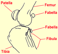
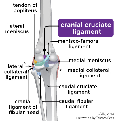
The knee is a fairly complicated joint. It consists of the femur above, the tibia below, the kneecap (patella) in front, and the bean-like fabellae behind. Chunks of cartilage called the medial and lateral menisci fit between the femur and tibia and offer some cushion-like support. An assortment of ligaments holds everything together, allowing the knee to bend properly.

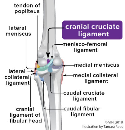
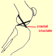
There are two cruciate ligaments that cross inside the knee joint: the anterior (or, more correctly in animals, cranial) cruciate and the posterior (in animals, the caudal) cruciate. They connect from one side of the femur on top to the opposite side of the tibia on the bottom, the two ligaments forming an X (hence the name cruciate) inside the knee joint. They are named for their attachment site on the tibia (the cranial cruciate attaches to the front of the tibia and the caudal cruciate attaches to the back of the tibia).

This may be hard to visualize based on the description but the illustration above shows the orientation of the two crossing ligaments effectively. The anterior/cranial cruciate ligament prevents the tibia from slipping forward out from under the femur.

Finding the Rupture

The ruptured cruciate ligament is the most common knee injury of dogs; in fact, chances are that any dog with sudden rear leg lameness has a ruptured anterior cruciate ligament rather than something else. The history usually involves a rear leg suddenly so sore that the dog can hardly bear weight on it. If left alone, it will appear to improve over the course of a week or two but the knee will be notably swollen and arthritis will set in quickly. Dogs are often seen by the veterinarian in either the acute stage shortly after the injury or in the chronic stage weeks or months later.

The key to the diagnosis of the ruptured cruciate ligament is the demonstration of an abnormal knee motion called a drawer sign. It is not possible for a normal knee to show this sign.

The Drawer Sign

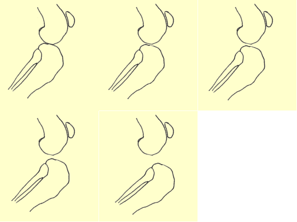
The veterinarian stabilizes the position of the femur with one hand and manipulates the tibia with the other hand. If the tibia moves forward (like a drawer being opened), the cruciate ligament is ruptured. (See a video of this manipulation.)

Another method is the tibial compression test where the veterinarian stabilizes the femur with one hand and flexes the ankle with the other hand. If the ligament is ruptured, again the tibia moves abnormally forward.

If the rupture occurred some time ago, there will be swelling on side of the knee joint that faces the other leg. This is called a medial buttress and is a sign that arthritis is well along.

It is not unusual for animals to be tense or frightened at the vet’s office. Tense muscles can temporarily stabilize the knee, preventing your veterinarian from being able to try the drawer sign during an examination. Often, sedation is needed to get a good evaluation of the knee. This is especially true with larger dogs. Eliciting a drawer sign can be difficult if the ligament is only partially ruptured so a second opinion may be a good idea if the initial examination is inconclusive.

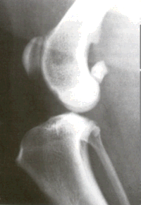
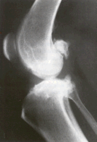
Since arthritis can set in relatively quickly after a cruciate ligament rupture, radiographs to assess arthritis are helpful. Another reason for radiographs is that occasionally when the cruciate ligament tears, a piece of bone where the ligament attaches to the tibia breaks off as well. This will require surgical repair and the surgeon will need to know about it before beginning surgery. Arthritis present prior to surgery limits the extent of the recovery after surgery though surgery is still needed to slow or even curtail further arthritis development.

How Rupture Happens

Several clinical pictures are seen with ruptured cruciate ligaments. One is a young athletic dog playing roughly who takes a bad step and injures the knee. This is usually a sudden lameness in a young large-breed dog.

A recent study identified the following breeds as being particularly at risk for this phenomenon: Labrador retriever, Golden retriever, Rottweiler, Neapolitan mastiff, Newfoundland, Akita, St. Bernard, Chesapeake Bay retriever, and American Staffordshire terrier.

On the other hand, an older large dog, especially if overweight, can have weakened ligaments and slowly stretch or partially tear them. A partial rupture may be detected or the problem may not become apparent until the ligament breaks completely. In this type of patient, stepping down off the bed or a small jump can be all it takes to break the ligament. The lameness may be acute but have features of more chronic joint disease or the lameness may simply be a more gradual/chronic problem.

Larger, overweight dogs that rupture one cruciate ligament frequently rupture the other one within a year’s time.

An owner should be prepared for another surgery in this time frame.

The cranial cruciate rupture is not limited to large breed dogs. Small dogs can certainly rupture their ligaments as well and, while arthritis is slower to set in when the patient is not as heavy, there is an association with cruciate rupture and medial luxating patella that is very common in small breed dogs. With the patellar luxation, the kneecap flips in and out of the patellar groove. If the condition is relatively mild, it may not require surgical correction but it does stress the cranial cruciate ligament and can predispose it to rupture and need to correct both conditions surgically.

What Happens if the Cruciate Rupture is Not Surgically Repaired

Without an intact cruciate ligament, the knee is unstable. Wear between the bones and meniscal cartilage becomes abnormal and the joint begins to develop degenerative changes. Bone spurs called osteophytes develop resulting in chronic pain and loss of joint motion. This process can be arrested or slowed by surgery but cannot be reversed.

Osteophytes are evident as soon as 1 to 3 weeks after the rupture in some patients. This kind of joint disease is substantially more difficult for a large breed dog to bear, though all dogs will ultimately show degenerative changes. Typically, after several weeks from the time of the acute injury, the dog may appear to get better but is not likely to become permanently normal.

In one study, a group of dogs was studied for 6 months after cruciate rupture. At the end of 6 months, 85% of dogs less than 30 pounds of body weight had regained near normal or improved function while only 19% of dogs over 30 pounds had regained near normal function. Both groups of dogs required at least 4 months to show maximum improvement.

What Happens in Surgical Repair?

There are three different surgical repair techniques commonly used today. Every surgeon will have their own preference for which technique is best for a given patient’s situation.

Extracapsular Repair

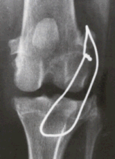
This procedure represents the traditional surgical repair for the cruciate rupture. It can be performed without specialized equipment and is far less invasive than the newer procedures described below. First, the knee joint is opened and inspected. The torn or partly torn cruciate ligament is removed. Any bone spurs of significant size are bitten away with an instrument called a rongeur. If the meniscus is torn, the damaged portion is removed. A large, strong suture is passed around the fabella behind the knee and through a hole drilled in the front of the tibia. This tightens the joint to prevent drawer motion, effectively taking over the job of the cruciate ligament.

  • Typically, the dog may carry the leg up for a good two weeks after surgery but will increase knee use over the next 2 months eventually returning to normal.
  • Typically, the dog will require 8 to 12 weeks of exercise restriction after surgery (no running, outside on a leash only including the backyard).
  • The suture placed will break 2 to 12 months after surgery and the dog’s own healed tissue will hold the knee.

Tibial Plateau Leveling Osteotomy (TPLO)

This procedure uses a fresh approach to the biomechanics of the knee joint and was developed with larger breed dogs in mind. The idea is to change the angle at which the femur bears weight on the flat “plateau” of the tibia. The tibia is cut and rotated in such a way that the natural weight-bearing of the dog actually stabilizes the knee joint. As before the knee joint still must be opened and the damaged meniscus removed. The cruciate ligament remnants may or may not be removed depending on the degree of damage.
This surgery is complex and involves special training in this specific technique. Many radiographs are necessary to calculate the angle of the osteotomy (the cut in the tibia). This procedure typically costs substantially more than extracapsular repair as it is more invasive to the joint.

  • Typically, most dogs are touching their toes to the ground by 10 days after surgery although it can take up to 3 weeks.
  • As with other techniques, 8-12 weeks of exercise restriction are needed.
  • Full function is generally achieved 3 to 4 months after surgery and the dog may return to normal activity.

Tibial Tuberosity Advancement (TTA)

The TTA similarly uses the biomechanics of the knee to create stability though this procedure changes the angle of the patellar ligament. This is done by cutting and repositioning the tibial crest where the patellar ligament attaches and implanting a titanium or steel “cage,” “fork,” and plate as well as bone grafts to stabilize the new angle. Like the TPLO, bone is cut, and special equipment is needed including metal implant plates. Similar recoveries are seen relative to the TPLO.

Which Procedure is Better?

The TTA and TPLO are much more invasive, much more expensive, and require special equipment and specially trained personnel. They have a greater potential for complications. Are they worth it? For dogs under 45 lbs, it is generally accepted that there is no clear advantage of the newer procedures over extracapsular repair. For larger dogs, there is great controversy. For all the theories behind TPLO and TTA, results in one year post-operative seem to be the same regardless of which of the three procedures the dog had performed. There is some evidence that recovery to normal function may be faster with the newer procedures. Controversy continues and there are strong opinions favoring each of the three procedures. Discuss options with your veterinarian in order to decide.

General Rehabilitation after Surgery

Rehabilitation following the extracapsular repair method can begin as soon as the pet goes home. The area can be chilled with a padded ice pack for 10 minutes a couple of times daily. (Do not try to make up for a skipped treatment by icing the area longer; prolonged cold exposure can cause injury.) Passive range of motion exercise where the knee is gently flexed and extended can also help. It is important not to induce pain when moving the limb. Let the patient guide you. Avoid twisting the leg. After the stitches or staples are out (or after the skin has healed in about 10 to 14 days), water treadmill exercises can be used if a facility is available. This requires strict observation and, if possible, the patient should wear a life jacket. Rehabilitation for patients with the intracapsular repair is similar but slower in progression.

Rehabilitation after TPLO or TTA is gentler. Icing as above and rest are the main modes of therapy. After 3 to 4 weeks, an increase in light activity can be introduced. A water treadmill is helpful. No jumping, running or stair-climbing is allowed at first. Expect the osteotomy site to require a good 6 weeks to heal.

What if the Rupture Isn’t Discovered for Years and Joint Disease is Already Advanced?

A dog with arthritis pain from an old cruciate rupture may still benefit from a TPLO surgery and possibly from the TTA. Ask your veterinarian if it may be worth having a surgery specialist take a look at the knee. Most cases must make do with medical management. Visit our section on arthritis treatment.

Meniscal Injury

We mentioned the menisci as part of the knee joint. The bones of all joints are capped with cartilage so as provide a slippery surface where the bones contact each other (if the bones contact each other without cartilage, they grind each other down). In addition to these cartilage caps, the stifle joint has two blocks of cartilage in between the bones. These blocks are called the menisci and serve to distribute approximately 65% of the compressive load delivered to the knee. The only other joint with a meniscus is the jaw (temporomandibular joint).

When the cruciate ligament ruptures, the medial (on the inner side of the knee) meniscus frequently tears and must either be removed, partly removed, or ideally repaired. This is generally done at the time of cruciate ligament surgery and we would be remiss not to mention it.

Pets with meniscal damage may have an audible clicking sound when they walk or when the knee is examined, but for a definitive diagnosis, the menisci must actually be inspected during surgery. It is difficult to access the menisci and thus repairing a tear in the meniscus is problematic; furthermore, the poor blood supply to the menisci also makes good healing less likely. For these reasons, removal of the damaged portion of the meniscus is the most common surgical choice. This leaves some meniscus behind to distribute the compression load on the knee but removes the painful, ineffective portion.

Areas of current research include techniques to improve blood supply to the healing meniscus so that repair can be more feasible. If meniscal damage has occurred in a cruciate rupture, arthritis is inevitable and surgery should be considered a palliative procedure.

Enhancing Recovery after Surgery

Confinement

Enhancing recovery post-operatively is largely about strict confinement early. This cannot be over-emphasized. Be prepared to crate your dog or employ a pen such as a child’s playpen depending on the dog’s size. A corral of sorts can be constructed with boxes and a baby gate. Be sure you understand the instructions with regard to a gradual return to exercise over several months.

Adequan Injections

A series of Adequan injections can help with joint inflammation as well as lubrication. Typically injections are given twice a week for a total of eight injections.

Glucosamine

Oral joint supplements such as glucosamine contain cartilage building blocks to help the body repair cartilage damage. This is an excellent time to begin supplementation and there are numerous brands.

Weight Management

Overweight dogs have an increased risk for arthritis and cruciate rupture. A weight management program can reduce the potential for arthritis and can reduce the risk of rupture of the opposite cruciate ligament. If your dog is overweight, ask your vet about a weight management plan that might be started during the recovery period.

Professional Rehab/Physical Therapy

Nothing compares to professional rehab for a return to function. If you are lucky enough to have such a facility in your area, consider utilizing their services. A list of home exercises may be obtained and/or the dog can visit weekly or a few times weekly for exercise and treatment. Some facilities allow the dog to board and have daily treatment. Ask your vet about this option.

In Summary

  1. In a dog’s knee joint there are two cruciate ligaments that cross, forming an X shape. When either ligament is not intact, the knee is unstable and painful. Usually the ligament at the front ruptures.
  2. Two types of patients are usually seen. The most common is an adult dog that has had mild lameness that suddenly increases in severity. Sometimes the patients are overweight but not always. It is thought that the ligament has degenerated over time. In these cases, approximately 50% will have the same problem in the opposite knee. Less commonly we see patients who have been involved in some sort of accident that has caused an acute rupture.
  3. If left alone, it will appear to improve over a week or two but the knee will be notably swollen and arthritis will set in quickly. More importantly, lameness and pain will persist.
  4. Three different surgical repair techniques are commonly used today: extracapsular repair; tibial plateau leveling osteotomy (TPLO); and tibial tuberosity advancement (TTA).
  5. The extracapsular repair involves using an artificial ligament replacement. TTA and TPLO are much more invasive and require metal plates and screws. These are more expensive as they require specialized equipment and personnel and can have more complications. For dogs under 45 lbs, there is no clear advantage to the newer procedures. For larger dogs, the choice of which procedure is best is controversial.
  6. Extracapsular repair: The ruptured ligament is removed and sutures are put through a hole drilled in the tibia; the dog needs 8 to 12 weeks of exercise restriction (outside on a leash only, including the backyard); the suture will break 2 to 12 months after surgery and the dog’s own healed tissue will stabilize the knee.
  7. TPLO: The tibia is cut and rotated; most dogs are touching their toes to the ground by 10-21 days after surgery; 8-12 weeks of being outside on a leash only including the back yard; full function normally returns within 3 to 4 months.
  8. TTA: The tibia is cut and repositioned, and titanium, steel, and bone grafts stabilize the new angle of the joint. Most dogs are touching their toes to the ground by 10-21 days after surgery; 8-12 weeks of being outside on a leash only including the backyard; full function normally returns within 3 to 4 months.
  9. After surgery, patients need strict confinement (this cannot be overemphasized), medications, and weight loss if needed. Water treadmills and other physical therapies are helpful. Rehabilitation is important no matter which method is used.
  10. Results a year after surgery seem to be the same regardless of which procedure was used. Osteoarthritis will develop in all cases and will cause a degree of lameness.
4128139

Roundworms in Dogs and Puppies

Toxocara Canis and Toxascaris Leonina: Roundworms of Dogs and Puppies

There are two species of roundworms affecting dogs and puppies: Toxocara canis and Toxascaris leonina. Both are treated with the same medication protocol, so when eggs are seen on a fecal flotation exam, it may not be necessary to determine which species is present. 

While T. canis infects only dogs, T. leonina is also able to infect cats and kittens. Treatment protocols for both worm species are the same. Still, if there are feline household members, it may be useful to specifically identify the roundworm species involved so as to determine which pets are potentially at risk. We will cover each species of roundworm separately, even though treatment is the same for each, their biology is different.

Fresh feces are not infectious. Fresh feces do not contain the infectious stage of T. Canis. Worm eggs require 30 days to become infectious, meaning it is contaminated dirt that is infectious to people and animals.

Toxocara Canis: How Infection Occurs

T. canis is the most common roundworm of the domestic dog, and it is not able to infect cats. Its presence can go completely without symptoms, although more likely it is going to create some degree of diarrhea and possibly vomiting or general unthriftiness in its canine host.

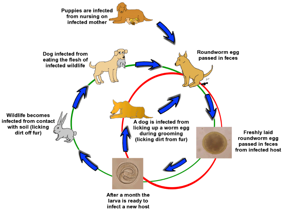
Its life cycle is somewhat complicated, as we are about to see.

  1. Consuming infective worm eggs from soil in the environment (generally through normal grooming//self-licking).
  2. Nursing from an infected mother dog.
  3. Consuming a prey animal (usually a rodent) that is carrying developing worms.
  4. During embryonic development when an infected mother dog is pregnant (most puppies are infected this way).

Cats cannot be infected with Toxocara canis but humans can.

Life As A Roundworm

Toxocara Canis has one of the most amazing life cycles in the animal kingdom. It is helpful to understand this life cycle if effective treatment and prevention are to be pursued. It gets complicated so we’ll start with a short synopsis.

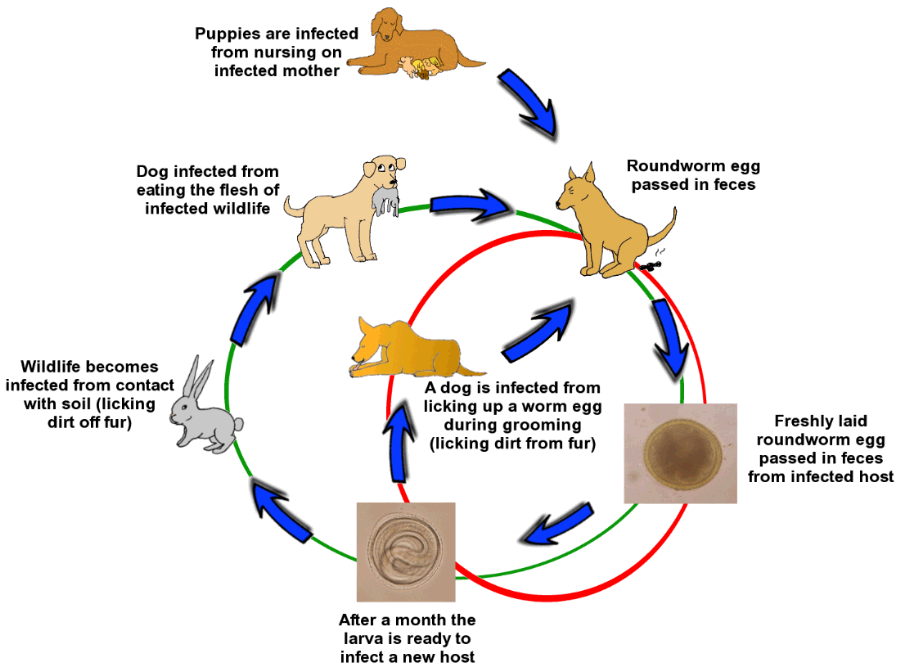
It all starts with an infected dog pooping on the ground, with an infected pregnant dog, or with an infected prey animal. In each of these scenarios, an uninfected dog or puppy comes into contact with a roundworm larva that has developed to its infectious stage. The uninfected dog becomes infected by licking contaminated dirt from its feet, from eating an infected prey animal, from developing in its infected mother’s womb, or from nursing its infected mother’s milk. The young roundworm migrates through the new host’s body, ultimately settling in the intestine where it mates and new eggs are produced. The cycle begins again. 

The biggest “take-home” points are these:  

  1. Fresh feces on the ground are not infectious, at least with regard to Toxocara, because the worm egg out in the world needs a good month to develop to an infectious stage. 
  2. Because the whole body migration drops worms into the intestinal tract over time, one deworming is not enough. Be sure not to skip the second (or even third) treatment. Even better, use a monthly parasite control product that includes a dewormer.
  3. Puppies can be assumed to be infected with roundworms when they are born. 
  4. Worm eggs can live months to years in contaminated soil.

Step One: Eggs Contaminate Environmental Soil

Toxocara eggs are passed in the host’s feces, where they can be detected if a fecal sample is tested. Feces and any worms’ eggs therein are deposited on the ground, where they are rained on, dried by the sun, stepped on, etc. The worms are developing during this time and are not infectious to new hosts until they have developed for about a month.

By that time, the original feces has long since melted away into the ground and is no longer evident. It is the dirt that contains infectious eggs. Toxocara eggs are famous for weathering harsh environmental conditions. Eggs can remain infective for months to years.

Fresh feces are not infectious. Soil contaminated with feces is infectious.

Step Two: A Host Eats an Egg and the Larva Encysts

The egg containing what is called a second-stage larva is picked up from the dirt by a dog or by some other animal, usually in the course of normal self-grooming. The egg hatches in the new host’s intestinal tract and the young worm burrows its way out of the intestinal tract to encyst in the host’s other body tissues. If the new host is a dog, the life cycle proceeds. If the new host is a member of another species, the larvae wait encysted until the new host is eaten by a dog.

Step Three: The Larva Awakens and Migrates Through the Host

These second-stage larvae can remain encysted happily for years. If the host is a puppy under age 6 months of age, the larvae mostly encyst in the host’s liver. In older dogs, the larvae encyst all over the body. When the time comes to move on, the larvae excyst and migrate to the host’s lungs where they develop into third-stage larvae. They burrow into the small airways and travel upward toward the host’s throat. A heavy infection can produce serious pneumonia.  When they get to the upper airways, their presence generates coughing. The worms are coughed up into the host’s throat where they are swallowed thus entering the intestinal tract for the second time in their development.

If the host is pregnant, the larvae do not migrate to the lung after they excyst; instead, they home to the uterus and infect the unborn puppies. The second-stage larvae make their way to the puppies’ lungs to develop into third-stage larvae.

If the host is a nursing mother, she secretes third-stage larvae in her milk for the first 3 weeks after giving birth. These larvae simply find themselves in the puppy’s intestinal tract where, at this stage, they do not need to migrate but can settle in and begin mating. Puppies can be infected by drinking their mother’s milk, though, due to the intrauterine cycle described above, the litter would probably already be infected.

Note: When dogs are dewormed with traditional dewormers, this affects only worms in the intestinal tract. It does not affect encysted larvae. It is difficult to prevent mother-to-puppy transmission, and routine deworming is not adequate. It is possible to prevent infection in unborn puppies by using a specific daily protocol of fenbendazole (your veterinarian can provide details) or selamectin (Revolution®).

Step Four: Finally Back in the Intestine and Ready to Settle Down

Once back in the intestine, the larvae complete their maturation and begin to mate. The first eggs are laid about one week after the larvae have arrived in the intestine and finished molting into their adult stages (about 4 to 5 weeks after the infection has first occurred). From here, the cycle repeats.

Why Is Infection Bad?

Roundworm infection can have numerous negative effects. It is a common cause of diarrhea in young animals and can cause vomiting as well. Sometimes the worms themselves are vomited up which can be alarming as they can be quite large with females reaching lengths of up to seven inches. The worms consume the host’s food and can lead to unthriftiness and a classical “pot-bellied” appearance. Very heavy infections can lead to pneumonia as the worms migrate and if there are enough worms, the intestine can actually become obstructed.

It should also be noted that human infection by this parasite is especially serious (see below). It is important to minimize the contamination of environmental soil with the feces of infected animals so as to reduce the exposure hazard to humans and other animals. In other words, dog feces should be removed and discarded promptly before worm eggs permanently contaminate the local dirt.

How Do We Know If Our Dog Is Infected?

Of course, there are ways to find out if your dog is infected. If a dog or puppy vomits up a worm, there is a good chance this is a roundworm (especially in a puppy). Roundworms are long, white, and described as looking like spaghetti. Tapeworms can also be vomited up, but these are flat and obviously segmented. If you are not sure what type of worm you are seeing, bring it to your veterinarian’s office for identification. You may not know if your dog is infected, and this is one of the arguments in favor of regular deworming. Regular deworming is especially recommended for dogs that hunt and might consume the flesh of hosts carrying worm larvae. Puppies are frequently simply assumed to be infected and automatically dewormed.

Fecal testing for worm eggs is a must for puppies and a good idea for adult dogs having their annual checkup. Obviously, if there are worms present, they must be laying eggs in order to be detected (and there are many reasons why they might not be laying eggs) but, by and large, fecal testing is a reliable method of detection.

How Do We Get Rid Of Roundworms?

Numerous deworming products are effective. Some are over-the-counter, and some are prescription. Many flea control and/or heartworm prevention products provide a monthly deworming that is especially helpful in minimizing environmental contamination.

Common active ingredients include:

  1. Febantel (the active ingredient in Drontal and Drontal Plus)
  2. Fenbendazole (the active ingredient in Panacur)
  3. Milbemycin oxime (the active ingredient in Interceptor, Sentinel, and Trifexis)
  4. Moxidectin (the active ingredient in AdvantageMulti, Coraxis, and Simparica Trio).
  5. Piperazine (the active ingredient in many over-the-counter products)
  6. Pyrantel pamoate (the active ingredient in Strongid, Nemex, Heartgard Plus, and others)
  7. Selamectin (the active ingredient in Revolution. Although not included on the US product label, recent studies show effectiveness in the prevention and treatment of canine roundworms.)

There are two important concepts to keep in mind about deworming. Medications essentially anesthetize the worm so that it lets go of its grip on the host’s intestine and passes out with the stool. Once it has been passed, it cannot survive in the environment and dies.

This means that you will likely see the worms when they pass, so be prepared as they can be quite long and may still be alive and moving when you see them.

The other concept stems from the fact that all the larvae in migration cannot be killed by any of these products. After the worms are cleared from the intestine, they will be replaced by new worms completing their migration. This means that a second and sometimes even a third deworming is needed to keep the intestine clear. The follow-up deworming is generally given several weeks following the first deworming to allow for migrating worms to arrive in the intestine, where they are vulnerable.

Do not forget your follow-up deworming.

Toxascaris Leonina

The life cycle of Toxascaris leonina is not nearly as complicated. T. leonina does not migrate through the body in the way that Toxocara canis does. Instead, the fresh egg is passed by the host in feces, develops into an infectious embryo in the environment, and is swallowed by the new host. The Toxascaris egg develops much faster in the environment than the Toxocara egg and can be infectious for its new host as soon as one week from the time it was passed. Once inside the host, however, Toxascaris development becomes slower. The young worm lives in the host intestine without migrating through the body and becomes a mature worm in 2-3 months. Like ToxocaraToxascaris can be picked up by wildlife, and the canine or feline host can be infected through hunting and consuming prey. The same dewormers listed above can be used on Toxascaris and must be repeated similarly.

Note: Toxascaris leonina can infect both dogs and cats alike. Unlike the Toxocara situation, unborn puppies cannot be infected by Toxascaris leonina.   

The Same Rules Apply

Basically, the same products listed above will kill Toxascaris leonina. As with Toxocara, removing feces from the environment promptly will greatly minimize contamination and the potential for new infections. Regular use of deworming products is preventive. Toxascaris leonina tends not to produce diarrhea and disease as badly as Toxocara, but we still want to get rid of Toxascaris anyway.

7497535

Rabies in Animals

Descriptions of rabies go back thousands of years as rabies has classically been one of the most feared infections of all time. It is because of this fear that dog ownership requires a license and documentation of rabies vaccination. There is still no reliably effective treatment for rabies infection once symptoms have started.

The virus does not survive outside of the infected host very long, so direct contact with fresh secretions is required for transmission.

In most cases, the disease is transmitted via a bite wound and it is mostly wildlife that gets infected. When wildlife starts coming into contact with the pet population, then pets become infected and when pets get infected, human infection is not far behind. Because this infection has nearly 100% mortality, preventing it in pets becomes crucial to preventing it in people. Of course, people can get infected by wildlife contact as well. Despite vaccination being readily available, every year the U.S. reports approximately 50 canine deaths, 250 feline deaths, and several human deaths from rabies. Worldwide, some 55,000 human deaths occur annually from rabies even now in the 21st century.

The most common wildlife species to spread rabies to domestic animals and humans in the Northern Hemisphere are skunks, bats, raccoons, foxes, and coyotes. Wildlife (especially bats) are able to gain access to indoor areas where they can come in close contact with both pets and people. Indoor pets are not without risk.

Many people do not realize how fast death occurs from rabies.

While it may take a long time for the virus to incubate, once even mild symptoms begin, death occurs within 10 days.

The injection treatment most people have heard of only works during the incubation period; once symptoms have started, survival chances are slim.

The virus in the infected animal’s saliva enters the victim’s tissues during the bite. The virus attaches to the local muscle cells for a couple of days before penetrating local nerves and beginning its slow ascent to the brain. Once within nervous tissue, the virus is not accessible to the immune system and may safely proceed toward the brain. The journey is slow and can take up to a year but the average time is three to eight weeks depending on the species. Virus ultimately reaches the brain and in two to three days more is evident in all body secretions, including saliva. At this point, the disease becomes transmissible and symptoms begin.

It can take up to a year from the time of the initial bite before symptoms begin to show. In dogs, typical incubation periods are 21-80 days. In cats, it’s 28-42 days. Once symptoms show, treatment is nearly impossible.

Prodromal Stage (The First 2-3 Days After Symptoms Have Started)

A change in personality is noted. Friendly animals become shy, etc. The larynx begins to spasm and a voice change may be noted (especially true in rabid cattle). Most infected animals will actively lick or scratch the site of the original bite.

Excitative Stage (Next 1-7 Days)

Classically, this would be the “mad dog” stage, though, in reality, most animals skip this phase altogether. The animal has no fear and suffers from hallucinations. If confined, the animal often attacks the bars of the cage.

Paralytic or Dumb Stage (Next 2-4 Days)

Weakness/paralysis sets in. The larynx is paralyzed, resulting in an inability to swallow, thus drooling and “foaming at the mouth” result. The animal dies when the intercostal muscles that control breathing are paralyzed. It is from animals in this stage where most human exposure occurs. There is no treatment for animals or humans once clinical signs appear.

Once the virus has been released to body secretions, it is again accessible to the immune system; however, the patient dies before an adequate immune response is mounted.

The classical symptoms of rabies described above may not be obviously recognizable, making diagnosis difficult if not impossible in a living animal. Long quarantines are often needed to determine if the infection has occurred.

When human exposure to the animal in question is involved, what happens depends on an assortment of criteria. If the animal in question is dead, its brain can be tested for rabies. There is no test for rabies in a living animal but since we know that death follows quickly after the virus becomes contagious, a living animal can be confined for 10 days. If the animal is still alive 10 days after biting a person, then the bite could not have transmitted rabies.

Prevention

For Our Pets

Happily, rabies prevention is accomplished with vaccination and limiting exposure to wildlife. The standard killed virus vaccines are available for both dogs and cats and, after the initial dose which is good for one year, subsequent doses are generally good for three years. Because tumors have developed in cats who received the killed virus vaccine, a recombinant product is now available that uses a portion of rabies viral DNA cloned into a harmless canarypox virus. This arrangement allows the pet to realize the benefits of live virus vaccination without any risk of exposure to the living rabies virus.

Rabies vaccination protocols are typically controlled by municipal regulations. Most communities legally require the vaccination of all dogs. The American Association of Feline Practitioners recommends rabies vaccination as a core vaccine for all pet cats.

For pets not current on rabies vaccination that have been exposed to biting wildlife, the Texas Post-Exposure Rabies Prophylaxis Protocol has been particularly helpful. In this situation, the pet should be vaccinated for rabies as soon as possible after the wildlife bite with booster vaccines given three weeks post-bite and eight weeks post-bite. The pet should be strictly isolated for 90 days. This protocol has been extremely successful in preventing rabies symptoms and contagion when normal rabies vaccination had lapsed. 

Treatment for Humans (Post Exposure Prophylaxis or PEP)

Treatment for Humans (Post Exposure Prophylaxis or PEP)

As mentioned, once symptoms have started, treatment can be attempted but truly there is little chance of survival. The only way to survive rabies is to exploit the long incubation period and basically get vaccinated quickly. Doing this involves a very big piece of information: knowing you were (or may have been) exposed. 

Every year a small number of people die of rabies in the U.S. Most of these people were bitten by a dog, bat, or some other animal and did nothing other than basic first aid. Recall that it takes many weeks for the rabies virus to reach the brain. This allows time for the vaccine to be given so that when the virus “comes out” of its neurologic hiding place and is ready to infect the brain, a substantial immunologic response is waiting. The virus is foiled and clinical rabies does not result.

If you are bitten by an animal and its rabies vaccination history is unknown, there are definite steps to take. A fresh bite wound should be washed out with water quickly as this may wash out viral particles. The time it takes for the virus to reach the brain depends on the amount of virus in addition to how close the wound is to the head. This simple step can be life-saving.

If the biting animal is alive, its vaccination status should be confirmed as soon as possible and it will need to be confined according to local law. The bite wound should be reported to the health department as soon as possible. Only rodent and rabbit bites are not reportable.

If the animal is dead then its brain can be tested for rabies. The head of the biting animal is submitted to the health department for fluorescent antibody testing for the rabies virus. This process takes a matter of hours so that any bite victims can know right away if they will require rabies treatment.

If the animal is not available or its vaccination status is in question, further treatment may be needed. Hyperimmune (antibody-rich) serum is flushed into the wound in hope of inactivating the virus before it may penetrate to the nerves. The patient receives a vaccination on a regular schedule for about a month, as described above.

Veterinarians have a rabies exposure risk of more than 300 times that of the general population. 

Anyone pursuing a career with animals should consider vaccination against rabies.

The Law Regarding Animal Bites (Against Humans)

In the U.S., states and counties have different regulations regarding rabies vaccines for companion animals and bites to humans. Regulations for your area in the U.S. can be found through your county’s health department. In almost all states in the U.S., the biting animal must be confined for observation for 10 days at the owner’s expense regardless of vaccination status.

The purpose of the quarantine period is to determine if the rabies virus could have been in the animal’s saliva at the time of the bite. An animal infected with rabies and shedding virus will certainly be dead within ten days.

If the biting animal is known to have been exposed to wildlife, the situation is different. A vaccinated animal must be re-vaccinated within the time period required by local regulations and confined for observation for the specified duration. Unvaccinated animals may be confined for a specified time period according to local/regional law, or be euthanized and tested for rabies. In the U.S., all bites to humans that break the skin are reportable to local health departments. Rabies vaccination requirements vary from state to state, and country to country.  Contact your veterinarian if you think your pet has come into contact with or been bitten by a wild animal. Regardless of the vaccination status of your pet, wild animal bites may lead to painful wounds and infection.

Again, laws regarding biting dogs and rabies vaccination are highly regional. Check with your local animal regulation department, your veterinarian, or visit rabiesaware.org to find out what you need to know in the U.S..

Quarantines when Traveling

Great Britain, Hawaii, Japan, New Zealand, and several other island areas have successfully eradicated rabies from their territory. These places are EXTREMELY cautious about allowing in potential carriers of rabies. Because of the long incubation period of rabies, a very long quarantine is needed; however, this must be balanced by the expense associated with quarantine and owners’ reluctance to be separated from their pets. Most places that have eradicated rabies have protocols for avoiding or minimizing quarantine. Typically, a microchip is implanted in the pet for identification purposes, a rabies antibody titer (a measurement of vaccine-induced protection) must be performed at an approved laboratory, and rabies vaccine documentation is necessary.

For listings of what each state requires for entry, go to the USDA. 

For travel to another country check with that country’s consulate, but guidelines are also available at USDA.

Other Links

The CDC’s rabies home page has, in addition to basic information, a children’s education area that is particularly helpful for families who go camping.

The annual World Rabies Day calls attention to this problem to raise awareness and provides information about rabies in both humans and animals.

In Summary:

You can’t treat rabies once symptoms set in; all you can do is prevent it.

Rabies is a virus transmitted through saliva or brain tissue. Without rapid preventive treatment, it is almost always fatal. That’s why your pets are required to have rabies vaccines in many areas of the world.

The most common wildlife species to spread rabies to animals and people in the Northern Hemisphere are bats, skunks, raccoons, foxes, and coyotes. 

It is generally caused by a bite wound, and mostly wildlife gets infected.

Rabies affects a mammal’s central nervous system. It can take up to a year before symptoms begin to show, but the average time is 3-8 weeks depending on the species. In dogs, the typical incubation period is 21-80 days. In cats, it’s 28-42 days. Once symptoms begin, treatment is nearly impossible.

If you are bitten, you need a preventive injection immediately as it only works during the incubation period. Once even mild symptoms begin, death occurs within 10 days.

After symptoms have started:

  •  First 2-3 days: a personality change is seen, and the larynx spasms, resulting in a voice change.
  •  Next 1-7 days: this is the “mad dog” stage in which an animal has no fear and suffers from hallucinations, but many skip this stage.
  •  Next 2-4 days: weakness and paralysis set in. The larynx becomes paralyzed so the animal cannot swallow, causing drooling and foaming at the mouth.
  •  The animal dies when the muscles that control breathing finally become completely paralyzed. Most people get bitten in this stage.

Symptoms may not be obviously recognizable, making diagnosis difficult if not impossible. There is no diagnostic test for rabies in a living animal.

What happens after people are exposed depends on the situation. If the animal is dead, its head is sent to a laboratory so the brain tissue can be tested. In some circumstances/locations, a living animal can be quarantined for 10 days at the owner’s expense; if the animal is still alive, then the bite could not have transmitted rabies.

The vaccine protocols are typically controlled by municipal regulations. Most communities legally require all dogs to be vaccinated and recommend it for cats. Vaccination requirements vary from state to state, and country to country. 

Exposed pets not current on rabies vaccination should be given booster vaccines at 3 and 8 weeks post-bite. The pet should be strictly isolated for 90 days.

Exposed humans should wash the bite with water as it may wash out some virus particles; the time it takes for the virus to reach the brain depends on the amount of virus in addition to how close the wound is to the head.  This step could save a life.

Animal bites need to be reported to local health departments.

Veterinarians have a rabies exposure risk of more than 300 times that of the general population. Anyone in an animal-oriented job should consider preventive vaccination.

4128949

Runny Eyes (Epiphora) in Dogs

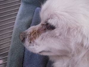
Watery ocular (eye) discharge is a common concern of pet owners. Excessive tears drain down the pet’s face, and in time, chronic exposure to moisture produces skin irritation, infection, and odor.  The fur becomes stained reddish brown due to tear pigments called porphyrins, an especially non-cosmetically appealing problem in white-furred pets. The medical term for this condition is epiphora, and it is one of the most difficult conditions to resolve in all veterinary ophthalmology.

There are two causes of epiphora (excess tearing) in pets: irritation to the eye and abnormal tear drainage.

Irritation to the Eye

One of the eye’s natural responses to irritation is to produce more tears. This helps flush away any irritants that might have contaminated the eye surface. If excessive tearing is accompanied by squinting or pawing of the eye, or if the excessive tearing should occur suddenly, this should be taken as an obvious sign of eye pain, and veterinary attention should be sought at once. Any accompanying loss of vision is also an emergency.

It is the more chronic cases that are harder to manage. Eyes can certainly become chronically irritated from viral conjunctivitis (probably the most common reason for excessive tearing in cats), glaucoma (painful increase in eye pressure in the eye), reaction to certain eye medications, or, more commonly, from eyelash or eyelid abnormalities.

Some breeds of dogs naturally have hair in their eyes, and this does not cause irritation in most cases, but in some cases, it can. Hairs can grow from the face at an angle so that they rub against the eye.

Eyelashes can also grow at abnormal angles and rub on the eye. Eyelashes can even grow on the inner surface of the eyelids or corner of the eye and cause irritation. Often magnifying instruments are needed to discover these tiny hairs, and delicate surgical procedures are needed to address them. Complicating the situation, however, is the fact that breeds that tend to have eyelid and eyelash problems also tend to be the same as those with faulty tear drainage anatomy (see below), which makes it hard to determine which of many possible causes is to blame.

Allergy, irritating dust or smog in the air, trauma, or infection can lead to excessive tears from conjunctivitis.

These problems, indicated below, are treated medically.

  1. Corneal ulcer
  2. Feline upper respiratory infection
  3. Airborne allergy
  4. Brachycephalic breeds

Normal Tear Drainage

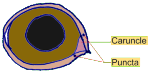
Once we are certain that there is no painful condition, we consider that the eye’s drainage ducts may not be normal. The normal eye is most efficient at draining tears. Looking at the inner corner of the eyelids (the side nearest the nose) one can note the pink, moist caruncle and on the eyelid margins, upper and lower openings called nasolacrimal punctae.

These are essentially drainage holes for tears. The punctae are the openings to small passages called canaliculi, which in turn open into the lacrimal sac.The lacrimal sac drains into the nasolacrimal duct, which drains tears into the nasal passages and throat. (This is why we get runny noses and sniffles when we cry and why we can taste our tears when we cry.)

There are many problems that can occur along this drainage route. One common problem, especially in poodles, bichon frises, and brachycephalic breeds, is simply that the eye socket is shallow. This means that tears overflow from the corner of the eye because the eyelid space there is not deep enough to contain them. These tears never make it to the punctae and instead, spill down the sides of the nose. This condition cannot be repaired; it is simply the conformation of the dog’s face.

Alternatively, the eyelids may be turned inward (a condition called entropion) blocking the puncta and preventing drainage. Another problem may be long hair acting as a wick drawing tears from the eye to the skin, especially in breeds where hairs grow on the caruncle. This hair may be kept trimmed; though, if the hair is part of the nasal skin fold of a brachycephalic breed, surgery may be needed to remove or alter the skin fold.

Old infections or injuries may cause enough scarring to close the punctae, canaliculi, or nasolacrimal ducts. Sometimes vigorous flushing of saline through the ducts, performed under general anesthesia, can re-open them. Sometimes the puncta are congenitally closed (common in poodles and cocker spaniels) and can be surgically re-opened.

It is fairly easy to determine if there is a problem with drainage in a non-invasive way which is sometimes called the Jones test. A stain for eyes called fluorescein is dropped onto the eye, and if the tear drainage system is intact, the dye should be evident at the nostrils after a couple of minutes.

Surgery to reopen drainage is a procedure that not all veterinarians are comfortable performing. Discuss with your veterinarian whether a referral to a specialist would be best for you and your pet. In this case, special equipment is needed. Ask your veterinarian for a referral if needed.

Addressing The Staining

An assortment of remedies has been suggested to resolve the unsightly reddish stains that result from chronic tear drainage. The following are some comments on suggestions we have heard.

Low Doses Of Tetracycline Or Tylosin


Variable success has been reported but the problem is that using antibiotics in this way constitutes less than responsible use of antibiotics, and resistant bacterial strains may result from this practice. It is generally frowned upon by pharmacologists. Furthermore, tetracycline should not be used in immature animals as it will cause teeth to stain.

Regular wiping of the area with hydrogen peroxide basically serves to bleach the area and requires regular use to make a difference.

Douxo Micellar Solution is also reported to cleanse away staining debris from the hair with use over time. This product is used on the fur and skin, not on the eye.

4128812

Ringworm in Dogs and Cats

What Kind of Infection is it?

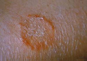
Many people are surprised to find that ringworm is not caused by a worm at all but by a fungus. The fungi involved are called dermatophytes, and the more scientifically correct name for ringworm is dermatophytosis. The dermatophyte fungi feed upon the dead cells of skin and hair, causing in people a classic round, red lesion with a ring of scale around the edges and normal recovering skin in the center. Because the ring of irritated, itchy skin looked like a worm, the infection was erroneously named. 

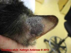
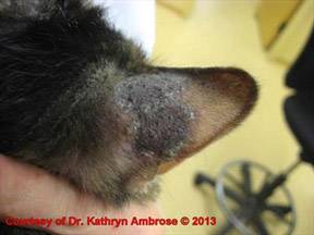
The characteristic ring appearance is primarily a human phenomenon. In animals, ringworm frequently looks like a dry, grey, scaly patch but can also mimic any other skin lesion and have any appearance. 

Where Would My Pet Pick Up This Infection?

The spores of dermatophyte fungi are extremely hardy in the environment; they can live for years. All it takes is skin contact with a spore to cause infection; however, the skin must be abraded, as the fungus cannot infect healthy, intact skin. This means that freshly shaved, scraped, or scratched skin is especially vulnerable.

Infection can come from direct contact with an infected symptomatic animal, direct contact with an asymptomatic carrier, or contact with spores in the environment. Infected symptomatic animals have skin lesions rife with fungal spores. Carriers may be infected animals who do not have obvious lesions (a common scenario towards the end of treatment), or they may be animals who are not actually infected per se but simply have spores on their hairs, just as a couch might have spores on its surface. Infection is transmitted when spores bind to abraded skin. Skin lesions typically appear one to three weeks after exposure.

There are several species of dermatophyte fungi. Different species come from different kinds of animals or even from the soil, thus, determining the ringworm species can help determine the source of the fungal infection. Predisposing factors towards infection include age (puppies and kittens are at higher risk than adult animals), lifestyle (free-roaming or hunting animals being predisposed), and local climate (pets living in warmer, more humid climates are predisposed). Immune suppression from the FIV or Feline Leukemia Virus turns out not to be a predisposing factor as one might expect, especially since immune suppression is a human risk. Still, there are two breed predispositions of note: Persian cats and Yorkshire terrier dogs. Infection rates are higher in these breeds, as are treatment failures.

Can I get This Infection?

Yes, ringworm is contagious to people; however, some people are at greater risk than others. The fungus takes advantage of skin belonging to those with reduced immune capacity. This puts young animals and children, pregnant women, elderly people and pets, those who are HIV-positive, people on chemotherapy or taking medication after transfusion or organ transplant, and highly stressed people and animals at high risk. In general, if you do not already have ringworm at the time your pet is diagnosed, you probably will not get it. Keep in mind that skin must be irritated to become infected.

How Does the Doctor Know This is Really Ringworm?

In some cases, we know for sure that the pet has dermatophyte fungi, while in other cases, we are only highly suspicious. Ringworm lesions on animal skin are rarely the classic ring-shaped as in people (in fact, in animals, lesions are often not even itchy) thus, some testing is usually necessary, as we will describe.

Wood’s Light (Fluorescence)
A Wood’s light is a lamp designed to emit light in a specific range of wavelengths. It looks like a black light but is actually entirely different. Ringworm fungi of the genus Microsporum (the most common genus in small animal ringworm cases) demonstrate a chemical reaction when they bind to hair shafts. This chemical reaction fluoresces apple green under the Wood’s light. Fungal spores will not fluoresce without infection, so an uninfected carrier will not fluoresce, nor will debris that is not attached to the hair.

There is controversy regarding what percentage of Microsporum infections will fluoresce. A commonly published statistic is that approximately 50 percent will fluoresce, but other information suggests that 100 percent of Microsporum infections will fluoresce at least at some point in their course. Fluorescence first becomes detectable five to 18 days post-infection. In many cases, using Wood’s light uncovers numerous additional skin lesions that were not visible to the naked eye.

Most veterinary hospitals are equipped with Wood’s lights and use them to screen pets for ringworm lesions. Unfortunately, fluorescence may be difficult to find, and complicating matters, many topical products and non-infectious debris will also fluoresce. Further testing is often needed.

Microscopic Examination

Your veterinarian may wish to examine some hairs for microscopic spores. This involves plucking hairs and inspecting them under a microscope. If spores can be seen on damaged hairs, then the diagnosis of ringworm is confirmed; however, as spores are difficult to see, especially in darker hair, many veterinarians skip this step.

Fungal Culture

Some hairs and skin scales are placed on a culture medium in an attempt to grow one of the ringworm fungi. The advantage of this test is that it not only can confirm ringworm but can tell exactly which species of fungus is there. Knowing the identity of the fungus

may help determine the source of infection. The disadvantage, however, is that fungi require at least 10 days to grow out. Unfortunately, false negative cultures are not unusual.

Fungal culture does not depend on a visible skin lesion. A pet with no apparent lesions can be combed over its whole body and the fur and skin that are removed can be cultured. Carrier animals are usually cats living with several other cats.

A specific growth-medium, called dermatophyte test medium, is commonly employed to distinguish ringworm fungi from other fungi. Ringworm fungi classically produce a white fluffy colony and will turn the orange growth medium red within two to 14 days. When the colony is mature, the material can be harvested from it and examined under the microscope for ringworm spores.

PCR Testing

The newest diagnostic method involves testing hairs for dermatophyte fungus DNA. The benefit is that it is much faster than the culture but is still able to confirm the infection as well as determine the species of ringworm fungus involved. This makes PCR testing an excellent way to make the diagnosis of ringworm initially but can pose a problem in determining the end of treatment. The downside of PCR testing is that it tests for fungal DNA, not for live viable fungi. When the pet is first diagnosed, if there is fungal DNA  on a skin lesion, we can assume the fungus is causing infection. After treatment, however, the fungus is killed or damaged to the point of being harmless, but its DNA will still be there, creating a positive PCR test. For this reason, PCR is best used for detecting fungus in an untreated patient, but culture is probably best at determining when treatment can be discontinued.

Biopsy

Sometimes the lesions on the skin are so uncharacteristic that a skin biopsy is necessary to obtain a diagnosis. Fungal spores are quite clear in these samples, and the diagnosis may be ruled in or out. Depending on the outcome of preliminary tests, your veterinarian may begin ringworm treatment right away or postpone it until after more definitive results are available.

Treatment

Commitment is the key to success, especially if you have more than one pet. Infected animals are constantly shedding spores into the environment (your house) thus disinfection is just as important as treatment of the affected pet. The infected pet will require isolation while the environment is disinfected and should not be allowed back into the clean area until a culture is negative. Ideally, all pets should be tested and isolated until they are deemed clear of infection, at which point they can be allowed back into the clean area.

Infected pets generally require oral medication, which may be supplemented with topical treatment (dipping, lotion, or both). Localized lesions might get away with topical treatment only.

Oral Medication for Infected Pets

Oral medication provides the foundation for treating ringworm as it is an oral medication that renders the fungus unable to reproduce and spread. With the spread of infection controlled, only the pre-existing fungus remains and generally can be removed with topical therapy as described later on.

Currently, two medications are primarily recommended to treat ringworm:  Itraconazole and terbinafine.  (Griseofulvin is also available and has been the traditional anti-ringworm oral medication for decades. While griseofulvin is still as effective as the other medications, the newer products appear to be safer, and griseofulvin is rapidly becoming only a historical note.)

Treatment with oral medication typically should not be discontinued until the pet’s cultures are negative. Stopping when the pet simply looks well visually frequently invites the recurrence of the disease.

Itraconazole

This medication is highly effective for ringworm. Recently, it has become available in an oral suspension (liquid) approved for cats, which is most likely going to be the form your veterinarian prescribes. Itraconazole is also available as a human product, in either capsules or liquid. The human product is not practical for pet use as the capsules are too strong and the liquid too weak. If the human product is to be used, it is important to obtain it through a compounding pharmacy into appropriately sized capsules using the brand name Sporonox®, rather than from generic. The reason for this is bioavailability (how much of the consumed drug actually makes it into the body after swallowing it). Generics and bulk products simply have poor bioavailability and are not recommended.

Compounded itraconazole is expensive and compounded itraconazole from a brand name product is even more expensive, but investing in a medicine that is not bioavailable is even worse so it is important to get either brand name Itrafungol® made for cats or brand name Sporonox® made for humans (and reformatted into a pet-sized dose). On average, cats treated with itraconazole and nothing else were able to achieve a cure two weeks sooner than cats treated with griseofulvin. 

After deciding which form of medication to use, there are several dosing regimens that have been used: daily, one week on/one week off, two weeks on/two weeks off, and the list goes on. The bottom line is that itraconazole is effective against ringworm in any of the protocols. As with any drug, side effects are possible, including nausea. 

Terbinafine
This is a newer antifungal on the scene and seems to be effective against ringworm fungi. While originally expensive, the generic form is currently relatively inexpensive. Terbinafine is best given with food and cannot be used during pregnancy or nursing.

Griseofulvin
This medication must be given with a fatty meal in order for an effective dose to be absorbed by the pet. Persian cats and young kittens are felt to be sensitive to its side effects, which usually are limited to nausea but can include liver disease and serious white blood cell changes. Cats infected with the feline immunodeficiency virus commonly develop life-threatening blood cell changes and should never be exposed to this medication. Despite the side effects, which can be severe for some individuals, griseofulvin is still the traditional medication for the treatment of ringworm and is usually somewhat less expensive than itraconazole. Treatment typically takes one to two months.

Lufenuron – Not Effective against Ringworm


Lufenuron is an oral product used in flea control. It works by inhibiting the insect’s ability to make chitin, an important component of its exoskeleton. It turns out that dermatophyte fungi also have chitin in their cell walls and some initial research suggested that lufenuron was a helpful adjunct to other more conventional treatments. This has not panned out in the long term and its use has been largely abandoned. Lufenuron is the flea-sterilizing ingredient in both Program and Sentinel.

Topical Treatment for Infected Pets

While the oral products suppress the infection on the host, they do not kill the spores. Topical treatment acts by directly killing fungal spores.  This is not only valuable in preventing environmental contamination by the infected animal but also is important in preventing infection in animals who come into contact with the infected animals. Topically treated hairs will not be infectious when they drop into the environment. In situations where it is difficult to confine the infected animals away from the non-infected ones, topical therapy becomes especially important. So what sort of options are available?

Lime Sulfur Dip

Dips are recommended twice a week and can be performed either at the hospital or at home. If you attempt this kind of dipping at home, you should expect:

  1. Lime sulfur will stain clothing and jewelry
  2. Lime sulfur will cause temporary yellowing of white fur
  3. Lime sulfur smells strongly of rotten eggs.

The dip is mixed according to the label instructions and is not rinsed off at the end of the bath. The pet should be towel dried. Shampooing is not necessary.

Miconazole-Chlorhexidine Rinse or Shampoo

Miconazole (an antifungal) and chlorhexidine (a disinfectant) synergize with each other when combatting ringworm. They are available as a combination rinse as well as shampoo.  The rinse, which is left to dry on the pet, is effective in killing ringworm spores though in the field lime sulfur seemed associated with a faster cure (median 48 days vs. 30 days with lime sulfur).  Allow a 10-minute contact time for a miconazole-chlorhexidine shampoo. Twice weekly application of either rinse or shampoo is the currently recommended frequency of use.

There are also products where chlorhexidine and miconazole are used as single agents. Chlorhexidine alone is not effective and miconazole alone is effective but is vastly more effective when synergized with chlorhexidine. It is best not to use these products separately.

Topical Lotions and Ointments

There are numerous antifungal products available to treat isolated lesions. Miconazole, clotrimazole, and other anti-fungal topicals can be applied in this way but these treatments should be considered adjuncts to other therapies.

Environmental Treatment

The problem with decontaminating the environment is that few products are effective. Bleach diluted 1:10 will kill 80 percent of fungal spores with one application and any surface that can be bleached, should be bleached. It should be noted, however, that bleach cannot disinfect anything if there is any dirt or grime. General cleaning should always precede disinfection. Vigorous vacuuming and steam cleaning of carpets will help remove spores and, of course, vacuum bags should be discarded. Wood floors can be decontaminated with the daily use of an electrostatic cloth, such as Swiffer, and twice weekly wood soap cleaning. Laundry can be decontaminated by running it through a washing machine twice; bleach is optional. The rest of the house can be disinfected during this confinement period. Be sure to clean areas with a detergent or soap to remove organic debris as disinfection will not work if the surface is not clean first. Cultures of the pet are done monthly during the course of treatment.

To reduce environmental contamination, infected cats should be confined to one room until they have cultured negative.

The following specific recommendations for environmental disinfection come from the Dermatology Department at the University of Wisconsin School of Veterinary Medicine. This cleaning protocol should be used in the room where the affected individuals are being housed:

  • The hairs and skin particles from the infected individual literally form the dust and dirt around the house and are the basis for reinfection. The single most important aspect of environmental disinfection is vacuuming. Target areas should receive good suction for at least 10 minutes and hard surfaces should be cleaned with a Swiffer or similar product. (Many people like to use an inexpensive vacuum that can simply be thrown out when the ringworm episode is over.)
  • Affected animals should be confined to one room which should be cleaned twice a week.
  • Areas that have been contaminated should be cleaned with soap and water and rinsed with water. This process is performed at least three times weekly. For carpeting, a steam cleaner can be used. The steam is not hot enough to kill ringworm spores but should help clean the dirt and remove the contaminated particles.
  • After the triple cleaning with soap and water, a 1:10 solution of bleach should be used on surfaces that are bleachable. The surface should stay wet for a total of 10 minutes to kill the ringworm spores. Bleach will not kill spores in the presence of dirt so it is important that the surface be properly cleaned before it is bleached.
  • Wood floors can be decontaminated by daily use of disposable cleaning cloths such as a dry electrostatic cloth. The floors are then cleaned twice weekly with wood soap.

To determine if an area has been properly decontaminated, use the following process: Use a piece of electrostatic cloth on the area to be tested, and dust for 5 minutes or until the cloth is dirty. 

Once a cat cultures negative and is removed from the contaminated room, decontamination should be achieved in one to three cleanings.

The ringworm fungus can remain infective in the environment for up to 18 months, maybe longer.

Identifying Carriers

When there is a pet with ringworm in the home, all other pets should be tested. A carrier of ringworm is one that is infected but not showing lesions. Usually, this will be the pet that has been treated for a while and appears visually to be cured but, in fact, is still infected or one that is simply carrying the fungus on its fur in the same way an inanimate object might have fungal spores on its surface. Both types of carriers must be identified as they are both capable of spreading the infection.

The MacKenzie Toothbrush Test is the best approach for the pet with no obvious lesions. Here the pet is combed with a clean toothbrush, and the hair that comes off is cultured for ringworm. This allows sampling of the whole cat when no lesions are visible either with the naked eye or with the Wood’s lamp.

Will Ringworm Go Away by Itself?

There have been several studies that showed this fungal infection should eventually resolve on its own. Typically, this takes 4 months, a long time in a home environment, for contamination to be occurring continuously. Actively treating the infection is considered a better approach than simply waiting for it to go away while environmental contamination progresses.

What to Change if the Outbreak Seems to Go on Forever (as in more than 100 Days)

After a couple of months of medication and dipping, the outbreak is generally over.

If the outbreak is still going strong, then it is time to look for corners that may have been cut and holes in the program that need patching:

  1. If you are using visual lesions as the endpoint for treatment, it is important to change to fungal culture as the standard.
  2. Dipping is labor intensive, and people tend not to do it twice a week as is optimal. Twice a week dipping should be instituted if there is trouble eradicating the infection.
  3. The environment must be properly decontaminated, and this includes not just identification but confinement of affected pets. If infected pets are not confined, they will contaminate the environment and keep getting re-infected.
  4. Consider whether the pet has a defective immune system. If the pet has a second disease, it must be controlled if the pet is to recover.
  5. Itraconazole compounded from bulk products does not have the same bioavailability as itraconazole compounded from prescription products. This means, in short, that it does not work as well. Changing to compounded prescription products or to terbinafine may make a big difference.
  6. Lastly, it is important to consider that the diagnosis may be wrong if only visualization were used to make the diagnosis. Proper testing as outlined above is crucial to the diagnosis of dermatophytosis. A biopsy may be needed.

If you become infected, contact your doctor to receive treatment. Veterinarians are not able to make recommendations for human disease or infection, even if the infection came from a pet.
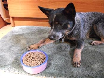
5261039

Retained Baby Teeth in Dogs and Cats Need Surgical Extraction

Puppies normally have 28 deciduous (baby) teeth that erupt during the first six months of life, and adults have 42 permanent teeth. Kittens have 26 baby teeth, and adult cats have 30.

In order for the baby teeth to fall out, specific cells resorb (dissolve) the roots of the primary tooth. This allows the permanent teeth to erupt normally. We don’t understand what triggers the resorption, so we don’t know why some baby teeth don’t fall out the way they are supposed to. 

When the baby teeth don’t fall out to make way for the permanent teeth, they are called retained deciduous teeth or persistent teeth. They should be extracted soon after they are discovered because otherwise they will cause dental problems (overcrowding in the mouth, plaque buildup, malocclusion, etc.).

As dentists like to say, two teeth should never try to occupy the space intended to contain one tooth.

When both deciduous (persistent) and permanent teeth are trying to occupy the same spot, this double row of teeth overcrowds the mouth, and food gets trapped between the teeth. This trapped food causes periodontal disease. In addition, the double set of roots can prevent the tooth socket from developing normally and eventually erode support around the adult tooth.

These difficulties can be prevented by extracting the retained deciduous teeth as soon as they are seen. If the tooth is extracted early enough, the adult tooth usually will move to its correct position; if it isn’t, there is a greater chance that the adult tooth will be in the wrong position, which can cause damage to the tongue, palate, mandible, etc.

It is much easier to position the permanent tooth while it is erupting, not after it has erupted. Repositioning after eruption requires orthodontic care to keep your pet’s mouth healthy.