4126803

Seizure Disorders in Dogs

Watching your dog experience a seizure is both frightening and disturbing, especially if it is unexpected. There is collapse, involuntary movement, and often loss of consciousness followed by a period of daze and disorientation. Prolonged seizure activity constitutes an emergency. You are presumably reading this because your dog has had some kind of involuntary fit and you want to understand what it means and what can be done to prevent future episodes so let’s cover some basics.

What is a Seizure and How Do you Know if your Dog has Had One?

A seizure results from excessive electrical activity in the cerebral cortex of the brain. The electrical activity starts in one area (called the seizure focus) and spreads in a process called kindling. Classically, the patient loses consciousness, collapses, becomes stiff at first and then begins paddling or struggling but seizures can take many forms. Any involuntary behavior that occurs abnormally may represent a seizure. Seizures are classified into several categories.

Generalized (Grand Mal) Seizures

This is the most common form of seizure in small animals. The entire body is involved in stiffness and possibly stiffness/contraction cycles (tonic/clonic action). The animal loses consciousness and may urinate or defecate. See a grand mal (violent) seizure. 

Focal Seizures (Also Called Partial Motor Seizures)

Focal seizures involve involuntary activity in only one body part. Consciousness may or may not be impaired. A classic example would be the “chewing gum” fit that frequently is seen in canine distemper infections but can be seen in other seizure disorders as well. See a chewing gum seizure in a Maltese with mild epilepsy. 

Psychomotor Seizures

Psychomotor seizures are focal seizures where the seizure is more like an episode of abnormal behavior than an actual convulsion. The pet’s consciousness is disturbed by this type of seizure as the pet appears to be hallucinating or in an altered state. The seizure may include episodes of rage or aggression where the pet does not recognize family members or may be as simple as a brief episode of disorientation or spacing out. Fly-biting is an example of a psychomotor seizure. See a psychomotor seizure.

Seizures (neurological events) are often difficult to tell from fainting spells (cardiovascular events). Classically, true seizures are preceded by an aura or special feeling associated with a coming seizure. As animals cannot speak, we usually do not notice any changes associated with the aura. The seizure is also typically followed by a post-ictal period during which the animal appears disoriented, even blind. This period may last only a few minutes or may last several hours. In contrast, fainting animals are usually up and normal within seconds of the spell, with no post-episode disorientation.

Post-ictal disorientation is the hallmark of the seizure.

Causes of Seizures and Diagnostics

There are many potential causes of seizures: toxins, tumors, genetic disease such as epilepsy, infections, even scarring in the brain from past trauma. Seizures resulting from metabolic problems or toxicity (i.e. when the brain itself is normal) are called reactive seizures.  Seizures resulting from identifiable brain abnormalities are called structural seizures. Seizures for which no clear cause can be found are called primary seizures and the patient is said to have epilepsy.

It turns out that dogs of certain age groups tend to have common causes for their seizures. This means that certain diagnostic tests are especially important in dogs of one age group while other tests are going to be more important for dogs in another age group. Here are some basic concepts concerning how age is an important consideration:

  • Reactive Seizures: Seizures resulting from metabolic problems or toxicity (i.e., when the brain itself is normal).
  • Secondary Seizures: Seizures resulting from an identifiable brain abnormality.
  • Primary Seizures: Seizures for which neither of the above problems apply (i.e., when no cause can be found).

Animals Less than Age Six Months

In this age group, seizures are usually caused by brain infections. For dogs, the most common infectious diseases would be canine distemper or a parasitic infection such as with Toxoplasma or Neospora. Analysis of cerebrospinal fluid, obtained by a tap under anesthesia, would be important though now that PCR technology is available for detecting DNA of infectious agents, less invasive testing may be recommended depending on the infectious under suspicion.

Animals Between Ages 6 Months and 6 Years

The most common reason for a dog, particularly a purebred dog, to begin having seizures in this age range is genetic epilepsy (also called primary epilepsy.) Epilepsy is diagnosed when no other cause of seizures can be found, there are no neurologic symptoms between seizure events, and the first seizure episodes begin in this age range. Usually basic blood work is done to rule out metabolic causes of seizures but more sophisticated and expensive testing (such as advanced brain imaging) is forgone as the presentation is fairly classic.

Schnauzers, basset hounds, collies, and cocker spaniels have two to three times as much epilepsy as other breeds. Labrador retrievers and Golden retrievers are also predisposed to epilepsy but tend to begin their seizures relatively late, closer to age five.

Animals More than 5 Years Old

In this group, seizures are usually caused by a tumor growing off the skull and pressing on the brain (meningioma would be the most common tumor type). Consult your veterinarian about treatment options. Such tumors may be operable or can be treated with radiation if found early. A CT scan or MRI would be the next step. Referral is usually necessary for this type of imaging. For patients where surgery is not an option, corticosteroids may be used to reduce swelling in the brain. Treatment to suppress seizures may also be needed using one of the medications discussed below.

When to Begin Treatment

In 2016, the American College of Veterinary Internal medicine published a consensus statement on this very subject.

If the dog fits into any of these criteria, medication to suppress seizures should be initiated:

  1. When seizures occur in clusters, which is more than 3 seizures within a 24-hour period.
  2. When two or more isolated seizures occur within a 6-month period.
  3. If a seizure has lasted 5 minutes or more.
  4. If the seizures or their post-ictal disorientation periods are particularly severe.
  5. If the dog has a visible structural lesion on a CT, MRI or even a radiograph.
  6. If the dog has a history of brain injury or trauma.
  7. It should be noted that the German shepherd dog, border collie, Australian shepherd, golden retriever, Irish setter, and Saint Bernard breeds are notorious for difficulty in seizure control. It is best not to wait for frequent seizures in these cases as each seizure makes the next more difficult to control. Often medication is started in these individuals after the first seizure. The more seizures the patient experiences, the more difficult control becomes in the future.

Treatment Choices: Medication

There are currently four main medications that are used in suppressing seizures in dogs in the United States: phenobarbital, potassium bromide, levetiracetam, and zonisamide. If adequate control cannot be achieved with one medication, often two or even three are combined. The ideal first line anti-convulsant medication is effective, reasonably priced, convenient to administer, and has limited side effects potential. Most dogs are started on either phenobarbital or potassium bromide but we will take a moment to review the pros and cons of all four of these medications.

Phenobarbital

Phenobarbital has been the first-line therapy for canine seizure control for decades as it is effective, reasonably priced, and can be given twice daily which is relatively convenient. When dogs with seizures are started on phenobarbital, approximately 31 percent of them can be expected to become seizure-free. Approximately 80% of dogs on phenobarbital will experience a greater than 50% decrease in seizure frequency. Approximately 20-30% of dogs on phenobarbital will require a second anti seizure medication to achieve acceptable seizure control.

The downsides are phenobarbital use stem from its side effects potential. Phenobarbital blood levels need to be periodically monitored as higher levels are associated with the development of liver disease. The phenobarbital dose must maintain the phenobarbital blood level within a safe therapeutic range and be adjusted accordingly. There is some expense associated with such testing.

Side effects of the drug include sedation, which is usually temporary during the first one to two weeks of medication use and wanes as the patient’s body adjusts. The patient is likely to be unusually hungry and thirsty on phenobarbital. These side effects can be objectionable. Some lab test changes are associated with phenobarbital usage and need to be recognized as such. Phenobarbital is removed from the body by the liver so good liver function is essential for phenobarbital use and phenobarbital can alter the metabolism of numerous other medications. 

Potassium Bromide

Potassium Bromide was used for human seizure control nearly 100 years ago but was eclipsed by the development of phenobarbital. It turns out that while phenobarbital may be a superior seizure drug for people, potassium bromide may be superior for dogs. When dogs with seizures are started on potassium bromide, 52 percent of them can be expected to become seizure-free. Approximately, 70 percent will have greater than 50 percent reduction in seizure frequency.

Potassium bromide is associated with pancreatitis and probably should not be used in patients with a history of that disease. Potassium bromide takes many months to reach a stable blood level which could leave the patient vulnerable to seizures during that time. As with phenobarbital, there are monitoring tests associated with potassium bromide use and sedation is a side effect.

Levetiracetam (Keppra®)

Levetiracetam is popular for refractory epilepsy in dogs because it has been shown to be fairly reliable and has minimal side effects potential. It appears to work best in combination with other seizure medications rather than as a sole therapy but many dogs are able to use it as a single agent. There are no monitoring tests recommended for its use and an extended-release formula allows for twice-daily use.

Zonisamide (Zonegran®)

Zonisamide is a sulfa class anti-seizure medication that is rapidly becoming a first-line treatment choice but might also be used to supplement more traditional therapies. Because it is a sulfa, it is vulnerable to the side effects associated with sulfa antibiotics: mostly tear production/dry eye issues but also some immune-mediated reactions. (Sulfa side effects are reviewed more completely in our pharmacy library under the sulfa antibiotics such as trimethoprim sulfa). Zonisamide can be used twice a day in dogs but lasts long enough in the cat to possibly be used once daily.

Treatment Choices: Diet

In 2017, a veterinary therapeutic diet designed to supplement anti-seizure medications was released. The diet uses medium-chain fatty acids as a fat source (fats come in short, medium, and long-chain types which relate to the length of their chain of their carbon chain) and it turns out that MCTs have a direct anti-seizure effect. Dogs that were not able to achieve full seizure control with medication were able to improve control or achieve total control after a 3-month trial on this diet. It is not meant to replace medications by any means, just to give them a nutritional boost.

Seizures at Home (When is it an Emergency?)

A Single Breakthrough Seizure

It is a lucky pet that never has another seizure after beginning medications, but an occasional breakthrough seizure (as disturbing as it may be to watch) is rarely of serious concern. In most cases, one can simply give an extra dose of the oral anti-seizure medication that has been in use and consider the episode over with. The veterinarian should be appraised of the situation and the medication regimen evaluated to see if adjustments should be made to prevent further breakthrough seizures in the future.

A Second Breakthrough Seizure within 24 Hours

If a second seizure occurs within 24 hours, one might consider bringing the pet to the vet’s office for a “seizure watch” (which means the pet can receive medication to interrupt any further seizures) as well as for re-evaluation of the current medication protocol.

Since emergency care can be expensive, one might consider rectal administration of diazepam (valium®) as a means of first aid and tiding the pet over until one’s regular veterinarian is available. In anticipation of late-night seizing, one can request a set-up for rectal diazepam to keep on hand. The injectable product is delivered rectally with a special syringe that can be kept at home. The rectal route avoids any danger of being bitten while trying to administer medication. 

Recently compounding pharmacies have been able to produce diazepam rectal suppositories which may be easier to use than the syringe method, however, absorption rates are unknown with these products and most neurologists prefer using the injectable product. Rectal diazepam administration has been used successfully for many years in epileptic children; the technique has adapted well to veterinary patients. Diazepam can also be given nasally but there is a greater chance of being bitten.

It is important not to put yourself in danger around a seizing pet. Involuntary jaw motion may bite you and in the period of post ictal disorientation, the pet may not recognize you and may snap. 

As mentioned, an isolated seizure at home probably does not require more than staying out of the way and keeping the pet from hurting himself.

 That said, there are some emergency situations:

  1. Seizure activity non-stop for five minutes or more.
  2. One seizure after another repeatedly.

Either of these situations is called “status epilepticus” and is a “drop-what-you’re-doing-and-rush-the-pet-to-the-vet emergency.

  1. More than 3 seizures in a 24-hour period

This is considered “cluster seizuring” and definitely warrants seizure watch in a hospital setting.

Can Seizure Medication be Discontinued Eventually?

While there is some risk to discontinuing seizure medications, this may be appropriate for some patients. Dogs should be completely seizure-free for at least a year before contemplating stopping treatment. In breeds for which seizure control is difficult, it is probably best never to stop medication (German Shepherds, Siberian Huskies, Keeshonds, Golden retriever, Irish Setter, St. Bernard). Phenobarbital is a medication that cannot be suddenly discontinued; if you are interested in discontinuing seizure medication, be sure to discuss this thoroughly with your veterinarian.

Other Information

The Epilepsy Genetic Research Project

Veterinary neurologists at several universities are looking for a genetic answer to epilepsy. They seek DNA samples from epileptic dogs and their close relatives if possible.

Canine Epilepsy Network

Affiliated with the Veterinary School at the University of Missouri at Columbia, this site reviews canine seizure disorders, treatment, history and more.

6493513

Storm and Other Noise Phobias

Storm phobia, or fear of storms, is a common behavior in dogs. Many dogs are terrified of the noise and react by hiding, pacing, panting, trembling, peeing, pooping, drooling, and destroying things. Some dogs even take it to the extreme of hurting themselves by jumping through windows and doors. These behaviors can appear during a storm, or when they see or hear things such as thunder, lightning, rain, or formation of dark storm clouds.

Another type of noise phobia is the fear of fireworks and gunshots. Dogs with this fear show similar behaviors to storm phobic dogs. Some become so fearful they do not want to go outside the house or around the area they associate with these noises.

Treating Storm and Noise Phobia

Environmental Management

The first step in managing and treating all types of noise phobia in dogs is to give them a safe place to hide. This safe haven can be a windowless basement, closet, crate, or bathroom. It is important to make sure your dog has access to this area when no one is home.

The noise of a radio, television, white noise machine, fan, or air conditioner reduces how much they hear of rain, thunder, and other scary noises.

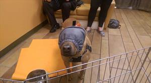
Classical music can be relaxing for some dogs. Anxiety wraps or close-fitting t-shirts may help calm your dog. Other useful devices are dog earmuffs to reduce noise sensitivity, and calming caps to decrease ability to see the storm.

Pheromones may be beneficial in some cases. These come in a diffuser, wipes, or spray. These pheromones can be placed on a bandanna, blanket, or a favorite toy to reduce anxiety.

Behavior Modification

Comforting: When it comes to the benefits of comforting your dog, the jury is still out. Many dogs enjoy the comfort of your closeness and reassurance whereas others become more anxious. Monitor your dog. If they become more fearful, stop trying to comfort them. If attention and touch calm your dog, there is no reason to stop.

Desensitization and counterconditioning are forms of behavior modification used to reduce the fear of noise and storms. Desensitization exposes the dog to a scary noise at such a low level that the animal is not scared. Counterconditioning changes the association of the noise with fear. Try to play with your dog, or do fun tricks he knows to distract your dog.

Food can also be a distraction, like an extra special treat that only comes out during scary events. Food puzzles (similar to these shown for cats) allow your dog to anticipate something fun and special when a storm is roaring overhead.

Short-Acting Medications

There are medications your veterinarian can prescribe to reduce the fear in your dog.

If medications are used, they must be given before the scary noise to be effective. Watch the weather closely and administer medication before the storm is predicted to arrive. It is better to medicate and no storm, than no medication and a storm. The same is true for other noise events.

Behavioral supplements may be beneficial in some dogs.

A magic pill to treat a noise or storm phobia does not exist. Treatment involves a combination of environmental management, behavior modification, and medication. Fear of noises can be reduced, but a cure is rare.

4128457

Sarcoptic Mange in Dogs

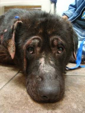
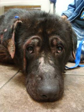
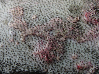
Sarcoptic mange, also called scabies, is an itchy disease in dogs caused by a mite (Sarcoptes scabiei var canis). Sarcoptic mites are small and not visible to the naked eye. They burrow into the skin and cause itching, redness, and skin crusts at the tips of the ears, elbows, hocks, chest and belly. In a severe infestation, Sarcoptic mites cause problems on the animal’s entire body.

How is sarcoptic mange (scabies) diagnosed? 

Your veterinarian will look for the mite by performing several skin scrapings on your dog and examining the debris under a microscope. Unfortunately, in many dogs with sarcoptic mange, the skin scrapings do not contain any mites. This is because only a small number of mites on your dog’s body can cause severe symptoms. If sarcoptic mange is suspected, your pet’s veterinarian will recommend treatment for the mites.   

How is sarcoptic mange (scabies) treated?


Several prescription medicines can be used to kill mites. These medicines come in various forms including:

  1. dips such as lime sulfur or amitraz
  2. spot-ons such as moxidectin (Advantage Multi®) or selamectin (Revolution®, Stronghold®)
  3. oral pills such as afoxolaner (Nexgard®), milbemycin oxime (Interceptor®), or sarolaner (Simparica®)

Your pet’s veterinarian may also prescribe other medicines to treat itching while the mite medication takes effect.

No matter which treatment is chosen, follow the instructions from your pet’s veterinarian. For some medicines, the amount needed to treat sarcoptic mange is different than the amount needed to treat other health conditions. If you give the medicine less often or in a smaller amount than what your pet’s veterinarian recommends, your pet’s sarcoptic mange will not get better.

Can my other pets or my family get sarcoptic mange (scabies)?

Yes. Sarcoptic mange is highly contagious and can spread from pet to pet or from pet to human. Treat all other dogs in the household. Although mites can only survive a brief time off the dog, clean your dog’s living quarters and wash or throw away the bedding. If your dog has sarcoptic mange and anyone in your family has red and itchy skin bumps, consult your family physician.

Your dog may remain contagious for two to four weeks after starting treatment. Please keep your dog confined and away from other dogs and unexposed persons until your dog’s re-check veterinary appointment.

4128955

Systemic Lupus Erythematosus (SLE) in Dogs

Systemic lupus erythematosus (SLE) is a fairly rare chronic and potentially-fatal autoimmune disease. The dog’s immune system fights itself by forming antibodies that “protect” it against its own cells and tissues. This results in inflammation and tissue damage in the skin, heart, lungs, kidneys, joints, nervous system, or blood. Usually several organs are affected. SLE can be managed, but not cured. While the disease itself is chronic, signs can be acute, chronic, or both. Lifelong treatment is necessary. It is different from discoid lupus erythematosus (DLE), which is a skin disease.

The word lupus is Latin for “wolf.” (Some humans get a facial rash that has a slight similarity with a wolf’s face.)

Breeds that most typically are affected by SLE include Afghan Hounds, Beagles, German Shepherd Dogs, Irish Setters, Old English Sheepdogs, Poodles, Rough Collies, and Shetland Sheepdogs. Dogs are usually middle-aged when they get SLE. Females are not more likely than males to get SLE.

The most common significant signs of SLE are a non-erosive polyarthritis (an arthritis that affects several joints), painful muscles; shifting lameness; skin sores and blisters; proteinuria (protein in the urine); decreased platelet and white blood cell count; fluctuating fevers, and immune mediated hemolytic anemia (IMHA). Some less-significant signs are mouth ulcers, inflamed lining of the chest cavity or heart, dementia, swollen lymph nodes, and seizures. Usually, dogs with SLE are first taken to the veterinarian because of the skin problems or lameness.

Blood tests are needed for diagnosis. While all dogs won’t have the same signs, dogs with SLE typically test positive for anti-nuclear antibodies (ANA). There are so many effects of this multi-systemic disease that it’s hard to rule any one disease in or out. Diagnosis is typically made using a combination of a positive ANA test with at least two significant signs. A positive ANA test, with one significant sign plus two less-significant signs is also indicative of SLE.

(SLE can be suspected if there is only one significant sign plus positive ANA, or if there are two significant signs with a negative ANA test.) It can be difficult to make a positive diagnosis since other problems, such as drug reactions and cancer, can have many of the same signs. (ANA can be positive with any chronic inflammatory disease, so if there are no other consistent pathologic or clinical signs, your veterinarian may suspect a false-positive ANA.)

Treatment is aimed at decreasing the inflammation and autoimmune activity, so treatment centers around anti-inflammatory and immunosuppressive medications such as corticosteroids (e.g. prednisone). Sometimes a second immunosuppressant, such as azathioprine, cyclophosphamide, or cyclosporine will be necessary. Specific problems, such as kidney or spleen damage, will also have to be treated. Treatment plans depend on what organs are affected.

The ultraviolet rays in sunlight can cause flare-ups of SLE, so exposure to sunlight should be limited.

Prognosis depends entirely on how each dog is affected, but owners can expect that their pets will require life-long treatment. Unfortunately, sometimes the damage caused by this disease can result in death.

4311221

Steroid Use in Dogs and Cats

Most people are familiar with cortisone and may even know that it is a steroid but, even though steroids are commonly prescribed to both people and animals, much more knowledge than that is not really mainstream. Steroid hormones are used to treat many types of diseases, and it turns out that by altering the dosage of a steroid, a completely different effect is achieved. This means steroids can treat inflammation, improve appetite, suppress an overactive immune system, reduce secretions, relieve itching, and more. 

Steroids can be short-acting, long-acting, intermediate-acting, oral, injectable, topical, or even inhalational. With so many uses possible, it is important to know which effect(s) your doctor is going for.

Let’s take a few moments to straighten out the vocabulary of steroid hormones to understand what is in that bottle of pills so frequently dispensed. A lot of terminology can be confusing, so it is good to have some basis for what your veterinarian is talking about.

Steroid: A hormone with a cholesterol-based structure is a steroid hormone. There are sex steroids (like anabolic steroids that bodybuilders should probably not be using) and adrenal steroids. The steroids that are most commonly used therapeutically are adrenal steroids.

Corticosteroid: Adrenal steroids can be produced by the center of the adrenal gland or by the outer cortex of the adrenal gland. Corticosteroids come from the outer cortex of the adrenal gland. There are two types: glucocorticoids (which address sugar metabolism) and mineralocorticoids (which address electrolyte metabolism). We are concerned with the glucocorticoids for this discussion.

Examples of Glucocorticoids

Cortisol: This is the natural hormone produced by one’s adrenal glands to regulate sugar, fat, and salt metabolism in times of stress.

It turns out cortisol can be made in the laboratory, and when it is, we call it hydrocortisone (or sometimes just plain cortisone.) Hydrocortisone is a common topical steroid used in assorted anti-itch products. Modifications of it make it stronger or longer acting, such as mometasone, which is used in human nasal products as well as veterinary ear medications.

Oral Glucocorticoids

Prednisone: In order for prednisone to become an active hormone, the liver has to convert it into prednisolone. Prednisone can be produced in the laboratory and is readily available as a medication.

Prednisolone: In order for prednisone to become an active hormone, it must be further activated by the liver into prednisolone. Again, prednisolone can be produced in the laboratory and is readily available as a medication.

Methylpredisolone (Medrol®): By altering the prednisolone molecule in the laboratory, the potential for side effects can be changed. One issue is the increased thirst side effect. By changing the prednisolone structure slightly, less salt retention occurs, leading to less thirst compared to what happens with regular prednisolone. Less thirst means less urination and less chance of an accident in the house.

Dexamethasone (Azium®): Adding still more methyl groups creates an even stronger steroid with even less salt retention side effects. Dexamethasone can also be manufactured in the laboratory and is readily available as a medication that can be used topically, orally, or as an injectable.

Injectable Steroids

We have already covered dexamethasone, which can be used as an injectable. Other modifications have led to:

Methylprednisolone Acetate (Depomedrol)This steroid is very strong and can maintain an anti-inflammatory effect for several weeks. This is helpful for patients (especially cats) who may not readily accept oral shorter-acting steroids every day. One shot can cover a long period of time, but the downside is that the injection makes for a high dose that wanes over months. The anti-inflammatory effect may last a few weeks, but the suppression of the natural adrenal system continues for months after that.

Triamcinolone (Vetalog)Intermediate-acting but strong, lasting a couple of weeks. Similar downsides as above.

So we have covered some of the benefits of steroid therapy.

There has never been a class of drug that has more application in disease treatment than glucocorticoids.  Indeed, this group is rivaled only by antibiotics in lives saved.

That said, side effects from the glucocorticoid group are numerous and can be classified into those seen with short-term and long-term use.

Short-Term Side Effects

A pet on glucocorticoids is likely to experience:

  1. Increased hunger
  2. Increased thirst and possibly urinary incontinence for dogs if there is inadequate access to an area for appropriate urination
  3. Panting (dogs)
  4. General loss of energy
  5. Hidden infection being unmasked, especially upper respiratory infections in cats.

These side effects are generally classified as inconvenient but not serious. If a short-acting oral steroid is being used, often simply changing the dosing schedule solves these problems. If a long-acting injectable is used, you simply have to wait for it to wear off.

But there are also more serious side effects:

  • Pre-diabetic animals may be tipped over into a diabetic state with steroid use. Often, in these cases, the diabetes resolves once the steroid wears off.
  • A similar situation exists for patients with borderline heart function (a situation for which cats are notorious). The extra salt retention from steroid use can prove to be just too much for a heart that can barely manage its normal fluid volume. The salt causes fluid retention, and a borderline heart may not be able to manage.

Even these serious side effects can resolve once the steroid wears off, but it is important to be aware of these possibilities. The above scenarios are rare, but if they happen to your pet, they will not feel rare to you.

Long-Term Side Effects 

Many conditions require long-term suppression of the immune system. Glucocorticoid doses generally include an anti-inflammatory dose that is lower and an immune-suppressive dose that is higher, though with long enough term use, lower doses will become immune-suppressive. When steroid use stretches out for more than four months, a new set of side effects, in addition to those listed above, becomes of concern:

  1. Latent urinary tract infections in up to 30% of patients. Monitoring for these with periodic urine cultures is necessary. The patient will not have the usual symptoms of urinary infection as the steroid will suppress the inflammation associated with the infection. Culture may be the only way to detect the infection.
  2. Development of thin skin, blackheads, and poor ability to heal wounds or grow hair
  3. Development of obesity and muscle weakness
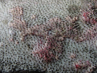
  4. Hard plaques of diseased skin called calcinosis cutis (see photo above). These plaques are calcium being deposited in the skin.
  5. Predisposition to infection of any kind/weakening of immune defenses
  6. Development of Cushing’s syndrome.

All of the above listed effects can be seen and be considered symptoms of this syndrome.

When long-term therapy is needed, monitoring tests become especially important; requesting refill after refill without regard for the potency of these medications is not appropriate. Periodic urine cultures, check-ups and even blood testing is part of responsible on-going corticosteroid use. Your veterinarian will suggest appropriate tests.

Steroid Alternatives

When a long-term dose of steroids is deemed to be excessive or if the short-term effects become problematic, it is time to seek another medication so as to spare the amount of steroids needed, if not replace the steroid altogether. 

4620034

Swimmer’s Tail in Dogs

Swimmer’s tail is known by many names, including limber tail, cold water tail, dead tail, broken wag and sprained tail. These names all refer to the same condition: a suddenly limp and flaccid tail. Swimmer’s tail most commonly affects large breed, hunting, athletic and working dogs such as Labrador retrievers and pointers.

Dogs with this condition have essentially strained the muscles in their tail. This is thought to result from overuse of the tail, which can happen when a dog swims for a long time, especially in cold water. Sometimes it happens just because the dog has had an exciting day or two and has spent far more time than usual wagging their tail.

For example, maybe they spent the weekend at the lake or the grandchildren came to visit. Other risk factors for swimmer’s tail include intense physical activity, prolonged transport in a cage and being in cold and wet weather. After these activities, signs usually appear quickly, within hours to days.

The primary sign of this condition is a limp tail that droops between the dog’s legs. In some cases the entire tail is flaccid, but in other cases the tail is stiff at the beginning and becomes flaccid towards the end. Because swimmer’s tail can cause pain and discomfort, affected dogs may pace, yelp when lying down or defecating, frequently change position while lying down, and eat less. Some dogs may struggle to stand up because the tail helps with balancing. Other potential symptoms include chewing the tail and raised hair on the top of the tail due to swelling.

To diagnose this condition, your veterinarian will rely on information about your dog’s recent activities, clinical signs and physical exam findings. Your veterinarian may also take X-rays to rule out other causes of your dog’s symptoms, such as tail fractures.

Once swimmer’s tail is diagnosed, your dog should rest and exercise less to allow for recovery, just as you would do if you sprained a muscle. Your veterinarian may also recommend giving anti-inflammatory drugs to reduce the pain associated with this condition. Warm compresses on the tail may also help with pain and quicken the recovery process. Swimmer’s tail has an excellent prognosis, as dogs generally get better within 2-14 days. However, having swimmer’s tail once does not mean your dog can’t get it again. Relapses are possible.

Potential ways to prevent swimmer’s tail include not overexerting your dog, especially if they aren’t trained for it, and, during transport, letting your dog out every 4 hours or so. There is no prevention for over-wagging when your dog is really happy, though!

7974141

Sleeping and Resting Respiratory Rates of Dogs and Cats with Heart Disease

Many heart diseases, when severe enough, result in congestive heart failure.  When the left side of the heart is diseased, this presents as a build-up of fluid in the lungs, causing breathing difficulties and shortness of breath which can be severe. However, before such signs occur, many animals start to have an increase in their breathing rate.  This can be subtle and is best detected when your pet is sleeping (because there are not a lot of things that affect your pet’s breathing rate when they’re asleep).

Your veterinarian can use your observational skills and record of your pet’s breathing to help manage your pet’s heart disease in three ways:

1. Preclinical heart disease.  Heart disease in dogs and cats that is destined to cause congestive heart failure progresses over time from mild disease to more severe disease. When it gets severe enough, fluid builds up in the lungs (congestive heart failure), requiring treatments to relieve this congestion. 

However, before it reaches that stage, your veterinarian might have you start measuring the sleeping respiratory rate (SRR) so that you catch congestive heart failure at the earliest stage possible.  Usually, such measurements start when the disease is moderate-to-severe, but not yet showing signs of congestive heart failure.  Your veterinarian will use tests such as chest X-rays (radiographs) or cardiac ultrasound (echocardiography) to determine when measuring the SRR is appropriate.

2. Diagnosis of congestive heart failure.  Sometimes signs of congestive heart failure are subtle and easily confused with other non-cardiac diseases. In such cases, your veterinarian might get you to measure your pet’s sleeping respiratory rate (SRR) to help rule out a diagnosis of congestive heart failure – if the rate is normal, congestive heart failure is extremely unlikely and your veterinarian can focus on other causes of your pet’s clinical signs.

3. Management of congestive heart failure (pulmonary edema).  If your veterinarian diagnoses your pet with congestive heart failure and institutes treatment for it (with diuretics and other drugs), they will probably get you to monitor the response to medications by measuring your pet’s sleeping respiratory rate (SRR) at home. 

Studies are finding that measuring the SRR is a very sensitive way to tell if your pet’s heart failure is being well controlled by drugs or not.  When the drugs are working well, the pet should be feeling good, but more importantly, the SRR should be between 10-25 breaths per minute (in most cases, it will be less than 30 breaths per minute).  If the SRR increases or exceeds 30 breaths per minute, it might indicate that there is fluid building back up in the lungs.  Your veterinarian will generally recommend you return to the clinic to confirm this is the case, or they might instruct you to increase the dose of diuretics.

How to measure SRR

SRR and RRR (Resting respiratory rate) should be obtained when the pet is comfortable, in a comfortable environment (not too hot or cold), and not after any exertional activity.  If sleeping, the pet should be in a “deep sleep”, not in a twitching or “dreaming” state.

The respiratory rate should be counted for a full minute if possible, although 30 seconds is often sufficient.  One breath is made up of 2 components: breathing in (inspiration or inhalation) and breathing out (expiration or exhalation).

These videos show 2 dogs and a cat either sleeping or resting quietly.  The counter records each breath for over a full minute. Watch these to understand how this is done.

7565896

Spina Bifida in Dogs and Cats

Vertebrae and the Spinal Column

In vertebrates, the delicate spinal cord (the part of the central nervous system connecting the brain to the rest of the body) is surrounded and protected by a bony tube that is made up of a series of bones (vertebrae), linked together by discs made of cartilage, ligaments, and muscles. This entire grouping is referred to as the spine.

How Does Spina Bifida Develop?

The vertebrae form in the embryo (earliest stages of development) during the mother’s pregnancy. Sometimes, abnormal growth and development of the spine happen before birth with both puppies and kittens. The result is that some of the neural tube, the part of a fetus that eventually develops into the brain and spinal cord, fails to close into a complete tube. This usually occurs on the back side (dorsal or posterior side), resulting in a defect in the affected vertebrae. This congenital (trait present at birth) defect is called spina bifida. Sometimes only the bones are involved, and the defect may cause no symptoms. This is called spina bifida occulta and is usually found incidentally on X-rays taken for other reasons. In other cases, however, the defect in neural tube growth and closure involves the membranes surrounding the spinal cord (called the meninges) or leaves a defect in the spinal cord itself. These defects usually result in clinical signs termed spina bifida manifesta.

Central Nervous System 

  1. The brain and spinal cord make up the central nervous system (CNS). The CNS is the body’s processing center and coordinates what the entire body does.
  2. The CNS takes in sensory information, processes it, and sends out signals through nerves to make the body do something.
  3. Information is sent from various areas in our bodies (like when a dog steps on hot pavement), and the CNS makes the desired movement happen (moving the foot away from the hot surface).
  4. A cat can’t jump onto a couch without a signal first being sent to the brain from the eyes that a couch is nearby. The cat’s brain must also then signal the legs to jump.
  5. Nerve cells, called neurons, send these signals. There are billions of neurons in the body, and they communicate with each another to cause physical responses and actions.
  6. The inability to move, called paralysis, stems from problems in the nervous system.
  7. A signal from the nerves in the body is what causes any part of the body to move, so if the part of the nervous system that takes care of the legs has enough damage, the nervous system cannot tell the brain to move the legs.
  8. If signals cannot be sent to tell urinary and fecal muscles to wait a bit or to loosen now, the body will have no control over those muscles, and incontinence will happen.

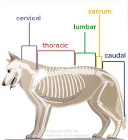
Because spina bifida manifesta most often affects the area of the low lumbar and sacral regions in affected puppies and kittens (most often Old English Bulldogs and Manx cats), the signs can include abnormal control of urination and bowel movements, as well as movement abnormalities in the pelvic (hind) limbs.

Another common finding in patients affected with spina bifida manifesta is tethered (spinal) cord syndrome, where the end of the spinal cord is abnormally attached to the spine. As the puppy or kitten grows, the spinal cord cannot expand along with the spine, stretching the spinal cord and leading to further neurological problems.

How Common is Spina Bifida?

Spina bifida is relatively rare in dogs and cats, and the disease can vary from mild to severe.

Human parents of babies with spina bifida are told that no two cases are alike because the symptoms vary in severity depending on where the vertebrae have not closed and by how much is exposed, and the same is true for dogs and cats.

Spina bifida can occur because of genetic factors, problems with nutrition, or if the mother was exposed to certain chemicals and toxins while pregnant. It’s possible that inbreeding and selective breeding practices may increase the likelihood of this condition. English bulldogs and the tailless Manx cats are the breeds most affected by spina bifida, but it can happen to any breed of dog or cat. In brachycephalic or short-faced dog breeds such as English bulldogs, French bulldogs, or Pugs, spina bifida tends to occur at the first thoracic (chest area) vertebra but can be found in different lower areas of the spine as well.

Cats and dogs with spina bifida may leak cerebrospinal fluid, or CSF, from sacs near their tail area. CSF is essential to cushion the spinal cord and keep the spinal cord healthy.

Screw tail dog breeds such as English bulldogs, French bulldogs, Pugs, and Boston Terriers are also known to develop a similar yet different spinal condition called hemivertebra.  With hemivertebra, vertebrae are deformed, and two or more may fuse together, which has the potential to cause walking and elimination (urination or feces) issues depending on the location of the deformity on the spine. However, most hemivertebra lesions are not associated with clinical signs. 

Diagnosis

Diagnosis includes X-rays, myelography, computed tomography (CT), or magnetic resonance imaging (MRI) of the spine. Myelography involves injecting a contrast dye into the fluid around the spinal cord before an X-ray is taken so lesions may be seen more clearly. It is used specifically to diagnose spinal cord issues. It is often used when other advanced imaging is unavailable.

Myelography is more informative to veterinarians than a regular X-ray but not as clear as a CT or MRI. Myelography can also be combined with CT (they are not mutually exclusive).  MRI is the most preferred imaging choice for spinal conditions and is preferable to plain X-rays, X-rays with myelography, or CT with myelography. 

Pets are generally sedated for these imaging procedures, so accurate imaging can take place without movement or undue stress to the animal. They may have an IV catheter placed, a sedative is given, and then wake up before returning home after their imaging.

Treatment

Spina bifida may be a minor or major issue for your pet, depending on how many vertebrae did not close properly and where those incomplete vertebrae are located.

Severe cases are considered untreatable, and unfortunately, the pet will have a very poor quality of life due to pain, paralysis, weakness, neurologic deficits, and little ability to control their bowel and bladder. Typically, puppies and kittens are euthanized immediately upon diagnosis of severe spina bifida, and signs that something is not normal can be seen as soon as the puppy or kitten begins to walk.

In minor cases, generally, no treatment is needed. Most of these cases are found coincidentally on X-rays taken for a separate concern. Supportive care can help manage fecal and urinary incontinence, urinary tract infections, and hygiene issues.

  1. Mild cases can often be treated with reconstructive surgery that removes tumors and cysts, closes sacs, and sometimes may restore part of the spinal cord. Discuss with your veterinarian whether referral to a surgical specialist is best for you and your pet.
  2. Medications can help with urinary and fecal incontinence, and your veterinarian can offer options for pain control.
  3. Physical therapy and rehabilitation can help with mobility and strengthen weak muscles.
  4. Wheelchairs and braces may be recommended.
  5. Diet may be modified to help manage specific issues and ensure your pet is getting adequate nutrition.

Prevention

Currently, veterinarians don’t know exactly how spina bifida is inherited. It is possible that exposure to chemicals during the growth of kittens and puppies in the mother’s womb could cause abnormal spine development. Still, more research is needed before any recommendations can be made. The only preventive measure currently is to avoid breeding affected dogs and cats.

Prognosis

Prognosis is good for clinically normal pets, and some with mild disease can lead good, functional lives. The prognosis for patients with severe nerve damage or lack of function is guarded, and euthanasia may be recommended if no quality of life is possible. If your pet has been diagnosed with spina bifida, your veterinarian can discuss with you how your pet may be impacted physically and what ongoing treatment options and lifestyle changes may look like for you and your pet so you can both have the best quality of life possible.

7559858

Skin Biopsies in Dogs and Cats

What is a Skin Biopsy?

In a skin biopsy, a small piece of skin is removed through minor skin surgery. The skin is then examined under a microscope by a veterinary pathologist to diagnose your dog or cat’s skin condition. Skin biopsies are also used to test a growth or mass for conditions such as cancer.

What Happens During My Pet’s Skin Biopsy?

The veterinarian may gently clip the fur near the skin biopsy. A sedative or anesthetic medicine may be given to make your pet more comfortable during the procedure. Different types of surgeries are performed during a biopsy. The type of surgery depends on where the biopsies are being performed and what skin conditions your veterinarian is concerned about. The most common surgeries for a skin biopsy include the following:

  1. wedge biopsy is used to surgically remove not only the skin but also the tissue under the skin (subcutis). The veterinarian angles a scalpel blade to meet as a “V” under the skin.
  2. An excisional biopsy is used if your pet has a growth (skin mass). The veterinarian uses a scalpel blade to cut the growth out of the skin. Excisional biopsies are used to diagnose many skin conditions.
  3. punch biopsy is used to remove the full thickness of a small, round piece of skin. The punch tool will be placed on the area where the skin sample(s) will be taken, pressed downward to cut the skin gently, and then the piece of skin will be removed. Punch biopsies are a common way to perform skin biopsies in dogs and cats. They can also be used to obtain a skin sample for a skin culture (bacterial culture and sensitivity) to select an antibiotic for your pet’s skin infection.
  4. shave biopsy is used to surgically remove only the top layer of skin. The veterinarian uses a scalpel blade or other tool to scrape or shave off the skin. Shave biopsies are used to diagnose skin conditions that only affect the top layers of the skin.

What Can I Do to Help Prepare My Pet for a Skin Biopsy?

The veterinarian will provide you with specific instructions for preparing your pet. This may include not feeding your pet in preparation for the sedative or anesthetic medication. For the skin, do not pick at your pet’s skin or remove any crusts or scabs. The veterinarian will also tell you when to stop bathing or applying medications to the skin before the biopsy.

What Will Happen After My Pet’s Skin Biopsy?

When the procedure is over, your pet will be able to go home, and the veterinarian will provide you with specific instructions to care for your pet. Your pet may have bleeding, redness, or swelling after the biopsy. If your pet has stitches where the biopsy was performed, the veterinarian will tell you if and when the stitches need removal. If the fur was clipped for the biopsy, the fur will be shorter but will grow back.

What Happens to the Skin Sample after a Biopsy?

The skin sample is cut into thin sections and put onto glass slides. The slides then go to a laboratory for testing. A veterinary pathologist (a veterinarian trained in interpreting biopsy samples) will check the slides under a microscope to help make a diagnosis. For skin biopsies, a veterinary pathologist who has received additional training in diagnosing skin diseases called a veterinary dermatopathologist, is often used.

5223011

Shar-Pei Recurrent Fever Syndrome

Also called swollen hock syndrome, Shar-Pei auto-inflammatory disease (SPAID), or familial Shar-Pei fever syndrome (FSF).

It would seem the Chinese Shar-Pei might have enough issues to contend with, given its potential for wrinkle-related skin and eyelid issues, but there is a special syndrome that every Shar-Pei owner needs to be familiar with: Recurrent Fever Syndrome.

As the name implies, the syndrome is characterized by fevers that seem to arise out of nowhere, run their course, and may or may not be responsive to anti-inflammatory medications.

The dog will feel bad during the fever episodes: listless and without appetite. Fevers typically last 12 to 36 hours and can go as high as 107ºF. Often the dog’s ankles (hocks) become swollen during these episodes. It is normal for a Shar-Pei to have a large skin fold around the ankle called a sock; the swelling that occurs during the fever is different and only lasts during the fever. The muzzle may also swell and become painful, and sometimes there is some associated nausea or diarrhea. It is important to learn how to take your dog’s temperature so you can monitor the situation. 

A fever of 106ºF is a medical emergency. It is good practice to know how to take your dog’s temperature and is especially important if your Shar-Pei seems listless. Normal canine body temperature is 100-102ºF.

The fevers are unpleasant and can be dangerous if the fever rises to 106ºF but what makes this syndrome a serious problem is the accompanying kidney damage. An abnormal protein called amyloid is laid down in the kidney, destroying the kidney’s ability to filter protein. The valuable blood proteins are thus lost in urine along with waste chemicals. The dog becomes thin from the loss of body proteins, develops a propensity to throw abnormal blood clots throughout the body (from urinating out the proteins that would normally prevent this), and high blood pressure results.

All Shar-Pei should be regularly screened for urinary protein loss with a urinalysis. Red flags include urinary protein and dilute urine (specific gravity less than 1.020).

Why This Happens: Wrinkles Gone Wrong

The characteristic skin wrinkles that make the Shar-Pei what it is are caused by excessive production of hyaluronan. Hyaluronan is a structural protein in everyone’s skin but a mutation in the Shar-Pei leads to multiple copies of the genes regulating the production of hyaluronan. The result is a whole lot of extra hyaluronan puffing up the skin and creating all the wrinkles that characterize this breed.

All Shar-Pei have this mutation; without this mutation, the dog cannot really be a Shar-Pei but not all Shar-Peis have this mutation in the same way. Some have a mutation that leads to variable qualities of hyaluronan. In other words, not all the hyaluronan produced is of a healthy quality. Poor quality hyaluronan breaks down rapidly in a process that generates inflammation which, in turn, creates fever and damages organs. The mutation that creates all this poor-quality hyaluranan is often called the meat mouth mutation.

It might seem that you could simply look at a Shar-Pei’s face to determine if the dog is a candidate for fever syndrome but the situation is more complicated.

How do I Know if my Shar-Pei Has the Syndrome?

A fever can develop in a Shar-Pei from a wound or other source of infection, just as it can in any other breed of dog. Some effort should be made to find another source of the fever and this generally requires a complete physical examination and some blood testing.

As for screening tests, there is a genetic test available at Cornell University. The mutated genetic variant that creates the meat mouth appearance is typically in multiple copies within an affected Shar-Pei. The more copies the dog has, the more reactive the inflammation. The genetic test determines the number of mutated copies present and reports the “copy number variant.” The higher this number, the more trouble for the dog. In the absence of a test, a diagnosis is made based on the clinical findings. One episode of fever is enough to make the diagnosis. Fevers classically begin before 18 months but can begin at any age. 

The test requires a blood sample and a six-page information form filled out by both the owner and the veterinary professional drawing the blood. A microchip number or tattoo is required to confirm the identity of the patient. Learn more about testing.

Though testing had been temporarily suspended as of August 21st, 2020, it seems that Cornell’s latest information says SPAID testing has been revived as of December 15, 2022.

Treatment

During a fever episode, anti-inflammatory medications provided by your veterinarian can be used to control high fevers. Pain medication is often needed to control the discomfort during the episode. The real challenge, however, is to prevent kidney damage in the long term. A dog can have substantial kidney damage before the first fever episode even happens therefore, it is important to begin therapy after the first episode and to regularly screen for urinary protein loss in any Shar-Pei whether fevers have occurred or not.

The medication central to preventing amyloid deposition in the kidney is colchicine.

Cell division, a process more scientifically known as mitosis, requires microscopic protein fibers acting like structural cables to pull dividing cells apart. These cables are called mitotic spindles, and colchicine interferes with their formation. The ability of colchicine to interfere with this sort of structural protein formation has led to its use in abnormal protein depositions such as amyloidosis. This means that colchicine can prevent the kidney damage that occurs in recurrent fever syndrome.

Recently, legal issues have forced generic colchicine off the market, leaving only the brand name product Colcrys®. This medication is problematically expensive for most pet owners. An arrangement has been made for the owners of recurrent fever syndrome dogs. An application can be obtained at www.needymeds.com; a valid prescription is required and your veterinarian will need to fill out part of the application. Colchicine can also be obtained through a compounding pharmacy. A prescription is still necessary.

Other Therapy

Antioxidants are important in amyloid prevention in that they help preserve cell membrane fatty acids. Current recommendations include supplements in omega 3 fatty acids (fish oil) and a quality multivitamin. Antioxidants such as vitamin C, lecithin granules, and glucosamine supplements to improve the hyaluronan quality are also recommended. Consult your veterinarian for a specific protocol for your dog. Early intervention is the goal with this condition.

Herbal antioxidants have also been recommended. Normalizing proper hyaluronan metabolism may require magnesium supplementation. Consult your veterinarian for specifics.