8229321

Ticks Are Arthropod Parasites for Mammals

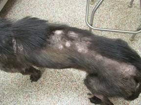
Ticks are skin parasites that feed on the blood of their hosts. Ticks like motion, warm temperatures from body heat, and the carbon dioxide exhaled by mammals, so they are attracted to such hosts as dogs, cats, rodents, rabbits, cattle, small mammals, etc. The bite itself is not usually painful, but the parasite can transmit diseases and cause tick paralysis, which is why tick control is so important. (Removing the ticks leads to rapid improvement of the paralysis.)

The minimal amount of time it takes for an attached tick to transmit disease is not known. The time it takes to transmit diseases is affected by the type of disease organism, the species of tick, etc. However, in general, if a tick is removed within 16 hours, the risk of disease transmission is considered to be very low. Therefore, owners can usually prevent disease transmission to their pets by following a regular schedule to look for and remove ticks.

The Tick Life Cycle

Most types of ticks require three hosts during a 2-3 year lifespan.  Each tick stage requires a blood meal before it can reach the next stage. Hard ticks have four life stages: egg, larva, nymph, and adult.

Larvae and nymphs must feed before they detach and molt. Adult female ticks can engorge, increasing their weight by more than 100-fold. After detaching, an adult female tick can lay approximately 3,000 eggs.

During the egg-laying stage, ticks lay eggs in secluded areas with dense vegetation. The eggs hatch within two weeks. Some species of ticks lay 100 eggs at a time, while others lay 3,000 to 6,000 per batch. Once the eggs hatch, the ticks are in the larval stage, during which time the larvae move into the grass and search for their first blood meal. At this stage, they will attach themselves for several days to their first host, usually a bird or rodent, and then fall onto the ground. The nymph stage begins after the first blood meal is completed. Nymphs remain inactive during winter and start moving again in spring. Nymphs find a host, usually a rodent, pet, or human.

Nymphs are generally about the size of a freckle. After this blood meal, ticks fall off the host and move into the adult stage. Throughout the autumn, male and female adults find a host, which is again usually a rodent, pet, or human. The adult female feeds for 8 to 12 days. The female mates while still attached to her host. Both ticks fall off, and the males die. The female remains inactive through the winter and in the spring lays her eggs in a secluded place. If adults cannot find a host animal in the fall, they can survive in leaf litter until the spring.

What are the best ways to deal with these blood-sucking parasites?

Outdoor Environmental Control

Treating the yard and outdoor kennel area with acaricides (tick pesticides) is an important tool in the arsenal against ticks. Some products can be used to spray the outdoor area. However, you should not rely on spraying to reduce your risk of infection.

If you have health concerns about applying acaricides, check with local health or agricultural officials about the best time to
apply acaricides in your area, identify rules and regulations related to pesticide application on residential properties, and consider using a professional pesticide company to apply pesticides at your home.

You can also create a tick-safe zone in your yard by using some simple landscaping techniques that can help reduce tick populations:

  1. Remove leaf litter.
  2. Clear tall grasses and brush around homes and at the edge of lawns.
  3. Place a 3-ft wide barrier of wood chips or gravel between lawns and wooded areas to restrict tick migration into recreational areas.
  4. Mow the lawn frequently.
  5. Stack wood neatly and in a dry area. (This discourages rodents who would be used as tick hosts.)
  6. Keep playground equipment, decks, and patios away from yard edges and trees.
  7. Discourage unwelcome animals (such as deer, raccoons, and stray dogs) from entering your yard by constructing fences.
  8. Remove old furniture, mattresses, or trash from the yard that may give ticks a place to hide.

Indoor Environmental Control

If ticks are indoors, flea and tick foggers, sprays, or powders can be used. Inside, ticks typically crawl (they don’t jump) and may be in cracks around windows and doors. A one-foot barrier of insecticide, where the carpeting and wall meet, can help with tick control.

Preventing Ticks from Attaching

If your pet goes outside regularly, you can use some type of residual insecticide on your pet. Talk to your veterinarian about what works best in your geographic area. If you use a liquid spray treatment, cats and skittish dogs typically prefer a pump bottle because of the noise from aerosol cans. Doubling the amount of anti-tick product, or using two at once, may cause toxicity problems. 

Any spray insecticide labeled for use on clothing should not be sprayed directly on pets.

Powders are fairly easy to apply, but they can be messy. (Avoid topical powders if your pet has a respiratory condition.) Shampoos are useful only for ticks that are already on your pet. A tick collar might be somewhat more water-resistant than a residual insecticide, so if your dog likes to swim, the collar might be a better choice. Flea combs can be used to help remove ticks and wash your pet’s bedding frequently.

Finding and Removing Ticks

The best way to find ticks on your pet is to run your hands over the whole body. Check for ticks every time your pet comes back from an area you know is inhabited by ticks. Ticks attach most frequently around the pet’s head, ears, neck, and feet, but they are not restricted to those areas.

There are several tick removal devices on the market, but a plain set of fine-tipped tweezers will remove a tick quite effectively.

How To Remove A Tick With Tweezers

  1. Use fine-tipped tweezers to grasp the tick as close to the skin’s surface as possible.
  2. Pull upward with steady, even pressure. Don’t twist or jerk the tick; this can cause the mouth parts to break off and remain in the skin. If this happens, remove the mouth parts with tweezers. If you can’t remove the mouth easily with clean tweezers, leave it alone and let the skin heal.
  3. After removing the tick, thoroughly clean the bite area and your hands with rubbing alcohol, an iodine scrub, or soap and water.
  4. Dispose of a live tick by submerging it in alcohol, placing it in a sealed bag/container, wrapping it tightly in tape, or flushing it down the toilet. Never crush a tick with your fingers.

Risk of disease transmission to you, while removing ticks, is low but you should wear gloves if you wish to be perfectly safe. Do not apply hot matches, petroleum jelly, turpentine, nail polish, or just rubbing alcohol alone. These methods do not remove the ticks and they are not safe for your pet. Rubbing alcohol could be used to disinfect the area before or after removing the tick.

Watch for Infection and Diseases

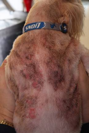
After you pull a tick off, there will be a local area of inflammation that could look red, crusty, or scabby. The tick’s attachment causes irritation. The site can get infected; if the pet is scratching at it, it is more apt to get infected. A mild topical antibiotic, such as over-the-counter triple antibiotic ointment, can help but usually is not necessary. The inflammation should go down within a week. If it stays crusty and inflamed longer than a week, it might have become infected.

Various diseases can be transmitted by ticks including anaplasmosis, babesiosisehrlichiosis, Lyme disease, tick paralysis (or tick toxicosis), and others. Not all species of animals (pets or not) are affected similarly by every tick-borne illness.

Although ticks can transmit diseases contracted from a previous host to pets and humans, they are usually nothing more than a nuisance. The best approach is to prevent them from embedding and once embedded, to remove them quickly. If you notice your pet experiencing signs of illness or acting differently after a tick bite, it is always best to see your veterinarian. If you stay on top of the situation, your pets should cruise right through the tick season with no problems.

4128995

Taenia Species Tapeworms in Dogs and Cats

After gaining some pet-owning experience, the average pet owner has heard of tapeworms transmitted by fleas and knows to watch for sesame seed-like segments around their pet’s nether regions or on the surface of stools. In fact, there is a part two to this story as there is another type of tapeworm to which dogs and cats are vulnerable. These other worms are members of the Taenia genus.

Our article on the common tapeworm (Dipylidium caninum) has generated some confusion regarding tapeworms that are not contracted from fleas. There actually is another tapeworm besides Dipylidium whose segments might be seen on a pet’s anal area or on their feces. These other worms are members of the Taenia genus.

There are several members of the Taenia genus with which one may come to be acquainted:

  1. Taenia solium, which infects humans when they consume undercooked pork
  2. Taenia saginata, which infects humans when they consume undercooked beef
  3. Taenia hydatigena, which infects dogs when they consume undercooked livestock or venison or feed from dead livestock or deer they find out in the world
  4. Taenia taeniaeformis, which infects cats when they consume rats and mice 
  5. Taenia ovis, which infects dogs after they consume dead sheep or undercooked lamb
  6. Taenia multiceps, which infects dogs that eat the brains of infected sheep
  7. Taenia crassiceps, which infects dogs when they consume rats and mice 
  8. Taenia serialis and Taenia pisiformis, which infect dogs when they consume dead rabbits.

To keep things simple, we will stick to the term Taenia to refer to all of them. There are, as you might guess, other Taenia species that infect animals other than dogs and cats but we will leave them out of this discussion for simplicity. In short, pets at risk for a Taenia infection are those that eat raw meat, either through predation or raw feeding.

The Taenia Life Cycle

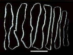
The life cycle of Taenia tapeworms starts in the host’s intestine, the host being a dog or cat. The worm can be unbelievably long (up to 5 yards for Taenia hydatigena) and is made of segments. Each segment contains an independent set of organs, with new segments being created at the neck while older segments drop off the tail. As a segment matures, its reproductive tract becomes more and more prominent until it consists of a bag of tapeworm eggs. These segments, called proglottids, are passed with the feces into the world, where an unsuspecting intermediate host (mouse, rabbit, deer, sheep, etc.) swallows one while eating.

The young tapeworm hatches in the new host’s intestine and escapes into the blood supply, with the next stop being the liver. (Remember, this new host is a prey animal such as a mouse, rabbit, deer, etc. We have not gotten to the dog or cat predator yet). The larval tapeworm wanders through the liver, leaving bloody tracks behind. It ultimately falls into the abdominal cavity, where it forms a sac and waits. After about two months of development in this location, the larval tapeworm is ready to continue its development, but it will need a new host to do so. When the host (prey species) dies or is killed by a predator, the sac and its young tapeworm inside may be consumed incidentally when the body of the host is eaten.

About two months later, inside the predator (dog, cat, or even human, depending on what kind of animal the prey was), the young tapeworm is now mature and is beginning to shed its first segments, and the cycle begins again.

How Do you Know What Kind of Tapeworm the Segments Are from?

In most cases, tapeworm segments seen are from Dipylidium caninum, which is not called the common tapeworm for no reason (i.e., it is very common). The segments of Dipylidium are longer than they are wide and are said to look like grains of rice. The segments of a Taenia tapeworm are wider than they are long.

As with most tapeworms, it is hard to find tapeworm eggs using the normal fecal testing we use to screen animals for worm infection. This is because tapeworms shed their eggs in discreet packets (the proglottids) rather than releasing them freely. Unless a proglottid breaks open and releases its eggs, it is likely that eggs will not be found. Diagnosis is usually based on seeing the proglottids with the naked eye.

Is it Important to Recognize Which Type of Tapeworm the Pet Has?

The good news here is that the same medication, praziquantel, kills both types of tapeworms efficiently. Where it becomes useful to know one type of worm from another is prevention. Dipylidium comes from swallowing a flea; Taenia comes from swallowing carrion, hunting prey, or feeding raw food. Knowing where the tapeworm came from tells you what to do to prevent the next infection: stop feeding raw, restrict access to prey, or beef up flea control.

Tapeworms do not cause significant symptoms and are largely of cosmetic concern.

If you see tapeworm segments on your pet’s fur or feces, see your veterinarian for a tapeworm treatment.

A note on the Echinococcus tapeworm:

There is a much more dangerous (to humans) genus of tapeworms called Echinococcus. Humans can be infected via contact with canine feces. We bring this up because under the microscope, Taenia eggs and Echinococcus eggs when they are found, appear identical. They can be distinguished with more advanced testing should this event occur.

A Few Words About Echinococcus multilocularis:

There is another type of tapeworm to be aware of if you live in a woodsy area, especially if your dogs and cats roam or hunt out in nature. This very small tapeworm, Echinococcus multilocularis, employs a fox (or potentially a dog or cat) as its host. The host sheds tapeworm segments that are too small to see in its feces. The egg-containing segments will likely be mixed in with dirt.

The usual life cycle involves a rodent consuming the egg, developing destructive tapeworm cysts in its liver, and ending up as lunch (cysts and all) to a dog, cat, or fox. The dog, cat, or fox then becomes the new source of tapeworm eggs. Should the contaminated soil end up on human hands instead of in a rodent’s mouth, the potential for human transmission exists, and the tapeworm cysts in the lungs and liver can be lethal. To prevent your roaming dog or cat from becoming a source of these dangerous eggs, regular praziquantel dosing should be included in the monthly parasite protection program for the pet. Check with your veterinarian to see if you live in an area where Echinococcus multilocularis is native.

4127201

Toothbrushing and Dental Prophylaxis in Cats and Dogs

Why Should I Brush My Dog’s Or Cat’s Teeth?

Daily removal of plaque is the key to an oral hygiene program. Unless your pet’s teeth are brushed daily, plaque, which is an accumulation of bacteria, will build up at the gum line. Eventually calculus forms, further irritating the gums, and then infection progresses to loosen and destroy the tooth’s attachment. In addition to loose teeth, infection under the gumline can spread to the liver, kidneys, and he

How Can I Brush My Animal’s Teeth?

It is usually an easy and fun procedure. First pick a soft-bristled or finger toothbrush. Next, get toothpaste from your veterinarian. Do not use human toothpaste because it has detergents that should not be swallowed.

How About Wipes?

Dental wipes can be effective in removing plaque. Be sure to wipe the area where the tooth meets the gum line.

What Is The VOHC?

The Veterinary Oral Health Council (VOHC) is a non-profit organization composed of board certified veterinary dentists that reviews studies and accepts dental products that have been shown to reduce the accumulation of plaque and/or tartar. They list their approved products,and the products have a VOHC seal of approval on the packaging.

How Often Does My Pet Need To Have Teeth Cleaned By The Veterinarian?

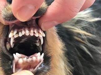
It depends on the degree of plaque and tartar accumulation. You need to examine your pet’s teeth monthly. Look for an accumulation of yellow or brown material at the area where the tooth meets the gumline especially over the cheek teeth and canines.

Once you notice plaque or tartar accumulation, it is time for a professional cleaning. Do not wait.

Attached to the tartar are bacteria, which irritate gum tissues. When treated, the inflammation will resolve. When gingivitis is left untreated, it will progress to periodontitis, which is non-curable.

The intervals between teeth cleaning procedures will depend on how often you can brush your pet’s teeth. Once or twice daily cleaning is optimum. If you cannot brush the teeth, then your pet will probably need two or three teeth cleaning visits yearly.

Can I Just Take My Fingernail Or A Dental Scaler To Remove The Calculus?

Dental disease occurs below the gum line. By removing calculus from the tooth, you are not removing disease below the gum line. In order to thoroughly help your pet, plaque and calculus must be removed from below the gum line.

Do You Have To Use Anesthesia To Clean My Pet’s Teeth?

Anesthesia is necessary when performing teeth cleaning. Anesthesia provides three important functions: immobilization in order to clean below the gum line, pain control, and the ability to place a tube into the windpipe, so bacterial products do not enter the respiratory system.

I Am Concerned About The Anesthesia. Is It Safe?

We take every effort to provide safe anesthesia. We use gas anesthetic agents; dogs and cats are given pre-operative tests depending on their age and condition to qualify them for anesthesia; and patients are monitored while anesthetized both visibly and with similar monitoring devices as used in human hospitals.

What Is Involved In The Teeth Cleaning At Your Hospital?

Each Oral Assessment, Treatment, and Prevention (Oral ATP) visit has twelve separate steps:

  1. general exam before anesthesia, pre-operative organ testing
  2. oral exam under anesthesia
  3. gross calculus removal
  4. subgingival (below the gumline) scaling, root planing, curettage where indicated
  5. tooth polishing
  6. irrigation
  7. fluoride / OraVet application
  8. post cleaning exam and dental x-rays to evaluate the areas below the gum line
  9. dental charting to create a treatment plan
  10. therapy if necessary
  11. home care instructions
  12. no-fee follow up appointment to see how well you are performing home care.

How Much Does A Tooth Cleaning Procedure Cost?

It is impossible to determine what the procedure will cost because we do not know the status of your pet’s teeth and gums. There are four levels of teeth cleanings at our hospital. Fees are based on severity plus costs for preoperative testing, anesthesia, necessary therapy, and medication. Fees for all dental services are available by e-mail or fax. The doctor or staff will provide an initial treatment plan based on exam room findings and a follow up treatment plan with fees after a tooth-by-tooth exam is conducted under anesthesia.

What Is The Best Food For My Pet?

Hard food will help remove plaque from teeth. There are special diets specifically manufactured to help control plaque. Feeding the special diets in conjunction with daily brushing is the best to keep the teeth clean. Diet alone will not control plaque, but it will help.

What Toys Should I Avoid To Protect My Pet’s Teeth?

Chewing on objects harder than teeth may lead to dental fractures. Be especially careful with cow and horse hoofs. They commonly cause fractures of the upper fourth premolars. Tug-of-war games must not be practiced, especially in young dogs and cats in order to avoid moving growing teeth to abnormal locations. Throwing dogs discs can also cause trauma to the teeth, resulting in pulpitis (an inflammation of the pulp).

What Are Cat Cavities?

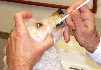
Many cats get painful lesions at the gum line that invade teeth. They are properly referred to as tooth resorptions. Unfortunately we do not know what causes tooth resorption, and the most effective treatment involves extraction of the affected tooth. Check to see if your cat has an advanced resorption by pressing a cotton-tipped applicator to the gum line. If a painful lesion is noted, your cat will chatter its jaw.

How Can I Tell If My Pet Is Suffering From Periodontal Disease?

The leading sign is bad breath. Dogs and cats should not have disagreeable mouth odor. Bad breath comes from infection. If your pet’s breath does not smell like roses, let us examine its mouth and advise care.

What Type Of Tests Are Done To Diagnose Dental Disease?

If your pet has periodontal disease or a fractured tooth, an oral exam is performed while under anesthesia. A periodontal probe is used to evaluate bone loss around each tooth. X-rays are taken to evaluate if the teeth can be saved or need to be extracted.

When Do I Have To Start Worrying About Dental Problems With My Pet?

As soon as puppy or kitten teeth emerge, it’s time to start brushing. Although baby teeth are replaced with adult teeth, the puppy or kitten gets used to the brushing procedure, which continues for life.

What Can Be Done If My Pet Has Periodontal Disease?

Periodontal disease occurs when tooth support structures are affected by infection. In the beginning stages, cleaning above and below the gum line as well as removal of calculus attached to the tooth will help restore periodontal health. In advanced cases, either periodontal surgery or extractions are performed. Antibiotics given monthly also help to control the progression of periodontal disease.

Which Animals Are At Most Risk For Periodontal Disease?

Smaller breeds are more prone than larger because the teeth are closer together in small dogs, and they usually live longer. Terriers, Maltese, and Shih Tzus are especially prone to periodontal disease.

What Can You Do To Fix A Broken Tooth?

If your dog or cat breaks a tooth, there are two treatments: root canal therapy or extraction. You cannot leave the tooth alone with an exposed nerve. In addition to pain, infection will soon develop that can spread to the rest of your pet’s body.

portfolio-1

Thrombocytopenia in Dogs and Cats

Platelets (thrombocytes) are produced in the bone marrow. Platelets circulate in the blood. When your pet gets cut, platelets stick to the edges of the cut and to one another, forming a plug. They then release chemicals that react with fibrinogen and other plasma proteins, leading to the formation of a blood clot.

If your pet has a condition called thrombocytopenia, his platelet count is low. When the platelet count is low, it is harder for clotting to occur.

Signs of thrombocytopenia vary, but can include petechial (small, pinpoint) hemorrhages in the skin or mucous membranes (lining of the mouth, for example), nosebleeds, hematuria (blood in the urine), melena (blood in the feces), and bleeding into the joints or body cavities. The lower the platelet count is, the more likely it is that your pet will show signs. (Some animals may show no warning signs at all, and the thrombocytopenia may be discovered only during routine health exams.)

Major causes of thrombocytopenia are immune-mediated thrombocytopenia (an autoimmune disease), bone marrow cancers, infectious diseases, hereditary diseases, drug/vaccination reactions, vasculitis (leading to deep vein thrombosis), and disseminated intravascular coagulation.

Thrombocytopenia is not common in dogs and cats, although there are several breeds of dogs that are predisposed to it. Animals with cancer are at higher risk than those without cancer, but it can occur in a dog/cat of any age, breed, or sex.

Diagnostic tests include: blood tests (platelet count, complete blood count, serum biochemistry), urinalysis, chest or abdominal radiographs (to look for diseases that could be causing the thrombocytopenia), tests for specific infectious diseases (Rocky Mountain spotted fever, ehrlichiosis, etc.), bone marrow aspiration, and tests for immune system function.

Treatment is based on the cause of the thrombocytopenia, and may include antibiotics, corticosteroids, blood transfusions, etc.

Prognosis depends on the cause of the thrombocytopenia. A mild, self-limiting vaccine-induced thrombocytopenia has an excellent prognosis, while a thrombocytopenia due to cancer might have a much worse prognosis.

5261039

Tremoring or Shivering in Dogs

Dogs are often brought in for tremoring or shivering episodes. The patient can be any age or any size. Tremors can involve only certain muscles or body areas or the entire dog. Finding the reason behind tremors is tricky as tremoring occurs for many reasons.

The first step is to consider normal reasons for shivering. The patient is:

  1. cold
  2. frightened
  3. asleep and dreaming.

Usually, these can be excluded from being a disease by providing a blanket or sweater, comforting the anxious patient, or rousing the sleeping patient.

Elderly dogs with muscle atrophy tend to suffer muscle fatigue and muscles will shake when over-worked. This type of tremoring is often pretty easy to distinguish as the patient is elderly and the tremors begin when the patient has been standing longer than they would prefer. When a muscle becomes fatigued, tremoring helps release stored glycogen/glucose to permit continued muscle contraction. Tremoring generally stops when the muscle is allowed to relax and the patient rests.

Mild shivers are rarely a significant symptom; it is the more violent involuntary tremors and twitches that indicate neuromuscular disease.

Conditions When Only Certain Body Parts Tremor

If only certain body parts tremor, there are some neuromuscular diseases to consider.

Intention Tremors

Diseases involving the cerebellum of the brain will lead to tremors when the patient directs his attention to a task. The patient may sit quietly without tremors, but if a bowl of food is put out, tremors of the neck and head erupt when the patient reaches for the food.

This sort of tremoring generally goes way beyond what might be described as shivering or even twitching. A classic example of cerebellar intention tremors is seen in kittens born with cerebellar hypoplasia, a neurologic condition stemming from the mother becoming infected with or vaccinated for feline distemper during pregnancy.

Cerebellar disease certainly does not have to be congenital. Any damage to the cerebellum (trauma, tumor, infection, etc.) can produce an intention tremor. An intention tremor is highly suggestive of disease in the cerebellum, though, and diagnostics should be pursued to uncover the nature of that disease. See a video of these wobbly kittens.

Idiopathic Head Tremors

No one really knows why this occurs but some dogs have episodes of head bobbing (usually up and down but can be side to side). Breeds that seem over-represented are Boxers, English bulldogs, beagles, and Doberman pinschers. This condition is totally unresponsive to seizure medications and the best way to curtail an episode seems to be to focus the dog’s attention on a toy or treat. Episodes tend to get milder with age. See this boxer’s head tremors.

Distemper Myoclonus

Dogs that survive a canine distemper infection may suffer from seizures, paralysis, or muscle twitching long term. Often a young dog is adopted from the shelter or rescue, and her history is unknown. Distemper myoclonus appears as involuntary twitches of muscle groups anywhere on the body. Multiple areas can be involved. These twitches are not seizures and do not respond to seizure medications. Numerous medications have been used with inconsistent results. The most successful treatment has involved injections of cosmetic Botox® into the muscle group involved, but this therapy is expensive and many dogs do not seem particularly inconvenienced by their myoclonus. The black dog is showing myoclonus of his jaw and neck muscles.

Orthostatic Tremors

This condition is observed only when the dog is standing and involves tremors of the leg muscles. The patient is typically a young, very large-breed dog (Great Dane, mastiff, Weimaraner). The condition tends to progress in severity over time and may come to require treatment. It is generally responsive to phenobarbital and/or to gabapentin.

Conditions When the Whole Body Tremors

If the entire dog is shivering and shaking, then we must consider diseases that involve the entire dog. Normal muscles rely on oxygen, nutrients, and electrolytes from the bloodstream as well as normal electrical stimulation from the nervous system.

We need to look at problems with these systems:

  1. Hypocalcemia (low blood calcium, especially if nursing puppies)
  2. Hypoglycemia (low blood sugar)
  3. Toxins (especially snail bait and insecticides)
  4. Little White Shaker Dog syndrome (which does not actually require that the patient be a white dog), is also called non-suppurative meningoencephalomyelitis or steroid-responsive tremors.
  5. Seizures/epilepsy.

Severe tremors can elevate the body temperature dangerously and cause brain damage. Patients for which tremoring is an emergency are generally clearly mentally distressed by the involuntary motions or disoriented from the underlying neurologic disease.

Many toxins can cause twitches and involuntary muscle tremors, so it is important to let your veterinarian know what products are in your home. What flea products do you use? Do you have pesticides in the garden? These products are frequently neurotoxins that produce seizures and twitching. Snail baits containing metaldehyde are particularly common, as are permethrin-based flea products (generally safe for dogs but cats are uniquely sensitive). Tremors are a notable feature of chocolate toxicity. Obviously, poisoning is an emergency so you will need to see your veterinarian or local emergency service right away.

Brain diseases can produce seizures that can manifest as persistent but fine tremoring or more violent convulsions. Metabolic diseases, such as low blood sugar or low blood calcium, can lead to tremors and even seizures. Muscle diseases such as tetanus can lead to involuntary muscle contraction. Twitches and tremors may be intermittent and are certainly not always emergencies; however, blood testing and possibly advanced neurological testing may be needed.

Your first step is going to be an evaluation with your veterinarian to narrow down the search and complete the necessary basic tests. Not all veterinarians are comfortable diagnosing neurologic disease, so discuss with your veterinarian whether referral to a specialist would be best for you and your pet. The best treatment plan can be made from there.

The small white dog in this video has White Shaker syndrome. It is generally controlled with immune-suppressive doses of corticosteroids. The prognosis (outlook) is generally good.

5482526

Traumatic Brain Injury in Dogs and Cats

If your pet has been involved in any event that may have caused head trauma, always see your veterinarian or an emergency practice for immediate attention.

What Is a Traumatic Brain Injury?

Traumatic brain injury (TBI) occurs when a sudden event, such as a blow or fall, happens to the head. In both humans and animals, a head injury severe enough to damage the brain is referred to as traumatic brain injury or TBI.

Humans are diagnosed with concussions (a mild form of TBI), and veterinarians may use that terminology to describe a dog or cat with an externally caused brain injury. A concussion is a mild form of TBI that occurs when there has been a rapid back-and-forth movement of the head, essentially causing the brain to come in contact with the skull.

There has been increased attention on TBI in humans as sports injuries have become more recognized and acknowledged, and you may hear the term TBI more often now than in the past.

How Does it Happen?

TBI can occur in any age group or breed of dog or cat. Pets spending time outdoors may be more vulnerable to traumatic brain injury as they are likely exposed to more injury-causing risks. One fortunate advantage that dogs and cats have over humans is that their skulls are relatively thicker and, thus, a bit more protective of the brain. However, violent trauma can easily overcome the protective effect of a thicker skull.

An auto accident, fall, blunt trauma, a bite, kick or violent shaking, or any other sudden injury to the head can result in TBI.

How is TBI Diagnosed?

If your pet is not stable upon arriving at the veterinary hospital, it will be stabilized. A clinical examination will be completed, and your veterinarian may recommend various tests depending on your pet’s status. Your pet may have also been injured in other parts of the body, and your veterinarian will check for those. Blood and urine testing can help determine the extent of the injuries and establish a baseline for further evaluation. Neurological examinations and images of the head and brain using X-rays, MRI, or Computed Tomography (CT) may be needed when there is a suspected history of head trauma. Blood pressure may be monitored.

Evidence of an injury may be apparent. Cuts, scrapes, or broken bones may help tell the story if you were not present when the trauma happened.

If your pet is showing signs consistent with TBI, seeing these signs exhibited can be frightening. More so if you have no idea what is causing them.

Suppose the potential TBI happened in the past, and the history of the trauma that caused the head injury is available. In that case, your veterinarian will use this information to help evaluate the effects of the original injury.

There can be a variety of neurological symptoms from brain trauma that your pet may experience. Motor control issues such as circling in one direction, head tilting or displaying rapid or abnormal eye movement, or a difference in the size of the pupils may be present. Your pet may experience seizures.

Another severe and recognizable symptom of TBI is known as decerebrate posture. The limbs are held unnaturally, straight out, and ridged, with the head back and neck arched. This behavior generally suggests a more serious brain injury.

X-rays will usually be recommended as your pet may have a skull fracture or other broken bones (in other areas). Continuous electrocardiogram (ECG) monitoring may be needed to check your pet’s heartbeat. Additional imaging such as ultrasound, MRI, or CT scanning may be indicated so that your veterinarian can see your pet’s brain and other areas, if needed, in better detail.

Generally, TBI is divided into two categories: primary, which refers to results from the initial injury, and secondary, referring to damage caused by the primary injury.

Primary Injury

There is swelling where there is an injury to any of the body’s tissues. Swelling reduces blood flow, and compressed blood vessels cause pressure to build up in and around the brain. Broken blood vessels cause blood to pool, forming clots (hematomas).

If there is a skull fracture, brain damage can be worse. Surgery may be considered in this case. The severity of the condition is determined by how bad the initial injury and trauma were.

Depending on the overall injury causing a TBI, your pet may also experience difficulties with physical movement or injuries to other parts of the body.

Secondary Injury

Changes within the brain occur immediately after the primary injury, affecting the processes, or metabolism, at work in the entire body.

Because metabolism uses energy to drive many functions of the body, the changes are linked and result in numerous systems reacting, furthering the damage to the brain. Poor blood flow and bleeding in the brain add to these issues.

After the initial injury, a cascade of issues may develop over time. Brain swelling and inadequate blood and oxygen flow occur not only in the brain but also in other organs, such as the heart. Increased pressure within the cranium and brain swelling are major causes of death from TBI in dogs and cats.

Further laboratory testing and follow-up exams help your veterinarian evaluate additional damage to the whole body in addition to the brain that may be present.

Treatment

Treatment for TBI involves mainly supportive care, which may include hospitalization with continuous monitoring, medications, appropriate wound care, blood work, and follow-up X-rays.

Prognosis

Recovery for your pet depends on how severe the primary injury was. Patients who survive the first 24 hours following injury will most likely recover but may need treatment and monitoring depending on the injuries. If referred to a neurologist, you may need to take your pet for checkups or additional therapy, depending on the situation. Brains, both human and animal, have amazing powers of recovery, and positive outcomes happen all the time. Working closely with your veterinarian and/or veterinary neurologist and knowing what signs to look for (both good and bad) can help your pet have the best chance at recovery and, if possible, get back to their normal activities.

Depending on the location and extent of brain injury, the animal may recover fully or be left with varying degrees of altered mental or motor functions. One possible long-term result of TBI is epilepsy, which is recurring seizures. These can usually be managed with medications so that the animal’s quality of life remains good.

7649235

Teeth Chattering in Dogs

Teeth chattering in dogs can be caused by several different things. Some veterinarians believe that the source of teeth chattering is usually dental or oral (mouth) pain until proven otherwise. An abscess, broken teeth, or gingivitis can cause pain; even some ulcerations or growths that don’t touch the teeth can cause chattering as a response to that pain.

Dogs can also suffer from pain in the temporomandibular joint (TMJ).

A dog with arthritis around that joint or who has or has had a jaw fracture may chatter their teeth in response to that pain. Teeth can rub together if the jaw is clenched or from grinding the teeth. Imaging such as X-rays or computed tomography (CT) (both done under anesthesia) may be recommended to help your veterinarian better visualize what is happening in the jaws or mouth.

Even though the most common cause is somewhere in the mouth, the source can be elsewhere, including the gastrointestinal tract, or involving a neurologic disease. In some greyhounds, and likely in some other breeds, it’s a relatively common sign of anxiety and often ends as the cause of anxiety ends, such as a veterinary appointment.

Generally, the first step in treatment is a tooth-by-tooth exam under anesthesia with dental imaging, either X-rays or a CT scan.

Tooth Resorption

Tooth resorption is when the dentin, or the harder outside of a tooth, is inflamed and disintegrating. Teeth need to be evaluated closely for any tooth-resorbing lesions, as oral lesions may not be visible even on dental radiographs.

Identifying tooth resorption can be difficult, and choosing which teeth to pull requires experience and judgment. You and your veterinarian should discuss whether referral to a veterinary dentist is in your pet’s best interest.

Gastrointestinal Issues

With some gastrointestinal issues, dogs may become nauseous, vomit, drool, or have gastroesophageal acid reflux. Lab work and imaging, such as ultrasound or endoscopy (a camera scope fed into the GI tract) or surgery, might be recommended. In cases caused by gastrointestinal issues, medications may help stop the chattering within the first week.

Focal Seizures

Focal seizures are sometimes a neurological cause of teeth chattering. A focal seizure is a type of seizure that only affects a certain area in one half of the brain, not both halves. A dog with focal seizures may chatter their teeth or display what may be referred to as “fly-biting behaviors,” where they snap at the air as if chasing an insect. However, unlike other causes of teeth chattering, a dog experiencing focal seizures will not respond to you as they normally would when you call them by name, make noise, or so on, and they may seem to stare off into space.

Before this type of seizure, your dog may act unusual, such as walking back and forth or clinging to you. If your dog stops chattering their teeth during a short trial of anti-epileptic drugs, the cause of the chattering is likely related to the seizures.

Other less common causes of teeth chattering exist, such as pain in other parts of the body or even hormonal changes. A physical examination and consultation with your trusted family veterinarian, along with appropriate testing, can help pinpoint the cause of chattering and help guide treatment.

Additionally, taking videos for your veterinarian and listing all signs can be helpful to get them started on the best diagnostic path for your pet.

5356517

Snail Bait Poisoning in Dogs

Snails and slugs are frustrating garden pests wherever anyone is trying to grow tender flowers or ornamental plants. While effective non-toxic methods abound on the internet, snail bait and snail poisons are readily available in most garden supply stores and pose a significant hazard to the pet dogs that share the garden.

Snail bait poisoning continues to be a frequent toxicity issue for dogs in any area where snails are common garden pests.

Not that malicious poisoning is the issue; most cases are accidental as many gardens with snail problems also have resident dogs. Snail bait is typically formulated in pellets and flavored with molasses or bran to attract snails. Unfortunately, the pellets resemble kibbled dog food and the molasses and bran are also attractive to dogs. Snail baits are also available as liquids and powders that can get onto paws and be licked off in normal grooming. Very little metaldehyde snail bait is required to cause poisoning: less than a teaspoon per 10 pounds of body weight.

Many snail bait products also contain insecticides that make them even more toxic. If at all possible, find out what specific product has been involved in the poisoning so that the veterinarian and animal poison control can determine proper treatment.

In a 2002 retrospective study by the ASPCA Animal Poison Control Center, 28 percent of all metaldehyde poisoning cases happened in California.

Obvious Twitching

Signs of poisoning begin fairly quickly after the poison is consumed. The dog will begin anxious twitching, at first only slightly and then uncontrollably. This progresses to seizures and potentially to death. The muscle contractions of the twitches raise body temperature so high that brain damage can result, leading to the colloquial emergency room term “shake and bake” syndrome.

Patients can also exhibit racing heart rates, vomiting, diarrhea, and rigidity, and respiratory failure.

A liver failure syndrome occurs in some patients approximately two to three days after poisoning, so it is important for liver enzymes to be monitored by blood tests throughout the recovery period. Additional liver support medicine may be needed.

Making The Diagnosis

Generally, the appearance of a twitching patient is characteristic even if there is no known history of snail bait exposure. Testing of stomach contents or urine for metaldehyde can be done, but is generally not necessary.

Treatment

There is no direct antidote for metaldehyde toxicity; treatment is aimed at controlling the muscle contractions. If less than one hour has passed since exposure, it may be possible to induce vomiting. If the patient is already twitching badly, the stimulation involved in inducing vomiting may not be in the patient’s best interest. In this case, the patient can be anesthetized and the stomach pumped. Activated charcoal can be given to prevent absorption of metaldehyde into the body from the intestine. Cathartics (used to induce diarrhea) can also be used with the activated charcoal to help remove the metaldehyde from the intestinal tract promptly.

Twitching can be controlled with methocarbamol (a muscle relaxant) or injectable benzodiazepine sedatives such as diazepam. Fluid therapy and body temperature monitoring will be needed throughout the recovery period.

Potential for complete recovery depends on how much poison was ingested, how quickly therapy was begun, and the general health of the patient. While this is a serious type of poisoning, most patients have a good chance at recovery if treated properly.

At home, the yard should be hosed down with water to dissolve the remaining metaldehyde, and the dog should be restricted from the treated area for a two-week period.

This page is dedicated to Justin, a companion to Saeeda, who did not survive his exposure to metaldehyde. We promised to put up a web page in his honor to help educate dog owners and hopefully save the lives of some snail-bait poisoned dogs around the world.

4127017

Snake Bite Prevention and Treatment for Dogs

Most snakes will try to avoid you or your pets; snakes typically bite only as a last resort. But while you may wisely decide to simply walk away when you encounter a snake, dogs and cats will often harass the sliding invader — and may get bitten as a result. Snakes help protect your garden and yard plants because they eat rodents and insects. Most of the snakes you will find are not a problem and they don’t harm property.

While hiking, stay on open paths. Off-trail hiking can stir up snakes. Keep your dog on leash and away from high grass and rocky outcrops in which snakes like to rest. Don’t let the dog explore holes or dig under rocks or logs. Remember that rattlers are nocturnal, so daytime hikes are safer. If you hear a rattling noise, keep your dog at your side until you figure out where the snake is, and then move away with your dog. Your best bet is to stay vigilant and keep control of your dog when walking in snake habitats.

If you see a snake that sees you, remember that a snake can strike only a distance of half its body length. Give the snake time to just go away. Snakes are not looking to interact with people or pets, either defensively or aggressively.

Don’t let your pet examine “road kill” snakes. Dead pit vipers can have some muscle contractions after death, and thus have been known to “bite” even after they’re dead. Those bites can still envenomate. Don’t handle a dead or injured snake – not even a decapitated head.

If your pet is bitten by a snake that you think might be venomous, get medical attention immediately. It’s better to go in and be checked out rather than wait and be sorry.

Repellants and Removal

Most wildlife experts believe there is no such thing as an effective snake repellant, although people have tried mothballs, vapor barriers, ultrasonics, sulfur, poison, etc. What can help is setting up physical barriers (e.g., fences that are set a foot or two into the ground), cutting off the snake’s food supply and shelter, mowing closely around the house, storing firewood away from the house, removing junk piles, removing weeds and brush, plugging up holes in the ground, etc. And if you need to have dangerous snakes removed from your property, contact a wildlife control operator to trap and remove the snakes. This is not a job for an inexperienced person.

Snake Identification: Venomous or Nonvenomous?

Most nonvenomous snakes have a large smooth cap over the top of the head past the eyes, divided scales on the underside of the tail, no pits, and no long fangs. Rattlesnakes, copperheads, and cottonmouths are all venomous pit vipers, and have a pit between and slightly below the eye and nostril. Pit vipers also have long movable fangs, a “cat’s eye” pupil, undivided scales under the tail, and a large triangular-shaped head with a small shiny cap over the nose.

Learn about the snakes in your area. Familiarize yourself with what the local snakes look like by reading through a field guide of reptiles and amphibians for your area. Compare the drawings and photos of the local venomous snakes with the non-venomous species so that you can remember what they look like, in general. A single species of snake may have a wide range of colors and patterns.

Herpetology field guides can be purchased at book stores and science/nature stores, both off- and online. One online field guide is eNature. When you register (it’s free) on the site, you can store your zip code, and access all the species (plants, invertebrates, mammals, lizards, etc.) found in your region or state, if the species is found throughout the state. Each species has a photo, written description, and locale.

Kaplan believes everyone should have a herp field guide if they live where there are venomous snakes and poisonous amphibians because:

  1. Drought and periods of prolonged dry heat drive prey into residential neighborhoods and irrigated parks from wild/undeveloped/non-irrigated areas; their predators will follow, including venomous and nonvenomous snakes.
  2. Long-established residential neighborhoods can become inundated with prey and predators when an old house is torn down; ditto for vacant lots undergoing clearing and development.
  3. Long-established residential neighborhoods and, increasingly, business parks situated in areas where the city/county planners keep or create green belts of parks and nature trails become highways for animals, providing shelter, cover and food for all sorts of species who can then move into the more central areas of town. Squirrels, opossums, raccoons, and rodents of many types will come in, followed by those who dine upon them.

People with pets or kids need to know what they are looking at and what their pets or kids may be about to poke at or pick up, says Kaplan.

Preventive Options

Training your Dog

In areas where poisonous snakes are common, classes to teach dogs to avo snakes are usually offered. They teach a dog to keep out of harm’s way by training him to recognize and avoid the smell, sight and sound of venomous snakes. These classes are effective. If you live in an area where rattlesnakes or other poisonous snakes are endemic, ask your veterinarian about these classes.

Vaccination


A snake-bite vaccine may be useful if you’re in a part of the country that has lots of venomous snakes. One has been created by Red Rock Biologics. There have been no controlled studies on the effectiveness of the vaccine, but the vaccine may create protective antibodies that might be useful in neutralizing some of the injected venom.

The vaccine may lessen the severity of signs if a dog is bitten. However, because there is so much variability in signs due to the different amounts of venom injected during bites, it can be difficult to determine if the vaccinated dog was helped by the vaccine or by the fact the snake injected little (or no) venom into the dog.

Even if the dog has been vaccinated, he will still need to be examined by a veterinarian to determine just how much treatment will be necessary.

  1. The vaccine is venom-specific. (It was developed for Western Diamondback envenomation only.)
  2. The vaccine can cause a sterile abscess at the site of injection.
  3. The vaccine is not labeled for use in cats.

Treatment

Antivenin is a commercially produced serum that neutralizes the effects of injected venom. A specific antibody is needed for each type of snake, so knowing what kind of snake bite your pet is critical. Antivenin is extremely expensive and can have side effects in some individuals.

Prognosis

A significant factor in the outcome of a venomous snake bite is how much venom was injected, but there is no way to determine that amount. If the bite was dry, the animal will survive even though it was not vaccinated, given antivenin, etc. If the bite injected a large amount of venom, even a vaccinated dog given massive post-trauma treatment may not survive.

However, we do know that quick action on the owner’s part improves the prognosis. A venomous snake bite is a life-threatening emergency. Irreversible effects from venom begin immediately after the animal is bitten, so speed of treatment is critical. The size of the snake can sometimes indicate how much venom was injected, and the size of the snake relative to that of the pet is significant; pound for pound, smaller animals are more likely to have more problems because of the “dose of venom” per pound of their body weight.

Your veterinarian’s knowledge and experience will help determine the appropriate treatment for your pet. Snake-bite envenomization treatment is complicated, and your pet’s well-being is best served by taking your bitten pet to your veterinarian as soon as possible.

4129009

Sebaceous Adenitis in Dogs

What is a Sebaceous Gland?

Everyone has heard skin described as dry, oily, or normal, which refers to the amount of natural oil on the skin surface. The oils of the skin are important moisturizers as they hold water inside the skin and prevent its evaporation. The oil also acts as an important part of the skin’s protective barrier keeping irritants or even infectious organisms away from the skin cells below. The skin’s barrier function is complex and multi-faceted but it is the oil portion that concerns us with sebaceous adenitis.

The oils that coat the skin come from “sebaceous glands” which are oil secreting glands that feed into the hair follicles. Hair follicles are small holes from which a hair grows. The oil is secreted into the follicle, eventually making its way out of the follicle and onto the surface of the skin.

Sebaceous Adenitis Means Inflammation of the Sebaceous Glands

In early stages of sebaceous adenitis, skin biopsies show glands in the throes of the inflammatory process. In later stages, the inflammation has more or less destroyed the glands so they are simply absent from the biopsy sample. Knowing the stage of the disease is important in determining what therapy will help: if the inflammation is still active, there is some chance at saving at least some of the glands.

No one knows what causes sebaceous adenitis, but we know that certain breeds are predisposed (which means there is most likely a genetic component). In the Standard Poodle, sebaceous adenitis is likely a recessive genetic trait, though not all dogs who are genetically able to express the disease will actually go on to actually express the disease. We think a similar situation exists for the Akita.

Affected dogs should be registered with the Orthopedic Foundation for Animals so that the heredity of the trait can be tracked. Other breeds that seem to have a genetic predisposition to sebaceous adenitis include the Chow Chow, Samoyed, Viszla, and Havanese (particularly prone to lesions on the ears) but any dog breed can be affected.

What Does an Affected Dog Look Like?

This depends a great deal on whether the hair is short or more plush/fluffy. The condition is generally not itchy unless there is an accompanying bacterial infection in the skin, which probably happens in about 40% of affected dogs. In other words, sebaceous adenitis may or may not be itchy, depending on whether there is a concurrent infection.

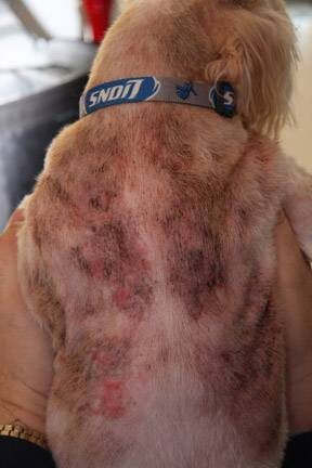
For short-haired dogs, a fine white dandruff begins on the head and ears that ultimately progress to the whole body. 

 Scaling areas tend to be round or S-shaped. For long-haired or plush-coated breeds, the scale is most obvious as a clump of dandruff sticking to a group of hairs. The coat is dull and brittle and tends to develop a reddish tint. In time, bald spots develop. Again, usually, the head is where problems start.

Biopsy of the skin is required for diagnosis.

Treatment

Response to therapy largely depends on whether there are still living sebaceous glands in the biopsy sample or not. If there are no glands left and they have all been destroyed, it will be much harder to get a response though it is possible for glands to regenerate with treatment. The inflammation that destroyed (or is destroying) the sebaceous glands must be stopped. Furthermore, new hair growth depends on a coat of sebum (skin oil), so if the patient’s skin is not making adequate skin oil, the oil must be replaced.

Topical

Oil replacement treatments are performed frequently at first and taper down to less frequent applications after the first month. Every dermatologist seems to have their own preferred regimen, so here are some typical recommendations:

Mineral oil, mineral bath oil (such as Alpha Keri®), or propylene glycol mixed with water, sprayed onto the coat or affected area of the coat, rubbed in, and shampooed out with a degreasing shampoo after a one-hour soak. 

This is performed typically once a week for the first month, then as needed. There are topical products that enhance the barrier function of the skin, and these can be used to assist therapy. Omega 6 fatty acid topicals are also becoming more popular as an easy way to replenish skin oil as they are easily applied as a top spot.

Oral Supplements

Oral omega-3 fatty acids are almost always included in the treatment regimen. Vitamin A has been advocated for sebaceous adenitis but seems to be fading out of popularity with the advent of other oral treatments (see below).

Oral Medication

Cyclosporine seems to be at the heart of treatment for the more severely affected dogs; further, on cyclosporine, dogs with only two percent of their sebaceous glands left have been able to increase this number to 40 percent. Cyclosporine is an immunomodulator that is able to suppress the inflammation that is destroying the sebaceous glands. Patients may need ongoing low dose therapy.

Synthetic retinoids have held a great deal of promise in treating sebaceous adenines, but they are tightly regulated by the government and are not readily available for veterinary patients.

Immunomodulation with doxycycline is an emerging therapy but is still in the investigational stage. Doxycycline is, first and foremost, an antibiotic but has numerous immunomodulating properties as well and is used widely to treat immune-mediated disease.

Managing a dog with this condition can be labor-intensive for the dog’s lifespan. The amount of work needed at home depends on the individual dog’s response to therapy. Marked improvement is typically realized within 4 months of treatment, but it is important to realize this can be a high-maintenance skin disease.