4128818

An Overview of Sarcoptic Mange (Scabies) in Dogs  

The Organism and How It Lives

Sarcoptic mange is the name for the skin disease caused by infection with the Sarcoptes scabiei mite. Mites are not insects; instead, they are more closely related to spiders. They are microscopic and cannot be seen with the naked eye.

Adult Sarcoptes scabiei mites live three to four weeks in the host’s skin. After mating, the female burrows into the skin, depositing three to four eggs in the tunnel behind her. The eggs hatch in three to 10 days, producing larvae which, in turn, move about on the skin surface, eventually molting into their nymphal stage and finally into adults. The adults move on the skin surface where they mate, and the cycle begins again with the female burrowing and laying eggs.
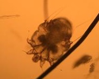
Appearance 

The motion of the mite in and on the skin is extremely itchy. Furthermore, burrowed mites and their eggs generate a massive allergic response in the skin that is even itchier.

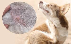
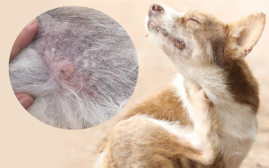
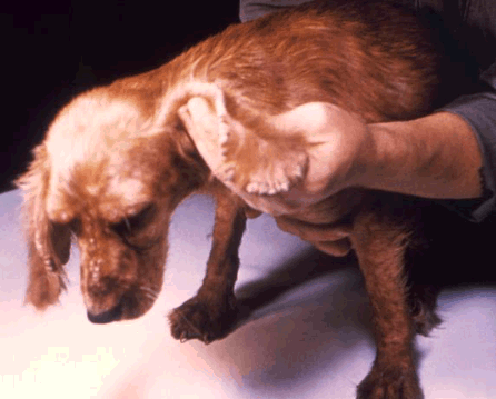
Mites prefer hairless skin, and thus, the ear flaps, elbows, and abdomen are at the highest risk for the red, scaly, itchy skin that characterizes sarcoptic mange. This pattern of itching is similar to that found with environmental allergies (atopy) as well as with food allergies. Frequently, before attempting to sort out allergies, a veterinarian will simply treat a patient for sarcoptic mange as a precaution or change to a flea product that will cover sarcoptic mange mites. This is an easy way to be sure sarcoptic mange is crossed off the list of possible skin disease causes. It is easy to be led down the wrong path and pursue allergies aggressively if sarcoptic mange is considered too unusual or unlikely.

As the infection progresses, eventually, most of the dog’s body will be involved. Classically, though, the picture begins on the ears (especially the ear margins), elbows, and abdomen.

The term scabies refers to mite infestations by either Sarcoptes scabiei or other closely related mite species. While Sarcoptes scabiei can infect humans and cats, it tends not to persist on these hosts. When people – including some veterinarians – refer to sarcoptic mange or scabies in a cat, they are usually referring to infection by Notoedres cati, a mite closely related to Sarcoptes scabiei. In these feline cases, it is more correct to refer to notoedric mange, though the treatment for both mites is largely the same.

Scabies vs. Sarcoptic Mange: Are They The Same Thing?

The term “scabies” refers to mite infections caused by Sarcoptes scabiei mites or other mites closely related to them (infections caused by the Sarcoptiform mite family). For dogs, this is usually Sarcoptes scabiei. For cats, this is usually Notoedres cati, but could be Sarcoptes scabiei. In humans, “scabies” usually refers to infection with Sarcoptes scabiei hominis, a mite that infects only people, although the canine mite discussed here (Sarcoptes scabiei) can most certainly infect people. 

How The Infection is Spread

While mites can live off of a host for days to weeks depending on their life stage, they are only infective in the environment for 36 hours, which means that decontamination of the environment is generally not necessary. Spraying, deep cleaning, etc., is not necessary, although machine washing bedding is advised.

Different varieties of Sarcoptes scabei mites infest their specific mammals. Sarcoptes scabiei var. canis mites infest dogs, while Sarcoptes scabiei var. hominis infests humans. Although the mites can transfer from one species to another, the infections are typically self-limiting (i.e. they go away on their own). The mites cannot complete their life cycles (they cannot reproduce and live continually) on the “wrong” hosts.

As previously discussed, when dogs are infected with Sarcoptes scabiei, those mites can transfer to people. The mites burrow into human skin, causing significant itchiness and irritation which sometimes leads to secondary skin infections (from humans scratching themselves and introducing bacteria into the skin). The mites are most active where the skin is warm, such as in bed, or where clothing is snug. Human infestations caused by dog-specific mites usually go away on their own within 12-14 days.

Always see your physician if you have concerns about your skin while your pet is experiencing a skin condition.

If a pet affected by sarcoptic mange is in the home, it is a good idea to wash any bedding in the washing machine (or replace it with new bedding) and wash any collars or harnesses.

Diagnosis

Skin Scraping


Classically, mite infection is diagnosed by scraping the skin surface with a scalpel blade and examining the skin debris under a microscope for mites. If the mite’s presence is confirmed by skin scraping, then you know immediately the cause of the itching; you need not be concerned about allergy possibilities or other diseases, and the condition can be addressed with confidence.

When an animal with sarcoptic mange scratches himself, he breaks open the tunnels that the mites have burrowed into, and the mites are killed, although the itch persists due to toxins in the skin. The result is that the mites can be difficult to confirm by skin scraping tests. (Probably mites are confirmed in 50 percent or fewer of sarcoptic mange cases.)

Medication Trial


Since negative test results do not rule out mite infection, a “maybe mange” test is frequently performed. This consists simply of treating for sarcoptic mange and observing for resolution of the signs within two to four weeks. Treatment is simple and highly successful in most cases, so it is fairly easy to rule out sarcoptic mange with a trial course of medication. See below for treatment options.

Biopsy


Mange mites are rarely seen on a skin biopsy sample, although if the sample is read by a pathologist who specializes in reading skin samples, the type of inflammation seen in the sample can be highly suggestive of sarcoptic mange. As a general rule, if the skin is biopsied, it is a good idea for the veterinarian to request that a dermatohistopathologist read the sample.

Treatment

While sarcoptic mange is difficult to diagnose definitively, it is fairly easy to treat, and a number of choices are available.

Remember, all dogs in a household where sarcoptic mange has been diagnosed should be treated.


At this point in time, many common flea products will get rid of a scabies infection handily. Using one of them will not only treat the scabies but will prevent future episodes as well. Some of these common products can be used the way they are normally used to prevent fleas, while others need an “off-label” dosing schedule that could be an issue for dogs with the MDR1 mutation.

Some individuals have a mutation of the MDR1 gene that interferes with the dog’s ability to metabolize many drugs, including those related to ivermectin, a broad-spectrum antiparasitic. This is not an issue at label doses but can be really toxic at the doses used to kill mites. These individuals are usually of the Collie family: Collies, Shetland Sheepdogs, and Australian Shepherds are classically affected. There is now a DNA test that can determine if any dog has the mutation.  If there is any question, a medication that does not require an off-label dose adjustment should be used for mange treatment.

The Isoxazolines


Oral flea products that cover both fleas and ticks are popular and include Nexgard®, Simparica®, Bravecto®, and Credelio®. Any of these will readily kill sarcoptic mange mites in one dose just as readily as they handle fleas and ticks. The MDR1 mutation does not come into play.

Selamectin (Selarid® or Revolution®)


Selamectin is an ivermectin derivative for dogs to control fleas, ticks, heartworm, ear mites, and sarcoptic mange mites. Normal monthly use of this product should prevent a sarcoptic mange problem, but to clear an actual infection, an extra dose is usually needed after 2 weeks for reliable results. This extra dose could possibly be an issue for dogs with the MDR1 mutation.

Moxidectin (Advantage Multi) – Moxidectin is yet another ivermectin derivative. In Advantage Multi, it is combined with imidacloprid, a flea-killing topical, to create a product used against heartworm, hookworm, roundworm, whipworm, and fleas. In the U.S., this product is now FDA labeled for sarcoptic mange and is also a good choice where there is concern about the MDR1 gene mutation as one regular dose should handle the mange mites. 

Milbemycin Oxime (Interceptor®, Sentinel®, or Trifexis®


Milbemycin oxime is approved for heartworm prevention as a monthly oral treatment and is available combined with oral flea products as well. Happily, it also has activity against sarcoptic mange, and several protocols have been recommended. There could be issues with the MDR1 mutation, depending on the protocol.

Ivermectin


This is one of the most effective treatments against Sarcoptes scabei yet is off-label as far as the FDA is concerned. There are several protocols due to its long activity in the body. Typically, an injection is given either weekly or every two weeks in 1-4 doses. Because there are so many safer and more convenient derivatives at this point, we mostly mention it for its historical significance because, before its development, mange treatment was labor intensive with repeated dipping and bathing. Ivermectin opened the door for a simple treatment for this condition.

Dipping 


Here, a mite-killing dip is applied, usually following a therapeutic shampoo. Weekly mitaban dips (Amitraz), or lime-sulfur dips are usually effective. The disease typically resolves within one month. Dipping is troublesome, messy, and rarely done anymore as the other products are easier and more rapidly effective. We only mention it since it had been a standard mange treatment for decades prior to ivermectin.

In the Meantime

During the time it takes to control the mite infection, the pet will be very itchy. Controlling secondary bacterial infections with antibiotics is important. Also, since the body’s reaction to the mite is one of hypersensitivity (essentially an allergic reaction), a cortisone derivative is worthwhile to quell the itchiest symptoms. Ask your veterinarian about which prescriptions are appropriate for your pet. As for anti-itch shampoos, rinses, and other forms of itch relief, see itch-relieving ideas for additional suggestions.
4127790

Alopecia X is a Pattern of Baldness

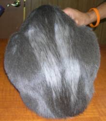
Alopecia is the medical term for baldness. The term does not specify a particular type, pattern or cause of baldness; it is a general term. There are many types of alopecia/baldness. One particular type of baldness has been described in the Nordic or plush-coated breeds whereby the dog develops coat loss on the main body as well as darkly pigmented skin in the bald areas. The legs and head are generally spared and retain a normal coat.

The condition we call “alopecia X,” however, is not associated with the hormone imbalances that normally create endocrine alopecia. Its causes remain mysterious hence the name alopecia X. Given that there are numerous therapies that work for some cases and not for others, and that many of these therapies seem to be in complete opposition, it may be that alopecia X is not one disease but several and we simply do not know how to distinguish them.

Alopecia X goes by many names:

  • Black skin disease 
  • Growth hormone-responsive alopecia 
  • Castration-responsive alopecia 
  • The coat funk 
  • Pseudo-Cushing syndrome 
  • Biopsy-responsive alopecia 
  • Follicular dysplasia of the Siberian husky 
  • Adrenal sex hormone alopecia 
  • Hair cycle arrest

The following is what is currently believed about this confusing condition.

The good news is that alopecia X is a cosmetic condition only. There is no downside to the dog except for looking funny. For this reason, treatment with medications is frequently discouraged because drugs can have bodywide effects while the disease itself has none. Furthermore, treatment has been fraught with partial responses and can be frustrating if not expensive, depending on what therapy is selected.

The skin biopsy is particularly important in making a diagnosis of alopecia X. If possible, a pathologist who specializes in reading skin tissue should be requested. The biopsy will identify structures typical of alopecia X hair follicles and help rule out concurrent allergy or infection that might mimic alopecia X.

The University of Tennessee Hormone Profile

One option in the pursuit of effective alopecia X therapy is the adrenal sex hormone panel available at the University of Tennessee. This test is done by drawing a baseline blood panel, administering a pituitary hormone called ACTH, and drawing a second blood sample an hour later to compare. Samples are shipped to Tennessee for evaluation for numerous adrenal sex hormones. The results show not only which hormones respond abnormally but the university will make suggestions as to which therapy might be likely to work.

Testing is not inexpensive and results can take several weeks to obtain but may help in selecting what therapy makes sense to try next. Often, results are ambiguous and difficult to interpret. Different specialists have different opinions on the usefulness of information obtained. This blood test may be recommended by your veterinarian as part of the alopecia X work up so we mention it here.

The Typical Patient    

The typical Alopecia X patient is a Spitz or Nordic breed such as an American Eskimo, Chow Chow, Pomeranian, Alaskan Malamute, Elkhound, or similar. Poodles have also been over-represented. Hair loss begins in early adulthood, usually by age three years. First, the long primary hairs go, leaving a fuzzy, puppy-like coat but eventually that goes, too. The bald skin becomes hyperpigmented but is not itchy, and the skin does not usually get infected.

Diagnostic Testing

Part of the problem is that all hormone-based hair losses can look exactly like this, so some testing is needed to determine which of several conditions are occurring.

Expect your veterinarian to begin with:

  • A blood panel 
  • A urinalysis 
  • Some kind of thyroid testing 
  • Some kind of adrenal hormone testing 
  • A skin biopsy

The purpose of this rather broad testing is to rule out diseases that look like alopecia X but for which well-defined treatment protocols exist. This means that two conditions must absolutely be ruled out before proceeding with the trial and error process of alopecia X treatment.

  • Cushing’s disease
  • Hypothyroidism

Both these hormone imbalances lead to endocrine alopecia and while they look like alopecia X, they have their own specific treatments.

Now that it has been determined that the dog in question has Alopecia X, it would be great if we could wake up the sleeping hair follicles and regrow some hair. It turns out there are several therapies to choose from. The problem is that while some dogs will respond with hair growths to a therapy course, other dogs will not respond at all. Let’s see what the choices are:

Sterilization


Alopecia X seems to be a sex hormone imbalance in at least some cases and didn’t earn the name castration responsive alopecia for nothing.

For this reason, the first step in treatment is to sterilize the patient; unspayed females should be spayed, and intact males should be neutered. There are health benefits to sterilization regardless of whether or not there is a hair loss issue, and many animals will grow their hair back (though possibly not permanently) so this is where we start rather than investing in complex and confusing diagnostics. If this does not work or the pet is already sterilized, then we move on to melatonin supplementation.

Melatonin


Melatonin can be obtained in 3 mg tablets at most health food stores or vitamin retail outlets. Approximately 40 percent of dogs will show some response within six to eight weeks. The medication should be given for at least two or three months before giving up but if hair regrowth occurs, the medication is continued until hair growth seems to have plateaued. After maximal hair regrowth has been achieved, the dose is gradually tapered down to a weekly dose over several months.

Some dogs can ultimately discontinue medication though it is important to realize that if you discontinue the medication and the hair falls out again, the condition may not be responsive to melatonin a second time. Recently melatonin implants have become available in the U.S. These are implanted in a similar manner to a microchip and dissolve under the skin over time. These can be used as an alternative to the oral product.

Melatonin Side Effects and Issues

  • Melatonin has been used as a sleep aid. Some owners find the sedating side effect to be unacceptable. Consider giving it at bedtime so that drowsiness is less noticeable.
  • Melantonin should not be used in diabetic patients as it has been found to create insulin resistance.
  • Beware of melatonin brands containing xylitol. Xylitol is an innocuous sugar substitute for people but is a poison for dogs.

Since melatonin is a nutritional supplement, rather than a prescription medication, the FDA does not insist on the same quality control it does for drugs. There may be tremendous differences in the amount of melatonin contained in pills between brands. Nature’s Bounty® brand has been a preferred brand but any major supplement brand should be acceptable.

If neither sterilization nor melatonin have been fruitful and we know the dog does not have Cushing’s disease or hypothyroidism, then it is important to realize that the therapies left to try have potential side effects.

Consider this: Alopecia X is a cosmetic condition. It may make the dog look funny, but it does not cause harm.

You will need to weigh the potential side effects of therapy against your pet’s appearance. That said, there are other therapies that can be attempted.

Micro-Needling


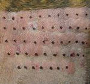
Alopecia X is sometimes called biopsy responsive alopecia. After the skin biopsy samples are taken in the diagnostic process described above, some dogs will regrow a small tuft of hair in the biopsy area. The theory is that the inflammation associated with healing has awakened the hair follicles in that local area. This concept is applied more broadly with micro-needling, in which a roller of tiny needles is rolled over the skin creating tiny punctures and putting the skin into a healing mode. This procedure is widely used in human aesthetics to generate collagen, remove wrinkles and brighten skin complexion. The needles can be painful or at least uncomfortable so the patient may require sedation or numbing with topical anesthetics.

The skin is somewhat tender for a day or so after the procedure. Different studies report different success rates ranging from 40 to 90 percent. Because some discomfort is afforded the patient and sedation is typically needed, plus alopecia X is of cosmetic concern only, using this treatment is somewhat controversial.

Deslorelin Implants


Deslorelin is a veterinary hormone that curtails the production of estrogen and testosterone and is usually used to time ovulation in mares. It is available as an implant and was recently tested in alopecia X. Within three months, 60 percent of the unneutered male dogs experienced hair regrowth. None of the spayed females experienced hair regrowth. No side effects were noted during the one-year period of testing. This may pan out as a treatment strategy for unneutered male dogs.

Medroxyprogesterone Acetate


Injections of this long-acting progesterone were used monthly for four months in a group of Pomeranians with alopecia X. Some responded partially, one completely regrew hair, and some did not respond at all. None of the dogs had side effects during the time of the study; however, progestins are famous for inducing mammary tumors, uterine infections, and diabetes mellitus so their use should be weighed against their potential for problems.

Trilostane


Cushing’s disease is a disease of adrenal cortisone excess. The production of cortisone is complex and some of the pathways are common to sex-hormone production. For this reason, therapy for Cushing’s disease sometimes works on alopecia X. Trilostane is a medication used to treat Cushing’s disease to suppress steroid hormone production, which includes the sex steroids. Trilostane has the potential to create a dangerous deficiency in adrenal steroids so it is not without risk even though it has been effective in causing hair regrowth in some patients.

Alopecia X is a frustrating condition and will remain frustrating for years to come. Research is ongoing and progress comes gradually.

7633845

Allergies: Atopic Dermatitis in Dogs and Cats

Atopy

Atopy is defined as the genetic tendency to develop allergic diseases. These diseases are due to a heightened immune response to common substances in the environment, known as allergens.

In atopic dermatitis, allergens, including pollens, molds, dust particles, etc. make their way to the skin and then gain entry through a defective skin barrier. Inhaling is not necessarily involved. The immune system becomes reactive to the airborne allergens and soon the inflammation, itching, and scratching begins. Even if the allergens come from the air, the itch is felt in the skin, leading to scratching, hair loss, redness, and skin thickening.

Allergens (pollen, dander, etc.) are harmless to someone who is not allergic to them. Atopic dermatitis has a genetic basis.

Breeds predisposed to develop atopy include Dalmatian, Golden retriever, West Highland white terrier, Shar Pei, Labrador retriever, Cairn terrier, Lhasa Apso, Shih Tzu, Boxer, and Pug.

Features of Atopy in Dogs

There are many reasons for pets to itch: parasites, allergy to flea bites, food allergy, secondary infection, and the list goes on.

The following are findings in the history and examination of the patient that might lead to a diagnosis of atopy.

In fact, meeting five criteria from this list yields an 85 percent accuracy for the diagnosis of atopic dermatitis, at least for dogs:

  • Young Age of Onset
    Seasonal itchiness due to atopy tends to begin early in a pet’s life (between ages 1 and 3 years in 70 percent of dogs diagnosed with atopy). Food allergy tends to begin later (more like age 5 or 6 years in dogs) or earlier (less than 6 months of age). The age at which itching first manifests is not as reliable a sign in cats as in dogs.
  • Mostly Indoor Lifestyle
    It is indoors where many airborne allergens are concentrated; however, it is important to note that allergens in the air are in the air for miles so it is not easy to escape allergens by simply going outdoors or by simply going indoors.
  • Good Response to Steroids
    Whether the patient is a dog or cat, itchiness due to atopy responds rapidly to cortisone-type medications (prednisone, betamethasone, dexamethasone, etc.) as does itching due to insect bite allergy. Food allergy is more variable in its response; it may or may not respond well.
  • Chronic or Recurring Yeast Infections in the Skin
    Yeast (Malassezia pachydermatis) live on the surface of the skin normally but with all the changes allergy causes to the skin’s microenvironment, yeast will proliferate and create a stinky, thickened, pigmented skin that resembles elephant skin or even tree bark. Environmental allergy is a strong predisposing factor in developing yeast dermatitis.
  • Front Feet Involved
    Whether it is chewing the top of the feet or between the toes, foot licking is a classic feature of atopic dermatitis.
  • Ear Flaps Involved
    The non-haired (inside) portion of the ear flap (the concave pinna) is commonly involved in atopic dermatitis regardless of whether the inflammation extends down the ear canal and leads to what we usually think of as an ear infection. The concave pinnae becomes greasy, red, thickened, smelly, or any combination of the above.
  • Ear Margins Not Involved
    Ear margin involvement is suggestive of sarcoptic mange and would lead one away from airborne allergies.
  • Lower Back Area Not Involved
    The lower back is the flea bite zone. There are few conditions other than flea bite allergy that cause itching in this area, so if this area is involved, make sure flea control is achieved before further evaluating the skin.
  • Seasonality
    Seasonality of the itching is also a clue towards an airborne-related allergy, but since there are many climates where seasons are ambiguous, this is not necessarily a hard and fast feature of atopic dermatitis. It should also be noted that while skin testing and blood testing are used to make immunotherapy sera to treat atopic dermatitis, reactions and positive results on these tests do not contribute to making a diagnosis of atopic dermatitis. Many normal animals will have positive results. These tests are for animals where the diagnosis of atopic dermatitis has already been made based on the above criteria as well as ruling out other itchy diseases.


Testing for Atopic Dermatitis

Unlike other diseases where a test of some sort can be performed, atopic diagnosis is a clinical diagnosis, which means the diagnosis is made based on symptoms and findings such as those listed above. Allergy testing, which is done either by skin test or by blood testing, is not a test for atopic dermatitis; instead, this type of testing is done after the diagnosis has been confidently made based on findings in the patient. Allergy testing is conducted to determine how best to make an allergy shot serum for a specific patient; it is not part of disease diagnosis.

In cats, the irritation pattern is not as characteristic. There are four common manifestations of atopy. Unfortunately, these same irritation patterns can be found in numerous other skin conditions and, in fact, up to 25 percent of atopic cats have multiple types of allergies.

Treatment Options

Many people are frustrated because treating atopic dermatitis focuses on controlling the symptoms. Unfortunately, finding out what a pet is allergic to is not entirely possible and even when it is, because pollens and other material circulate in the air for miles, you can’t just avoid allergens in the air. Only hypersensitization treatment stands a chance at actually changing the patient’s immune system so that they are no longer allergic, but this is far from a sure thing. The good news is that at the end of the day, the goal is healthy comfortable skin and there is a lot that can be done towards achieving this. How big a project this becomes depends on an individual patient’s ability to respond to different therapies or combinations of them. Here are some of the options.

Clearing Secondary Infections

Before doing anything else, it is important to clear up secondary infections. Secondary infections involve bacteria (usually Staphylococcal) and/or yeast (Malassezia) at the site of the itchiest areas on the body. These organisms live naturally in the skin but when the skin is irritated, they gain access to inner tissue layers and proliferate. Sometimes they generate further allergic response in the skin. These infections tend to recur and are the usual cause of recurrence of itch symptoms in a patient who was previously controlled.

Hyposensitization (Allergen Specific Immunotherapy)

Hyposensitization, more commonly known as allergy shots, is by far the treatment of choice for atopic dermatitis. All the other medications are basically just itch relief; only hyposensitization actually changes the immune system. Some dogs are eventually able to go off all treatments and are no longer allergic after they have been on hyposensitization long enough. Most dogs experience at least enough improvement to require fewer additional treatments but there are some caveats before making an appointment for allergy testing.

  • Allergy shots require approximately 6 to 12 months to begin working.
  • 25% of atopic dogs will not respond (these are usually the animals allergic to multiple allergens).
  • 25% will require prednisone or a similar steroid at least at some times.
  • You will most likely have to give the allergy shots yourself.
  • Referral to a veterinary dermatologist may be necessary.

In hyposensitization, the patient is injected with small amounts of allergens on a regular basis. As time passes, the amounts of allergens increase and injections are given at longer intervals. The selection of allergens is made based on the results of either an intradermal skin test (as described above), an in vitro test (a blood test) or a combination of the results of both tests. The younger the pet is when this treatment starts, the better it works.

Steroid Hormones

These cortisone-type medications (prednisone, prednisolone, triamcinolone, dexamethasone, etc.) have been useful as the first line of defense against itchy skin for decades, and they are still widely used. There are negative side effects with higher doses as well as with long-term use, so the trend is to use other medications to either remove the need for steroids or reduce the amount needed for itch control.

Side effects include:

  • Excess thirst
  • Excess hunger
  • Excess urination, which could lead to house-breaking issues
  • Suppression of the immune system/bringing out latent infection, especially urinary tract infection and upper respiratory infection.
  • Raising blood sugar/destabilizing a borderline diabetic (especially a problem for cats if long-acting injectable steroids are used). In the short term, side effects can be controlled by adjusting dosage but in the long term, these medications are more problematic and if possible their use should be minimized.

Steroid hormones are useful for acute flare-ups as well as for long-term management of atopic dermatitis (assuming limits are placed on how long they are used). Side effects are greatly minimized when steroids are used topically.

Cyclosporine

Cyclosporine is a modulator of the immune response that has been helpful in both human and non-human organ transplant patients. It has been found to be reliably effective in atopic dermatitis and does not carry the unpleasant side effect profile that steroids do. That said, it is not without side effects of its own. It is used mostly in dogs but can also be used in cats. It is for long-term management and is not helpful for acute flare up control.

Oclacitinib (Apoquel)

This is a new medication best used for itching relief and blocking itch symptoms. Apoquel is popular as it works fast. It does not address the inflammation in the skin; it just stops the itch sensation. This means that any skin infection causing the itch will still need to be controlled. 

Canine Atopic Dermatitis Immunotherapeutic (Cytopoint®) Injections

This is a new treatment that uses vaccine technology to eliminate one of the main mediators of itch sensation. The injections provide relief from itching for one month in 80% of dogs (many dogs get longer relief) and usually show effectiveness within 24 hours.

  • Bathing the pet weekly to remove allergens from the fur may be helpful in reducing allergen exposure, plus tepid water is soothing to itchy skin. There are also many therapeutic moisturizing shampoos that can be used to restore the skin’s natural barrier or to assist in general itch relief. 
  • Avoid stuffed toys, wash bedding regularly. This minimizes dust mite exposure. Also, remove the pet from the area when vacuuming or dusting.
  • Use air-conditioning and/or an air filter system.
  • Keep the pet away from the lawn while it is being mowed.
  • Minimize houseplants.

Omega 3 Fatty Acid Supplements

These products are not analogous to adding dietary oil to the pet’s food, such as olive oil, coconut oil, corn oil, etc. Instead, these special fatty acids act as medications, disrupting the production of inflammatory chemicals within the skin. By using these supplements, it may be possible to postpone the need for steroids/cortisones or reduce the dose of steroids needed to control symptoms. It takes a good 6 weeks to build up enough omega-3 fatty acids in the body to see a difference.

Ultra-Microsized Palmitoylethenolamide or PEA (Redonyl®)

Plants and animals make a natural substance called PEA. In animals, it helps restore balance to the skin’s biochemistry and prevents the release of the biochemical causes of itching. The commercial product uses a soybean extract to obtain its PEA and creates a super concentrate as it would not be possible to eat enough soybeans to get a therapeutic dose of PEA. The super small ultra-micronizing allows it to be absorbed better. It is available as a flavored chew for dogs and can also be used in cats.

Antihistamines

Antihistamines have been popular for many years for pets, and it seems their effectiveness does not stand up to scrutiny. They provide neither short-term relief nor reliable long-term relief. They may be helpful in combination with other products in that their use may reduce the need for other products. They may work better in cats than in dogs.

Solid Flea Control

Allergies are additive. This means that when a patient has multiple allergies, each allergy alone may not be enough to cause itching but the allergies all active together probably will. Consequently, taking away one of the active allergies may be enough to allergy reduction to resolve the itching. Flea bite allergy is extremely common. We now have so many effective products available that there is no reason for an animal to contend with a flea bite allergy. This simple bit of therapy (flea control) may be enough to bring the pet below her itch threshold without having to contend with any of the therapies listed above.

Itchy skin has been the scourge of dogs, cats, and their owners for decades if not centuries. We are now armed with a great understanding of immunology and have many tools to address allergy symptoms. Your veterinarian can guide you further with regard to a proper regimen. If your veterinarian decides treatment is not working as well as hoped, discuss whether or not a referral to a veterinary dermatologist is in your pet’s best interest.

4128352-1

Atrial Fibrillation in Dogs and Cats

What Makes a Normal Heart Beat?

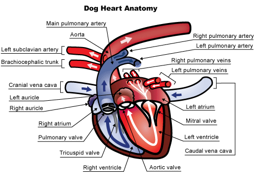
The heart is a large muscle with four chambers. There are two top chambers (left and right) called the atria and two bottom chambers (left and right) called the ventricles. These chambers fill with blood from the body and then contract to circulate or deliver poorly oxygenated blood to the lungs (right side) or well-oxygenated blood to the rest of the body’s tissues (left side).

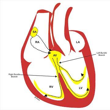
The heart muscles contract after they are stimulated by an electrical impulse. A small area of special tissue in the right atrium called the sinoatrial (SA) node starts an electrical impulse (it’s like the heart’s spark plug) that travels out through the atrial muscle, stimulating that muscle, and, simultaneously down special electrical tracts (AV node, Bundle Branches, Purkinje Fibers – the heart’s “wiring”) to the ventricles and ultimately out into the ventricular muscle (see Figure 2 below).

The electrical impulses cause the heart to contract in a coordinated fashion: the atria contract first, because they get stimulated first. As they contract, they push blood into the ventricles. Then, as the electrical signal reaches the ventricles, they also contract and push blood to either the lungs or the rest of the body. This coordinated electrical activity can be recorded on an electrocardiogram (ECG or EKG). There are several components to the normal ECG, called waves or complexes. They are labeled P, QRS and T. The P waves represent the atrial stimulation, while the QRS waves represent the ventricular stimulation. The T waves represent the re-setting of the ventricular muscles, so that they can be stimulated by the next impulse. The short pause between the P waves and the QRS waves is the time that the impulse is traveling along the wiring system from the atria to the ventricles.

Atrial fibrillation (sometimes called “A fib”) is an arrhythmia, an irregularity of the heart’s rhythm.  Instead of the SA node (the spark plug) starting and coordinating the electrical signal, many areas within the atria send out electrical impulses in a rapid, disorganized manner.  This whirlwind of electrical impulses or wavelets spreads over the atrial tissue causing the atrial muscle to quiver or fibrillate, instead of contracting in an organized, structured manner (Figure 3). Only a fraction of the electrical impulses are able to jump onto the “wiring system” and get across the AV node and down into the ventricles to make the ventricles contract.  The pattern with which all these abnormal atrial impulses are conducted through the AV node is irregular making the ventricles beat irregularly. 

What Causes Atrial Fibrillation?

In humans, atrial fibrillation can occur for no apparent reason (often in athletes or older people). This is because the size of our hearts is large enough to allow such abnormal rhythms to develop (veterinarians also see this type of atrial fibrillation in horses, which have large hearts). However, most pets have much smaller hearts than humans, so for atrial fibrillation to occur in your pet, the atrial chambers must usually be bigger than normal. This enlargement of the atrial chambers occurs with many types of heart disease, such as degenerative valve disease of the mitral valve or cardiomyopathy, dilated cardiomyopathy and hypertrophic cardiomyopathy. Generally, the smaller your pet, the more significant the heart disease must be to cause atrial fibrillation.  Thus, atrial fibrillation is much less common in cats than in dogs. And it’s less common in small breed dogs than large breed dogs.

Occasionally, giant and large-breed dogs develop atrial fibrillation without any other heart disease, similar to what we see in humans and horses (let’s face it, some Great Danes are almost as big as horses!). This is termed “lone” or primary atrial fibrillation.  In some cases, this is “lone” atrial fibrillation turns out to be the first indication of dilated cardiomyopathy; in other cases, the arrhythmia exists without underlying heart disease.

How Is Atrial Fibrillation Diagnosed?

Often, the first idea that a pet has atrial fibrillation is when your veterinarian detects an erratic heart rhythm while listening to the heart with a stethoscope. Many veterinarians describe the sound as bongo drums or shoes in a dryer. Your pet’s pulse quality may also be irregular. However, to confirm the diagnosis an electrocardiogram (ECG) needs to be performed (because other arrhythmias can sound like atrial fibrillation. There are specific ECG criteria that must be met prior to making the diagnosis.

At times, a pet with severe heart disease may suddenly become weaker, or have a relapse of heart failure upon development of atrial fibrillation, prompting you to visit your veterinarian. Again, the veterinarian will auscultate the abnormal rhythm and diagnose atrial fibrillation via an ECG examination.

In giant breeds with lone atrial fibrillation, the arrhythmia may be detected on routine examination – often the owners are unaware that any problem exists. Some dogs may develop mild exercise intolerance when they develop atrial fibrillation.

What Happens When a Pet Develops Atrial Fibrillation?

People with lone atrial fibrillation typically require treatment to re-establish a normal heart rhythm because they usually have exercise intolerance or shortness of breath, symptoms of palpitations, and are at an increased risk of having a stroke. If a normal rhythm cannot be re-established, people are often placed on “blood thinners” to reduce the risk of stroke. However, dogs with lone atrial fibrillation rarely show exercise intolerance or shortness of breath and do not suffer from strokes, so treatment is rarely required. Their heart rate remains normal in most cases.

However, in a pet with serious heart disease, atrial fibrillation causes a worsening of clinical signs, and can further damage the heart. Cardiac output, or the amount of blood pumped out of the heart to the rest of the body, is reduced and often congestive heart failure develops or recurs. These pets usually have very fast heart rate (more than 200 beats per min) which, if not controlled, can lead to additional heart muscle damage and dysfunction.

How Is Atrial Fibrillation Treated?

Treatment in people is aimed at re-establishing and maintaining a normal rhythm, because this greatly reduces the risk of strokes. This is usually achieved by delivering a “shock” to the heart (termed electrocardioversion), or by using antiarrhythmic drugs. Recent studies in people suggest that re-establishing a normal rhythm is not essential and medical therapy might suffice if clinical signs are tolerable or absent.

In pets, cardioversion is generally not feasible. Several studies have examined electrocardioversion or chemical cardioversion in dogs with lone atrial fibrillation – none has shown consistent success. Newer studies are being conducted that may ultimately provide a technique that allows a normal rhythm to be established in these pets.

Pets with severe heart disease are generally not candidates for cardioversion. In these pets, a normal rhythm can rarely, if ever, be re-established. Instead, the veterinarian attempts to reduce the high heart rate with antiarrhythmic drugs. By reducing the heart rate (which is often more than 200 beats per minute), the veterinarian allows the heart to pump more efficiently, and prevents some of the damage that the high heart rate can produce. Additionally, the clinical signs associated with the development of atrial fibrillation (that is, congestive heart failure) resolve when the rate is controlled. The drugs that decrease the heart rate need to be given indefinitely to maintain rate control and will generally be added to drugs already being administered. Studies have shown that dogs in which the heart rate is reduced to 120 beats per minute (average over a day) do better than dogs in which the heart rate is not as well controlled. In other words, the closer to “normal” your veterinarian can bring the heart rate, the better the outcome.

People with atrial fibrillation are usually treated with anti-coagulants, such as warfarin, or other similar drugs (commonly, but incorrectly, termed blood thinners), to reduce the incidence of strokes, which can be devastating or fatal. However, dogs with atrial fibrillation do not appear to have increased risk of strokes and are therefore not treated with anti-coagulants. Cats with atrial fibrillation usually have severe heart disease, and are often treated with anti-coagulants whether they have atrial fibrillation or not.

What is the prognosis for a pet with atrial fibrillation?

Lone atrial fibrillation, like we see in large or giant-breed dogs, is well tolerated, even if we cannot convert it back to a normal sinus rhythm. In dogs with underlying severe heart disease, 50 percent of those with higher heart rates (more than 125 beats per minute) in one study had died from their heart disease within four months. On the other hand, 50 percent of those with lower heart rates (less than 125 beats per minute) lived to at least three years. Whether or not these numbers reflect what will happen with all dogs is hard to say, but we can say that bringing the heart rate close to normal improves survival.

4128968

Ataxia in Dogs and Cats

When your pet is stumbling and staggering around, almost as though drunk, the condition is called ataxia. It is an inability to make normal, coordinated, voluntary movements but is not caused by muscle weakness, involuntary spasms, or too little strength to move. Generally speaking, ataxia is a symptom caused by some type of central nervous system problem in which the brain cannot correctly tell the body what to do.

Ataxia affects a pet’s ability to coordinate their head, legs, and body. Perhaps the pet cannot place their foot appropriately while walking, resulting in the pet knuckling their feet and dragging their toes on the ground. Since the pet does not realize where their feet are, they cannot just flip the feet over to walk normally. Dragging knuckles on the ground can damage the tops of their feet. Some pets with ataxia do not knuckle their toes, but their foot placement is exaggerated.

Ataxia needs to be differentiated from lameness or weakness due to musculoskeletal disease or generalized illness.

Three types of ataxia can be seen in dogs and cats, depending on whether the inner ear, brain, and/or spine is involved: 

  • Cerebellar (brain)
  • Proprioceptive (brain or spine)
  • Vestibular (inner ear)

Cerebellar

The cerebellum is the part of the brain that coordinates voluntary movement, allowing your pet to reach toward the water bowl or you to move your arm appropriately when you want to pick something up. Common causes of cerebellar ataxia in pets are congenital defects and inflammatory diseases. Other causes include degenerative neurological diseases, brain tumors, and strokes.  

The pet’s head and torso sway and stagger (see video) and their feet step up high and wide in an exaggerated walk as though they are about to go up steps even though they’re not. They may have head tremors.

Proprioceptive

Proprioception is an awareness of where your body is positioned in space, strengthening coordination. It tells us how much force to use when pushing or pulling. Proprioception is how your pet knows without looking if their tail or foot is up or down, and you know if you are standing up straight or slanted. Proprioceptive ataxia is caused by a problem in the cerebral cortex of the brain or the spine. Common causes include brain tumors, infectious diseases of the brain or spinal cord, traumatic brain injuries, injured disks, and strokes. 

The pet’s unsteady body is seen swaying, knuckling, feet crossing over, and with a wide-based stance. You would see an inability to get the limbs into their normal positions. This form can affect one or more limbs and the torso.

Vestibular

Vestibular ataxia is caused by a problem in the vestibular system in the inner ear or within the balance center in the brain, both of which contribute to balance. The vestibular system helps your pet stay upright when walking forward and lets you walk or run on uneven ground without falling. The vestibular systems signal to the brain to let the eyes and extremities know what they are supposed to do. Here, the ataxia is characterized by leaning and falling to one side or, less commonly, both sides. It affects the head, trunk and limbs. It’s often the easiest form of ataxia to recognize and often is worse if the animal is removed from contact with the ground.

Pets with vestibular ataxia may have a head tilt, walk like they are going in a circle rather than straight ahead, and may have odd back-and-forth eye movements called nystagmus. Drooling, leaning, rolling, and falling are common. Nausea or vomiting may occur as a result of dizziness.

Diagnosis

Your veterinarian will start by taking a detailed history of what has been happening to identify likely causes and will do a neurological examination (neuro exam) to determine where the problem is located. A neuro exam is quite simple and has two objectives: (1) Confirm that the pet has a neurological condition, and (2) see where the problem is located. A neuro exam may sound like you need to see a specialist, but you do not as the exam does not require special knowledge or expensive equipment.  Your veterinarian will watch your pet walk, move, check reflexes, see what happens when they lift your pet’s leg, and other tests to see what your pet’s physical reaction is. The neuro exam checks on:

  • Mentation (level and quality of consciousness)
  • Posture of head, limbs, and body
  • Gait (how the pet walks)
  • Postural reactions (incoordination)
  • Spinal reflexes (normal, diminished, or increased)
  • Cranial nerves (e.g., vision, eye movements, facial sensation, etc.)
  • Palpation of the spine to look for swelling or atrophy
  • Nociception (ability to sense pain)

A neuro exam will suggest the location of the problem but cannot by itself determine which disease your pet has. The history you provide is the most important information in determining which diseases are likely causes of abnormal signs.

Treatment

Treatment depends on what is happening with your pet. Your veterinarian needs to figure out where the problem is located, and which diseases are most likely, to determine which tests and treatments to recommend. Hospitalization with some medication to help the pet’s specific signs, such as vomiting, and some IV fluids may be of most use, especially early in the game. Good nursing both at the clinic and at home can be helpful. 

The prognosis for pets with ataxia depends on where the lesions are located and what the specific disease is that is causing the abnormal signs. Some diseases are benign, and recovery only requires time and supportive care. Other diseases are serious and may be difficult to resolve.

5695881

Anaplasmosis in Dogs and Cats Is Tick-Borne

Anaplasmosis is a disease of dogs and rarely of cats caused by Anaplasma phagocytophilum. This infectious organism is an unusual type of bacteria known as a rickettsial agent/bacterium. The disease is transmitted to dogs and cats by a tick bite. As with some other tick-borne diseases, a tick must remain attached to the pet for more than 24 hours for the infection to be transferred.

It has been reported from around the world in a variety of animals.

Signs of Anaplasmosis

Signs usually begin 1 to 2 weeks after the tick bite. Anaplasmosis organisms enter the bloodstream and live in the animal’s white blood cells, which normally aid in fighting infections.

This invasion of circulating white blood cells creates inflammation throughout the body. Signs can include poor appetite and fever. Joints are commonly affected, making the pet seem stiff or painful or appear to have trouble walking. Sometimes pets may have signs associated with bleeding as well. A bloody nose, dark bloody stool, or bruising may be seen.

Not all pets will have symptoms. Some pets will only appear sick for a short time, then start to improve. We don’t fully understand why this happens, but it may be associated with a pet’s ability to fight off the infection. 

Diagnosing Anaplasmosis 

A veterinarian’s physical examination is often not useful as a sole means of diagnosis; however, in pets showing signs, it will point the veterinarian in the right direction for testing. A complete blood count, also known as a CBC, may indicate a decrease in red blood cells (anemia), or a decrease in platelets (thrombocytopenia). Sometimes anaplasmosis can be diagnosed by finding the organisms in a blood sample by using a microscope.

Additional lab tests may need to be sent off to confirm the diagnosis. Occasionally, these tests can be positive in a dog or cat who is not showing symptoms. This does not necessarily mean the pet has the disease, but it can mean that they were exposed to it at one point. The veterinarian will then need to take into account the exam, signs, and the rest of the lab work to make a proper diagnosis. 

Treatment and Recovery 

Anaplasmosis is treated with certain types of antibiotics, most commonly doxycycline. Treatment is usually for 2-4 weeks; the longer course tends to be given if the pet also has Lyme disease, but it all depends on the specific case. Pets usually feel better within a few days after taking the antibiotic. If the anemia or thrombocytopenia are severe, a blood transfusion may also be needed. Most dogs and cats make a full recovery.

Prevention

No vaccine is available to prevent anaplasmosis. The best form of prevention is tick control, either through treating the yard or through applying tick preventive medications to the pet. Consult your veterinarian for the best prevention options for each pet.

4128349

Anaplasmosis

Anaplasmosis is a tick-borne disease. Two forms of anaplasmosis are known: granulocytic anaplasmosis and infectious cyclic thrombocytopenia. Granulocytic anaplasmosis is more common. A dog can have both infections at the same time.

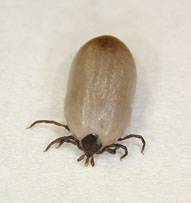
Transmission is via a tick vector (a vector is an organism that can passively carry and transmit disease). A tick needs to be attached for a minimum of 24 hours to transmit the organism. Incubation time is about 1 to 2 weeks. Reservoir hosts (a source of infection and possible reinfection that sustains a parasite) are usually small rodents, deer, etc.

Granulocytic Anaplasmosis

Granulocytic anaplasmosis is an infection of white blood cells. It is caused by Anaplasma phagocytophilum.

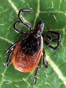
Anaplasma phagocytophilum is transmitted via a bite from an Ixodes tick. Ixodes scapularis (often commonly called the deer tick, blacklegged tick, or bear tick) is the primary vector in the Midwest and Northeastern United States; Ixodes pacificus (commonly called the Western blacklegged tick) is the primary vector in the Western United States; and Ixodes ricinus (commonly called the castor bean tick) is the primary vector in Europe.

Granulocytic anaplasmosis is seen most commonly in dogs in the Northeastern, upper Midwest, and coastal Western United States. Since granulocytic anaplasmosis requires the Ixodes tick as its vector, seasonal outbreaks of the disease can occur from spring through summer. Clinical disease is most often seen in adult dogs, and golden retrievers and Labrador retrievers appear to get it more than other breeds.

A. phagocytophilum can infect a wide range of mammals. In addition to dogs, the clinical disease has been documented in cats, cattle, sheep, goats, llamas, and humans.

Because of common vectors and rodent reservoirs, co-infection with Borrelia burgdorferi can be seen and can lead to more severe illness.

Clinical Signs and Prognosis

Many dogs exposed to granulocytic anaplasmosis do not get obvious signs of the condition. If signs are seen, they most often occur during the acute phase of infection, which is 1 to 2 weeks after transmission. The signs may be vague and include lethargy, lack of appetite, and fever. Some dogs may become lame because their joints are painful. Less common signs include vomiting, diarrhea, coughing, and difficulty breathing.

The prognosis for granulocytic anaplasmosis is quite good.

Infectious Cyclic Thrombocytopenia

Infectious cyclic thrombocytopenia is an infection of blood platelets. It is caused by Anaplasma platys.

Anaplasma platys transmission has not been fully determined, although tick vectors are probable. The organism has been found in Rhipicephalus and Dermacentor ticks.

Clinical Signs and Prognosis

Signs include lack of appetite, lethargy, fever, bruising on the gums and stomach, nosebleeds, and weight loss.

Many dogs with infectious cyclic thrombocytopenia have only mild clinical disease, so the prognosis is generally good.  

Diagnosis

Blood tests and urinalysis are the main diagnostic tools for anaplasmosis. The blood tests usually include a complete blood count, blood smear evaluation, biochemistry panel, serology to look for antibodies and polymerase chain reaction (PCR) assays. If the dog is lame, radiographs and analysis of joint fluid are usually included.

Treatment

Treatment includes antibiotics, pain relievers, and anti-inflammatory drugs.

Doxycycline is the most commonly used antibiotic. Most dogs respond within one to two days after they first take doxycycline. Other antibiotic options are tetracycline or minocycline.

Analgesia and anti-inflammatory drugs may be needed for joint pain. Let your veterinarian choose the anti-inflammatory, rather than choosing and dosing it yourself because dogs metabolize these medicines differently than humans do. Your veterinarian will have the most appropriate medication.

Disease Prevention

Because there is no vaccine for anaplasmosis, appropriate tick control is critical to preventing this disease. Preventing ticks from attaching and removing any ticks from your pet within a few hours of attachment is vital.

Note: Dogs with anaplasmosis may also be infected with other tick-borne organisms (Ehrlichia, Borrelia, etc.), so infected dogs should be screened for those diseases also.


6021696

Anal Glands and Anal Gland Abscess in Dogs and Cats

What are anal glands?

Anal glands are two sac-like structures that release an unmistakable, foul-smelling fluid. It’s really unfortunate when veterinarians get that fluid on their clothing because then they smell like that all day, as does the exam room. Dogs and cats use this fluid to mark their territory when they poop. Anal glands are not important for your pet’s health and can be removed if medically necessary (i.e, constant infections, recurrent abscess formation), but not without risking incontinence.

What animals have anal glands?

  • Female and male cats, dogs, and many other animals
  • People do have them. They are like sweat glands and much smaller vs. cat or dog anal glands.

What is the anatomy of an anal gland?

Each animal has two glands located on the left and right side of the anus. Each gland connects to the end of the anal canal through a small connecting tube (duct). When your pet is defecating (pooping), the feces (poop) passes through the anal canal and squeezes the sacs, releasing the smelly fluid.

What is an anal gland abscess?

An anal gland abscess is a painful infection of the anal glands. During an infection, pus builds up in the sac. The infection prevents the foul-smelling anal gland fluid from leaving the sac. The anal gland swells with the fluids and may even burst. This abscess is extremely painful and should be treated immediately.

Can my pet get an anal gland abscess?

Animals with anal glands can have an anal gland abscess. There is no breed, gender, or age group that is more likely to have this happen. There are a variety of potential explanations of why the anal gland swells and even ruptures, such as underlying diseases or allergies. By working together, you and your veterinarian will create the best treatment plan to prevent abscesses from forming in the future.

What are the symptoms?

  • Scooting across the floor to put pressure on the anal gland and release the fluid
  • Trauma and biting of the tail and anal area
  • Matting of the hair at the anal area
  • Reluctance to sit and poop
  • Constipation and anal discharge
  • Crying, whimpering or signs of pain
  • Being withdrawn and lethargic, not wanting to eat
  • Swelling and redness of anal area

Should I treat the anal gland abscess?

Yes, and treat it immediately. Although this is not a medical emergency, make an appointment with your veterinarian as soon as possible. An abscess is painful for your pet and infection can spread to different parts of the anus. The veterinarian will clean the abscess and treat the infection. In some cases, anesthesia or sedation is needed to clean it. Treatment only stops after your veterinarian has rechecked your pet to determine if the abscess is healing properly. The goal of this treatment is to minimize the swelling and discomfort to your pet. Treatment may include:

  • Cleaning and unclogging of the anal duct to prevent fluid buildup
  • Anti-inflammatory medications to reduce swelling and pain
  • Antibiotics to treat infection
  • Recheck by your veterinarian is necessary

Is there any additional testing needed?

There may be additional testing depending on each case. Standard tests that may be needed include complete lab work, especially if anesthesia is needed. In complex or unusual cases, culture or biopsy may be needed.

Does my pet need surgery?

These sacs only produce scent markers and are not necessary for your pet’s health. If your pet continues to have infected and abscessed anal glands, a more permanent treatment may be needed. This involves removing the anal glands entirely. The surgery may affect sphincter strength, causing incontinence. This severe side effect makes surgery a last resort for chronic anal gland abscess offenders.

How can I prevent it?

If your pet continues to have anal gland swelling and infection, your veterinarian may suggest high-fiber diets, hypoallergenic diets, or surgical removal of the anal sacs. Knowing the cause of your pet’s continued anal gland abscesses is important for coming up with a prevention method. Your veterinarian will work with you to come up with the best plan to prevent discomfort and pain in your pet.

6778455

Allergic Conjunctivitis in Dogs and Cats

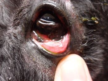
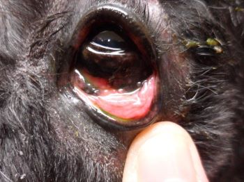
Conjunctivitis is inflammation of the conjunctiva in the eye. The conjunctiva is a pinkish mucous-secreting membrane, similar to the lining of your mouth and nose, that covers the eyeball and lines the inner surface of the eyelids. The conjunctiva also covers the third eyelid, or nictitating membrane, that dogs and cats have but you do not. Inflammation, as indicated by the suffix ‘-it is’, is tissue that is red, warm, swollen, and usually painful as a result of disease or injury.  

In conjunctivitis those membranes become red and swollen. Dogs and cats with conjunctivitis normally have cloudy, yellow, or greenish discharge from the eyes; a lot of blinking or squinting; and redness and swelling around the eye. Green or yellow discharge often indicates a bacterial infection; clear or whitish discharge is more likely to be caused by allergies or a bit of debris in the eye. Usually both eyes are affected, but not always. Sometimes other signs are seen, such as itching, hairlessness around the eye, discharge from the nose, sneezing, or coughing.

What we normally call “pink eye” in people is a type of conjunctivitis. The condition can be caused by several reasons, such as bacteria, herpes virus or allergies.

Any breed of dog or cat may develop allergic conjunctivitis. All breeds predisposed to atopic dermatitis, a hypersensitivity or over-reaction to a variety of commonplace and otherwise harmless substances like pollen, are also predisposed to allergic conjunctivitis. Allergic conjunctivitis is most common in young adults but can occur at any age.

In this allergic condition, the following are frequent contributors:

  • Allergies
  • Atopy (genetic tendency towards allergies)
  • Dust
  • Food allergy
  • House dust, molds

Treatment

Allergic conjunctivitis is most commonly treated with eyedrops or ointments containing corticosteroids, such as dexamethasone or hydrocortisone. Your veterinarian may check first for a scratch on the cornea (called a corneal ulcer) by instilling a yellow dye called fluorescein. If the eye has a scratch, medication without steroids is used, as steroids can delay healing of the scratch. Oral corticosteroids and/or antihistamines may also be helpful, especially in those pets with associated skin disease. Over-the-counter topical antihistamines may be beneficial, particularly in itchy dogs, but ask your veterinarian which one is best for your pet’s situation. However, topical mast cell stabilizers and antihistamines have variable clinical results.

A sterile saline eye wash can be used once or twice a day to clean the eyes and remove the accumulated discharge. Saline may also reduce surface irritation by flushing dirt, dust, debris, and pollens away from the eye.

Preventing damage to the eye is important as pets with allergic conjunctivitis may scratch at their eye with their paws or rub their face on furniture. This rubbing can lead to a corneal ulcer and associated pain and damage to the eye. Using an Elizabethan collar to prevent scratching may be part of the treatment plan from your veterinarian. This is especially important in short-faced breeds like pugs and shih-tzus, who are particularly prone to eye injuries. A recheck examination may be recommended by your veterinarian after treatment. If signs have not improved, your veterinarian may suggest sending your pet to a veterinary dermatologist or ophthalmologist for a work-up or recommending allergen testing to try and determine what your pet is allergic to. This step is usually reserved for dogs or cats with severe allergies.

Prevention

Allergic conjunctivitis is caused by allergies, so the best way to prevent it from happening again is to remove whatever your pet is allergic to from your home, if possible. Even if you can figure out what allergen they are reacting to and remove it or deal with it, you may not be able to eliminate it. If dust is found to be a cause, keep household dust to a minimum, and consider using an air purifier to filter out dust.

Allergic skin testing or food trials may be needed to find out the cause of the inflammation. In those cases, you may be able to determine if your dog or cat is reacting to something like dust or an ingredient in their food.

In many cases, allergic conjunctivitis can’t be cured, but with the help of your veterinarian (and perhaps a few steps taken around the house) it can be managed and you can make your pet more comfortable.

Grove Center Services

Adverse Reactions to Spot-on Flea and Tick Products  

Spot-on flea and tick products for dogs and cats have greatly improved the safety and convenience of controlling external parasites on our pets. Using pesticides that are many degrees less toxic and frequently more effective than the organophosphate or carbamate dips and sprays of the past, these spot-on products help keep our pets (and our homes) free of annoying pests that can also carry serious diseases.

When used according to the label directions, spot-on products are well tolerated by most pets. However, as with any product that is applied directly on the skin, there is the possibility that certain individuals will have adverse local reactions to one or more ingredients in the product. Some individuals will have similar reactions to many different spot-on products with different active ingredients, suggesting that their sensitivities may be to some of the inactive ingredients. These reactions are restricted to the area of skin that comes in direct contact with the product, so they do not reflect a systemic toxicosis but rather a local hypersensitivity. Skin reactions to spot-on products can vary from mild tingling sensations to actual chemical burns of the skin in especially sensitive individuals.

The mildest form of skin reaction to spot-on products is epidermal paresthesia, which is defined as an abnormal sensation such as an itch or prickling of the skin. Paresthesia occurs when the applied product “tickles” the nerve endings in the skin, causing the characteristic sensation. Although paresthesia may occur with any spot-on product, it is most commonly associated with products containing concentrated pyrethroids such as permethrin, cyphenothrin, and etofenprox. Pyrethroid paresthesia is a syndrome that has been well documented in both humans and animals. Human descriptions of pyrethroid paresthesia vary from “tingling” to “pins and needles” to “burning” sensations in the skin at the site of pyrethroid contact. These sensations begin within 30 minutes of application and may last 8 to 24 hours if untreated. Some pets appear to experience similar discomfort following application of spot-on products; the fact that the products are generally applied between the shoulder blades can make the sensation particularly annoying to the pets. Affected pets, cats in particular, may become hyperactive and agitated as they try to walk away from the sensation; others may become quiet, subdued and reluctant to move. Skin at the product application site will appear normal (if redness is noted, you’re dealing with contact dermatitis, see below).

Fortunately, treating epidermal paresthesia is fairly simple and entails bathing off the product with a mild dish soap (pet shampoos are too mild to remove all of the product). For pyrethroid paresthesia, applying vitamin E to the affected skin can provide quick relief— just use scissors to snip open a vitamin E capsule (the kind used as a vitamin supplement) and squeeze the oily contents onto the pet’s skin and rub it in.

A rare but more significant skin reaction (in terms of sensitivity) to spot-on products is contact dermatitis, in which an inflammatory reaction develops in response to the topically applied product. These responses – sometimes referred to as hypersensitivity reactions – can occur upon the first use of a product, but more commonly occur after several uneventful exposures to the product. The body sets up an inflammatory response to the site of application, and the skin will appear red and irritated. In more severe cases, wheals or blisters may develop and the skin may actually ulcerate. Unlike paresthesia, which tends to occur within 30 minutes of application, oftentimes the onset of contact dermatitis is delayed for several hours, with the full extent of the injury taking 12 to 24 hours to develop. Depending on the degree of the inflammatory reaction, the level of discomfort can range from mild to quite severe. After bathing off the spot-on product, pets who develop more than mild redness at the application site should be seen by their veterinarians for further treatment to reduce the inflammation and discomfort.

The good news is that skin reactions to spot-on flea/tick control products are quite uncommon, and most pets will have no problems when these products are used. Unfortunately, it is not possible to predict which individual will react to a given product until the reaction has occurred. For this reason, it is a good idea to observe your pet closely for at least an hour following the application of any spot-on product for any signs of discomfort.

Although not life-threatening, skin reactions can range from mildly annoying to very painful; for this reason, a pet that has had a reaction to a spot-on product should never have the same product applied in the future. If a spot-on product is still desired for flea and/or tick control, try a product with different active ingredients than the one that caused the reaction. Following application, watch the pet carefully and be ready to bathe at the first sign of problems. Unfortunately, there are rare individuals that cannot tolerate any of the topical spot-on products, and other flea control options such as sprays or dips may be needed.

More serious problems can result when spot-on flea/tick products designed for dogs are inappropriately applied to cats. Certain concentrated pyrethroids (e.g. permethrin) that are not harmful to dogs can cause life-threatening tremors and/or seizures if applied to cats—these are not really adverse events but are instead true poisonings since the product was misused.  Always read the label before using any topical flea/tick product on cats, and never apply products intended for dogs onto cats.   

Frontline (fipronil) is toxic to rabbits. 

If your pet has a reaction to a flea or tick control product of any kind, you should report the reaction. All spot-on product labels will have a phone number that you can use to report the reaction to the manufacturer. By law, the manufacturer is required to send monthly reports of adverse reactions to the appropriate federal regulating authority. Alternatively, you can go to the agencies’ websites to find out how to report reactions directly to them. Report reactions to flea/tick products containing heartworm preventatives, such as Revolution and Advantage Multi, to the Food and Drug Administration; report flea/tick products that do not contain heartworm preventatives to the Environmental Protection Agency.