6618065

Alternative Diets for Dogs and Cats

You’ve spoken with your veterinarian and are thinking about personally making your pet’s meals. There are about as many diet options for your pet as there are for you, so which one is best? Breed, age, weight, allergies, medical conditions, and finances are some of the contributing factors that need to be considered when choosing an alternative diet.

If you decide to home cook your pet’s food, we highly recommend first consulting with a veterinary nutritionist to ensure that your pet’s nutritional needs are adequately met. Once there is a recipe available, it is important to follow it exactly. Any changes could affect its nutritional value.

Sometimes, especially in the case of dietary adverse responses, your veterinarian may recommend a limited ingredient home-cooked diet. The purpose is to identify those foods to which your pet is sensitive; there will be trial and error. When discussing your pet’s diet history with your veterinarian, be as thorough as possible to shorten the time that your pet has to be on a limited-ingredient diet.  During this period, do not give anything outside of the specific diet or the results will not be accurate.

When purchasing foods and/or ingredients, look at the nutrition label, just as you do for your own food. “Organic” is a USDA certification that refers to how a food is grown and processed. For example, organic vegetables may be grown without pesticides and organic meats may come from animals that were allowed to graze or are antibiotic/hormone free. “Natural” products are those without synthetic ingredients, added coloring, and often are minimally processed.

A common misconception is that corn is a filler and causes significant allergies in pets. However, a study was done in 2016 on 297 dogs. Of those,  only about four percent had a corn allergy. Corn is a source of carbohydrates, proteins, vitamins, minerals, fiber, and fats. Corn provides energy and helps digestion and in most cases is completely fine for dogs to eat.

“Grain-free” means that the product does not include grains such as rice, corn, or wheat. Therefore, the source of carbohydrates are peas, legumes, potatoes and/or lentils. Grain-free diets and their possible association with dilated cardiomyopathy (DCM) are continuously being researched. Caution is advised at this time before making any choices about grain-free diets.

Gluten is the protein in grain that is left over after all the starch is removed. Celiac disease is an intolerance to gluten. Celiac disease in dogs is incredibly rare (as it is in people) and has not been documented in cats as of November 2019. Dogs reported to have celiac disease were mainly from a single line of inbred Irish Setters in the United Kingdom. Unless your dog has a known sensitivity to gluten, there is no need to avoid it in their diet.

Raw pet food is commonly either frozen or freeze-dried. Prepackaged raw diets that are not marked as complete may need to have vitamin and mineral supplements added to provide a balanced meal. Do not exclude the parts of the meal that your pet selectively ignores, as this can also lead to deficiencies and subsequent clinical disease; make sure, one way or another, that the pet eats all of it.  Nutrient deficiencies can occur if a meal is not made correctly. Bones can potentially cause obstructions, constipation, and tooth fractures. A real risk of feeding raw food is bacterial contamination, especially by Salmonella and E. coli. Contamination can affect your pets, healthy humans, and can be devastatingly harmful to pregnant, young, old, or immunocompromised people in the house. Raw diets also increase the risk of exposure to parasites. Raw feeding is not recommended according to the official AVMA position, which strongly discourages raw feeding.

Some veterinarians recommend that cats be able to eat various foods, including non-dry, non-kibble food to provide moisture in their diets. If your feline arrives to your home as a kitten, make sure to feed a variety of foods and textures as kittens will imprint on what they were fed as kittens. If your feline arrives as an adult, do your best to encourage a variety of foods. For some medical conditions, feeding an all-canned diet may be recommended. 

Although raw feeding is not recommended, if you have made the decision to feed a raw or alternative diet, it is time to hit the store! A great way to make consistent meals for your pet is to print and laminate instructions from your veterinarian and veterinary nutritionist. You can purchase a cutting board and bowls/plates that are specifically for your furry friend’s food to lower the risk of cross-contamination. Many people find they can reduce their costs by purchasing large quantities of each ingredient and storing the meat in a freezer purchased specifically for pet food. Some butchers understand the needs of pet owners, and you may be able to arrange with them to have the best possible price.  Dedicate a part of your pantry and fridge to ingredients specifically for your pet; Your pet will thank you!

4127791

Abdominal Ultrasounds in Pets  

An abdominal ultrasound (previously known as a sonogram) is a diagnostic tool for looking at the organs and structures inside your pet’s belly. It can see the liver, spleen, kidneys, bladder, reproductive organs, stomach, pancreas, intestinal tract, adrenal glands, and lymph nodes. An x-ray is a still photograph, but an ultrasound is like a video.

Ultrasound uses high frequency sound waves to create live images, allowing veterinarians to get an idea of what these organs looks like and how they are functioning.

The ultrasound can help discover where and why a problem is occurring so that appropriate treatment can be started. In some cases, the ultrasound findings may result in your pet needing additional testing. This does not mean that the ultrasound was unhelpful. It usually just means that the findings pointed the veterinarian in the right direction to correctly solve the pet’s problem.

Why Does my Pet Need an Ultrasound?

If your pet has been showing symptoms associated with a problem in one of those belly organs, an ultrasound may be needed. This can include diarrhea; throwing up; belly pain; abnormal urination; a mass that can be felt when pressing on the belly; or if internal injuries are suspected, such as after being hit by a car. Sometimes the veterinarian will find something specific during a pet’s physical exam, such as feeling fluid buildup in the belly, that could be seen by ultrasound. Other situations in which an ultrasound may be necessary are if lab work or x-rays show something unusual.

Abdominal ultrasounds can also be used to take samples of fluid or tissues in order to get a diagnosis of the problem. This can include guiding a needle to biopsy unusual masses or removing fluid for analysis. Veterinarians can either view these samples themselves or send them to a pathologist for testing.

As with people, ultrasounds can also be used to check for pregnancy. Pregnancy is best diagnosed after roughly 20 days from the last heat cycle. Intestinal contents and gas can occasionally cause inaccurate results. Ultrasound is not particularly useful for estimating litter or individual fetus size, so x-rays are more commonly performed. X-rays also tend to be less expensive.

The Procedure

Ultrasounds are typically done with the pet lying on a table. The veterinarian holds a transducer or probe against the belly skin. As the transducer is moved over the skin, it sends sound waves to the structures inside, which are then translated to black and white real-time images on a screen. Hair does not conduct sound waves well, so the pet’s belly is usually shaved beforehand. Alcohol or ultrasound gel may be used to provide better conduction.

Ultrasounds are usually painless and often performed in a quiet, dark room. Most pets are able to lie comfortably without stress and with minimal restraint during the brief procedure. Most pets do not need sedation. Almost all pets can safely undergo an ultrasound, regardless of medical concerns.

4128885

Aspergillosis in Dogs

Fungi of the Aspergillus genus are well known to most people as the lacy, fluffy mold that grows on foods kept a bit too long. There are over 180 species of Aspergillus fungi, and they generally do not cause disease unless the host has a compromised immune system or has a big exposure to the fungus. The most common species to cause problems in veterinary patients are Aspergillus fumigatus (which typically infects the nose and nasal passages) and Aspergillus terreus (which causes systemic/whole-body disease).

Under the microscope, Aspergillus looks like a beautiful flower, but note the spores (called conidia) it sheds. These float in the air to colonize organic matter or infect a host when they land.

Sinonasal (Nose) Aspergillosis

Sinonasal aspergillosis is the most common type of Aspergillus infection. Sinonasal infections can be very invasive or less invasive, the very invasive kind unfortunately being much more common. Invasive infections actually destroy the delicate bones of the nasal sinuses while the less invasive infections involve formatting a big mucous wad of fungus called a “fungal ball” or “aspergilloma.” Advanced imaging can detect either of these, but more on that later. What the owner usually sees is a snotty nasal discharge that often stinks, lasts for months, does not respond to antibiotics, and classically only comes from one nostril.

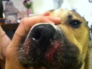
Nose bleeds occur intermittently, and the edges of the nostrils are often ulcerated. Both the more invasive and less invasive forms create similar signs; it is imaging and/or rhinoscopy that usually tell them apart, and sometimes they occur together.

Classically, the affected dog is of a breed with a long nose (collie, greyhound, dachshund, etc.), though in one study, retrievers and rottweilers showed the highest numbers, possibly suggesting they do more sniffing in fungal-contaminated areas. Any age dog could become infected, though Aspergillus infection is unusual in dogs under the age of one year.

To diagnose sinonasal aspergillosis, two out of the following criteria must be met:

  • Imaging (ideally CT or MRI) should be compatible with a fungal infection. This means bone destruction is visible. Radiographs can also be used, but CT and MRI offer superior imaging.
  • Fungal colonies should be visible with rhinoscopy (a technique where a narrow needlelike camera is inserted in the nose).
  • Aspergillus organisms are seen in or cultured from either a tissue biopsy or the nasal discharge. 
  • A blood test (either antibody level, test for fungal cell wall galactomannan, or PCR test) or a urine test for galactomannan antigen is positive against an Aspergillus species.

A typical approach to a dog with a nasal discharge suggestive of fungal infection will likely involve imaging, followed by rhinoscopy. General anesthesia is needed for both of these procedures and referral is likely to be recommended as specialized equipment will likely be necessary.

The treatment of aspergillosis has been challenging for many years; fungal infections, as a rule, are slow to clear, and for a long time, the only available drugs had toxic side effects. Today, sinonasal aspergillosis has a good prognosis thanks to a unique treatment plan. The patient is anesthetized, and the back of the throat is closed off with gauze and inflatable balloon catheters called Foley catheters. The nostrils are also closed off (the patient breathes through an endotracheal tube that is placed through the mouth) and a one percent solution of clotrimazole, a topical antifungal lotion, is infused into the nose and frontal sinuses. The solution incubates for an hour, and the patient is periodically turned to be sure all the nooks and crannies of the sinuses are treated. At the end of the incubation, the clotrimazole is drained through the nostrils and suctioned out.

This treatment is highly effective, with an 86 percent success rate, though about one-third of patients require several treatments. Most of the time, the nasal discharge has resolved within a couple of weeks, but if there is still evidence of continuing infection one month after the treatment, the patient should be rechecked, and another bout of treatment will most likely be needed.

If there is evidence that the infection has eroded through the sinus bones and has penetrated the brain, the treatment described above cannot be used, and oral medication is needed. Common medications used are itraconazole and fluconazole. Several months of therapy are needed, and a 60 to 70 percent success rate has been reported.

Disseminated Aspergillosis

As shown in the photograph, Aspergillus fungi shed microscopic spores that float in the air and which we inhale all the time. Fortunately, we have an assortment of safety mechanisms built into our bodies that prevent infection (sinuses to trap inhaled debris, the ability to sneeze, the mucus lining of the entire respiratory tract to trap debris, cilia to remove mucus with trapped debris, and an immune system to fight infection). If Aspergillus spores called conidia escape these mechanisms and begin to grow, they become more difficult for the body to remove. In disseminated aspergillosis, the fungus penetrates the respiratory tract and may travel to other organs via the bloodstream, creating a more serious fungal invasion. 

German Shepherd Dogs seem to be predisposed to this type of dissemination.

Signs experienced by the patient depend on where the fungus settles. Bone infection is common, and the intervertebral discs of the spine also tend to be favorite spots for Aspergillus. This means that clinical features commonly include lameness, weakness, and incoordination. Frequently, there are draining tracts (holes with pus or bloody discharge oozing out) in the areas of infection. Fever, weight loss, appetite loss, and uveitis (deep inflammation of the eye) are also features. Sadly, most dogs are already terminally ill by the time they are first examined.

Diagnosis is tricky. Bone destruction patterns on radiographs may be suggestive of fungal infection. If you’re lucky, the organism can be identified in some of the draining fluid or in a tissue sample. If the organism is not visible, it may be cultured from a fluid or tissue sample. Ideally, there would be a blood test for Aspergillus antibodies that could make the diagnosis non-invasively, and while such antibody tests exist, it is hard to interpret them. There are accuracy issues, plus different species of Aspergillus do not cross-react, so one must test separately for each Aspergillus species.

Treatment is particularly frustrating for disseminated aspergillosis. At present, posaconazole seems to be the best choice, but years of treatment are likely required (very likely the medication will be for life), and many dogs still die of their disease before the medication can get the condition controlled. If the body part involved can be surgically removed (such as amputation of a bone), this can be curative, and, as distasteful as it may sound, aggressive surgery could be a dog’s only chance of survival.

4127749

Abscess: First Aid

Animal teeth and claws easily puncture skin, introducing bacteria under the skin. If the skin heals quickly, the bacteria become trapped and create pockets of infection commonly referred to as abscesses.

Abscesses may be very small or extraordinarily large, and may feel swollen and warm. These are often extremely painful. Typically, animals with abscesses become depressed and may tend to hide in inconspicuous places. Occasionally abscesses may rupture prior to the onset of any other signs.

Abscesses of the anal glands are common and can be mistaken for rectal bleeding if they rupture. They may cause the pet to “scoot” the rectal area on the ground.

Tooth root abscesses typically form just below the eye, and start as a bump or swelling. They may break open and bleed, and the pet may stop eating due to the pain or experience pain upon chewing.

Small, uncomplicated abscesses may respond to medical therapy, while larger and more extensive abscesses may require surgical treatment. Proper surgical management of abscesses often requires placing a drain or latex tubing (either under sedation or general anesthesia) to provide an escape route for secretions from the wound’s damaged tissues.

Tooth root abscesses require tooth extraction – antibiotics alone will only lead to a temporary solution, but the abscess will recur unless the affected tooth is pulled. This surgery requires general anesthesia.

What to Do

Abscesses should be examined by a veterinarian as soon as possible and within 24 hours.

  • Fluid may normally drain from the site of an abscess. An abscess does not drain through the tube, but rather around the latex tubing. Therefore, it is important for you to clean the area around the drain if directed to do so by your veterinarian.
  • Apply a warm compress to the affected site at least two times daily for 3 to 5 days after your pet leaves the hospital. Wet a clean washcloth with warm water and place it directly over the affected site, and then apply gentle pressure ideally for 5 to 10 minutes. Test it on your skin – if it is too hot for you, it is too hot for your pet.
  • Be sure you and/or other family members wash your hands thoroughly after contacting any fluids draining from the abscess site.
  • Be sure to give all prescribed medications exactly and completely as detailed by your pet’s veterinarian. Some patients may appear to feel better after only a few days of treatment; however, it is crucial for medications to be administered according to schedule to prevent the infection from recurring.
  • Restrict your pet to indoor activities until the infection has resolved completely.
  • Notify your pet’s doctor should your pet experience any of the following:
    – Increased redness and/or heat from the site of the abscess
    – Failure of the abscess to heal
    – Worsening of your pet’s general health
    – Loss of appetite lasting longer than 24 hours

What NOT to Do

  • Do not attempt to open the abscess yourself.
  • Do not attempt compressing the wounds of an animal that may bite you. Your safety is of utmost importance to us. Contact your veterinarian for assistance should this situation arise.
  • Do not apply topical medicines (such as ointments or creams), give any human medications (such as Tylenol/acetaminophen or Advil/ibuprofen), or use any home remedies, unless directed by a veterinarian.

Browse the complete Veterinary Partner First Aid collection.

  • Introduction: First Aid
  • Abscess: First Aid
  • Bandaging: First Aid
  • Bee Stings and Insect Bites: First Aid
  • Bleeding: First Aid
  • Bloat: First Aid
  • Burns: First Aid
  • Can’t Breathe: First Aid
  • Cardiopulmonary Resuscitation (CPR): First Aid
  • Chemical Injuries: First Aid
  • Choking: First Aid
  • Dehydration: First Aid
  • Diarrhea and Vomiting: First Aid
  • Difficult Birth: First Aid
  • Drowning or Near Drowning: First Aid
  • Electrocution: First Aid
  • Eye Injuries: First Aid
  • Fainting and Dizziness (Syncope): First Aid
  • Fever: First Aid
  • Fractures and Injuries: First Aid
  • Hyperthermia (Heat Stroke): First Aid
  • Hypothermia: First Aid
  • Impalement and Penetrating Injuries: First Aid
  • Nosebleed: First Aid
  • Paralysis: First Aid
  • Physical Exam Checklist for Pets: First Aid
  • Poisoning in Dogs and Cats
  • Preventing a Health and Safety Crisis: First Aid
  • Seizures and Convulsions: First Aid
  • Shock: First Aid
  • Snakebite: First Aid
  • Straining to Eliminate: First Aid
  • Sunburn: First Aid
  • Transporting an Injured Pet: First Aid
  • Wounds: First Aid
  • Wrapping Up First Aid

4128589

Adrenal Tumor Treatment in Cushing’s Syndrome 

Let’s assume an adrenal tumor has been confirmed with either blood testing, imaging, or both. Two questions must be answered next:

  • Is the tumor benign or malignant?
  • Should you choose surgical treatment or medical management?

Benign vs. Malignant

While only approximately 15% of canine Cushing’s syndrome patients have adrenal tumors, half of them will have benign tumors and half will have malignant ones. The choice of therapy may well depend on which one the patient has.

If imaging has not yet been performed, this is the time to do so. Chest radiographs will be important as malignant adrenal tumors tend to spread to the chest. If such spread is seen, the tumor can be assumed to be malignant. Absence of such findings, however, does not mean the tumor is benign. Ultrasound of the abdomen, if this has not already been done, (or even CT scanning, MRI imaging, or nuclear medicine scanning) will be needed to determine the size of the tumor, and to check for invasion of local abdominal tissues (especially in the liver).

Between evaluation of the chest and the abdomen, it may be possible to non-invasively determine if the tumor is malignant. It should be noted that the absence of tumor spread does not mean that the tumor is benign. If there is obvious spread to other organs, medical management is the only meaningful hope for the patient. Discuss with your veterinarian whether a referral to an oncologist for the most current medication plan is in your pet’s best interest.

What if Imaging Fails to Confirm That the Tumor Is Malignant?

As mentioned, it is not possible to say that the tumor is benign simply because tumor spread has not been detected. Still, no evidence of spread is about as close to determining that the tumor is benign as we can get without actually obtaining tissue samples. If the adrenal tumor is benign, there is an excellent chance for complete recovery if the tumor is surgically removed. The smaller the tumor, the easier the surgery, though the surgery involves delicate tissue in a difficult area.

What if Imaging Indicates the Tumor Is Malignant?

If there is obvious tumor spread, surgery may be too risky. The decision to proceed with medical therapy will depend on how debilitated the patient is, and the degree of tumor spread versus the severity of the clinical signs of Cushing’s disease. Relief, but not cure, of the clinical signs may be achieved by removing part of the tumor. Medical management with trilostane or with high doses of lysodren would be fair alternatives.

What You Should Know About Surgery

  • Removing the adrenal gland is a relatively difficult surgery and may be best left to board surgeons who perform this surgery with some regularity. (The adrenal gland is located between the aorta, which is the body’s largest artery; the renal artery and vein, which are the sole blood supply to the kidney; and the phrenicoabdominal artery. This vascular area is half-jokingly referred to by surgeons as the Bermuda triangle. Surgery here is not for the inexperienced.) Removing an adrenal tumor is generally considered to be one of the most difficult surgeries in veterinary practice.
  • Risk of bleeding is higher for larger tumors, especially if they are malignant and have invaded local structures. It is quite possible that the full extent of such invasion will not be apparent prior to surgery.
  • Animals with Cushing’s syndrome have poor healing ability and tend to have high blood pressure. Several months of medical therapy (i.e., lysodren or trilostane) prior to surgery may be a good means to strengthen the patient, especially if the tumor is believed to be benign. 
  • The dog’s natural cortisone mechanisms will have been suppressed by the tumor. This suppression means the patient will not be able to produce adrenal steroids on their own for a while and they will need to be given as medications. Several months of prednisone will likely be required at home. Some patients require florinef as an additional supplement. ACTH stimulation test are used to monitor the need for medication.
  • Adrenal tissue is notoriously difficult for pathologists to grade as benign or malignant. It is possible that a tumor initially graded as benign will later turn out to be malignant.
  • In a statistical survey of 63 dogs undergoing surgery for adrenal tumors:
    • 6% (4) had inoperable tumors and were euthanized on the surgery table.
    • 29% (18) died either in surgery or shortly thereafter due to complications.

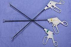
The most common serious complication is thromboembolism (abnormal blood clots), which typically happens in the first 24 hours after surgery. This risk is tremendously reduced if the surgery can be performed by laparoscopy. Incisions are smaller, organ manipulation and bleeding is less and recovery is faster. Laparoscopy uses several small holes in the belly wall through which instruments, including a small video camera, can be inserted. Surgery takes place inside the belly and is viewed on a video screen or through a lens.

Medical Therapy for the Adrenal Tumor

As can be seen from the above discussion, adrenalectomy is a high-risk surgery. It may not be a good choice for an elderly debilitated patient, especially if the tumor is large or has evidence of spread. Further, a 24-hour specialty center is usually needed for this sophisticated procedure and these centers may not be accessible, plus there is a great deal of expense involved in a surgical adventure of this magnitude. For these reasons, medical management may be selected as an alternative.

Lysodren is a chemotherapy drug that is able to erode the cortisol-producing layers of the adrenal gland. This ability has made lysodren, for decades the traditional medication for treating pituitary Cushing’s disease, helpful in treating adrenal tumors as well. It turns out the adrenal tumors will respond to higher doses, The higher doses needed to control adrenal tumors tend to produce more lysodren reactions than are seen when treating pituitary tumors. The average survival time for this type of therapy is 16 months.

Trilostane can also be used for functioning adrenal tumors though it will not actually diminish adrenal tissue (which could potentially diminish the tumor). Trilostane acts by inactivating excess steroid hormones and thereby managing the symptoms of Cushing’s disease. You would think that this would not be effective since it does not directly affect the tumor, but at least one study showed a comparable survival time to that of patients on lysodren. As with lysodren, a protocol is needed that is different from those using the same drugs to treat pituitary-dependent Cushing’s disease.

Something Different: Hypofractionated Stereotactic Radiotherapy

A study presented in 2014 by a group of Italian researchers treated 12 dogs with adrenal tumors with this special type of radiotherapy and all 12 of them responded. After 12 months, 11 patients were not only still alive but their Cushing’s syndromes were controlled as well (the 12th patient had died of unrelated causes). This type of therapy was considered to be far less risky than surgery and the researchers recommended further investigation. Stereotactic radiotherapy involves highly specialized equipment and is not readily available but might be worth travel depending on where it might be located. An oncologist should be consulted for more information.

4128729

Addison’s Disease (Hypoadrenocorticism)

Adrenal Hormones

The adrenal gland is so named because it is located just forward of the kidney (renal means kidney). The center of the gland is called the medulla and the outer area is the cortex. While both areas produce hormones, Addison’s disease concerns the hormones produced by the cortex; these hormones are called corticosteroids.

Corticosteroids are the hormones that enable us to adapt physiologically to stress. The glucocorticoids (such as cortisol and related synthetics, prednisone, dexamethasone, and numerous others) act on the mechanics of sugar, fat, and protein metabolism.

They gear the metabolism towards the preparation of burning, rather than storing, fuels so as to be ready for a fight or flight situation.

The mineralocorticoids (such as aldosterone and related synthetics, fludrocortisone acetate, and desoxycorticosterone pivalate) influence the electrolytes sodium and potassium.

You may wonder what electrolytes have to do with fight or flight but it is important to remember that where there is sodium (salt), water soon follows. Conserving sodium will pull water from other tissues and bring it into circulation ready to support blood pressure should a life-threatening bleed occur. The mineralocorticoid hormones instruct the kidney to retain sodium so that the circulatory changes of stress can be handled smoothly. As sodium is retained, potassium is lost in the exchange.

Corticosteroid hormones are needed to adapt to stressful situations and without these hormones, even small stresses could lead to physiologic disaster.

Hypoadrenocorticism (Addison’s Disease) is a Deficiency in Corticosteroid Hormones

In animals with Addison’s disease, there is a deficiency of corticosteroid hormones. There are several reasons why this might happen: the adrenal gland might be damaged or drugs may be involved. We usually never find out what caused an individual patient’s Addison’s disease but the good news is that we don’t need to know why it happened. Treatment is straightforward: we simply provide the hormones that the body is not making on its own.

Clinical Signs

Patients are usually young (age four to five years) but any age dog can be affected. (This disease can occur in cats but is very rare.) There is a genetic predisposition for Addison’s disease in the standard poodle and bearded collie, Nova Scotia Duck Tolling Retriever, and Portuguese water dog. Female dogs are affected twice as often as males.

At first, signs may be vague: listlessness, possibly some vomiting, or diarrhea. The dog just does not seem to feel right but not in an obvious way and may seem more or less normal most of the time as symptoms wax and wane with stress. This vague waxing and waning go on and on with the dog never really getting fully sick but never staying well either. Eventually, if the disease is not diagnosed, it can come to a head in a phenomenon known as an Addisonian crisis. The animal collapses in shock due to an inability to adapt to the caloric and circulatory requirements of stress. Blood sugar may drop dangerously low. Potassium levels can soar and disrupt the heart. Heart rate slows and arrhythmias result. The patient may not survive this episode.

About 30% of dogs with Addison’s disease are diagnosed at the time of an Addisonian crisis. Approximately 90% of the adrenal cortex must be non-functional before clinical signs are observed.

Making the Diagnosis

Because of the numerous symptoms Addison’s disease can present with, Addison’s disease has earned the medical nickname “The Great Imitator.” One would think that one could simply look for an increase in potassium and/or a drop in sodium on a basic laboratory blood panel, but it turns out spot checks of electrolyte values like this are not reliable enough for a diagnosis of Addison’s disease.

Shock

Veterinarians can be presented with a young animal in shock. There is usually no history of trauma or toxic exposure so general treatment for shock is initiated. This consists of rapid administration of fluids (usually Lactated Ringers solution which has little potassium and a moderate amount of sodium) plus some glucocorticoids. By coincidence, this also happens to be similar to the specific treatment for Addison’s disease so often the patient simply recovers without the veterinarian really knowing why.

Imitating Kidney Disease

The blood panel often comes back showing elevations in the renal parameters (BUN and creatinine), and, thus the elevated potassium is suggestive of acute renal failure, a condition with an extremely poor prognosis. The veterinarian may become suspicious of another diagnosis as the patient will respond well to fluid administration and most renal failure patients do not respond as well.

Low Blood Sugar

Addison’s disease may present in more unusual ways. The inability to maintain normal sugar levels (ultimately manifesting as a seizure disorder) may be strongly suggestive of an insulin-secreting pancreatic tumor but before major abdominal surgery is planned, it is important to test for Addison’s disease.

Megaesophagus or Recurring GI Disease

In a similarly unexpected way, regurgitation of undigested food due to abnormal nerve function in the esophagus (a condition called megaesophagus) can be caused ultimately by Addison’s disease. Megaesophagus has many causes but it is important not to forget Addison’s disease. Of course, Addison’s disease can also manifest with chronic waxing/waning diarrhea and/or poor appetite which would suggest a gastrointestinal problem such as inflammatory bowel disease. Before investing in the expense and potential anesthetic risk for endoscopy and intestinal biopsy, a screening test to rule out Addison’s disease is prudent.

Sorting it out with the ACTH Stimulation Test

Your veterinarian may use a resting cortisol test to rule out Addison’s but the only definitive test for Addison’s disease is the ACTH stimulation test. The patient receives a dose of ACTH, the pituitary hormone responsible for the release of corticosteroids in times of stress. A normal animal will show an elevation in cortisol in response to ACTH while an Addisonian has no corticosteroids to respond with. This lack of response is diagnostic for Addison’s disease; however, a false positive may be obtained if corticosteroids have been used in the treatment of the crisis prior to the test. Of all the commonly used corticosteroids, only dexamethasone does not interfere with the assay for cortisol; if any other steroid has been used, the test will not be valid for at least 24 hours.

Treatment

The most important aspect of treatment for hypoadrenocorticism is replacing the missing mineralocorticoid hormones. One way to do this is with oral fludrocortisone (brand name Florinef®). Fludrocortisone is given twice a day at a dose determined by the patient’s sodium and potassium blood tests. At first, these electrolytes are monitored weekly. When levels seem stable, these blood tests are repeated two to four times per year. Often with time, it will be found that the fludrocortisone dose needed will increase. This increase is unfortunate as the medication is relatively expensive. Since fludrocortisone has glucocorticoid activity as well as mineralocorticoid activity, only 50% of patients need to receive additional glucocorticoids. Using a compounding pharmacy or looking into the cost of generic fludrocortisone may be helpful in managing the costs of this particular medication, especially in a larger dog.

Another way to treat this condition is with an injectable medication called DOCP desoxycorticosterone pivalate, (brand names Percorten-V or Zycortal®). This treatment is given approximately every 25 days. Electrolytes are measured prior to injections at first but testing can usually eventually be tapered to once or twice a year. There is some feeling among experts that DOCP produces better regulation of electrolytes than oral Florinef. Dogs on DOCP, however, do require glucocorticoid supplementation (such as a low dose of prednisone).

What is Atypical Addison’s Disease?

Most dogs become Addisonian when they lose the ability to produce both mineralocorticoids and glucocorticoids. They need both types of hormones replaced. It turns out that there is a subset of Addisonian dogs that are able to control their sodium/potassium imbalance and only the glucocorticoids need to be supplemented. These patients have what is called atypical Addison’s disease. They can have all of the symptoms of typical Addison’s as described above (chronic relapsing diarrhea/appetite loss, low blood sugar crisis etc.) but not the electrolyte abnormalities. Treatment is supplementing with glucocorticoid hormones, such as prednisone.

Atypical Addison’s patients can progress to the typical form, which means they will eventually need more aggressive treatment with fludrocortisone tablets or DOCP injections. The current recommendation is to monitor sodium and potassium levels every couple of months in atypical patients for up to one year, to watch for the switch to the typical (and more serious) form of Addison’s disease. Becoming atypical Addison’s can still occur after one year. If an atypical Addisonian patient becomes ill, sodium and potassium should be checked to see if they have progressed to the typical form.

What is Secondary Addison’s Disease?

Addison’s disease is most often caused by the destruction of the adrenal glands.  In some cases, the problem is a failure of the pituitary gland.  ACTH is a hormone made by the pituitary that tells the adrenal glands to produce and release more glucocorticoids.  Without ACTH, the adrenal glands atrophy and stop producing glucocorticoids.  Measuring the hormone ACTH in the blood can distinguish adrenal and pituitary failure.  This is not simply of academic interest.  A patient with secondary Addison’s disease (i.e., pituitary failure) will never progress to typical Addison’s disease and does not require long-term, periodic monitoring of electrolytes.

What is Pacific Rimism?

Dog breeds originating in the Pacific Rim, such as the Akita and Shiba Inu, commonly have elevated potassium levels on blood tests. This can be confusing when a patient has symptoms that suggest Addison’s. These patients will have normal ACTH stimulation test results if they do not have Addison’s disease.

Whipworm Infection?

Whipworm infection has been known to create a syndrome nearly identical to Addisonian crisis, complete with abnormal sodium and potassium values. These patients will have normal ACTH stimulation tests but because whipworms only periodically shed eggs, fecal testing may not detect whipworm infection. If there is any question about whipworm infection, treatment should be instituted.

4128603

Anorexia, or Lack of Appetite, in Dogs and Cats

Appetite loss and appetite reduction are important factors in illness assessment, and they must be recognized and reported as soon as possible. Acceptance of favorite foods often determines if a pet should be hospitalized or not. A couple of off days is generally not a big problem as long as the pet maintains hydration one way or another and recovers promptly, but recurring episodes of poor appetite or slow, gradual reduction in appetite are important and frequently point to a chronic progressive illness. Nutritional support not only helps the pet recover but buys time to keep the pet stable while diagnosis and treatment are worked out. The pet must be fed in order to get well.

The term anorexia simply means eating no food. Many people get confused by the human eating disorder anorexia nervosa. When the veterinarian says the pet has anorexia, it does not mean the pet has a distorted body image; it simply means the pet is not eating.

So how do we get a pet to eat? We will review some techniques here. Be sure to check with your veterinarian regarding the proper amount of food to feed and which foods are acceptable. Eating only a few bites of food or drinking the gravy does not constitute a good appetite so when asked if your pet is eating, this isn’t really a yes or no question. Be specific about quantity.

Don’t wait for the appetite to completely disappear before seeking veterinary assistance.

Pets with a poor appetite are sick, and if you wait until the appetite is completely gone, it may be too late for recovery. This is particularly true for cats. As the appetite fades, the pet must depend on stored fat for nutrients. When large amounts of fats are mobilized to meet energy demands, they must be processed by the liver before being used for calories. The feline liver is not designed to handle large amounts of fat and will fail in a condition called hepatic lipidosis.

First Offer Canned Food

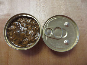
If you think your pet’s appetite is poor but are offering only kibbled food, your first step is to get some canned food and offer that. Most animals find canned diets far more palatable than dry foods, and you may find that this step alone fully alleviates the problem. There is a misconception that canned food is somehow of poor nutritional quality. In fact, canned food and dry food differ primarily in their water content and, thus in texture. If you consider the food without water, the unprepared diet is basically a powdered meat mix similar to a flour.

It can be baked into a kibble or steamed into a canned food. Canned foods differ in quality just as dry foods do. See if the pet will eat a canned food or a mixture of dry and canned food. Adding a flavored broth or cooked egg is also helpful in enticing the pet to eat a kibbled diet.

Second, Offer a Delicacy

Foods that are generally regarded as delicacies among pets include canned chicken, cooked egg, and canned tuna. Therapeutic recovery formula diets are generally well accepted.

With the exception of the recovery diets, these treats are not nutritionally complete but can make a good jump starter for pets. A pet who has not been eating may feel continued discomfort until eating begins again. Something tasty may be necessary to get the appetite restarted.

Do not simply put the food in a dish in front of your pet. Instead, rub a small amount on the teeth or spoon a little in the mouth so that the pet can get a taste. Don’t be surprised if he spits it out; we are just trying to get the taste of the food in his mouth. Hold the bowl up to the pet’s nose so that the aroma is inescapable. You may find that coaxing in this way gets the appetite started.

A Note on Gourmet Cat Foods (Yes, Dogs Eat Them, too)

Several “extra tasty” products available in the grocery store can be really helpful in tempting a pet with a poor appetite. Fancy Feast by Purina comes in numerous textures and flavors, each can containing approximately 100 calories. The diet is complete and balanced for cats and is often a good appetite jump starter. Temptations®, by Mars Petcare, are especially well-accepted treats made for cats. Because many cats will not eat anything else, they have been balanced to be nutritionally complete for cats and can be used as a cat’s sole diet if necessary.

A Note on Starting Prescription Diets

If the pet is supposed to eat a prescription diet but refuses, do not attempt to starve the pet into eating the prescription food.

Many prescription diets are relatively bland and pets do not wish to eat them, particularly if they are used to eating large amounts of table scraps (always a bad practice) or a more flavorful regular food. Starving the pet will only make him sicker. Try a gradual change from the regular food to the new food over a week or so. If the pet simply will not accept the new food, be sure to let your veterinarian know this. There may be an alternative flavor to try; further, prescription diets are guaranteed by the manufacturer meaning you can get a full refund on the bag or case if the pet does not accept the food. Sometimes it is necessary to forgo the therapeutic aspect of the special diet just to get the pet to eat, but your veterinarian will help you with these guidelines.

Provide Privacy

Be sure other pets at home do not bully or distract the sick pet. In a multi-pet home, it may be difficult for the sickly or elderly pet to eat without the younger pets taking his food. Many animals wish to eat at their leisure, particularly if they do not feel well. Consider giving your pet a private area and her own dish. Never feed multiple pets from the same bowl as one is sure to get the lion’s share of the food to the other’s disadvantage.

Many pets like to eat overnight when no one is watching.

Medical Assistance

Appetite-stimulating medications are available.

There are several products available that can create a sense of hunger:

Capromorelin: This is an oral liquid that mimics the body’s natural system that generates the sense of hunger. It is best given daily rather than only on bad days. Capromorelin is available in both a canine and a feline formula. 

Mirtazapine: This is a pill originally developed as an anti-depressant for humans but was found to have appetite increase as a side effect. It is available as a tablet, typically given daily for dogs or every two to three days for cats, or as a transdermal gel for cats which is applied daily to the inner surface of the ear.

Cyproheptadine: This is a psychoactive anti-histamine that has an appetite stimulation side effect. It is available as a pill and is typically used twice daily.

Prednisolone/Prednisone: This hormonal anti-inflammatory has a number of uses in the treatment of immune mediated conditions, inflammatory conditions, cancer therapy and many other situations. One of its side effects is increased appetite and it is sometimes used for this effect, though because it affects so many body systems, it is generally not used for its appetite effect alone.

These medications represent non-invasive ways to medically improve appetite.

Assisted Feeding

Non-invasive methods are all well and good but the bottom line is that an animal that does not eat will get sicker and ultimately die. Receiving nutrients is the basis of recovery and one cannot simply wait around for days for medications to work. Nutrients must be delivered ideally in a way that is not stressful for either the pet or the human caretaker.

Feeding Tubes

Feeding tubes are the least stressful method of delivering nutrition and can be placed through the nose, throat, directly into the stomach or into the intestine. Liquid diets are delivered through the tube, usually with no resistance from the patient whatsoever since the mouth and face are not manipulated.

Gaining in popularity is the esophagostomy or E tube. This tube is placed in the esophagus via an incision in the side of the neck. A bandage or special cloth collar may be used to hold it in place though many patients do not require a wrap of any kind. The placement procedure is fairly short, and the tube is comfortable for the pet. Protective cone collars are not generally needed and the pet can go about his or her life with the tube in place.

The larger size of the tube allows for blenderized diets which can be prepared at home for less expense than prepared liquid diets. Feeding does not require fussing with the pet’s face and thus is more comfortable. Bandages must be kept clean around the area, and tubes must be kept in place for a minimum amount of time to allow for proper scar tissue to form, sealing the feeding hole to the outer tissue. When the time comes, the tube can be pulled, and the hole seals up. These kinds of tubes require brief surgical placement, and thus anesthetic risks apply. 

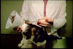
Nasogastric tubes (N-G) that go through the nose require no anesthesia to place but do require an Elizabethan collar to prevent the pet from yanking the tube out. They can accommodate only a liquid diet because of their small diameter and are difficult to manage at home. Most animals will not want a piece of equipment attached to their face and will try to remove it. N-G tubes are best used for patients that are too sick to try to pull out the tube. Alternatively, they can be placed for the feeding process and then removed.

Feeding tubes can similarly be placed in the stomach (G-tube) and protrude from a belly bandage. The G-tube has all the advantages of the E-tube and is favored by some doctors. Again, the tube diameter is large enough to accommodate a slurry rather than a liquid, and the tube is comfortable to wear and use. A brief surgery under general anesthesia is required to place the tube.

Syringe or Force Feeding

In the past, syringe or force-feeding was used in an effort to get calories into a pet. There are several disadvantages to this technique and no advantages. First, the pet is not going to like it, and there will be struggling, stress, and mess. The pet may be too sick to withstand stressful feedings, and creating ongoing unpleasant feeding experiences can result in what is called “food aversion,” where the pet may not ever have a normal appetite again. 

 Feeding too quickly can lead to choking or food aspiration if the feeder is too aggressive. Talk with your veterinarian before using this method, as it can be detrimental to your pet.

Nutritional support is essential to proper recovery and it is important to realize that there are several techniques available to see that the pet does not suffer extra debilitation from malnourishment. If you think your pet has a problem with weight loss or inadequate appetite, do not wait until the problem is extreme; see your veterinarian promptly.

4127654

Acute Hemorrhagic Diarrhea Syndrome (AHDS or HGE)  

The term hemorrhagic gastroenteritis basically means stomach and intestine inflammation with bleeding. This kind of bleeding inflammation might be seen in patients with parvovirus infection, pancreatitis, inflammatory bowel disease, or any number of other conditions but when the term HGE is used, it usually refers to a more specific canine syndrome and is an entity in and of itself. To clarify terminology, HGE has been renamed AHDS, which stands for acute hemorrhagic diarrhea syndrome. Because this change in nomenclature is recent, you may hear veterinary personnel using either term, but regardless they are referring to the specific syndrome that we will now describe.

Acute hemorrhagic diarrhea syndrome is a potentially life-threatening intestinal condition of an otherwise healthy dog, which manifests as sudden onset bloody, watery diarrhea. The symptoms are extremely dehydrating, often much more than expected from the amount of diarrhea, and if it is not promptly treated, the dog can go into shock and die.

Vomiting is often part of the syndrome. In fact, 80% of patients experience vomiting approximately 10 hours before the dramatic bloody diarrhea, and about half the time, vomiting contains obvious blood. The good news is that with hospitalization and treatment, most dogs recover rapidly. It is crucial to identify AHDS and rule out other serious conditions promptly so that the dog can get the correct treatment and the pet’s family can have proper expectations.

Smaller dog breeds seem to have a predisposition towards AHDS though any dog can be affected.  Stress and hyperactivity seem to be predisposing factors.

How is this Condition Diagnosed?

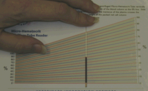
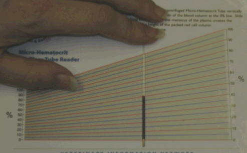
There are no specific tests for AHDS, but a test called a packed cell volume (also called PCV or hematocrit) helps make the diagnosis. This test can be performed in most veterinary hospitals using a few drops of blood. The percentage of the blood volume made up of red blood cells is measured. A normal PCV for a healthy dog would be 37-55%, meaning 37-55% of the blood volume should be red blood cells; the rest is fluid and white blood cells. When the patient becomes really dehydrated, there is less fluid in the bloodstream, and the percentage of blood fluid drops (and consequently, the percentage of red blood cells rises). In other words, the seriously dehydrated patient has a very high PCV.

The average AHDS patient will have a PCV of at least 57%. (It will even be greater than 60% in approximately 30% of patients.)

The measurement of the PCV also usually includes some sort of measurement of total protein (sometimes called total solids). In AHDS, the total protein measurement from the blood sample is paradoxically normal or low.

There are several aspects to the AHDS syndrome, and they do not all have to be there to make the diagnosis. The syndrome elements your veterinarian will be looking for are:

  • Elevated PCV of 57% or more
  • Relatively low total protein
  • At least one episode of vomiting (with or without blood) approximately 10 hours before the diarrhea
  • A watery, very bloody diarrhea that looks almost like pure blood
  • Relatively young dog (median age is five years)
  • Relatively small dog (median size 25 lbs)
  • Rapid response to intravenous fluids

As noted, there are many causes of bloody diarrhea like this, including an intestinal foreign body that requires surgery. Expect your veterinarian to need other tests to rule out other potentially more serious conditions.

What Causes AHDS?

There are still many unknown factors, but the cause appears to begin with infection from an intestinal bacterium called Clostridium perfringens Type A. A PCR test for Clostridium perfringens Type A can be used to determine for sure. This organism produces two toxins called NET E and NET F, which are different from the usual Clostridial enterotoxin that is well known to cause diarrhea. The toxin ulcerates the intestinal lining, and the intestinal blood vessels become permeable to fluid. In this way, fluid is lost into the intestine, dehydrating the patient, and the PCV rises while the ulcers are responsible for the bleeding.

Treating AHDS

The heart of therapy is aggressive fluid replacement. The idea is to get the PCV back to the normal range and keep the patient out of shock. Medication for nausea and pain control are included as is a low-fat diet as soon as the patient is willing to eat. As dramatic as the diarrhea is, most patients have an equally dramatic improvement in the first 24 hours of hospitalization and the average dog is out of the hospital after three days total.

Antibiotics are surprisingly controversial in this syndrome, and their use depends on many patient factors (such as whether or not there is a fever, white blood cell count, whether or not the expected improvement is achieved, etc).

The stool can be expected to gradually return to normal over about one week.

Probiotics (cultures of healthy bacteria) have been recommended to help repopulate the bowel, with “good” bacteria which improves the function of the intestines. They may be recommended for 2-4 weeks, and your veterinarian will let you know how long to give probiotics.

7624966

AAFCO Pet Food Labeling

Pet food marketing can get confusing. The labeling does not provide detailed nutritional information and it’s easily misunderstood by consumers. Try as you might, sometimes reading the label doesn’t give you enough answers. However, the more you know about what pet food labels indicate – and don’t – the better off your pet will be.

What is AAFCO?

AAFCO is the go-to organization for understanding animal feed, including food for pets. It does not regulate or inspect anything, nor is it a government agency. It is a group whose members are government agencies representing the 50 states, Canada, and the federal government. It is not a regulatory body and has no ability to monitor or enforce specific food manufacturing procedures. Animal feed specialists indicate to them what is appropriate for a specific species, and AAFCO provides model feed laws. AAFCO does not approve or certify any pet food, treat, or supplement products. The FDA, not AAFCO, regulates pet food, and FDA is a member of AAFCO.

State feed control officials can choose to adopt these model feed laws – if they do, then monitoring and enforcement duties are the responsibility of the individual state. Pet food manufacturers that sell food must comply with all relevant federal or state laws. Most pet foods sold in multiple states will comply with the AAFCO guidelines.

AAFCO relies on current scientific knowledge to provide guidelines for pet food composition which, if followed, minimize the risk of malnutrition. On the other hand, the FDA enforces food safety through the Federal Food Drug and Cosmetic Act, which demands that pet foods, like human foods, be “pure and wholesome, safe to eat, produced under sanitary conditions, contain no harmful substances, and be truthfully labeled.” Therefore, pet food manufacturers are legally responsible for providing nutritionally adequate, safe, and wholesome products that conform to local, state, and federal law.

AAFCO requires nine components to be printed on a pet food label:

  1.   Brand and product name
  2.   Net quantity statement
  3.   Manufacturer or distributor information
  4. Calorie content statement
    1. Energy content on a kcal/kg and kcal/common unit as fed basis as well as how this was determined (measured or calculated).
  5. Nutritional adequacy statement
    1. Specifies whether the product carries a complete and balanced claim, and if so, how the claim was substantiated as well as the species and life stage for which the product is intended.
  6. Guaranteed analysis
    1. Minimum crude protein and crude fat and maximum moisture and crude fiber (as fed basis) are required; other nutrients are optional.
  7. Ingredient list
    1.  Must be listed in descending order of weight; ingredients must be as defined by AAFCO; however, it is not possible to tell the relative contributions of ingredients to any particular nutrients (the ingredient list is not a recipe). Note that ingredients, as defined in AAFCO, are not often similar to those typically used/defined for human foods; for example, “chicken” must include both flesh and skin and can include bone.
  8.   Species designation
  9. Feeding directions
    1. Must be given for each life stage if the diet is formulated for more than one.
    2. Target animal weights can be given as wide ranges, and no requirement for a specific equation to use to determine the energy needs of target animals. 

What are the different AAFCO nutritional adequacy designations for pet foods?

There are four possible AAFCO nutritional adequacy statements that can appear on pet food labels. These must be printed verbatim as one of the following:

  1. [Diet X] is formulated to meet the nutritional levels established by the AAFCO [Dog /Cat] Food Nutrient Profiles for [gestation/lactation/growth/maintenance/All Life Stages]
  1. If the life stage is growth or all life stages, the claim must also include one of the following depending on the calcium content of the product (maximums vary):
    1. “including the growth of large size dogs (70 lb or more as an adult)”
    2. 2. “except for growth of large size dogs (70 lb or more as an adult)”

How are AAFCO feeding trials conducted?

The guidelines for an AAFCO feeding trial vary depending on the diet type being evaluated. There are four protocols: maintenance, growth, gestation/lactation, and all life stages. Although the minimum standards for each type of trial must be met, companies may add on other components to a feeding trial, such as plasma amino acid measurement, digestibility studies, full blood chemistry panels, and complete blood counts to gain additional information about the suitability of their diets for the life stage being tested.

Foods can be put through AAFCO feeding trials even if they do not qualify for a “formulated to meet AAFCO” feeding statement due to deficient or excessive levels of nutrients. One example is veterinary therapeutic diets formulated for chronic kidney disease, which are intentionally formulated to contain phosphorus (+/- protein) at concentrations below the AAFCO Food Nutrient Profiles for adult maintenance; many of these have passed maintenance feeding trials and thus have a “feeding trial” nutritional adequacy statement on the bag. If they have not undergone feeding trials, they must carry the statement: “This product is intended for intermittent or supplemental feeding only.” Such diets are not allowed to reference the AAFCO Food Nutrient Profiles on the label. Theoretically, the flip side is that diets that fail feeding trials but contain nutrient levels within the specified minimum and maximum concentrations per AAFCO Food Nutrient Profiles can be marketed with a “formulated to meet” adequacy statement. Ideally, diets are formulated to comply with the concentrations specified by the AAFCO Food Nutrient Profiles and then pass appropriate feeding trials.

For adult maintenance canine and feline diets, the feeding trial guidelines are:

  • 8 animals older than 1 yr. must start the test.
  • At the start all animals must be a normal weight and health, and weight is monitored weekly. Complete veterinary examinations are mandated at the initiation and completion of the test.
  • A blood test is to be taken from each animal at the start and finish of the test for four parameters: (hemoglobin, packed cell volume, alkaline phosphatase, and albumin).
  • For 6 months, the animal must only eat the food being tested.
  • The animals finishing the test must not lose more than 15 percent of their body weight.
  • During the test, none of the animals used are to die or be removed because of nutritional causes.
  • 6 of the 8 animals starting must finish the test.
  • Data are compared to a concurrent control group or to historical colony averages

For growth canine and feline diets, the feeding trial guidelines are:

  • 8 animals NO older than 8 weeks must start the test. Juvenile animals should be obtained from at least 3 dams.
  • At start all animals must be normal weight and healthy, and weight is monitored weekly. Complete veterinary examinations are mandated at the initiation and completion of the test.
  • A blood test is to be taken from each animal at the start and finish of the test (hemoglobin, packed cell volume, alkaline phosphatase, albumin).
  • For 10 weeks, the animal used must only eat the food being tested.
  • During the test, none of the animals used are to die or be removed because of nutritional causes.
  • 6 of the 8 animals starting must finish the test.
  • Data are compared to a concurrent control group or to historical colony averages

For reproduction canine and feline diets, the feeding trial guidelines are:

  • 8 animals older than 1 year and on at least their 2nd heat must start the test
  • At the start all animals must be normal weight and healthy, and weight is monitored weekly. Complete veterinary examinations are mandated at the initiation and completion of the test. Offspring are examined within 72 hours of birth and at the end of the test.
  • A blood test is to be taken from each animal at the start and finish of the test (hemoglobin, packed cell volume, alkaline phosphatase, albumin).
  • Litter size is recorded.
  • From the onset of estrus to 4 weeks after parturition, the animal (and offspring) used must only eat the food being tested.
  • During the test, none of the animals used are to die or be removed because of nutritional causes.
  • 6 of the 8 animals starting must finish the test.
  • Data are compared to a concurrent control group or to historical colony averages.

Do AAFCO feeding trials accomplish what they claim to accomplish?

Many pet owners and clinicians have raised the concern that AAFCO feeding trials do not represent what really happens in a pet’s life, i.e., that feeding a diet for six months is not the same as feeding a diet for 10 years. That is true. However, although AAFCO trials are imperfect, they do identify most serious acute and semi-acute nutritional deficiencies or excesses, and some minor ones. Furthermore, manufacturers are free to supplement the minimum protocol requirements with additional measures of nutritional adequacy. Also, consider that diets for growth, gestation/lactation, or all life stages get tested more rigorously compared to maintenance since the nutritional demands of those animals are greater.

Evaluation of diets under AAFCO guidelines also includes an examination of the nutrient content of the diet being tested. The nutrient content can be determined either by testing a sample of the finished diet or by using the nutrient content of the individual ingredients in the food to calculate the diet composition. The latter is less desirable, since ingredients are naturally variable in nutrient content and because processing and nutrient interactions could alter bioavailability (how much can actually be absorbed and used by your pet’s body) in the final diet. There is no requirement for either in vivo or laboratory testing of pet food products if the formulation method of substantiating nutritional adequacy is used.

Some companies have performed other in vivo testing (in-vivo meaning taking place within a living thing) and even lifelong feeding trials to demonstrate that their diets provide complete and balanced nutrition for the expected feeding duration (i.e., “for life”).

How can you identify the pet foods that have undergone “more rigorous” testing of their pet food, rather than just the AAFCO statement on the bag?

Unfortunately, you can’t. Even veterinarians can’t. The pet food label is really a legal document and is not designed to convey significant nutritional information. As much as some sources (such as internet ranking lists) would like it to be possible, you cannot evaluate ‘quality’ from the label, especially from ingredient lists. You need to consider the manufacturer (reputation, experience, investment in AAFCO trials and research, etc.), cost, availability, and how your pets are doing on various diets.

Board-certified veterinary nutritionists in clinical practice are alerted to dietary issues with specific diets because of extensive interaction with clients, practitioners, and each other. Therefore, these specialists are often a reliable resource for determining which diets are causing disease problems.

In addition, the World Small Animal Veterinary Association Nutrition Toolkit developed by the WSAVA Global Nutrition Committee provides many useful nutrition resources including Recommendations on Selecting Pet Foods. This document lists eight questions to help owners assess pet foods:

Factors to consider about a brand:

  1. Employment of a full-time qualified nutritionist (Ph.D. in animal nutrition or board-certified by the American or European specialty veterinary colleges).
  2. Who formulates the diets and what are the credentials of this individual
  3. Details of quality control measures and finished products
  4. Availability of any product research (published in peer-reviewed journals or elsewhere).

Things to look for on a pet food label:

  1. Nutritional adequacy statement: Is the diet balanced, and does it meet the needs of the individual pet (is a diet intended for a puppy suitable for meeting the needs during growth?)
  2. Calorie content
  3. Company contact information
  4. Who makes the food?

It is ideal for a manufacturer to have a qualified nutritionist as a full-time employee rather than just as a consultant. Dr. Cailin Heinze, a board-certified nutritionist, additionally prefers companies that perform testing above and beyond AAFCO trials, especially with veterinary therapeutic diets. Furthermore, Dr. Heinze looks for company longevity as a sign of a successful track record and is critical of “guerrilla marketing” in pet supply stores (aggressive, emotion-based strategies directly to consumers). Finally, she looks for companies willing to share full nutritional profiles (e.g. full or typical analysis) of their diets rather than just a guaranteed analysis, and provide updated information with regular batch testing.

Understanding what’s best for your pet isn’t always easy. Oftentimes the only way to get a clear answer to your question is to telephone (not email) the manufacturer. However, the best chance for your pet’s good health is to begin with food bearing an AAFCO nutritional adequacy statement on its label. 

4127773

Aural Hematoma in Dogs and Cats

Why is an Aural Hematoma a Problem?

A hematoma is swelling created by a broken blood vessel after bleeding has occurred inside a tissue. Hematomas in the earflaps (aural hematomas) occur when head shaking breaks a blood vessel within the earflap. The earflap may partially or completely swell with blood. The swelling may be so large that the opening of the ear canal is occluded. The extra weight of the earflap may be uncomfortable and may lead to a permanent change in the carriage of the ears. This condition is more common in dogs but can occur in cats as well. The earflap will feel fluctuant and fluid-filled, like a water balloon.

A small hematoma may not actually be a problem and may not require repair. There are several situations where the hematoma should be repaired.

  • The hematoma is so big that the ear canal is occluded (blocked). If this is the case, the ear cannot be evaluated for infection, nor can any infection be treated. In this situation, the hematoma must be relieved before the ear canal can be accessed.
  • The hematoma is in a location where natural healing will create scarring in such a way that the ear canal will be permanently narrowed. A permanently narrow ear canal can predispose the patient to a lifetime of ear infections. This is particularly a problem in cats.
  • The hematoma should be repaired if the owner feels the heavy ear flap is unacceptably uncomfortable for the pet.
  • To maximize the ability of the ear flap to stand up straight after hematoma resolution or to prevent excessive scarring in the ear flap for cosmetic reasons.

What do we do to Relieve it? 

There are probably as many ways of correcting ear hematomas as there are veterinarians. Some veterinarians have good results using medical therapy, treating with a corticosteroid. Some veterinarians will use surgery or low-level laser therapy in the treatment regimen for aural hematomas. You and your veterinarian need to discuss the best way to treat your pet. The following are some of the commonly performed procedures.

Aspiration

This procedure involves simply using a syringe to remove the fluid contents from the hematoma. The problem is that a space is left behind when the fluid is removed, and this space readily refills with more fluid leading to only temporary results. The benefits of the aspiration method are that it is inexpensive and relatively easy to perform, but the disadvantages are that it may introduce infection and may require multiple attempts. If the clot in the hematoma is already well organized and on its way to scarring, there may not be much fluid left to aspirate, and the technique may not work at all. Usually, other methods are utilized.

Pie-Crusting Sutures

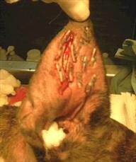
Here, an incision is made in the earflap surgically. The hematoma is drained of fluid and blood clots. To prevent the hematoma from refilling with fluid, multiple sutures are placed in the hematoma area either vertically or horizontally, either partly or completely through the earflap, with or without ear cartilage removal. Sometimes bandages are applied post-operatively, sometimes not. Sutures are generally left in place for three weeks to allow good scarring to take place so that refilling will not occur. The earflap is essentially quilted to close any space where fluid might refill.

Teat Cannula Placement

A teat cannula is a small device used in the treatment of udder inflammation in cows. It can be placed in the opening of the teat to allow drainage of milk or infected discharges. Teat cannulas can also be surgically placed in a dog’s aural hematoma if the earflap is large enough to accommodate the device. The hematoma is drained of fluids and allowed to heal over the next several weeks. This method is generally successful but does involve the dog tolerating a gadget inserted in its earflap for several weeks as well as accompanying fluid drainage.

What if there is a Concurrent Ear Infection?Usually, there is a reason why a dog has been shaking his head: an ear infection. This means that the ear infection must be treated along with the hematoma. The ear will need cleaning, a microscopic examination of the discharge, and medication. Sometimes ear shaking just happens and there is no underlying infection, but be prepared for the expense and trouble of treating an ear infection along with that of the hematoma.

What if We Leave it Alone?

If left alone, an ear hematoma will resolve by itself. The fluid will be re-absorbed back into the body and the earflap will no longer bulge. The problem is that a lot of scarring is associated with this process and the ear is often not cosmetically appealing afterward (i.e. it becomes a “cauliflower” ear). Resolution of a large hematoma can take several months during which it may be uncomfortable for the pet. If the patient is a poor anesthetic risk, it is certainly reasonable to forgo surgery. Corticosteroids, such as prednisone, can be used to reduce scarring.

Aural Hematoma in Cats

The situation in cats is somewhat more complicated than in dogs largely because the cartilage in the feline ear is more sensitive to inflammation and scarring is more severe. This makes the untreated hematoma more likely to form a permanently narrowed ear canal and long-term ear infection potential. Cats’ ear cartilage tends to experience more healing deformity than dogs’ ear cartilage and more curling and softening of the thinner pinnal areas is seen.

What this comes down to is that there is less leeway in letting the ear heal on its own in cats than dogs. Surgical repair is especially important as there is a greater tendency for a cat’s canal to narrow. That said, a more natural cosmetic appearance of the actual ear flap is harder to achieve in cat than dogs. It is more important to focus on the function.

As with dogs, a cat’s hematoma is generally brought about by ear infections and subsequent head-shaking. (In cats, most ear infections stem from ear mites but there are plenty of exceptions.) Bandaging is often used post-operatively as is the Elizabethan collar to protect the ear from being scratched. The cat will need confinement during a healing period of approximately 3 weeks.