4478882

Breast Cancer Happens in Companion Animals

Print an 8.5″x11″ pdf of this poster. 

Everyone reading this is a mammal (unless you are a particularly sentient anemone, coelacanth or axolotl), and one of the things that pegs us as mammals and unites us all is…well, boobs. Yes, even the men and boys. Boob-bearers, all of us.

Let me explain. 

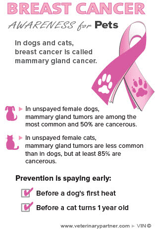
But it still seems to shock people to learn that dogs, cats, rabbits, rats and a host of other domestic species can get breast cancer, too. Since the aim of Breast Cancer Awareness Month is to raise awareness, our aim here is to wave the flag for those who can’t speak for themselves and raise awareness of mammary cancer in companion animals. We also want people to know that breast cancer and mammary gland cancer are the same disease; it’s just called by a different name in other species. Check out the poster we created for use in veterinary hospitals that we hope will help pet owners understand that they’re the same.As a pre-amble ‒ and the way this is going it could be a “pre-ramble” ‒ October is Breast Cancer Awareness Month.

I think people are fairly keyed in to the fact that humans can get breast cancer, or mammary cancer, as it is called in companion animals; its gnarled and potentially deadly hand has touched the lives of many of you, I presume. My own mother had surgery to have a tumor removed that was, luckily, low grade.

Some numbers may help to illustrate the risks to dogs and cats. For humans, breast cancer happens more frequently in more developed countries, and accounts for about 10 percent of all deaths in women between 20 and 59 (source: World Health Organization).

Data for dogs and cats and other domestic species are harder to come by as there is no World Doggy Health Organization database to be accessed as easily as WHO data, but estimates are that 1 in 4 dogs who are not spayed and experience more than one heat cycle will develop mammary tumors. Those stakes are high enough that Vegas would avoid this bet like the plague.

Happily, for dogs or cats who are spayed before they are sexually mature (the usual timeframe for a spay is before 6 months of age) mammary cancer is virtually unheard of. Learn more about the specifics of the disease and treatment in dogs and cats.

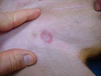
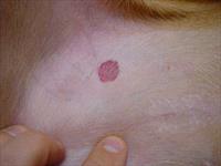
The terminology surrounding lumps, tumors and cancer can be a little daunting sometimes, and is often a source of confusion. One thing to keep in mind is that the word tumor does not always mean cancer. Cancer implies malignancy, or a predilection to grow rapidly and spread to other parts of the body: it’s a process known as metastasis, in which a tumor says, in essence, I’m outta here and decides to infiltrate new and exciting regions for growth. A tumor can be benign or malignant, but cancer is always malignant.

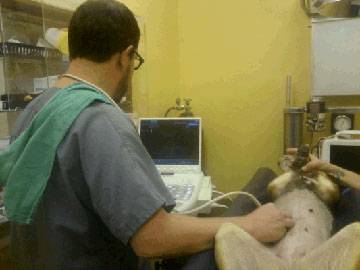
So how do we know which lump is bad, and which is not likely to be a problem for our pets? Sadly, there’s no easy way. Any lump found on a pet’s mammary gland has the potential to be cancerous, so having your veterinarian remove the lump (or a piece of it, in what is known as an incisional biopsy) and submit it for analysis is an important part of keeping your pets safe from this disease. 

A biopsy is a piece of tissue that can be preserved and looked at under a microscope by a pathologist to determine what the lump is (mammary tissue vs. some other tissue that just decided on a whim to form a lump) and whether the lump is cancerous, or as doctors call it, malignant. You can’t tell if tissue is cancerous just by the way it looks, how it feels or where it is.

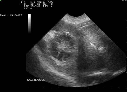
There is one intermediate step that can offer information to your veterinarian on how extensive or radical surgery to remove the lump should be. This is known as a fine needle aspirate, or FNA. With an FNA, you don’t get a piece of the mass, you just suck some cells up into the needle and place them onto a slide. It is much less invasive and doesn’t usually require any sedation or anesthesia, but the trade-off is that it is a less accurate test and less likely than a surgical biopsy to tell you the badness level of the tumor. An FNA is actually notoriously unreliable for mammary masses as far as predicting malignancy but can help make sure the lump is from mammary tissue and not something else that set up shop in the same area.

Medicine is often like that – the invasive stuff is the most accurate, and the easiest stuff gives you the least useable information. It’s like food where the good stuff tastes bad and the stuff that tastes great kills you.  The universe can seem like a vast, cruel joke some days.

I know this sounds complex, intimidating and potentially invasive – not to mention expensive. The good news is that the take-home on this is pretty simple: spay your pet before she goes into heat, unless you have a rock-solid breeding plan for your purebred pet, and have your veterinarian check out any suspicious lumps. That’s about it. Luckily, your trusted family veterinarian has the skills and knowledge to guide you and your pet through this maze to ensure the highest degree of success.

canine_Brachial Plexus_brachial_plexus_closeup_2

Brachial Plexus Avulsion in Dogs and Cats 

Brachial Plexus avulsion or brachial plexus injury can occur when an animal suffers an injury involving the front limbs or shoulders.

Caused by trauma such as an automobile accident, a bite or gunshot wound, a surgical complication, or a fall, brachial plexus avulsion or injury can happen to any breed or age of dog or cat.

Like people, dogs and cats have a network of nerves that arise from the cervical spinal cord in the region of the neck, cross each other in the armpit of the dog, and extend to supply stimulation to the muscles of the neck, shoulders, and front limbs. These nerves convey sensation from this region to the brain via the spinal cord. The brachial plexus is the term for the bundle of nerves in the armpit.

In the case of injury, damaged nerves can affect the function of a front leg. Usually, the damage is on only one side. 

Brachial plexus injuries usually involve more than one nerve. Nerve roots which arise from the spinal cord and contribute fibers to one or more nerves are more apt to be damaged than the nerve itself because the roots are less elastic.

When the nerve roots in the neck region are torn away from the spinal cord, the injury is called a brachial plexus avulsion. When the nerves located in the armpit are stretched or torn, the injury is called a brachial plexus injury.

Depending on the severity of the injury and which nerves are involved, your dog may be unable to move the injured shoulder or leg, may not be able to flex the elbow or toes, or may not put weight on the injured leg. They may lose feeling in the leg or paw. The seriousness of this type of injury depends on what kind of nerve damage your pet has.

How is it Diagnosed?

After hearing your pet’s history, a neurological examination will help your veterinarian determine if your pet has suffered a brachial plexus avulsion or brachial plexus injury and, if so, which type and how severe it is.

canine_Brachial Plexus_brachial_plexus_closeup_2

X-rays, ultrasound, and advanced imaging tests such as computed tomography (CT) and MRI may be recommended.

Treatment

Your veterinarian will treat any life-threatening injuries resulting from the accident or trauma first. If one or more nerves have been stretched rather than torn away from the spinal cord, recovery may occur given time. Treatment involves mainly supportive care. This care should be started early. Your veterinarian may prescribe analgesic drugs for pain as needed. Range-of-motion exercises to do when your pet is ready may be discussed and demonstrated for you. 

Monitoring

Watch your pet closely after this type of injury. An abrasion to the front of the paw may occur if the pet drags the limb. You may find a wound that was not visible at first. Your pet may fuss at or lick a wound or the affected leg. E-collars or other protective measures can be used if your pet starts to self-mutilate.

Your veterinarian will continue to check your pet’s progress and evaluate pain perception and the use of the injured leg.

Prognosis

How well your pet does will depend on the type and location of the injury and how severe it was. A mild injury may show signs of improvement within 1-2 weeks after the trauma happened. In severe cases, the affected leg may not improve even after several months. If there has been avulsion of multiple nerve roots, loss of sensation to the limb, or no progress is seen after several months, and lack of normal limb function is resulting in poor quality of life for the pet, amputation may be recommended.

Prevention is best: do not let your pets have free access to traffic or other hazards, and don’t let your dog ride in open truck beds.

5666579

Bladder Stones in Dogs and Cats

Bladder stones are solid mineral deposits that form inside the bladder of dogs and cats and are quite common. Stones start out as crystals that form in the urine. Crystals form when a combination of events take place, such as urine pH change (pH is a measure of acidity and alkalinity in urine), increased urine concentration, and changes in the mineral makeup of the urine produced by the body. Over time, the crystals combine and layer together to form bladder stones. The process of forming bladder stones is called urolithiasis or cystolithiasis, 

The number of bladder stones produced can range from one or two stones to hundreds. Some bladder stones are small and grit-like, while others can grow to be larger than two inches in diameter. Kidney stones, more common in humans, occur much less often in dogs and cats. Only 2% of stones found in the urinary tract of our pets are found in the kidney.

The two most common types of bladder stones are those made of calcium oxalate and those made of struvite (also known as magnesium ammonium phosphate). Urate, cystine, xanthine, calcium phosphate, and silica are other mineral types that can form bladder stones but are rare.

Types of Bladder Stones 

Struvite bladder stones can occur with bladder infections. Certain bacteria will change the urine’s pH to reproduce faster. This pH change causes the urine to be more alkaline, causing struvite crystals to form. Struvite stones can also form without an infection, which is seen more commonly in cats than dogs.

Less is known about why struvite stones form without an infection. They often play a role in idiopathic cystitis in cats (also known as feline lower urinary tract disease), a condition associated with stress and straining to urinate.

Calcium oxalate stones tend to form with a more acidic pH and are rarely caused by bacteria. Other types of stones can occur with toxins, such as antifreeze poisoning, or can be breed-related, as occurs in Dalmatians.

Clinical Signs

Signs related to bladder stones include:

  •       straining to urinate
  •       urinating small amounts more frequently
  •       dribbling urine
  •       urinating in unusual places
  •       vocalizing or crying when urinating
  •       licking the vulva or penis frequently
  •       urine may have a strong odor or show mucus or blood

Diagnosis

To diagnose bladder stones, your veterinarian will perform a thorough physical examination. Sometimes large stones can be felt by your veterinarian during an exam when they feel the belly (also called abdominal palpation) to check for normal organs within the abdomen. Urine can be obtained to look for crystals, pH changes, and evidence of infection. Radiographs, also called X-rays, are almost always necessary to confirm bladder stones. Unfortunately, some stones do not show up well on X-rays, so your veterinarian will also take into account any signs present during a physical exam and examination of your pet’s urine.

Ultrasound is sometimes used to look for bladder stones, especially those invisible on X-rays. It can also be used to view any damage to the urinary tract from bladder stones.

Determining the type of mineral in a stone is difficult when looking at its shape or appearance. Urine pH can provide clues, but this is not very accurate. To determine the stone’s composition, it must be sent to a laboratory that does urolith analysis, and results can take several weeks.

Treatment Options

Any infection will need to be treated. Depending on the type of stone, removing bacteria that change the pH can prevent new stones from forming. Very small stones may dissolve with normalized pH. Unfortunately, large stones create a perfect environment for bacteria to live, so antibiotics may not be able to completely kill off all the bacteria. Some stones are made of different layers of various minerals, so adjusting the urine pH by getting rid of bacteria may not have a big impact on these “combination” bladder stones. Also of concern, giving antibiotics in situations where all bacteria cannot be killed can potentially cause antibiotic-resistant bacteria. That can make infection harder to control, even after bladder stones are gone.

In some cases, usually with small stones and in combination with antibiotics for bacteria, struvite stones can be dissolved by feeding a therapeutic diet recommended by your veterinarian. This diet acidifies urine pH and restricts certain minerals such as magnesium and phosphorus. Dissolving the stones can take between one week to two months, depending on many factors. Calcium oxalate crystals cannot be dissolved with diet, but specific therapeutic diets can change the urine environment such that enlargement or new stone formation is less likely.  

Sometimes, bladder stones migrate along the urinary tract and become lodged in the urethra, which is the tube that leads from the bladder to outside your pet’s body (to the hole from which they urinate). In such cases, your pet is at serious risk of a urethral blockage, which prevents your pet from urinating. When this occurs, a method called retropulsion (also called retrograde hydropulsion or hydropropulsion) may be attempted. A urinary catheter is inserted into the urethra, and sterile saline (and sometimes lubricant) is injected into the catheter to try to push the urethral stones into the bladder to allow for easier removal.

Sometimes veterinarians will use retropulsion to try to remove small stones from the bladder itself (in which case it may be called voiding hydropulsion or hydropropulsion). The increased pressure of additional fluid in the bladder allows for small stones to pass easily and quickly out of the bladder and through the urethra with the extra fluid once the catheter is removed.

If hydropulsion does not work and diet change is not an appropriate option for your pet, surgery is needed to remove the bladder stones. This type of surgery is called a cystotomy. Your veterinarian will surgically open the abdomen and bladder to physically remove the stones. Sutures, also called stitches, or staples are used to close surgical openings.

After surgery, recovery can take two to four weeks. Pets will often need pain medication for at least one week. Pets are usually given antibiotics after surgery if they had a urinary tract infection prior to surgery. Remember, more stones may form if the infection is not cured. Your pet will need to be on restricted activity (this means no exercise, no running in the house, going outside only on a leash, even to use the bathroom) for one to two weeks following the surgery so they don’t damage the surgical sites or break internal sutures. Urine may be blood-tinged for several days following surgery. Straining to urinate should improve by two to three weeks after the surgery.

Lithotripsy, a method to fragment stones into a smaller size so they can be passed or removed through the urinary tract, is extremely uncommon in veterinary medicine. The procedure is only available at a few referral institutions and veterinary schools.

Increasing water intake by providing more water or adding canned food can also help with bladder stones. Increased water allows for increased flushing of the bladder and dilution of minerals within the urine.

Consequences

If left untreated, bladder stones can grow to the point that urinating is difficult or impossible. This difficulty is especially problematic if stones become stuck in the urethra. The inability to urinate is a life-threatening situation. Other issues associated with bladder stones are chronic pain and an increased risk for urinary tract infections.

The inability to urinate is a life-threatening situation.

Prevention

Once your pet has had bladder stones, therapeutic diets selected for that specific stone can help prevent recurrence. It is crucial that only the therapeutic food be given to your pet. No additional treats (unless specific to the diet), bones, or flavored chew toys can be given. Even a small change in the diet can change the pH and mineral content of the urine and lead to the stones reforming. Increase water consumption as much as possible to help dilute the urine to further decrease the chances of bladder stone formation. 

8202139

Black Fly Bites on Dogs

Black flies (buffalo gnats) are members of the Simullidae family. Their colors vary from black to gray to yellow to olive, and their thorax is humped over the head, giving the appearance of a bison’s hump. The female flies require a blood meal to lay eggs. Males are never blood-feeders; they feed on nectar from flowers. Black fly larvae (young) are carnivorous. There are more than 1,000 species of black flies, but only a few are considered important pests.

Black flies feed on all types of animals, including dogs, cats, cattle, horses, goats, sheep, wildlife, birds, and humans. Black flies are distributed throughout the world. The eggs are laid in fast-running water on stones or leaves just below the water. Larvae are nearly always found in swiftly flowing, well-aerated water. Some species breed in larger rivers; others live in temporary or semipermanent streams. Black flies are particularly abundant in the north temperate and subarctic zones.

Adult black flies may fly 8–11 miles (12–18 km) from swiftly flowing streams; migrating windborne swarms have been known to travel more than 155 miles (250 km). Black flies are active during the morning and evening hours, and during hot days, black flies rest near the ground on the underside of leaves.

Female black flies inflict painful bites, and animals’ ears, neck, head, and abdomen are favorite feeding sites. Local reactions such as redness, itching, and wheals (a raised, itchy spot) at the bite site are common. There can be severe effects in sensitive animals or if the animal has a large number of bites. Swarms of black flies have caused cattle to stampede and refuse to graze, and there have been cases of death in production animals. The bites may result in petechial hemorrhage (reddish spots indicating bleeding under the skin) and lesions can take weeks to heal, especially on the teats of cows and goats. Reduced milk, meat, and egg production may result from less extensive attacks.

Poultry can become anemic from a black fly attack. Death from a severe attack can result from a toxin in the fly’s saliva, which increases capillary permeability, meaning small blood vessels are compromised, permitting the fluid from the circulatory system to ooze into the body cavity and tissue spaces. Some species of black flies can transmit diseases.

Bite Prevention and Treatment

Control of black flies requires destroying breeding sites, moving animals away from the insects’ breeding grounds, and treating animals with insecticides and repellents. Black fly control is difficult because of the large number of flowing water breeding sites. Streams can be treated by using a microbial larvicide, Bacillus thuringiensisisraelensis, a product safe for mammals.

Adult black flies are small enough to pass through window screens or come indoors on or within a pet’s hair coat. The adult female flies prefer to feed outdoors and during the daylight hours. Pet owners concerned about black fly bites may use over-the-counter insect repellents. Aerosols containing pyrethrins may provide only temporary relief. Because area-wide control of black flies is difficult and expensive, people frequently use repellents daily to protect their pets and other animals.

Treatment is often unnecessary. The skin reaction usually goes away in 24 – 48 hours. But if your pet is very uncomfortable or having a more extensive reaction from the bites, your veterinarian may suggest antihistamines, steroids, anti-inflammatories, etc.

4128725

Biliary Mucocele is a Surgical Emergency in Dogs

It all starts with a sick, usually middle-aged or older dog. He is listless, not eating, and maybe has vomiting and/or diarrhea and fever. These are symptoms that could mean any number of things, but usually, when he reaches the veterinarian’s examination table, he has jaundice (yellow pigmentation visible in the whites of his eyes and possibly on his skin and gums). Hospitalization is recommended to rehydrate him and provide supportive care. Blood tests point to a liver problem. Medications are given to minimize the liver’s workload, but soon, an ultrasound is being discussed to image the liver, and costs may be rising quickly.

It all starts with a sick, usually middle-aged or older dog. He is listless, not eating, and maybe has vomiting and/or diarrhea and fever. These are symptoms that could mean any number of things, but usually, when he reaches the veterinarian’s examination table, he has jaundice (yellow pigmentation visible in the whites of his eyes and possibly on his skin and gums). Hospitalization is recommended to rehydrate him and provide supportive care. Blood tests point to a liver problem. Medications are given to minimize the liver’s workload, but soon, an ultrasound is being discussed to image the liver, and costs may be rising quickly.

Why Do We

Need an Ultrasound if We Already Know the Problem Is Liver Disease?

While blood testing can point to the liver, the fact is that numerous diseases can affect the liver. The liver can have an infection, cancer, scarring (cirrhosis), or any number of conditions. The more specific our diagnosis gets, the more specific treatment can be.

Ultrasound is a non-invasive way to evaluate the internal texture of the liver and gall bladder. By looking at the liver’s texture, it is possible to see a tumor and determine if removing it is possible or if it has invaded too far. Ultrasound can evaluate scarring and abscesses. Through ultrasound, it is possible to guide a biopsy needle to an exact area to sample tissue should this be deemed necessary. Ultrasound evaluates the gall bladder and bile ducts. 

One of the more important diseases to rule out promptly is the biliary mucocele because it is commonly a surgical emergency. As we will describe in more detail, a biliary mucocele is a wad of sludged mucus inside the gall bladder. If you wait a few days to see how the patient responds to general liver support, it may be too late for surgery. Furthermore, ultrasound can evaluate the gall bladder’s integrity; if the mucocele ruptures and spills bile into the belly, the surgeon will need to know that as additional treatment is needed. Ultrasound also evaluates concurrent abdominal conditions that might impact recovery, including pancreatitis, which can occur alone with similar symptoms or in addition to a biliary mucocele.

If the patient has a surgical disease, they will not get better until surgery is performed in the vast majority of cases. Furthermore, it may be a surgical emergency (not only will surgery be needed but it will be needed immediately). The sooner the need for surgery is identified, the better the chance of survival.

What Is the Gall Bladder and What Is the Biliary System?

The liver serves as a toxic waste processing center for the body. It filters bacterial products (as well as nutrients) entering the body from the gastrointestinal tract and it removes toxic waste products from the bloodstream. This material is bound to special biochemicals called bile acids and the body would like to get rid of it, bile acids and all.  The solution of bile acids, water, mucus, pigments, and cholesterol forms the greenish-yellow fluid we call bile.

Bile is made in the liver, and then collected into small ducts called bile ductules and bile ducts. The bile is then moved for storage into the greenish round organ called the gall bladder.

During food digestion, hormones cause the gall bladder to contract and squirt bile through the large common bile duct and into the intestine. The bile assists with digestion and carries toxins out of the body so they may be eliminated in feces. The gall bladder and its ducts represent the biliary system.

If the biliary tract becomes obstructed, the patient becomes rapidly jaundiced, painful, and sick.

What Is a Biliary Mucocele?

We mentioned that one of the components of bile is mucus. Normal bile is probably less than 3% mucus but when a mucocele develops, the bile becomes mostly mucus. Normal bile is liquid but mucocele bile is thick and goopy and will not flow easily through the common bile duct. The gall bladder distends trying to pass the mucocele bile and if it actually ruptures, the risk of serious complications and death is much higher. The gall bladder with a mucocele develops an appearance on ultrasound described as resembling the cut surface of kiwi fruit. The biliary mucocele is sometimes called a “kiwi gallbladder.”

When this is seen on ultrasound in a sick, jaundiced patient, surgery to remove the diseased gall bladder should be performed as soon as possible.

Why Does This Happen?

Mucocele development starts with delayed gall bladder contraction, which allows bile acids to accumulate. Altered fat metabolism is also associated with reduced gall bladder contraction, resulting in an accumulation of bile acid. With that accumulation, the lining of the gall bladder feels inflamed and responds by making more mucus and more mucus-producing glands. Pretty soon the bile is just a big wad of congealed mucus and it will not flow. 

Several hormone imbalances have been associated with altered fat metabolism and reduced gall bladder contraction: diabetes mellitus, hypothyroidism, and Cushing’s disease. Cushing’s disease patients have an incidence of biliary mucocele development that is 29 times the incidence of patients without Cushing’s disease. Mucocele development has been linked to pancreatitis (inflammation of the pancreas) as well.

Having Cushing’s Syndrome increases a dog’s risk of developing a biliary mucocele by 29 times.

No one knows why this might be so but it does correspond to the mainstreaming of diagnostic ultrasound into general practice.

The Shetland sheepdog, cocker spaniel, miniature schnauzer, and dachshund seem predisposed to developing biliary mucoceles.

Removing the Gall Bladder (Cholecystectomy)

Removing a dog’s gallbladder is a serious surgery that not all veterinarians are comfortable performing. Discuss with your veterinarian whether a referral to a specialist would be best for you and your pet.

The goal is to remove the gall bladder before it ruptures. If it has already ruptured, tissue damaged by the rupture must be cleansed or removed. Whether or not the gallbladder that has a mucocele is removed before or after rupture does not affect the rate of survival.

Mucocele surgery survival rate is 75-80%. However, if the gallbladder ruptures before surgery occurs, survival rate decreases significantly to 25-40%, depending on the study.

The gall bladder’s function is mainly one of bile storage. Without the gall bladder, bile simply dribbles into the intestine constantly rather than in controlled squirts. You might think this would be a problem, but it turns out not to be. Some patients require long-term medication for liver support but generally speaking, if the dog recovers from surgery, the prognosis is excellent, and the patient can return to normal life.

What if Surgery is Not an Option, or What if the Dog Isn’t that Sick?

There is no question that surgery is the best treatment choice, but there is more to the story. Biliary mucoceles can be an incidental finding in dogs who are not sick or who are sick from something else and are having an ultrasound of the belly for some other reason. In a patient that is not experiencing problems with a mucocele, medication may be able to stave off illness in some cases.

As for skipping surgery on a dog that is sick from its mucocele, this is a very risky move.

It is possible to attempt treatment with general liver support medications, a low-fat diet, and choleretics (medications to help liquefy bile, such as ursodiol), but the problem is that the gall bladder is obstructed with a big wad of goop. This goop is unlikely to liquefy in a timely fashion, if at all, no matter what we do. If medical management is attempted, it is important to regularly recheck the gall bladder by ultrasound to watch for any sign of progression that would indicate that surgery should no longer be postponed.

New Project (6)

Bicipital Tendinopathy in Dogs

Sometimes dogs develop a foreleg lameness without any witnessed injury. Initially, you may note only stiffness or limping when rising or after heavy play. Over time, the limping becomes consistent. One of the possible reasons for these subtle changes progressing to a lameness is tendon damage, which can range from uncomfortable to painful.

Tendons are connective tissue that attach muscle to bone and help the bones move. One tendon that dogs are likely to have problems with is the bicipital tendon, which attaches the bicep muscle to the shoulder blade and crosses through the shoulder joint.

The bicipital tendon allows the shoulder to extend and the elbow to flex. While people and dogs sometimes have problems with this tendon, people don’t walk on their arms, so it’s an entirely different problem for dogs.

Repetitive motion over time from running, jumping, and playing can lead to changes in the tendon, causing it to become tight and painful. In addition, an overweight dog will be putting more stress on the tendon. It is this repetitive trauma and/or overloading of the tendon that initially causes inflammation and tendon damage. The body will try to heal the tendon naturally; however, the lack of blood vessels at the origin of the tendon result in a response that is prolonged and lacks complete healing. This lack of complete healing can result in scar tissue forming, thus not allowing the tendon to work as well as it should.

With bicipital tendinopathies, pain and lameness increase with exercise and improve with rest. It gets worse after vigorous exercise. The condition is seen most often in medium- to large-breed dogs of most ages, and in athletic dogs (agility participants, racing and lure coursing dogs), and often in dogs who jump a lot, either over hurdles in flyball and agility or on and off the couch. Sometimes it is seen in small breed dogs too. However, it’s most common in out-of-shape dogs, those weekend warriors who sit all week and overdo on weekends. There are also some underlying diseases that can cause it, such as such as osteochondritis dissecans, medial shoulder syndrome, or developmental elbow disease.

Diagnosis

If the dog has a history of a subtle lameness of the front limb when rising or after play/exercise, an orthopedic examination is the first step. The shoulder will need to be localized as the source of discomfort. Your veterinarian will perform various manipulations to see if there is pain associated with the biceps tendon. Following an orthopedic examination, radiographs are the next step. A radiograph does a great job of giving us bony information to rule out many things; however, since the biceps is a soft tissue structure, unless it is calcified it can’t be seen on a radiograph. To truly diagnose a biceps tendinopathy, an ultrasound is used and in some cases magnetic resonance imaging (MRI) may be called for.

Treatment

Initially for an acute lameness or a first-time offender, it can be managed medically with rest, exercise restrictions, and nonsteroidal anti-inflammatories (NSAIDs) or other pain medications. The timeframe for healing is typically around 4-6 weeks.

In more severe cases or repeat offenders, a formal rehabilitation program and/or intra-articular injection of a steroid or biologic can be used. The timeframe for exercise restriction is going to vary between 8-12 weeks. That is a long, long time in which to walk the dog on leash to go potty, be restricted inside the house, and to forego all manner of squirrel chasing, playing, and running. However, that’s what lessens the inflammation. If a dog is not restricted to rest during that time frame, the inflammation is almost certain to return, and the rest period will need to start from day one again. The addition of formal rehabilitation therapy will aid in tendon healing and allows your pet to be intellectually stimulated during the down time of exercise restriction.

If the lameness does not go away after appropriate conservative management, surgery can be considered. The surgery aims to cut the tendon from its origin inside the shoulder joint (biceps release) to eliminate the discomfort that occurs from the tendon stretching across the joint. This surgery is commonly completed arthroscopically as this results in less tissue damage and a quicker return to function. Restricting activity and physical rehabilitation are still necessary after surgery, though. 

In most situations appropriate conservative management solutions result in a return to normal function. Unfortunately, because the tendon was damaged over a long period of time, the full recovery time is also prolonged. A full recovery takes approximately 4 to 6 months for a patient to go through the exercise restriction/rehabilitation phase as well as the return to activity/conditioning phases.

Weight control after healing is critical as an increased body condition causes overloading of the joint, which is damaging. After the dog has healed, take care to avoid repetitive concussive activities and weight gain.

6590175-1

Applying Eye Drops with Low Stress Handling™

At some point in many dogs’ lives, they will require eye drops. This might be a one-time event to examine the eye or it may be multiple times per day to treat chronic disease. Regardless of the cause, applying eyedrops need not be a struggle for you or the pet, whether at home or in a clinic setting. The same approach works in both places.

Gather everything you need:

A comfortable place for you and the pet. For small pets this might be on a table, on a chair, or even on your lap if the pet is comfortable there. For large dogs, leave them on the floor.

Items for positive reinforcement and distraction: typically food, but a toy will work well for some pets. You may want or need something the pet can lick the food off (for example, a spoon with a dab of peanut butter).

The eye drops/medication to be given.

Get set up comfortably:

Avoid bending over or toward your pet, it is best to crouch to the side or behind the pet.

Keep your posture as upright as possible so there is no sense of urgency due to impending back/neck pain. If you are prepared to take your time, your pet won’t pick up on your anxiety cues.

Have the dog seated or standing, looking at you or at a right angle. Large dogs can rest their chin on your leg, or the edge of the couch or a chair to decrease downward motion. These are behaviors that can be taught; in a pinch, you can lure a dog to do them with some food.

You are most stable with your hand and wrist on the dog’s head, which requires placing the target eye away from you.

Since you are slightly above the dog, make a kissy or smoochy noise, or say their name in a high-pitched happy tone to get them to look up. This opens their eyes wider for you.

Quickly drop in the drop/ ointment without touching the lid or the eye – gravity does most of the work for you.

Follow with food from your other hand or use it for the entire duration of the process if needed. There will be more head motion if the dog is eating while you attempt to drop in the drop/ointment, but your hand will move with their head.

If you need to put drops in both eyes, some dogs will allow you to shift immediately to the other eye. Some dogs need a short break, and you’ll need to repeat the process.

7905375

Aggression Between Familiar Dogs

Aggression between familiar dogs in the same household is a common occurrence and is one of the most common reasons that dogs are surrendered, rehomed, or euthanized. This aggression can be secondary to high arousal or excitement levels. When a fight occurs, damage to the relationship between the dogs can be difficult to repair.

There are a variety of factors that need to be considered when determining the future relationship between these dogs. For instance, aggression between female dogs is reported to be the most common and is the most difficult to treat due to the severity of injuries and intensity of aggression that occurs between the dogs. Fights between male dogs are often due to resource guarding and are the easiest to treat. Other indicators for poor outcome include:

  • fights escalating in intensity.
  • fights occurring between two female dogs of similar size and age.
  • no noticeable early warning signs or triggers.
  • level three bite or above.

As with any behavioral concern, the first step is looking for underlying medical causes contributing to the behavior.

Treatment Plan

When aggression occurs between familiar dogs, the initial goal of the treatment plan should focus on improving the welfare for both dogs. Although it can be difficult, there should be complete separation of the dogs with no visual contact. Leashes, gates, and tethers can be useful.

Monitor body language of each dog. Since dogs are often very subtle with body language monitor closely for hard stares or stiff posture that is signaling to another dog that they are uncomfortable. It is important that both dogs be completely relaxed in close proximity to each other. Direct stares (longer than three seconds) need to be quickly redirected to reduce escalating tensions.

Basket muzzle training is very important for safety. All dogs should be conditioned to wear a muzzle. This is not a punishment but rather a predictor of good things. Go slow and always wait for each dog to put their own nose into the muzzle. We never force it on, and the dog must willingly place their nose in the muzzle. We never advance it on them.

The dogs should never be together until they are 100% comfortable wearing their muzzles. A muzzle with a buckle is ideal as plastic clasps can break easily. A Baskerville Ultra or Jafco Muzzle with the end square for treat dispensing would be best.

Once comfortable in muzzles you can start walking them next to each other, one adult per dog. This is a great way to rebuild their relationship.

Teaching alternative behaviors such as “touch” is a great behavior to teach your dogs. It can be used to redirect a dog when needed. It can also be used to teach new behaviors and even as an emergency recall.

Teach your dogs to relax on a mat. This can be used later if the dogs are to be reintroduced.

Establish a safe haven such as a crate or room for all dogs. Make sure good things happen in this area. Food dispensing, puzzle toys, classical music and pheromones, can be helpful. A tether or gate may be necessary. If using a crate, be sure to cover and place them out of sight from each other.

Practice Cue-Response-Reward interactions with each dog. A Cue-Response-Reward interaction system encourages predictability and consistency therefore reducing overall anxiety and instability in the relationship between the dogs.

Phase II

Once both dogs are comfortable wearing muzzles and relaxing on their mat.

  • Initially, bring them out for short periods together.
  • One adult per dog and sit on opposite sides of the room. A tether may be necessary.
  • Give them something to do, like a long-lasting treat or toy giving the dogs an opportunity to be together, stay calm and relearn to like each other.

Always separate when they cannot be directly supervised with your entire attention.

Medications

In many cases medications for one or both dogs may be needed to lower anxiety.

5482316

Adopting a Shelter Dog

Deciding to add a new animal to your family is an exciting time! It can be very rewarding to choose to adopt a dog or puppy from an animal shelter or rescue program. Not only will you be adding a wonderful companion to your family, but you will also be saving a life. There are still millions of shelter animals being euthanized annually in the United States that would have made wonderful family pets. Here are a few things to consider before, during, and after the adoption that can help ensure success.

Before Adopting Make sure you’re ready

Adopting an animal means that you are agreeing to be responsible for the animal’s care for many years to come. It is always smart to think ahead and consider what might change in your life and how you will be able to ensure that you meet your new dog’s needs as a beloved family member. Veterinary care, nutritional needs, exercise requirements, and how well the dog will blend with children and other family members are all important considerations.

Research what type of dog is right for you and your family
Many factors may play into the type of dog you seek to adopt. Breed, energy level, sociability, haircoat, age, your personality, and lifestyle are just a few. Many shelters are experienced at helping to make lasting matches and may even have standardized, research-backed questionnaires for you to fill out to help with this process.

Learn about your local shelters and rescues


Most areas have more than one animal welfare organization and each may have different missions and philosophies. You may wish to give your adoption support to the agency your philosophies align with the best. Consider asking your regular veterinarian about the local shelters. Chances are, they’ve examined animals from most of the nearby facilities. They will have opinions about the health and well-being of newly adopted animals and about shelter operations. It can also be useful to ask friends what their experiences have been. Ultimately, you should not only feel great about the dog or puppy you are bringing home, but also about the organization you choose to support through your adoption. There are many things to learn about and consider!

During the Adoption

What to expect


Rescues and shelters take variable approaches to the adoption process. Some require you to fill out involved questionnaires and will call landlords and perform reference checks. Others take a more open, trusting, and conversational approach. Some shelters will send animals home the first day you visit and others take a slower approach. It can be helpful to familiarize yourself right away with how the shelter you visit works so you won’t be disappointed after you’ve matched with a dog.

Almost all shelters will require their animals to be spayed/neutered before adoption. Many shelters will have already performed the surgery before animals are made available for adoption, while others wait until adoption papers have been signed. This is also something you may want to find out about early on. Less commonly, shelters will send animals home with a voucher or other system and require spay/neuter at some point after adoption. It can be helpful to try to keep an open and compassionate mind during the adoption process. Remember that shelters are busy and chaotic places not only for the animals they house but also for the humans that work there.

What to ask


Once you have found a dog that you are interested in, ask the shelter for as much information as can be provided! Get a complete history, but recognize that sometimes there is not much information known. Find out when and where the shelter obtained the dog; was it brought in as a stray, surrendered by an owner, transferred from another shelter, or in some other situation? Ask what is known about the previous living situation and how the dog has behaved since being at the shelter. You should also be shown the medical records the shelter has kept since intake (and any previous records that might exist). You will want to ask about any ongoing medical issues, whether the dog is on any medication, and if there are known medical issues what anticipated care will be required. Inquire about what sort of follow-up services the shelter offers such as health insurance, microchipping, obedience training, or consulting for behavioral issues or medical issues. Finally, be sure to ask about return policies as it is important to know what options exist if the adoption does not work out.

After the Adoption Veterinary visit

Although many sheltering organizations have staff veterinarians, many do not. The level of veterinary care can be advanced to non-existent. No matter what exists in the shelter your dog comes home from, it is always wise to schedule an appointment to see your regular veterinarian soon after adoption. Illnesses such as upper respiratory infection and diarrhea are quite common in the early period after adoption due to the stress of shelter stays, and your veterinarian can help provide appropriate care so that your new friend recovers optimally.

Vaccination boosters, parasite examinations, microchip implantation, and other important preventive care may also be needed during the early time after arriving home.

Be patient


It can take weeks for a new dog to adjust to a new home, yard, family, and environment. Keeping things quiet for a week or so, learning about and employing crate training, and establishing a solid routine can help. Especially for an older dog, patience during the initial adjustment period, and offering enrichment (opportunities to play, learn and explore new things) often pays off in spades once the dog settles in!

Use your Community Resources

Finally, don’t be afraid to ask for help if things aren’t perfect initially or if you have questions. The shelter you adopted from will ideally be your first source of information and happy to hear from you and to provide support!

5770367

Alcohol Poisoning

Dogs and cats can get more than just drunk when they drink ethanol, isopropanol, or methanol — they can get a trip to the emergency room. Pets can die from ingesting alcohol. 

What are ethanol, isopropanol, and methanol?

Ethanol (ethyl alcohol) is most commonly formed from the fermentation of sugars. Ethanol is found in alcoholic beverages, some liquid medications and mouthwash bases, rotting/fermenting fruits, and rising (raw) yeast-containing bread dough. The “proof” of ethanol is twice the percentage of alcohol concentration (i.e. 80 proof = 40% ethanol). Although ethanol is also in some household inks, cleaners, and solvents, the levels are low enough to generally be of no clinical significance if these products are ingested by pets.

Isopropanol (isopropyl alcohol) is in rubbing alcohol (70%), some anti-freezes, some detergents, glass/window cleaners, perfumes/colognes, and alcohol-based topical sprays, including some pet flea-control and grooming products. Isopropyl alcohol is more than twice as potent as ethanol or methanol.

Methanol (methyl alcohol, wood alcohol) is most commonly found in automotive windshield washer fluids (20-80%) but is also in some gasoline additives, “canned heat” fuels (e.g., Sterno®), and some household solvents (e.g., paint removers).

Although the strength of these three alcohols varies, the clinical signs associated with their ingestion by dogs and cats are quite similar.

All poisoning problems boil down to the amount of alcohol ingested compared to weight, just like a dosage of medicine. Thus, when pets drink an alcoholic beverage that was left within reach or that was given to them intentionally by someone, it can cause a significant toxicity problem. In addition, significant absorption can occur through the skin or by inhalation.

Dogs are highly susceptible to the effects of alcohol. Signs of mild inebriation may occur with even minor exposure. Within 15 to 30 minutes after the pet has drunk the alcohol on an empty stomach (or within 1 to 2 hours on a full stomach), central nervous system (CNS) signs, such as staggering, excitement, or decreased reflexes, can begin. Behavioral changes can be seen, as can an increased need to urinate. As the problem gets worse, the pet may become depressed, have a slow respiratory rate, or go into cardiac arrest. Puppies and kittens are at particular risk because of their small size and immature organ systems. 

Alcohols are depressants, so many of the clinical signs associated with them are due to their effect on the CNS. Alcohols irritate the gastrointestinal tract. They also act as diuretics, and the alcohol and its metabolites are eliminated by the kidneys. Liver damage may occur after exposure, although it is much more common in animals that have repeated or chronic exposure. 

Clinical Signs

Clinical signs of intoxication can occur within 15-30 minutes of ingestion of alcohol. Signs include nausea, vomiting, thirst, urination, dehydration, lethargy, incoordination, disorientation, becoming cold (hypothermia), low blood pressure, and alcoholic bad breath. In severe cases, blindness, tremors, tetraplegia, respiratory depression, coma, or seizures may develop. Death is uncommon but may occur, especially if the pet has severe respiratory and cardiovascular depression, low blood pressure, and/or hypoglycemia. 

Diagnosis

Diagnosis is based on a history of exposure, combined with appropriate clinical signs and laboratory tests.  

Treatment/Management/Prevention

The treatments your veterinarian may use can include assisted ventilation, intravenous fluids (to improve elimination of the alcohol/metabolites, to provide cardiovascular support, and to correct electrolyte abnormalities), seizure control, and other supportive therapies. In addition, if the alcohol toxicosis is due to the ingestion of bread dough, your veterinarian may need to wash out your pet’s stomach with cold water to inhibit further alcohol production and to break up the dough mass for removal. In addition, your pet will be confined to prevent accidental self-injury.

Prognosis

Most dogs with alcohol intoxication can recover with adequate symptomatic and supportive care. Recovery may take 8-12 hours and tends to take longer if it was a severe intoxication or if isopropanol was involved.

Preventing Alcohol Toxicosis

All alcoholic beverages and alcohol-containing fluids should be kept out of reach of your dogs and cats. Consult your veterinarian before giving any ethanol-containing liquid medications.

Do not feed raw bread dough to your pets or leave bread dough out to rise in areas that can be reached by them. Always dispose of discarded bread dough carefully. 

The ASPCA National Animal Poison Control is available 24 hours a day at 888-426-4435. Expect an initial consultation fee of around $100.00 and additional follow-up is at no charge. You will be assigned a case number your veterinarian can use to communicate with a toxicology specialist before beginning treatment.