716164

Bleeding: First Aid

Pets often suffer blood loss as a result of trauma or poisoning. Some bleeding is visible, and is a result of a cut or laceration, while internal bleeding occurs inside the body (such as in the chest or abdomen) and is not visible to the naked eye. If bleeding is severe or continuous, the pet may lose enough blood to cause shock; loss of as little as 2 teaspoons per pound of body weight may cause shock, which doctors usually detect as a high heart rate and low blood pressure. Emergencies may arise that require you to control the bleeding, even if it is just during transportation to the veterinary facility. Pet owners should know how to safely stop hemorrhage (bleeding) if their pet is injured.

Techniques to Stop External Bleeding 

The following techniques are listed in order of preference. 

Use caution; avoid injury to yourself, and take appropriate precautions (such as the use of a muzzle, wrapping your pet in a towel or blanket, and having someone assist you if possible) to avoid being bitten. For all techniques below, seek veterinary attention immediately after stopping the bleeding, or on the way to the veterinary hospital.

Direct Pressure

Gently press a compress (a pad of clean cloth, feminine sanitary pad, or gauze) over the bleeding area, so it can absorb the blood and allow it to clot. Do not disturb blood clots after they have formed. If blood soaks through, do not remove the pad; simply add additional layers of cloth and continue the direct pressure more evenly. The compress can be bound in place using loosely applied bandage material, which frees the hands of the first provider for other emergency actions. If you don’t have a compress, you can use a bare hand or finger.

Direct pressure on a wound is the most preferable way to stop bleeding

Elevation

If a severely bleeding wound is on the foot or leg, gently elevate the leg so that the wound is above the level of the heart. 

Elevation uses the force of gravity to help reduce blood pressure in the injured area, slowing the bleeding. Elevation is most effective in larger animals with longer limbs where greater distances from the wound to the heart are possible. Direct pressure with compresses should also be maintained to maximize the benefits of elevation.

Elevation of a limb combined with direct pressure is an effective way to stop bleeding.

Pressure on the Supplying Artery

If external bleeding continues after you have used direct pressure and elevation, use your finger or thumb to place pressure over the main artery to the wound. Apply pressure to the femoral artery in the groin (on the inside of the thigh) for severe bleeding of a rear leg; to the brachial artery in the inside part of the upper front leg for bleeding of a front leg; or to the caudal artery at the base of the tail if the wound is on the tail. Continue application of direct pressure.  Seek veterinary attention immediately.

Tourniquet

The use of a tourniquet is potentially dangerous and it should be used only for a severe, life-threatening hemorrhage in a limb (leg or tail) not expected to be saved. If you see blood spurting or pumping from a wound, which is a rare occurrence, consider the use of a tourniquet. Use a wide (2-inch or more) piece of cloth and wrap it around the limb twice and tie it into a knot. Then tie a short stick or similar object into the knot as well.

Twist the stick to tighten the tourniquet until the bleeding stops. Secure the stick in place with another piece of cloth and write down the time it was applied. Every 20 minutes loosen the tourniquet for 15 to 20 seconds. Remember this is potentially dangerous and can often result in disability or amputation.

A tourniquet should only be used as a last resort and a life-saving measure!

Internal Bleeding

Internal bleeding is a life-threatening condition, but it is not obvious like external bleeding. In internal bleeding, blood pools in the abdomen or chest but does not result in blood in the stool or bleeding from the rectum.

There are, however, some external signs of internal bleeding:

  • The pet is pale (check the gums- if they are white or blue, this is an emergency). 
  • The pet is cool on the legs, ears, or tail. 
  • The pet may cough up blood. 
  • The pet is unusually subdued.

If you see any of these signs, immediately transport your pet to a veterinary facility for professional help. Most cases of internal bleeding will require intensive therapy in a veterinary hospital. Remember: internal bleeding is not visible on the outside.

4127746

Bee Stings and Insect Bites: First Aid  

Any insect or spider can cause problems if they bite or sting your pet. A bite or sting can cause swelling, redness, and itching.

Some animals can have an allergic reaction to a sting or bite that may result in a range of symptoms:

  • mild to severe hives,
  • facial swelling,
  • vomiting,
  • difficulty breathing,
  • or even collapse.

Some spider bites can lead to extensive skin necrosis (skin death) which may need treatment or surgery.

Contact a veterinarian right away if your pet is showing signs of an allergic reaction. Reactions can be severe and may require emergency treatment.

What to Do:

If the stinger can be found, scrape it out with a credit card or other stiff material. Alternatively, use tweezers by grasping the stinger, which is located below the venom sac. Studies have shown that speed is of the essence in removing the stinger. If the only way is to pinch it and remove it, then do so. You’ll do more good than harm and you’ll get the stinger out faster. Don’t worry about squeezing more venom into the area – this has been disproven.    

  • Apply cool compresses to the area 3-4 times daily for 3-4 days.  
  • To help neutralize some of the acidic venom, apply a paste mixture of baking soda and water to the sting area.
  • Prevent your pet from licking at the area by using an Elizabethan collar (cone) to prevent further trauma to the area.
  • In short-faced/brachycephalic breeds like pugs, boxers, etc., facial swelling can lead to damage to the eyes. Have your veterinarian examine your pet and prevent rubbing on carpet, furniture, etc.

What NOT to Do:

Do not administer any medications without first contacting your veterinarian or a veterinary emergency hospital. The veterinarian may need to examine your pet before recommending medications.

4128950-1

Bandaging: First Aid

Home bandaging is not a substitute for proper veterinary care and improperly applied bandages can result in limb loss.

We use bandages for several reasons: to protect wounds from the environment, protect the environment from wounds, and discourage a pet from licking or irritating a wound. Bandages may be applied as support for strains or sprains or to prevent motion. Proper application is important – an improperly applied or too-tight bandage can cause decreased blood flow and potential loss of the limb.

Cleaning the Wound

The process of bandaging begins with careful cleaning of the wound. All dried blood, dirt, and debris should be washed away using mild soap and lots of water. Hair should be clipped away so that it cannot lie in the wound. If possible, the area should be patted dry.  

Materials Needed

In an ideal setting, a bandage should have a contact layer, an absorbent layer, and an outer layer.

This may include (from the innermost layer to the outer layer):

  • antibiotic ointment
  • Telfa Non-Adherent Pad
  • cotton wrap
  • gauze wrap
  • Vetrap

The Contact Layer

After cleaning the wound, apply antibiotic ointment to the contact layer and use it first. Ideally, this layer should:

  • Be sterile and inert. 
  • Stay in close contact with, but not stick to, the wound.    
  • Be very absorbent.   
  • Be free of particles or fibers that might be shed into the wound.   
  • Conform to all shapes. 
  • Allow drainage to pass to the next layer without becoming wet.  
  • Minimize pain.

A Telfa Non-Adherent Pad, available at most pharmacies, comes closest to meeting these requirements.

It is desirable to apply an antibiotic ointment, such as Neosporin, to the pad but this is not absolutely necessary. Frequent bandage changes are more important. After cleaning the wound, place a new contact layer over the wound.

The Absorbent Layer

After the contact layer is in place, apply the second (absorbent) layer to hold the contact layer snugly, but not tightly, over the wound. This layer is usually a cotton or Dacron material that comes in various widths. Generally, 1-inch rolls are used for small limbs and the tail; 2-inch rolls are for medium-sized legs; and the 3- and 4-inch rolls are for large legs and the body. It is important to use the proper size. Materials that are too narrow often cause a tourniquet effect, especially if the wound becomes swollen.

If materials are too wide, they are difficult to apply smoothly. Any wrinkles or ridges may cause the bandage to become uncomfortable for your pet. Uneven pressure may cause necrosis (tissue death) of the underlying tissues.

Begin with just enough of an absorbent layer to hold the contact layer in place. If the wound is on a leg or the tail, wrap it from the toes or the tip of the tail towards the body. If you begin at the top of the leg or the tail, the bandage is more likely to restrict blood flow and cause swelling, which may cause tissue damage. Apply several layers of absorbent material, which will soak up the fluid from the wound and increase the patient’s comfort by cushioning the wound.

Gauze wrap can be applied next to hold the cotton wrap in place and to add extra support.  This step can be skipped for small wounds or for temporary bandages.

The Outer Layer

Finally, apply the outer (third) layer, usually made up of porous adhesive tape or elastic tape (i.e., Elastikon, Vetrap). Wrapped from the toes up toward the body, this layer should also be smooth and snug. Do not stretch elastic tapes to their limits as this will interfere with circulation and result in bandage failure. It helps to unwrap the Vetrap or Elastikon first and then rewind it to remove the tension from the wrap before placing.

The tape should be in contact with the skin (hair) at the bandage margins, anchoring the bandage so it will not slip.

The outer layer of a bandage should be applied smoothly and snugly, but not tight enough to cut off blood circulation.

Bandage Changes

Bandages should be checked frequently for any signs of swelling, skin discoloration or coolness, odor, or saturation of the bandage material. The bandage should be changed whenever any of the above are noticed or any time it appears to be uncomfortable for the pet. If a bandage becomes wet, soiled or slips out of position, it should be replaced.

Be very careful when bandaging at home. Wounds that are draining heavily may require bandage changes every one or two hours.

Bandages over wounds with little or no drainage should be changed every 24-72 hours, depending on your veterinarian’s advice and evaluation. Some wounds may require up to 6 weeks of bandage changes to fully heal.

5482316

Bloat: First Aid

Bloat is a life-threatening condition in which the stomach fills with air (dilatation) and/or twists upon itself (volvulus). It’s also called GDV – gastric dilatation volvulus.

What to Do 

  • Transport to a veterinary hospital or emergency facility immediately. In all cases, this condition requires professional assistance. Most cases will require surgery.

What NOT to Do

  • Do not attempt to relieve the gas from the stomach.  
  • Do not give anything by mouth.

It is imperative that this condition be recognized early. Your pet’s abdomen may not have a bloated appearance. Signs of bloat include:

  • Drooling of saliva 
  • Frequent retching and attempts to vomit (occasionally patients may be able to regurgitate a pool of foamy saliva) 
  • Anxiety, restlessness, and pacing 
  • Lethargy or agitation
  • Depression and shock.

Much has been learned about bloat in the past decade. Decades ago, a diagnosis of bloat was almost always a death sentence because only 25 percent survived. Today the survival rate is better than 80 percent with surgery. Part of the reason for this is increased owner awareness. The earlier the veterinarian gets started with treatment and takes the pet to surgery (after stabilization for shock) the better chance there is for survival. Extremely aggressive medical and surgical intervention early in the course of the disease has the most dramatic impact on overall treatment success.

Prevention

Research into this area is ongoing and many studies have produced conflicting results. No one intervention has been shown to prevent GDV.  Elevated feeding may actually increase the risk of GDV in some patients.  Smaller kibble size, feeding smaller more frequent meals, and not breeding animals with a history of GDV in their lineage may potentially decrease the risk of GDV for the animal and future generations.

In breeds with a high risk of bloat, such as Great Danes, German Shepherd Dogs, and Akitas (as well as other deep chested dogs), a preventive surgery called a prophylactic gastropexy can often be performed when the dog is being spayed or neutered. This involves surgically attaching the stomach to the inside of the abdomen to prevent rotation and can sometimes be done through ‘minimally invasive surgery’ or laparoscopy. Ask your veterinarian for details and advice if you would like to discuss preventive surgery for bloat.

6725556

Bloat – The Mother of All Emergencies 

Many injuries and physical disorders represent life-threatening emergencies, but there is only one condition so drastic that it overshadows them all in terms of rapidity of consequences and effort in emergency treatment: the gastric dilatation and volvulus – the bloat.

What is it, and Why Is It So Serious?

The normal stomach sits high in the abdomen and contains a small amount of gas, some mucus, and any food being digested. It undergoes a normal rhythm of contraction, receiving food from the esophagus above, grinding the food, and moving the ground food out to the small intestine at its other end. Normally, this proceeds uneventfully except for the occasional burp.

In the bloated stomach, gas and/or food stretches the stomach many times its normal size, causing tremendous abdominal pain. For reasons we do not fully understand, this grossly distended stomach tends to rotate, thus twisting off not only its own blood supply but the only exit routes for the gas inside. The spleen, which normally nestles along the greater curvature of the stomach, can twist as well, cutting off its circulation. The distended stomach becomes so large that it compresses the large veins that run along the back, returning the body’s blood to the heart, creating a circulatory shock. Not only is this collection of disasters extremely painful, but it is also rapidly life-threatening. A dog with a bloated, twisted stomach (more scientifically called gastric dilatation and volvulus) will die in pain in a matter of hours unless drastic steps are taken.

What Are the Risk Factors for Developing Bloat?

Dogs weighing more than 99 pounds have an approximate 20 percent risk of bloat. The risk of bloating increases with age.

Classically, this condition affects dog breeds that are said to be deep-chested, meaning the length of their chest from backbone to sternum is relatively long while the chest width from right to left is narrow. Examples of deep-chested breeds would be the Great Dane, Greyhound, and the setter breeds. Still, any dog can bloat, even dachshunds and chihuahuas.

Classically, the bloated dog has recently eaten a large meal and exercised heavily shortly thereafter. Still, we usually do not know why a given dog bloats on an individual basis. No specific diet or dietary ingredient has been proven to be associated with bloat. Some factors found to increase and decrease the risk of bloat are listed below:

Summary of Factors Increasing the Risk of Bloat

  • Increasing age
  • Having closely related family members with a history of bloat
  • Eating rapidly
  • Feeding from an elevated bowl
  • Feeding a dry food with fat or oil listed in the first four ingredients.

Factors that May Decrease the Risk of Bloat

  • Adding table scraps, canned food, or non-kibble supplements to the dog’s kibble diet reduced the risk of bloat in some studies. More research is needed to fully understand the implications of this.
  • Happy or easy-going temperament
  • Feeding a dry food containing a calcium-rich meat meal (such as meat/lamb meal, fish meal, chicken by-product meal, meat meal, or bone meal) listed in the first four ingredients of the ingredient list.
  • Eating two or more meals per day

Contrary to popular belief, cereal ingredients such as soy, wheat, or corn in the first four ingredients of the ingredient list do not increase the risk of bloat.

In a study done by the Purdue University Research Group, headed by Dr. Lawrence T. Glickman: 

  • The Great Dane was the #1 breed at risk for bloat. (The incidence of bloat in this breed is reported to be 42%. Preventive gastropexy should be considered as described below.)
  • The St. Bernard was the #2 breed at risk for bloat.
  • The Weimaraner was the #3 breed at risk for bloat.

In the 1993 study from Germany (see below), the German shepherd dog and the boxer had the highest risk for bloat.

How to Tell if Your Dog Has Bloated

Classically, the dog is distressed and makes multiple attempts to vomit, and the upper abdomen is hard and distended from the gas within, though in a well-muscled or overweight dog, the distention may not be obvious. There are other potential emergencies (sudden abdominal bleeding from a ruptured tumor, for example) that might have a similar presentation, so radiographs may be needed to determine what has happened. The hallmark presentation of bloat is a sudden onset of abdominal distention, distress, anxiety, and pain (panting, guarding the belly, anguished facial expression), and multiple attempts at vomiting that are frequently unproductive. Not every dog will have a classic appearance, and some dogs will not have obvious abdominal distention because of their body configuration. If you are not sure, it is best to err on the side of caution and rush your dog to the veterinarian immediately.

What Has To Be Done

There are several steps to saving a bloated dog’s life. Part of the problem is that all steps should be done at the same time and as quickly as possible.

First: The Stomach Must be Decompressed


The huge stomach is by now pressing on the major blood vessels, carrying blood back to the heart. This stops normal circulation and sends the dog into shock. Making matters worse, the stomach tissue is dying because it is stretched too tightly to allow blood circulation through it. There can be no recovery until the stomach is untwisted and the gas is released. A stomach tube and stomach pump are generally used for this, but sometime surgery is needed to achieve stomach decompression.

Also First: Rapid IV Fluids Must be Given to Reverse the Shock


Intravenous catheters are placed, and life-giving fluid solutions are rushed in to replace the blood that cannot get past the bloated stomach to return to the heart. The intense pain associated with this disease causes the heart rate to race at such a high rate that heart failure will result. Medication to resolve the pain is needed if the patient’s heart rate is to slow down. Medication for shock, antibiotics, and electrolytes are all vital in stabilizing the patient.

Also First: The Heart Rhythm is Assessed and Stabilized


A special and very dangerous rhythm problem called a premature ventricular contraction, or “PVC,” is associated with bloat and it must be ruled out. If this is the case, intravenous medications are needed to stabilize the rhythm. Since this rhythm problem may not be evident until even the next day, continual EKG monitoring may be necessary. If the disturbed heart rhythm is noted at the very beginning of treatment, this is associated with a 38% mortality rate.

Getting the bloated dog’s stomach decompressed and reversing the shock is an adventure in itself, but the work is not yet half finished.

Surgery

All bloated dogs, once stable, should have surgery. Without surgery, the damage done inside cannot be assessed or repaired, plus bloat may recur at any point, even within the next few hours, and the above adventure must be repeated. If the stomach has not untwisted with decompression, the surgeon untwists it and determines what tissue is viable and what is not. If there is a section of dying tissue on the stomach wall, this must be discovered and removed, or the dog will die despite the heroics described above. Also, the spleen, which is located adjacent to the stomach, may twist with the stomach, necessitating removal of the spleen or part of the spleen as well. After the nonviable tissue is removed, a surgery called a gastropexy is done to tack the stomach into its normal position to prevent future twisting.

If the tissue damage is so bad that part of the stomach must be removed, the mortality rate jumps to 28 – 38 percent.

If the tissue damage is so bad that the spleen must be removed, the mortality rate is 32 – 38 percent.

After the expense and effort of the stomach decompression, it is tempting to forgo the further expense of surgery. However, consider that the next time your dog bloats, you may not be there to catch it in time and, according the study described below, without surgery, there is a 24 percent mortality rate and a 76 percent chance of re-bloating at some point. The best choice is to finish the treatment that has been started and have the abdomen explored. If the stomach can be surgically tacked into place, the recurrence rate drops to 6 percent.

Surgery will prevent the stomach from twisting in the future, but the stomach is still able to periodically distend with gas. This is uncomfortable but not life-threatening.

Results of a Statistical Study

In 1993, a study involving 134 dogs with gastric dilatation and volvulus was conducted by the School of Veterinary Medicine in Hanover, Germany.

Out of 134 dogs that came into the hospital with this condition:

  • 10% died or were euthanized prior to surgery (factors involved included expense of treatment, severity/advancement of disease, etc.)
  • 33 dogs were treated with decompression and no surgery. Of these dogs, eight (24%) died or were euthanized within the next 48 hours due to poor response to treatment. (Six of these eight had actually re-bloated.)
  • Of the dogs that did not have surgical treatment but did survive to go home, 76% had another episode of gastric dilatation and volvulus eventually.
  • 88 dogs were treated with both decompression and surgery. Of these dogs, 10% (nine dogs) died in surgery, 18% (16 dogs) died in the week after surgery, and 71.5% (63 dogs) went home in good condition. Of the dogs that went home in good condition, 6% (four dogs) had a second episode of bloat later in life.
  • In this study, 66.4% of the bloated dogs were male, and 33.6% were female. Most dogs were between ages seven and 12 years old. The German Shepherd dog and the Boxer appeared to have a greater risk for bloating than did other breeds.

(Meyer-Lindenberg A., Harder A., Fehr M., Luerssen D., Brunnberg L. Treatment of gastric dilatation-volvulus and a rapid method for prevention of relapse in dogs: 134 cases (1988-1991) Journal of the AVMA, Vol 23, No 9, Nov 1, 1993, 1301-1307.)

Another study published in December of 2006 looked at 166 dogs that received surgery for gastric dilatation and volvulus. The point of the study was to identify factors that led to poor prognosis.

  • A 16.2% mortality rate was observed. The mortality rate for dogs over the age 10 years was 21%.
  • Of the 166 going to surgery, 4.8% were euthanized during surgery, and the other 11.4% died during hospitalization (two dogs died during surgery). All dogs that survived to go home were still alive at the time of suture removal.
  • 34 out of 166 dogs had gastric necrosis (dead stomach tissue that had to be removed). Of these dogs, 26% died or were euthanized.
  • Post-operative complications of some sort occurred in 75.9% of patients. Approximately 50% of these dogs developed a cardiac arrhythmia.
  • Risk factors significantly associated with death prior to suture removal included clinical signs of bloating for greater than six hours before seeing the vet, partial stomach removal combined with spleen removal, need for blood transfusion, low blood pressure at any time during hospitalization, sepsis (blood infection, and peritonitis (infection of the abdominal membranes).

(Beck, J.J., Staatz, A.J., Pelsue, D.H., Kudnig, S.T., MacPhail, C.M., Seim H.B, and Monnet, E. Risk factors associated with short-term outcome and development of perioperative complications in dogs undergoing surgery because of gastric dilatation-volvulus: 166 cases (1992-2003). Journal of the AVMA, Vol 229, No 12, December 15, 2006, p 1934-1939.)

It is crucially important that the owners of big dogs be aware of this condition and prepared for it. Know where to take your dog during overnight or Sunday hours for emergency care. Avoid exercising your dog after a large meal. Know what to watch for. Enjoy the special friendship a large dog provides, but at the same time, be aware of the large dog’s special needs and concerns.

Prevention: Gastropexy Surgery

Preventive gastropexy is an elective surgery usually done at the time of spaying or neuter in a breed considered at risk. The gastropexy, as mentioned, tacks the stomach to the body wall, which drastically reduces the stomach’s ability to twist. The stomach may distend with gas in an attempt to bloat, but since twisting is not possible, this becomes a painful and uncomfortable situation but nothing more serious than that. That said, gastropexy is not an absolute guarantee against twisting but we are talking about a recurrence rate of 76% without gastropexy versus 6% recurrence with gastropexy.  

A study by Ward, Patonek, and Glickman reviewed the benefit of prophylactic surgery for bloat. The lifetime risk of death from bloat was calculated, along with estimated treatment for bloat versus the cost of prophylactic gastropexy. Prophylactic gastropexy was found to make sense for at-risk breeds, especially the Great Dane, which is at the highest risk for bloat.

4128659

Bacterial Diarrheas in Puppies & Kittens   

Bacterial diarrheas are generally a nuisance for the adult animal but can be lethal to a small puppy, kitten, or even a human baby. Most of these problems stem from contaminated food or fecal contaminated environment. Feeding raw food diets to pets dramatically increases the exposure to infectious organisms to the humans in the family. Since these conditions are also human diseases, it is helpful to have some understanding of what they are, even if there are no young pets or immune-suppressed individuals (human or otherwise) at home.

Campylobacter

Campylobacter species are a group of bacteria capable of causing diarrhea in dogs, cats, humans, and other animals. They have a unique curved appearance under the microscope and are said to be sea gull-shaped.  They are difficult to isolate as they grow in conditions of low oxygen (making them microaerophilic as opposed to being aerobic or anaerobic bacteria). With regard to pets, Campylobacter are generally a problem for the very young. Puppies and kittens have not yet matured immunologically.

Plus, because they are small, fluid loss from diarrhea hits them much harder. Furthermore, puppies and kittens are more likely to be housed in groups where fecal cross-contamination is common so they may be more likely to become infected than adult animals. Adult animals commonly have Campylobacter organisms living in their intestines, but they do not experience any sickness due to it.

In humans, Campylobacter infection is a leading cause of gastrointestinal (GI) disease; infected dogs and cats can carry the organism and spread it even if they do not have symptoms themselves.  For this reason, pets used for therapy in assisted living communities and similar situations should be screened for Campylobacter by fecal culture before exposure to people with suppressed immunity. Humans are also infected by consumption of contaminated food, water, or raw milk; only 6% of human Campylobacter infections are attributed to dog exposure.  That said, exposure to a dog with diarrhea triples a person’s risk for developing enteritis from Campylobacter jejuni or Campylobacter coli. Studies screening pet animals for inapparent Campylobacter infections have found surprisingly high incidences of inapparent infection. In one study in the Midwest, 24% of 152 healthy cats were positive, for example.

After consuming Campylobacter organisms, they travel to the lower small intestine, attach, and multiply. They produce a toxin that destroys the lining of the intestine with the result being a bloody, mucous diarrhea (though occasionally a more watery diarrhea is described).  Sometimes a fever results, appetite becomes poor, and vomiting can occur.  Incubation is 2 to 5 days.  The organism can survive as long as a month in environmental feces.

Diagnosis is made by seeing the sea gull-shaped organisms under the microscope; however, there are so many bacterial organisms on a fecal sample that finding the culprit can be tricky. For this reason, a culture is often performed as a more accurate test. Because the organism is microaerophilic, specific culture requirements must be met; the facilities of a reference laboratory are needed.

Treatment is with appropriate antibiotics, such as erythromycin, chloramphenicol or tylosin.

Salmonella

Most people are somewhat familiar with Salmonella. They know it represents a type of food poisoning, probably know it is associated with diarrhea that can be severe, and may even know that Salmonella species are bacteria. Most human cases of Salmonella infection cause fever, diarrhea, and cramping that go away on their own, but in children, it can produce more severe disease. As with Campylobacter, the young are more susceptible to more severe illness because they are smaller and do not have mature immunity. 

The same is true with puppies and kittens; adult animals are almost never affected by Salmonella infection.

An important exception to the “Salmonella is rare in adult dogs” rule is the case of dogs fed a raw food diet. It has become popular to feed raw foods to pets with the idea that a raw food diet more closely approximates the natural diet that the feline or canine body evolved to consume, and thus, such a diet should be healthier than commercially prepared foods. In fact, cooking food is central to removing parasites, bacteria, and bacterial toxins from food. A recent study evaluating raw food diets found that 80% of food samples contained Salmonella bacteria and that 30% of the dogs in the study were shedding Salmonella bacteria in their stool. Adult dogs are often asymptomatic, but any infected animal or person will shed the organism for at least six weeks, thus acting as a source of exposure to other animals or people. Salmonella organisms are difficult to remove from the environment and easily survive three months in soil. Again, dogs used for therapy around the elderly or children should be cultured for Salmonella.

There are two syndromes associated with Salmonella: diarrhea and sepsis. Salmonella bacteria, once consumed, attach to the intestine and secrete toxins. The toxins produce diarrhea that can be severe and even life-threatening in the young. If this were not bad enough, some Salmonella can produce an even more serious “part two” (sepsis) should these bacteria invade the body through the damaged intestine, causing a more widespread and much more serious infection.

In young animals, the syndrome resulting is similar to that of canine parvovirus thus similar treatment is expected.

E. Coli

Escherichia coli may be the most common bacterial organism in the world. It lives in our intestines naturally and covers the world we live in. Unfortunately, some strains of E. coli are not so neighborly and are capable of producing diarrhea via toxin production. Like the other organisms we have discussed, this is a serious problem for the very young and more of a nuisance for adults.  There are three main types of unfriendly E. coli: Enterotoxic E. coli, enterohemorrhagic E. coli, and enteropathogenic E. coli.

Enterotoxigenic E. coli is a common cause of diarrhea in young animals as well as human infants and is responsible for the famous traveler’s diarrhea. These bacteria produce what is called an enterotoxin in the upper small intestine. This toxin, similar to the toxin of cholera, causes the intestine cells to secrete the body’s fluid into the intestine, creating spectacular watery diarrhea and what can be life-threatening dehydration for smaller living creatures.  Young pigs, cattle, and other livestock are commonly lost to this kind of dehydration. Again, the younger and smaller the patient is, the more serious this infection is.

Enteropathogenic E. coli also produce diarrhea in humans and animals. Rather than a secretory diarrhea as above, they simply destroy the intestinal cells where they attach. Diarrhea still results, but it creates more damage to the intestinal lining.

Enterohemorrhagic E. coli is similar to enteropathogenic E. coli but with more associated inflammation. This type does not seem to be a problem for small animals though they can carry it asymptomatically.

It would seem that antibiotics would be the obvious treatment for a bacterial disease, yet for E. coli it is surprisingly controversial. It seems that the use of antibiotics can enhance the synthesis of toxins by these bacteria; plus, oftentimes, antibiotic use only serves to make E. coli more resistant in the GI tract. Antibiotics are generally reserved for those animals (or people) who seem the most sick or who have evidence of bacterial invasion in the bloodstream. Basically, treatment is supportive care until the patient’s immune system regains the upper hand.

In Conclusion

Bacterial diarrheas are an especially serious consideration in the very young and in the weak or compromised, be they human or non-human. It is not unusual for apparently healthy animals to carry these organisms and shed them into the environment and feeding raw foods greatly increases the risk of this kind of latent infection. If you are considering raw food diets, especially if someone in the home is very young or has a compromised immunity, discuss prevention of these infections with your veterinarian.

4128715

Benign Sebaceous Gland Tumors

We receive a fair amount of emails related to the Papilloma article from people with older dogs with numerous “warts” wondering if their dog’s warts will go away as viral warts usually do. The problem is that in older dogs, what looks like a viral wart is probably a sebaceous gland tumor, while there is a very good chance it is benign, it will not be going away any time soon.

It is not uncommon for an elderly dog to develop scores of “warts” which are not warts at all but are sebaceous growths. Most sebaceous growths are benign but one cannot say for sure simply by looking.

Here are some reasons to remove a sebaceous growth:

  • when the growth has been bleeding.
  • when the growth is itchy or is in a location where it is bothering the pet.
  • when the growth is in a location where it interferes with normal grooming of the pet (i.e. the growth gets caught in the grooming clippers, etc.).
  • when there is a question as to whether the growth actually IS a sebaceous tumor and biopsy is needed to settle the question.
  • when you don’t want to take any chances that a sebaceous growth is malignant (as mentioned, you can’t tell by looking).

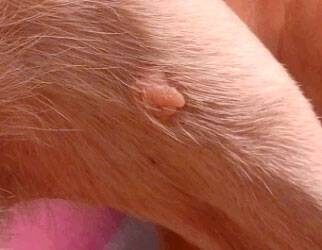
These growths are typically small (pea size or smaller) and originate from the skin’s sebaceous glands, the oil-producing glands of the skin. Because these growths are small, they are generally amenable to removal with local anesthetic. This is helpful since often patients are older and not good anesthesia candidates. It is usually not practical to remove a large number of sebaceous growths with local anesthesia at the same time but the most troublesome can be selected for removal.

Viral warts are different and occur primarily on the face of young adult and adolescent dogs. Sebaceous gland tumors occur in any location, often in large numbers, and usually in older dogs (and occasionally in older cats).

There are several types of sebaceous gland tumors:

Nodular Sebaceous Hyperplasia

About 50% of sebaceous growths are technically not tumors at all and are classified as excessive growth of the gland tissue. It is thought that growths of this group ultimately develop into actual benign sebaceous adenomas as described below. These lesions are round, cauliflower-like, and sometimes secrete material that forms a crust. Occasionally they even bleed. They are particularly common in Cocker spaniels, Beagles, Miniature Schnauzers, Poodles, and Dachshunds. This growth is technically not a tumor but is actually an area of excessive sebaceous cell division.

Sebaceous Epithelioma

Another 37% of sebaceous growths fit into this category. These look just the same as sebaceous hyperplasias to the naked eye but tend to occur in larger breeds and usually on the eyelids or head. They often pigment into a black color. They were formerly described as benign but it turns out they are able to spread in a malignant fashion if given enough time. Since it is not possible to distinguish a low-grade malignant epithelioma from hyperplasia, it is smart to remove any sebaceous growth and not take a chance.

Sebaceous Adenoma

These lesions also look the same as the others to the naked eye. These are also actual benign tumors that probably arose from areas of hyperplasia. As previously mentioned, if given enough time a sebaceous hyperplasia growth will develop into an adenoma. Both are benign.

Sebaceous Carcinoma

About 2% of sebaceous tumors are malignant and may be locally invasive but even malignant sebaceous tumors rarely spread. They have a greater tendency towards ulceration than benign growths. Cocker spaniels seem to be predisposed.

Again, in most cases, the removal of sebaceous gland tumors is straightforward and can frequently be done with a simple local anesthetic. In the event that further treatment is needed, your veterinarian will make recommendations or possibly refer you to a veterinary dermatologist.

4128320-1

Brushing your Dog’s Teeth

What would happen if you stopped brushing your own teeth? Even if you only ate hard food, as most dogs do, there still would be problems. You should be brushing your dog’s teeth twice daily if you can manage it; it’s not as difficult as you might imagine.

What are the benefits of brushing? Brushing removes daily accumulation of plaque from the teeth. Even though dogs do not commonly get cavities, they do suffer from periodontal disease. If untreated, gum disease can lead to bad breath, pain, and tooth loss.

How Do You Brush Your Dog’s Teeth?

Step one is to pick an appropriate pet toothbrush.

Save yourself time by not buying a child’s toothbrush, which is usually too hard for dogs. The ideal dog toothbrush will have a long handle, an angled head to better fit the mouth, and extra soft bristles. For dogs less than 30 pounds, a finger toothbrush that fits over the tip of your finger works well.

Step two is to select an appropriate toothpaste.

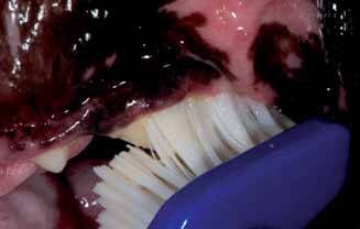
The best pet toothpastes contain enzymes that help control plaque. Try to avoid toothpastes with baking soda, detergents, or salt that are sometimes found in human pastes. Fluoride may be incorporated to help control bacteria. Place the toothpaste between the bristles as this allows the paste to spend the most time next to the teeth and gums.

Step three is to get the brush with paste into your dog’s mouth to get all the teeth brushed.

Most dogs accept brushing if they are approached in a gentle manner. If you can start when they are young, it’s quite easy, but even older pets will accept the process. Start slowly; you can use a damp washcloth or piece of gauze to wipe the teeth, front and back in the same manner you will eventually be using the toothbrush. Praise your dog during the process and offer teeth-healthy treats, such as dental chews, as a reward. Do this twice daily for two weeks, or until your dog is familiar with the approach. Then take the pet toothbrush, soak it in warm water and start brushing daily for several days. When your dog accepts this brushing, add the pet toothpaste.

Most attention should be given to the outside of the upper teeth. The toothbrush bristles should be placed at the gum margin where the teeth and gums meet at a 45-degree angle. The movement should be in an oval pattern. Be sure to gently place the bristle ends into the area around the base of the tooth as well as into the space between the teeth. Complete ten short back and forth motions, then move the brush to a new location. Cover three to four teeth at a time.

Dog home dental care should include daily brushing using an enzymatic pet toothpaste and a proper toothbrush.  Taking an active role in your dog’s dental care will help reduce dental disease, bad breath, and potentially life-threatening heart and kidney disease. Everyone wins.

4128649

Bad Breath in Dogs and Cats

What Is Halitosis?

Halitosis, also called bad breath, is as an offensive odor emanating from the oral cavity. Bad breath is a common presenting pet odor complaint. Common causes may be related to the mouth or, rarely, related to other health problems.

What Causes Halitosis?

The most common cause of halitosis is periodontal disease caused by plaque (bacteria). Bacteria is attracted to the tooth surface within hours of teeth cleaning. Within days, the plaque becomes mineralized producing calculus. As plaque ages and gingivitis develops into periodontitis (bone loss), bacteria changes from somewhat irritating strains to bone destroying types that produce hydrogen sulfide causing halitosis.

Other causes include eating malodorous food; metabolic disease (diabetes, uremia); respiratory disease (rhinitis, sinusitis, neoplasia); gastrointestinal (megaesophagus, neoplasia, foreign body); dermatologic (lip fold pyoderma); dietary (fetid foodstuffs, eating stool); non-periodontal oral disease (orthodontic, pharyngitis, tonsilitis, neoplasia); foreign bodies; trauma including electric cord injury; open fractures; caustic agents; infectious agents including bacteria, fungi, and viruses; autoimmune diseases; and eosinophilic granuloma complex.

What Are The Signs Of Halitosis?

Periodontal disease is painful. Some dogs and cats will have problems chewing hard food, others will paw at their mouths. Unfortunately most will not show any signs.

How Is Halitosis Diagnosed?

Halitosis is easily diagnosed by smelling your dog or cats breath. If there is a disagreeable odor, halitosis is present. A veterinary examination is necessary to diagnose the specific cause of bad breath. If the diagnosis is not obvious after oral examination, blood tests will be taken to check for internal disease.

How Is Halitosis Treated?

Halitosis treatment depends on the cause. There are four recognized stages of periodontal disease. The first two (early gingivitis and advanced gingivitis) are treated by professional teeth cleaning. As the disease advances bone loss occurs causing periodontitis, which may require surgery or tooth extraction.

Odor neutralization of hydrogen sulfide occurs with the use of zinc citrate.

What Is The Prognosis For Halitosis?

Once the underlying disease has been treated, halitosis will disappear. If due to periodontal disease, daily tooth brushing will help maintain good oral health and sweet breath.

5226265

Brucellosis from Raw Milk Consumption

Recently a case of brucellosis was diagnosed in a Texas man from drinking raw milk from a licensed raw milk dairy.  Brucellosis is a bacterial disease of cattle that was very common in Texas 50 years ago but is now rarely found due to vaccination and testing.  However, it is still in some areas and the bacteria is transmitted in the milk as well as fetal fluids at calving to other animals and people. There are also other diseases that can be carried in raw milk, which is the reason for pasteurization as this process kills the organisms.

Raw milk is not pasteurized.  Other animals commonly affected with brucellosis include sheep, goats, pigs and others.  The disease can also cause fistulous withers that is a draining tract at the withers in horses and is a cause of infection of the vertebrae in dogs. In humans, symptoms include fever, sweats, decreased appetite, headache, pain in muscles and fatigue, or basically flu-like symptoms that do not respond to normal treatment.  The condition is called undulant fever in humans because it can recur for long periods of time and can cause heart valve infection, swelling of the liver and spleen and abortion in pregnant women. 

Brucellosis is a very serious disease in animals and people and drinking raw milk should be discouraged as the risks of developing disease are not worth taking the chance.   Death rarely occurs with the disease but many people have recurring symptoms that can last a lifetime, which was common before antibiotics were discovered.  Fortunately, today the disease responds to antibiotics in most humans.  However, drinking raw milk or feeding it to your pets, just like feeding raw meat to your pets, is dangerous for you and your family and is not worth the risk.