4128436

Bladder Stones (Struvite) in Dogs  

There are many types of bladder stones, and each type forms under specific circumstances. In almost all cases, struvite bladder stones in dogs are caused by bladder infection with specially enabled bacteria.  Staphylococci (often simply called Staph) and Proteus bacteria are the usual culprits, and they gain access to the bladder by simply crawling up from the lower urinary tract.

  • 85% of patients with struvite bladder stones are female.
  • Breeds felt to have an increased risk for the formation of struvite stones are the Miniature Schnauzer, Shih Tzu, Yorkshire terrier, Labrador retriever, and Dachshund.

The average age of patients with struvite bladder stones is 2.9 years.

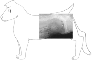
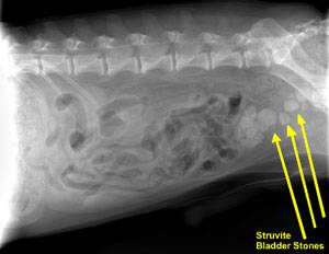
Some patients with bladder stones show no symptoms of any kind and the stones are discovered incidentally but there are some symptoms that might promote a search for stones. Bloody urine, recurrent bladder infection (especially by the same organism and especially if Staphylococci or Proteus is cultured), or straining to urinate all would raise suspicion. Fortunately, struvite stones are radio-opaque, which means they show up readily on radiographs.

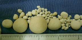
Occasionally stones are simply passed and discovered by the pet owner. If this occurs, it is important to bring the stone to your vet’s office for analysis, have the dog examined, and have radiographs taken to check for more stones.  Patient care will be highly dependent on the stone’s mineral composition.

When to Suspect Struvite Stones

Bladder stones come in several mineral compositions. The most common stone types are oxalate and struvite. Since the approach is different for each type, it is crucial to determine the stone type. The stone type can be confirmed if a sample stone is available (either passed naturally or obtained via surgery, voiding urohydropropulsion, or cystoscopy). A laboratory analysis can easily determine the content of the stone and even determine if the stone consists of layers of different mineral types. Without a sample stone, there are still some hints that can be obtained through other tests.

As mentioned, struvite stones in dogs are almost always formed because of the urinary changes that occur with specific types of bladder infection: almost always Staphylococcal infection but occasionally a Proteus infection. If a urine culture from a patient with a bladder stone should grow either staph or proteus, this would make struvite more likely than oxalate. Also, struvite requires an alkaline pH to form while oxalate requires an acid pH to form; urine pH is a part of any urinalysis and thus provides another clue as to the stone’s identity.

An educated guess is better than nothing but does not replace the analysis of a stone. Remember, occasionally a stone of one type forms around a stone of another type. A complete analysis is needed to effectively prevent a recurrence.

How Do Struvite Stones Form?

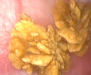
Struvite is the name given to the crystal composed of magnesium, ammonium, and phosphate. (Struvite is also occasionally referred to as “triple phosphate” due to an old erroneous belief that the phosphate ion was bonded to three positive ions instead of just magnesium and ammonium.) Struvite crystals are not unusual in normal urine, and their presence alone does not require treatment. Combine them with certain bacteria, however, and a stone is created.

Stone creation is made possible by an enzyme called urease that certain bacteria, particularly Staphylococci and Proteus species, can produce. Urea is a substance seen in large amounts in urine. Where does all this urea come from? In short, when the body breaks down amino acids, it must contend with the ammonium that is generated in this process. The ammonium, which would be toxic if left alone, is converted to urea, which is much less toxic and is readily soluble in water, making for its easy disposal in urine. Unfortunately, adding urease-positive bacteria into the urinary bladder converts the urea back into ammonium. The combination of infection and inflammation caused by the ammonium creates a matrix that traps the struvite crystals and gels into an actual stone. This reaction can only take place in alkaline urine but the ammonium creates the perfect pH for stone formation. In dogs, the general rule is, if there is no infection, there are no struvite bladder stones.

There are a few rare exceptions to this rule. Certain antacids and diuretics can produce struvite stones when there isn’t any infection. A hormone imbalance called hyperaldosteronism is associated with struvite stone development. The hereditary situation of the English cocker spaniel also represents an exception, as in at least one genetic line of this breed has the tendency to form a purely metabolic struvite stone has been documented. These situations are rare, and for most patients, the focus of struvite stone management is dealing with bladder infections and preventing future ones.

What Should Be Done About Struvite Bladder Stones?

Struvite stones can be removed surgically with a technique called voiding urohydropropulsion; removed with a cystoscope (if they are small enough); or dissolved by diet (also called dissolution). Stone dissolution with diet is the least invasive and probably the best option unless the patient really needs a faster treatment, such as if there is a risk of urinary obstruction in a male dog. Each approach has pros and cons. 

Dietary therapy to prevent new struvite stones is of secondary importance in dogs (except the English Cocker spaniel, for which this is a hereditary metabolic problem rather than a matter of infection).

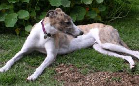
The English Cocker Spaniel has a unique genetic predisposition to make struvite stones even without any infection. Image courtesy of David Gjestson.

The focus is on preventing infection. If your dog has a history of struvite bladder stones, be sure to discuss long-term monitoring and understand what schedule of testing is best for your pet. Expect periodic urine cultures to be needed.

Dietary Dissolution

Dietary dissolution of the stone is not only possible with struvite bladder stones, it is actually the treatment of choice. There are several therapeutic diets available by prescription from your veterinarian that are designed to dissolve struvite bladder stones when they are in the bladder by creating urine that is not compatible with the solid state of struvite.

A therapeutic diet must be the only food fed until the stone is dissolved. Antibiotics are needed as long as stones are in the bladder (bacteria are encrusted within the stone and as the stone dissolves, they are released). Every 4 to 6 weeks, new radiographs are taken to evaluate the stone size. If it is at least 20 percent smaller, the process is continued. A urinalysis is also checked to be sure the proper urinary conditions for dissolution are being created by the diet.

On average, 2 to 3 months are needed to dissolve stones, but the diet should be continued for a full month after the stones are no longer visible on radiographs because small stones may not be large enough to see. Small stones are typically dissolved in just a few weeks as long as the infection is controlled.

After the stones are dissolved, periodic (usually quarterly) urine cultures are performed to check for infection recurrence. If stones do not shrink as expected on the dissolution diet, they may not be pure struvite stones, and another method described below should be selected.

The main advantage of the dissolution method is that it seems to be the lowest risk and most comfortable approach for the dog. That said, an important disadvantage of the dissolution approach is the possibility of urinary tract obstruction as the stone gets smaller and gets lodged in the urethra on the way out. This is potentially a life-threatening hazard for male dogs as they have a narrow urethra. The stone cannot be dissolved in the urethra as, for dissolution to work, the stone must be immersed in urine, which is not the case in the urethra. Furthermore, the inability to pass urine is an emergency, and the patient will die of uremic poisoning in a matter of days if urine flow cannot be restored. The stone can be pushed back into the bladder and dissolution re-attempted but at this point, it may be best to go for a faster resolution with surgery, lithotripsy, cystoscopy, or voiding urohydropropulsion.

Many dissolution diets are really high in fat and high in salt. They may not be appropriate for patients with a past or current history of pancreatitis, patients with heart disease, kidney insufficiency, or high blood pressure.

Surgery

Surgical removal is the most direct method. The advantage is that the stones are removed and healing may commence all in one day. The chief disadvantages are those inherent to surgery: anesthetic risks, post-operative pain, risk of contaminating the abdomen with infected urine, the possibility that not all stones will be removed, and the possibility that the bladder stitches will not properly hold. These risks are generally considered minor and complications associated with cystotomy (opening of the urinary bladder) are unusual. The patient usually stays in the hospital for a day or two to be sure urine production is normal, to properly confine the patient, and to assess pain.

If the dog has urethral stones, they can usually be flushed back into the urinary bladder for surgical removal. If the stone is lodged too tightly for this, it can be removed surgically from the urethra, although the potential for urethral scarring usually makes this a last-choice approach.

Voiding Urohydropropulsion

If the stones are small enough to pass, the bladder can be manipulated in a way to promote expelling them through the urethra. This is called voiding urohydropropulsion and involves filling the bladder, agitating it so the stones float freely in the urine, and then generating a high-pressure urine stream to force the stones out. The patient must typically be held vertically so that gravity may assist in the expulsion of the stones. This technique only works if the stones are small and the patient is not too large. Sedation or general anesthesia is needed. If there are numerous stones, several attempts are often needed if this is to be the only means of removal. Often, this technique is used to obtain a sample stone for analysis to determine if dietary dissolution is feasible.

Cystoscopic Retrieval/Laser Lithotripsy

If you wish to avoid surgery and the stones are small enough, a cystoscope can be passed into the patient’s bladder, and the stones retrieved with a basket (or fragmented via laser lithotripsy). This requires specific equipment most clinics do not have, and thus, usually needs referral to a specialty practice. It is generally more expensive than surgery, though recovery time for the patient is typically much faster.

Recurrence 

After stones are removed one way or another, the focus shifts to prevention. Often, patients are somehow predisposed to a bladder infection, which means they are also predisposed to form more struvite bladder stones. A stone can form as quickly as two weeks after infection if a urease-positive bacterium sets in.

After surgery, antibiotics must be continued until the infection is confirmed to have cleared (i.e., a negative urine culture is obtained). After this, a follow-up schedule of radiographs and/or urine testing is recommended. For a single stone episode, only a few follow-up visits may be necessary. Realize that some individual animals are predisposed to recurring bladder infections, and they may form new struvite stones repeatedly. Obviously, if stones were to recur, a more regular monitoring schedule would have to be revised.

Dietary therapy in the prevention of struvite stones is of secondary importance in dogs (with the exceptions being the rare situations mentioned above). The focus is on preventing infection. If your dog has had a history of struvite bladder stones, be sure to discuss long-term monitoring and understand what testing schedule is best for your pet. Expect periodic urine cultures to be needed.

1862771

Bladder Stones (Oxalate) in Dogs

There are many types of bladder stones, and each tends to form in a specific breed or species under specific conditions. 

Oxalate Bladder Stones in Dogs

  • 73% of calcium oxalate patients are male. 
  • Breeds at especially high risk include miniature schnauzers, Lhasa Apsos, Yorkshire terriers, miniature poodles, Shih Tzus, and Bichon Frises.
  • Most cases occur in dogs between ages 5 and 12 years of age.

How Are Stone Types Determined?

Although a urinalysis can provide a clue, the only way to know for sure that a dog’s bladder stone is an oxalate is to retrieve a stone and have a laboratory analyze it. If the stones are very small, flushing the urinary bladder and expressing it (known as voiding urohydropropulsion) may not only produce a stone sample for testing but remove all the stones and alleviate the current signs. When stones are too large to fit out the urethra, they can be removed to resolve signs and obtain sample stones by surgical removal from the bladder (the traditional method), retrieving them by sending a cystoscope up the urethra, or by using laparoscopy (percutaneous cystolithotomy – PCCL) to send a cystoscope through a small incision into the abdomen and then into the bladder and using a grabbing device in the cystoscope channel to retrieve them. Calcium oxalate stones cannot be made to dissolve over time by changing to a special diet (as can be done with struvite or uric acid bladder stones).

This endoscopic photograph of calcium oxalate bladder stones are in a dog. Bladder stones can be detected during periods of urinary discomfort or they might be discovered incidentally as a surprise. If allowed to remain in the bladder, they can lead to on-going infection, urinary obstruction or continuing discomfort so they must be addressed. Preventive measures must be taken to avoid the formation of new stones. Photo by Dr. Craig Stanton.

Why Would My Dog Form Calcium Oxalate Stones?

It shouldn’t be too surprising that there is a strong hereditary component to forming oxalate bladder stones; this is also true in humans. There is a substance (called nephrocalcin) in urine that naturally inhibits the formation of calcium oxalate stones.

This substance can be defective in both humans and dogs who form calcium oxalate bladder stones. This has been demonstrated in Miniature Schnauzers. Other breeds, such as English Bulldogs and related breeds have Hereditary Calcium Oxalate Urolithiasis Type 1, which is noteworthy for earlier age at the initial development of stones. There is also a Type 2 disorder from a different mutation. There are other potential genetic causes as well.

There is a bacteria that exists as part of the normal flora of the GI tract that metabolizes oxalate. Antimicrobial use may eliminate this good bacterium altogether. This would be expected to increase calcium oxalate stone risk and is being studied.

In humans, the genetic predisposition for stone formation is coupled with dietary issues (problem foods include spinach, peanuts, chocolate, dairy products, calcium supplements, vitamin C supplements, and tea). Dogs are more likely to eat simply a commercial brand of dog food without nearly the dietary variety that humans experience. This means that diet can often be used to manage the problem in the dog unless genetics (not diet) is the cause.

There are some metabolic diseases that might predispose a dog to create a calcium oxalate stone. For example, a dog with Cushing’s disease will be overproducing cortisol (commonly known as cortisone). This hormone increases calcium excretion in urine. The extra calcium present in the urine will promote a stone. Some animals may have an elevated blood calcium level (hypercalcemia) for any number of reasons, and this could lead to excess calcium in the urine as the body attempts to restore calcium balance. Again, extra calcium in the urine promotes a stone. Screening for such medical conditions is important and is generally part of the medical workup once stones are detected on a radiograph. If any such metabolic problems are uncovered, they will need to be treated separately. As these problems are resolved, no further stone prevention measures should be needed. Because of the complexity involving a variety of different causes and risk factors, this should be viewed as a group of diseases that result in the same endpoint, calcium oxalate stones. The specific cause does change what prevention measures are expected to help. Clues based on the breed can help; but it can be very difficult to know which disease and risk factors are present in any given dog, contributing to the high recurrence rate of these stones in dogs.

Why Can’t The Stones Stay Where They Are?

The most immediate concern for the dog with bladder stones is that the urinary opening may obstruct as the dog attempts to pass the stones. This is largely a male dog problem but the results can be life-threatening uremic poisoning. In such cases, the veterinarian will try to dislodge the stone, flushing it back into the bladder to restore the patency of the urinary opening. If the stone cannot be dislodged, a new urinary opening may have to be surgically created. The urethra (the narrow tube connecting the urinary bladder to the outside world) is a difficult place to perform surgery, so it is preferable to move the stone back into the bladder for removal rather than attempting removal from the urethra.

Bladder stones are irritating to the bladder simply by rubbing on the tender bladder lining. Bleeding typically results, and, of course, the chance of developing chronic bladder infections is markedly increased with the presence of bladder stones.

Because calcium oxalate stones cannot be dissolved by diet change, removing the stones is usually necessary. Studies have shown that 50% of dogs who have undergone such surgery will develop new calcium oxalate stones within three years unless proper prevention is instituted.

The following therapeutic plan has been devised to minimize the chance of recurrence after the stones have been removed.

Step One: Diet

While special diets cannot dissolve existing stones, they do help prevent new stones from developing. Prevention centers on creating a urinary environment with minimal calcium and oxalate as well as creating a urine pH that is not conducive to calcium oxalate formation.

Illustration by Wendy Brooks, DVM

Further, high protein, high sodium diets should not be used and diets low in phosphorus may stimulate vitamin D production and could increase calcium absorption. Commercial specialty foods with these requirements are available. Work with your veterinarian to find an appropriate diet.

For each of the commercially available diets, the canned form is preferable to dry to increase water consumption and thus help dilute the urine. It is especially important to avoid table scraps when caring for an oxalate stone-forming dog. Still, no one wants their dog to live life without treats.

The following treats are acceptable for oxalate stone-forming dogs:

  • Plain, cooked chicken
  • Plain, cooked turkey
  • eggs
  • rice
  • peas
  • pasta
  • white potatoes
  • cabbage
  • cauliflower
  • bananas
  • melon

Selecting a diet that actually prevents stones but is also desirable to the picky pet may be challenging. It may be possible to have a veterinary nutritionist provide a recipe for an appropriate home-cooked diet. Please visit www.balanceit.com or www.petdiets.com for details.

Some medications can increase the risk of calcium oxalate stone development, and these should be avoided. Prednisone and other cortisone-type medications (commonly used for itchy skin, arthritis, inflammatory bowel disease, and other inflammatory conditions) should be avoided if possible. Furosemide (brand name: Lasix or Salix) is a diuretic mostly used in the treatment of heart failure. Both these medications lead to excess calcium in the urine. Should a diuretic be needed for a patient at risk for calcium oxalate stones, one from the thiazide class (see later) would be a better choice. Supplementation with Vitamins D or C also increases the risk of forming oxalate stones; such supplementation should be discontinued.

Step Two: Urinalysis and Radiographs

Two to four weeks after surgery, a urine sample is checked and the bladder is radiographed. If the urine is not adequately diluted (meaning the specific gravity is less than 1.020), water consumption will have to be increased (either by adding water to the food or increasing the amount of canned food).

If urinary pH is less than 6.5 or if oxalate crystals are seen, it is time to go to Step Three. If all is well, a urinalysis should be performed every three to six months or so for the rest of the dog’s life (the University of Minnesota’s current recommendation).

Radiographs are also taken every 6-12 months so that any new stones will still be small enough to be retrieved from the bladder by flushing. If you skip this monitoring, you may find stones have developed to a large size, and surgery is again needed to remove them.

Step Three: Potassium Citrate

By taking potassium citrate pills, citrate levels increase in the urine. Calcium binds to the citrate instead of the oxalate. This is a desirable event since calcium citrate tends to stay dissolved, whereas calcium oxalate tends to precipitate out as a mineral deposit. Potassium citrate also helps create alkaline urine (in which calcium oxalate stones have difficulty forming). Potassium citrate supplements are typically given twice a day.

Step Four: Urinalysis Again

Two to four weeks after potassium citrate is added to the therapeutic diet, a urine sample is checked. If the urine is not adequately diluted, water consumption will have to be increased either by adding water to the food or increasing the amount of canned food.

If urinary pH is less than 6.5 or if oxalate crystals are seen, it is time to go to Step Five. If all is well, urinalysis and radiographs should be performed every 6-12 months or so for the rest of the dog’s life (the University of Minnesota’s current recommendation). In other words, if the urinalysis shows all the desired parameters and radiographs show no new stones forming, then therapy is working, and the pet can go on a regular monitoring schedule. If testing shows the patient is still predisposed to stone formation, it is necessary to proceed down this list until control is achieved. Again, radiographs should be taken every six to 12 months.

Step Five: Thiazide Diuretics

A diuretic is a medication designed to increase the amount of urine produced. The thiazide class of diuretics does this in a way that reduces the calcium content of the urine. This medication is typically given twice a day and electrolytes are generally measured via blood test within the first two weeks of this treatment.

There will always be some dogs who continue to form stones no matter what is done. With radiographic monitoring, it is hoped that new stones can be detected while they are still small. As research continues, new developments may arise that will not require such frequent monitoring.

A Note On The Bichon Frise

The Bichon Frise appears to have a unique situation with oxalate bladder stones in that they seem to begin to recur virtually as soon as they have been removed. In a study presented by Jody Lulich’s group at the University of Minnesota at the 2004 meeting of the American College of Veterinary Internal Medicine, 24 out of 33 Bichons had oxalate stone recurrence after cystotomy (37% had had their first recurrence in the first year after surgery, by the end of the second year 57% had recurred, and by the end of the third year 73% had recurred, some more than once). Steps to prevent recurrence need to be particularly aggressive from the beginning in this breed as they seem to have 20 times the risk of oxalate stone formation compared to other breeds. 

Preventing Male Dog Obstruction

In male dogs, a bone called the os penis is located in the penis, surrounding the urethra. Urine passes through this hollow bone like water through a pipe. Because this area is made of bone, it cannot stretch or expand to accommodate a stone trying to pass. A stone blockage of this type leads to an emergency and death if it goes untreated.

In a perfect world, controlling the bladder stones controls the risk of obstructions, but controlling the bladder stones is not always easy, and treating a dog over and over for obstruction becomes expensive. A surgical procedure called a urethrostomy can be performed to prevent obstruction. This procedure involves creating a new urinary opening in the area of the scrotum. This allows urine to be expelled earlier in its course so that passage through the os penis does not occur. The flexibility of the non-bony part of the urethra plus the surgically enlarged urinary opening allows for smaller stones to pass rather than stick in the os penis.

To perform this surgery, the male dog must be neutered (which can be done at the time of the urethrostomy). The creation of the new opening usually requires the removal of the empty scrotum.

While this is not as good an outcome as preventing stone recurrence, at least the emergency obstruction is prevented.

Calcium oxalate bladder stones can be frustrating. Not only do they tend to recur, but following monitoring recommendations involves a visit to the veterinarian’s office every three to six months. It is up to every owner how rigid to be with guidelines, but keep in mind that the trouble and expense of a stone surgery weigh against the much less trouble and expense of monitoring.

Laser Lithotripsy

A new technique of stone removal involves the use of a laser to blast the stone into pieces small enough to pass. This option (called laser lithotripsy) is especially useful when the number of stones present is small and/or when a stone is lodged in the urethra and cannot be flushed into the bladder. It will not be helpful for a large number of stones as the process would be too time-consuming relative to a more conventional approach but for the right patient, it is less invasive than other procedures. 

5313917

Breeding Information for People Who Want to Breed Dogs

Factors to Consider

  • Will your dog contribute excellent health, temperament, working ability or conformity to the breed standard?
  • Do you understand that spaying and neutering will prevent some health problems that you risk by keeping your dog intact?
     
  • Are you aware of any and all health and temperament problems in your dog’s pedigree, looking at both depth and breadth of pedigree?
  • Are you willing to search for the best dog to breed your dog to, even if you have to travel out of state?
  • Do you have carefully screened buyers and deposits for all the puppies you may produce?
  • Do you have money set aside in case the dam or puppies need emergency care?
  • Can you or another responsible adult be present 24 hours a day for the first 3 weeks in case hand feeding is needed?
  • Have you read about what to prepare and expect for canine pregnancy, whelping and puppy rearing? (rec source: Canine Reproduction: A Breeder’s Guide 3rd Edition, Phyllis Holst)
  • Are you willing to keep and properly socialize all the puppies until good homes are found?
  • Are you willing to take back any or all puppies any time in their lives that they may no longer be wanted?
  • Are you willing to serve as a lifetime resource for the buyers of your puppies?

Pre-Breeding Procedures

  • Annual CERF eye certification.
  • Wait until 2 years of age before breeding, then have OFA hip and elbow certification performed.
  • Have all breed-specific health clearances performed – check with veterinarian and national breed club (may include heart, thyroid, genetic testing, many others).
  • Have Brucella canis test performed 1 month in advance.
  • Have a complete physical examination performed on your dog prior to breeding. 
         – This should include a digital vaginal exam to check for vaginal band/stricture.
4126766

Birthing Puppies  

Prenatal Care

Preparing for your dog’s labor and puppy care can be both exciting and fun; still, awareness of potential problems is of paramount importance. It is a good idea to keep track of your dog’s breeding date so as to know when to expect what. We will first present some prenatal care suggestions, but for more details, read specifically about care during pregnancy; you may wish to begin there.

After about 35 days of pregnancy, the mother’s nutritional needs will begin to increase. In general, she should require about twice as much food as usual, whereas when she begins nursing, she will need three times as much food. The best nutritional plan is to buy a dog food approved for growth (i.e., puppy food) and feed according to the package; such diets are balanced and require no supplementation, plus they typically have the extra calories needed by the pregnant or nursing mother. Exercise of the pregnant mother need not be restricted until after the first 4-6 weeks of pregnancy. Do not supplement calcium as this can cause metabolic imbalances; also, excess vitamins may be harmful to the puppies.

Sometime around the 45th day, your dog should be examined by a veterinarian. At this time, the skeletons of the unborn pups will have mineralized and are thus going to be visible on an x-ray.  Your dog’s abdomen should be x-rayed so that you know how many pups to expect. This is important as you will need to know when her labor is finished so you can be sure none of the puppies have been retained. Ultrasound may be used to confirm pregnancy much earlier (after 25 days, the embryonic heart may be seen beating), but it is more difficult to count the number of pups using this method. A general pregnancy blood test can be performed around day 35 just to confirm whether or not she is pregnant, but neither this nor ultrasound will tell you how many puppies to expect; only radiographs can do that.

A comfortable area should be set aside for whelping (giving birth) and raising the puppies. The mother should feel at home here and should be able to come and go as she likes while the puppies must remain confined.

It is important that the mother be isolated from all other dogs for three weeks prior to labor through three weeks after delivery to prevent herpes infection. Herpes is spread by sniffing and licking between two dogs. Adult dogs rarely have any symptoms but the newborn or unborn puppies generally die.

The dog’s gestation period is considered to be 63 days, though this is not written in stone, and a normal range might be 58 to 68 days.

Impending Labor

When your dog’s due date is approaching, you should begin monitoring her rectal temperature. When her temperature drops below 100°F (normal canine temperature is 101-102°F), labor may be expected within 24 hours.

It is a good practice to know how to take your pregnant dog’s temperature as her due date approaches. Ask your veterinarian to show you how.

The First Stage of Labor

During this stage, uterine contractions begin. The mother will appear restless and may pace, dig, shiver, pant, or even vomit. This is all normal and all an owner can do is see that she has water available should she want it. This stage of labor is long, lasting 6 to 12 hours and culminates with full dilation of the cervix in preparation to expel a puppy.

The Second and Third Stages of Labor

Puppies are born covered in membranes that must be cleaned away or the pup will suffocate. The mother will bite and lick the membranes away. Allow her a minute or two after birth to do this; if she does not do it, then you must clean the pup for her. Simply remove the slippery covering and rub the puppy with a clean towel. The umbilical cord may be tied in a knot about one inch from the pup and cut with scissors on the far side of the knot. Be careful not to pull on the umbilical cord as this can injure the puppy. The mother may want to eat the placenta but this is probably not a good idea as vomiting it up later is common; it is best to clean away the placenta yourself.

Expect one pup every 45 to 60 minutes with 10-30 minutes of hard straining. It is normal for the mother to take a rest partway through delivery and she may not strain at all for up to four hours between pups. If she is seen straining hard for over 30 minutes or if she takes longer than a 4-hour break, consult a veterinarian.  This is where it is important to know whether she has delivered the entire litter that was counted on the X-ray. Expect some puppies (probably half of them) to be born tail first, which is normal for dogs.

Most of the time nature handles things according to plan and there are no complications. The important thing is to be prepared and know what constitutes a deviation from normal. During the delivery, a puppy can get stuck either because of size or positioning, the mom can get too tired or dehydrated to complete the mission without help, or any number of unexpected problems can arise. Problems can happen during the actual delivery or in the days following.

Call your veterinarian if:

  • 30 to 60 minutes of strong contractions occur with no puppy being produced.
  • More than 4 hours pass between pups and you know there are more inside.
  • She fails to go into labor within 24 hours of her temperature drop.
  • She is obviously in extreme pain.
  • Greater than 70 days of gestation have passed.

It is normal for the mother to spike a fever in the 24 to 48 hours following birth. Clinical signs of illness should not accompany this fever.

Normal vaginal discharge after giving birth should be odorless and may be green, dark red-brown or bloody and may persist in small amounts for up to 8 weeks.

Green discharge is a bit special as this is the discharge indicating separation of a placenta from the uterus. During the birth of a litter, many placentas are separating so there will be plenty of green discharge. Where this becomes important is on the very first puppy, as when the first placenta separates, its associated puppy will need oxygen very soon, so a live puppy should appear within 30 minutes of seeing green discharge. If not, there is a problem, and you should consult a veterinarian.

Problems to Watch for in the Following Days 

Metritis (Inflammation of the Uterus)

Signs of this condition are as follows:

  • fever
  • foul-smelling vaginal discharge
  • listlessness
  • loss of appetite
  • no interest in the puppies
  • decreased milk production

If these signs are noted, usually in the first day or two postpartum, a veterinarian should be consulted. Your dog may have retained a placenta or have suffered some trauma during delivery. Animals who require assistance with delivery are often predisposed to metritis. She will likely need to be spayed.

Eclampsia

This condition results when the mother has trouble supporting the calcium demand of lactation and is a particular concern for toy breed dogs. Calcium supplementation predisposes a new mother to this condition. Usually affected animals are small dogs. They demonstrate:

  • nervousness and restlessness
  • no interest in the pups
  • stiff, painful gait

This progresses to:

  • muscle spasms
  • inability to stand
  • fever
  • seizures

This condition generally occurs in the first three weeks of lactation and a veterinarian should be consulted immediately.

Mastitis (Inflammation of the Breasts)

Normal nursing glands are soft and enlarged. Diseased glands are red, hard, and painful. In general, the mother does not act sick; the disease is confined to the mammary tissue. The mother may be sore and discourage the pups from nursing; however, it is important to keep the pups nursing the affected glands. This is not harmful to the puppies and helps flush out the infected material.

Warm compresses may be helpful.

Agalactia (Not Producing Milk)

Milk production and secretion (let down) is essential for the puppies’ nutrition. If the puppies are nursing but it appears that milk is simply not flowing, there are a few simple things to try at home before going to the veterinarian. First, make sure the puppy room is not too warm and that the mother has plenty of food and water and that she seems to be healthy in other respects. If these issues seem controlled, the next step is to determine if milk is being produced and not “let down” or simply not being produced as different hormones are involved in each process. Your veterinarian will need to intercede with treatment for the mother. If the pups cannot so much as get colostrum, that all-important first milk that provides immunity from common infections, they may need to receive injections of canine plasma to replace the antibodies they did not get from their mother.

Most dogs are excellent mothers and problems are few. The basic rule is to seek veterinary care if she seems to feel sick or if she ceases to care for her young. Puppies nurse until they are about six weeks old but can begin solid foods as early as four weeks of age. A good age for adoption to a new home is eight weeks or later.

5313942

Brachycephalic Airway Obstruction Syndrome in Flat-Faced Dogs  

Dogs with Short Faces

Most people are not familiar with the term brachycephalic, but if you own a Pug, Boston Terrier, Pekingese, Boxer, Bulldog, Shih Tzu, or any one of the other breeds with pushed-in or short faces, you should become familiar with this word. The word comes from the Greek roots brachy, meaning short, and cephalic, meaning head.

Brachycephalic dogs have been bred so as to possess a normal lower jaw that is in proportion to their body size and a compressed upper jaw. In producing this cosmetic appearance, we have compromised these animals in many important ways, and you, as an owner, must be familiar with the needs of your pet.

The Respiratory System and Brachycephalic Airway Obstruction Syndrome

Brachycephalic breeds make a lot of snorting respiratory sounds, as a matter of course, simply because of the way their throats and faces are shaped. These sounds are generated from a combination of several anatomic deformities that will be described below. These deformities occur to varying degrees of severity. Most brachycephalic dogs are not hampered by their anatomy on a day-to-day basis, but they do have limitations that must be recognized. Severely affected individuals require surgical intervention.

Stenotic Nares

This is a fancy name for narrowed nostrils. The brachycephalic dog’s respiratory passage begins with very small, often slit-like, nasal openings for breathing. This leads to a great deal of open-mouth breathing and panting as the nasal breathing route is limited by its small opening. In selecting a puppy for adoption, it is a good idea to look at the nostril sizes on each member of the litter and look for the widest opening. Stenotic nares can be surgically corrected after the age of five months.

Enlarged Tongue (Macroglossa)

The brachycephalic dog’s tongue can be particularly thick and large, which contributes to the overall obstruction. The French and English bulldogs seem particularly predisposed to this issue.

Elongated Soft Palate

It is difficult to fit the soft tissues of the canine mouth and throat into the brachycephalic’s short face. As a result, the soft palate, which separates the nasal passage from the oral cavity, flaps loosely down into the throat, creating snorting sounds. Virtually all brachycephalics suffer from this, but actual respiratory distress is rare except in English Bulldogs. The English Bulldog tends to have more severe symptoms in almost all aspects of brachycephalic syndrome. Excess barking or panting may lead to swelling in the throat, which can, in turn, lead to trouble. Again, the soft palate can be surgically trimmed.

Tracheal Stenosis/Hypoplastic Trachea

The trachea is also known as the windpipe. The brachycephalic dog’s windpipe may be dangerously narrowed in places. This condition creates tremendous anesthetic risk and should be ruled out by chest radiographs prior to any surgical procedures, as anesthetic safety depends on the placement of a tube in the windpipe to secure air passage. If the trachea is too small, intubation may not be possible, and it is important to know this in advance of any planned procedure. The English bulldog is particularly predisposed to this anatomical defect.

Everted Laryngeal Saccules

The normal larynx has two small pockets called ventricles or saccules. When a dog has increased effort to breathe, over time, these little pockets will actually turn inside out inside the throat. When this occurs, it obstructs the throat. This condition can be corrected surgically; however, in many cases, it resolves on its own after the stenotic nares are corrected, so surgery is generally only performed in severe situations. The pug is especially predisposed to this situation.

Heat Stress, Excitement, and Exercise 

Because of all these upper respiratory obstructions, the brachycephalic dog pants inefficiently. A dog with a more conventional face and throat is able to pass air quickly over the tongue through panting. Saliva evaporates from the tongue as air is passed across and the blood circulating through the tongue is efficiently cooled and circulated back to the rest of the body.

In brachycephalic dogs, so much extra work is required to move the same amount of air that the airways become inflamed and swollen. This inflammation leads to a more severe obstruction, distress, and further overheating, which is potentially a life-threatening problem. It should always be a consideration during stressful situations, especially air travel. 

The American Veterinary Medical Association has travel guidelines for brachycephalic dogs.  

The American College of Veterinary Surgeons has further information on brachycephalic obstructive airway syndrome.

Brachycephalic dogs are the most likely candidates for heat stroke.

Altogether, the upper airways of the brachycephalic dog compromise the ability to take in air. Under normal conditions the compromise is not great enough to cause a problem; however, an owner should take care not to let the dog become grossly overweight or get too hot in the summer. Be aware of what degree of snorting and sputtering is usual for your pet, and should your dog require general anesthesia or sedation, your vet may want to take extra precautions or take radiographs beforehand to assess the severity of the syndrome. Anesthetic risk is higher than usual in these breeds, though under most circumstances the necessary extra precautions are readily managed by most animal hospitals.

To be clear, brachycephalic syndrome can be progressive if it is not corrected at an early stage. Severely affected dogs can experience collapse of the larynx and require a permanent tracheostomy (a hole in the throat for breathing).

Gastrointestinal Tract

There appears to be a constellation of stomach and swallowing issues that correlates to the respiratory obstruction. In brachycephalic breeds, the esophagus (the tube that conducts swallowed food from the throat to the stomach) experiences reflux of stomach contents backward. 

This reflux generates pain and inflammation. Compounding this problem is the retention of food in the stomach for prolonged periods. The pooling of food in the stomach creates a sensation of nausea and increases the potential for vomiting and/or stomach acid reflux. The English bulldog seems particularly predisposed to GI complications in this way. This sounds like a separate issue from the respiratory syndrome, but in fact, the reflux, regurgitation, and even herniation of part of the stomach into the chest cavity stems from the extreme inhalation efforts made against the upper airway obstruction that comes from the shape of the brachycephalic head. Medications are helpful for intestinal issues as is weight control, but often correcting the respiratory obstruction corrects the intestinal problems as well.

Eye Problems

With most of the nasal bones compacted, brachycephalic dogs tend to have trouble with the way their eyes seat in their heads. The eye sockets are shallow making the eyeballs especially prominent and vulnerable. Often a nasal fold of skin rubs on the eyeball and/or there are eyelashes rubbing on the eye.

Lagophthalmos

Sometimes, the eyes are so prominent that the lids cannot close all the way over them. This will lead to irritation and drying of the center of the eye unless surgical correction is performed. If you cannot tell by watching your pet blink, watch as your pet sleeps to be sure the eyelids close all the way. If the eyelids cannot protect the eyes, blinding pigmentary changes will form where the eyes become irritated.

Nasal Fold Irritation

Many brachycephalic dogs have a fold of skin between the nose and eyes. This skin fold may need regular cleaning as it tends to collect skin oil and moisture but it can also be prominent enough to rub on the actual eyeball. Chronic irritation will show as a pigmented area on the eye surface, especially on the side nearest the nose. This is hard to see without a bright light but if it is noted, a search for the cause is warranted. Depending on the location of the pigmentation, surgery may be recommended.

Entropion (Turned-In Eyelids)

The shortened face leads to rolling of the eyelids in such a way that the eyelashes or even haired skin can rub the eye. This is not only uncomfortable but will damage the eye. Some dogs have eyelids that droop or turn out in one area but turn inward in another area (usually the corner of the eye). Surgical correction may be needed to protect the eye and restore comfort.

Dry Eye (Keratoconjunctivitis Sicca)

Brachycephalic breeds tend to get more than their share of tear production problems. In this situation, inadequate tears are produced so a thick, goopy, yellow eye discharge results. In response to the irritation, the eye becomes pigmented and can become blind if treatment is not instituted. This condition is treatable with medication so it is important to recognize it before it progresses to a point where vision is lost and the goal is simply restoring comfort. 

Risk of Proptosis

Recognize the prominence of the eyes on brachycephalic. The bony eye sockets are very shallow. This means that any blow to the back of the head, even a fairly minor one, can cause an eye to pop from its socket (proptosis) and require surgical replacement or even removing the eye. This can happen also with too much pulling against the leash if the pet is wearing a collar. You may wish to consider a harness for your pet. Traumatic proptosis is frequently blinding to the eye that was popped out.

Other Concerns

The normal dog has 42 teeth in the mouth. The brachycephalic dog also has 42 teeth but a lot less space to fit them in. This means that the teeth will be crowded and growing in at odd angles which, in turn, traps food debris and leads to periodontal disease at a far younger age than in non-brachycephalics. The earlier you begin using home care dental products, the longer you will be able to postpone full dentistry under general anesthesia.

As mentioned, skin fold infections are common amid the facial folds of the brachycephalic breeds. Be sure to examine these areas periodically for redness. Even if the fold does not rub on the eyeball, it will likely need regular cleaning and topical disinfection.

The broad-headed nature of these breeds makes reproduction a tricky matter as a Caesarean section is frequently needed. Difficult labor is common and, as surgical assistance is often necessary, it is important not to breed females with tracheal stenosis (see above). Brachycephalic breeding is not for the inexperienced. 

Altogether, the brachycephalic breeds show plenty of personality and intelligence, just as all dogs do, but because of their specific needs, their owners need extra knowledge. If you have any questions about your brachycephalic dog, don’t hesitate to call your veterinarian.

4128609

Brucellosis in Dogs

Brucellosis is an important venereal disease in many species. It does not usually come up in pet ownership because most pet dogs are not bred. Once someone decides to breed their dog, though, they should know all about this disease, particularly since it can be transmitted to humans.

All dogs to be bred should have a brucellosis test. Active stud dogs should be tested every 6 months.

If you are planning to breed your dog, test your dog and insist that the owner of your dog’s mate produce results of a recent test for your inspection.

Which Type of Brucella

There are ten species of Brucella and while Brucella canis is of most concern for dogs. Dogs can certainly become infected with Brucella abortus from cattle; Brucella melitensis from goats; and Brucella suis from pigs if they are allowed to drink contaminated milk or eat leftover birth membranes, contaminated meat, or aborted young.

Other Brucella species are Brucella ovis, which affects only rams, Brucella neotomae, which affects desert mice, Brucella microti which affects rodents and voles, Brucella ceti and Brucella pinnipedialis, which affect marine mammals, and Brucella inopinata, which has only been found once in a human breast implant infection. Luckily for people, Brucella canis causes much less serious disease in humans than the livestock Brucella but the health department still considers any Brucella infection reportable.

How Dogs get Infected

Dogs like to stick their noses in all sorts of nasty places. They also chew up all sorts of disgusting things. Brucellosis can be contracted sexually, but it can also be contracted by inhalation (sniffing contaminated urine or fetal membranes), through the eyes, or orally (licking contaminated urine or urogenital secretions or chewing up fetal membranes). Urine and saliva from an infected dog are not nearly as rife with organisms as urogenital secretions; it is when breeding and/or whelping is in the picture that the transmission risk becomes high.

When a female dog aborts a pregnancy because of a Brucella infection, she continues to secrete fluids packed with Brucella bacteria for four to six weeks.

Brucella organisms are able to survive in the environment for months if conditions are moist, cool, and dark. Bleach is an effective disinfectant in the environment.

What Happens After Infection?

The organism requires three weeks on average to become evident in the bloodstream. After that, it localizes in the reproductive or urinary tract and either continuously or periodically seeds the bloodstream from there. Lymph nodes can enlarge and possibly the spleen or liver can become inflamed but generally the infected adult dog does not seem sick in the short term. Chronic disease from long-term immune stimulation can result. Diseases produced by long-term inflammation can include:

  • Diskospondylitis (inflammation of a disk in the spine)
  • Uveitis (deep eye inflammation)
  • Multiple joint arthritis
  • Glomerulonephritis (kidney inflammation and protein loss)

Most of the time, the only sign is aborted pregnancy between the 45th and 59th day of pregnancy (relatively late in the pregnancy). Classically, the aborted pups appear to have died at least several days prior to abortion as they do not look freshly dead. Abortion does not always appear in this most common form, though. Sometimes, the pregnancy is lost so early in its course that the problem is mistaken for infertility. Sometimes, puppies are stillborn. Sometimes, they are born live and infected.

Testing

Direct culture of the organism from a dead puppy, infected dog’s blood or from secretion is confirmatory but the organism is difficult to isolate in this way. This means we usually depend on immunologic tests. Which test is selected depends on what the test is being used for.

Screening before Breeding

The RSAT (rapid slide agglutination test) is a test that can readily identify negative dogs. That is, if the test comes out negative, the dog can be considered negative. If the test comes out positive, further testing is needed. Up to 60% false positives occur. A test kit is available inside the veterinary hospital and some facilities can perform this test while you wait.

The IFA (Immunofluorescent Antibody) test is a similar screening test, but it must be sent to the reference laboratory. The same guidelines apply negative means negative, and positive means do further tests. That said, if there is a classic history (such as an aborted litter of puppies) along with a positive IFA test, it may not be necessary to do further testing, as the situation is clear.

Further Testing for RSAT or IFA Positive Dogs


There are two tests that fit in this category. The most specific test (meaning the most trustworthy positive value) is the AGID (agar gel immunodiffusion) test. A version of the test called the CPAGID, named for the bacterial protein it detects, is the most accurate of all.

Another test is called a TAT (tube agglutination test). It looks for antibodies against Brucella canis. Antibiotic treatment with tetracyclines can drop antibody levels low enough for the TAT to be negative but this does not necessarily indicate that the infection has cleared. At this time the CPAGID is favored over TAT.

If a dog is to travel to Australia, a TAT test is required as part of the travel documentation. If a dog is to travel to New Zealand, an AGID test is required as part of the travel documentation.

Treatment

First and foremost, the dog in question must be removed from the breeding program by either spay/neuter or euthanasia. The dog can remain as a pet but probably should not be sold due to potential health risks to potential buyers.

 If the dog is to remain as a pet, there are several considerations that must be observed:

1) BRUCELLOSIS IS CONSIDERED TO BE A PERMANENT INFECTION! This means that precautions against human exposure must be maintained for the life of the animal.

2) The dog should be confined to the owner’s property for life. This means that veterinary services should be provided by a house call veterinary service and grooming services should be provided by a mobile service. Obviously, these service providers should be warned about the dog’s condition so that proper protective gear can be employed.

3) The dog can NEVER have contact with pregnant women, children, elderly people, people on chemotherapy or anyone considered to have a compromised immune system. Most human cases are mild and flu-like, but certain “at risk” human populations can have more serious consequences.

4) No exposure to any other dogs. All body secretions of an infected dog and contagious.

5) The infected dog will need to be on antibiotics (see below) as long as tests are positive. Ideally, testing (such as gel immunodiffusion) should be done monthly. Antibiotics can be discontinued as long as tests are negative but they will invariably become positive again eventually necessitating more antibiotics.

ANTIBIOTIC TREATMENTS

Tetracycline and Streptomycin


In this regimen, tetracycline is given twice daily for a month, and injectable streptomycin is given daily for the first and last week of the month. In one study, over half the dogs were considered cured with this regimen, but currently, streptomycin is difficult to come by (as it is considered old-fashioned and has few uses at this time).

Minocycline and Streptomycin


Minocycline and doxycycline are derivatives of tetracycline that can also be used to treat brucellosis, but neither is effective when used alone. Again, four weeks of treatment is needed. Minocycline is used once daily while the aminoglycoside is used daily during the first and last week.

Lack of availability of streptomycin has been a problem in treating this condition. Aminoglycosides such as gentamycin have been proposed as substitutes but they are not as effective. Quinolone antibiotics such as enrofloxacin have also been proposed as substitutes, and are much less toxic than aminoglycosides; they may emerge as the best replacement for streptomycin.

Doxycycline and an Aminoglycoside


This regimen requires daily injections of the aminoglycoside antibiotic for the first and last weeks of a four-week course of treatment. The aminoglycosides are a class of antibiotics that has some very real negative effects on the kidney and so a longer course is not without risk. Monitoring may be needed. Newer literature suggests replacing the aminoglycoside with rifampin.

Because of the potential for human exposure, euthanasia has been encouraged for dogs confirmed to be positive. In some states, euthanasia of positive dogs is mandatory. If you choose to keep a positive dog for a pet, see the links below to minimize contact with infected body fluids.

Keeping Brucella Out of the Kennel

A new dog for a breeding kennel should be isolated for one month. Two Brucella tests two to three months apart should be adequate to confirm negativity (it takes 8-12 weeks from infection for tests to turn positive).

If an infection is detected in a kennel, the entire kennel population should be evaluated and re-screened every 3 months. The kennel should be quarantined until all dogs have tested negative three times with 3 months between tests. Positive dogs should be removed from the kennel.

4126859

Bloody Nose (Epistaxis) in Dogs and Cats

Some blood-tinged droplets sneezed on the floor might be the only sign, or there might be a steady, inexorable bloody drip from one or both nostrils. These findings are alarming as well as messy in the home and we want to identify the cause and take care of it promptly if it is possible to do so. The problem is that there are many causes, and not all of them are localized to the nose, and many are very serious diseases. The following is a review of tests typically necessary to get to the bottom of the bloody nose as well as the conditions that might be responsible.

First Aid

So, you are at home with your pet and a bloody nose starts and does not seem to be stopping. Here are some tips to get the bleeding controlled in the time prior to your vet appointment:

  • Keep yourself calm. If your pet sees you getting frantic, they will, too. Excitement = higher blood pressure = more bleeding.
  • Get an ice pack and apply it to the bridge of the nose (obviously, be sure your pet can breathe around the ice pack). The cold will constrict small blood vessels, which will slow the bleeding.
  • Do not insert absorbent material or cotton swabs in the pet’s nose, as this will generate sneezing, which will make the bleeding worse. A dose of an oxymetazoline nasal spray such as Afrin may help constrict blood vessels and lead to relief.
  • If the pet has a condition that involves recurring nose bleeds, consider the oral use of the Chinese herb Yunnan Baiyo, which promotes blood clotting tendency. Ask your veterinarian for details.

If these steps do not stop the bleeding or the pet is having difficulty breathing, go to your vet’s office or local emergency clinic at once.

Don’t forget that a pet with a bloody nose will likely swallow a great deal of the draining blood. This may lead to an especially black stool or even vomit with blood clots in it.

After a bloody nose, such findings are usually just a reflection of the bloody nose and do not necessarily indicate bleeding in the GI tract.

Information Your Veterinarian Will Need

You can help your veterinarian tremendously by taking some time to think about the following information and bringing up anything pertinent.

  • Does your pet take medication? Non-steroidal anti-inflammatory medications (aspirin in particular) will inactivate blood clotting factors. Do not assume your vet knows all the medications your pet is taking; list them for your vet.
  • Do you have any rat poison or has your pet been consuming any dead rodents that might have been poisoned? Most rat poisons act by disabling the ability to clot blood.
  • Look closely at your pet’s face. Is there any deformity or asymmetry? Is the bridge of the nose swollen? Are either of the third eyelids elevated? Does one eye seem to protrude more. does one eye tear more? Does the nose leather (textured tip) look normal?
  • Could there have been any trauma to the nose? Does your pet play roughly with another animal?
  • Is your pet exposed to foxtails or other grass awns that could become lodged in the nose?
  • Has your pet been sneezing? Has the pet been rubbing at the nose?
  • Open your pet’s mouth if possible. Look at the gums under the lips. Is there blood in the mouth? Do the gums seem pale? If they are, this suggests a serious loss of blood and you may have an emergency on your hands.
  • Is there any evidence of bleeding anywhere besides the nose? You may see a black tarry stool with Intestinal bleeding may present with a black tarry stool. Any unusual bruising should be reported. Any unexplained swelling that might be bleeding under the skin should also be noted.
  • Is this the first nosebleed or have there been others?
  • Is the blood coming from both nostrils or only one?

Where to Start

After the veterinarian performs a general examination of your pet, some more specific tests are needed with the idea of prioritizing the most likely conditions and least invasive forms of testing.

Blood Tests First

A basic blood panel and urinalysis will probably be needed as a database for the animal’s health as well as to assess the degree of blood loss. This information also serves as a pre-anesthetic evaluation should rhinoscopy or nasal imaging become necessary. A platelet (a blood cell involved in blood clotting) count will be needed as will coagulation tests (common tests are the PT or prothrombin time; the PTT or partial thromboplastin time; the ACT or activated clotting time; and the buccal bleeding or symplate time.) These tests evaluate a complicated biochemical cascade responsible for clotting blood. The pattern of abnormalities found in these tests will sort out blood clotting disorders.

Other blood tests that may be helpful involve titers for fungal infections, a classic cause of the nosebleed. Fungi are inhaled and if the patient is immune-compromised or excessively exposed, the fungus can take root and begin to grow in the nasal cavity.

In cats, the most common nasal fungal infection is caused by Cryptococcus neoformans. The good news here is that a blood test for fungal antigen is very accurate. Any positive number is significant and warrants treatment.

In dogs, fungal infections are not so simple. The most common organisms are Aspergillus fumigatus and Penicillium species. Blood tests are not as accurate especially since there are other species of Aspergillus besides fumigatus and each requires its own blood test. Complicating matters is the fact that nasal tumors predispose a dog to fungal infections so a dog can easily have both problems in the same nose. Blood tests for fungal infections may be included in the initial battery of tests. A negative Aspergillus test does not rule out Aspergillus infection.

Blastomyces dermatitidis is another fungus that can get into a dog’s nose. Urine antigen testing is accurate for diagnosis and blood testing is also available if results are ambiguous. As with other fungi, treatment is long term and challenging.

Another condition worth mentioning is hyperviscosity syndrome. In this situation, an extremely high blood protein level makes the blood so thick that blood vessels break from the pressure. Certain types of cancer (multiple myeloma, lymphoma, and certain types of leukemia) as well as infection with Ehrlichia canis, a blood parasite can cause this syndrome.

A routine blood panel should show the unusual globulin levels that typify hyperviscosity syndrome.

Another relatively simple parameter to measure is blood pressure. High blood pressure can occur as a complication of numerous diseases. When blood pressure rises, small blood vessels begin to burst and bleed, not just in the nose but often in the eyes or nervous system as well. Do not be surprised if your veterinarian checks for retinal hemorrhage.

Tick-borne infections (Ehrlichia, Babesia, and others) commonly involve low platelet counts. Platelets are blood cells involved in clotting and when they become infected with blood parasites, they do not work properly in the clotting cascade. Tick panels are blood panels that screen for infection with numerous tick-borne parasites, most of which can be managed or eradicated with antibiotics.

The bottom line is that there are many causes of nose bleeds but many can be ruled out with non-invasive testing and it is the non-invasive tests that we want to perform first.

Blood Clotting Disorders of Pets

  • Rat poisoning
  • Von Willebrand’s disease
  • Hemophilia
  • Liver failure
  • Disseminated intravascular coagulation

Diseases Causing a Low Platelet Count

  • Immune-mediated thrombocytopenia
  • Anaplasma infection
  • Bone marrow disease
  • Drug reactions (methimazole, chemotherapy drugs, excess estrogens, sulfa class antibiotics) 
  • Feline leukemia virus infection
  • Feline immunodeficiency virus (FIV)
  • Ehrlichia infection (dogs) 
  • Rocky Mountain spotted fever
  • Hemangiosarcoma
  • Other cancers
  • Babesia

Cruising Towards Anesthesia

If the basic blood tests and clotting parameters are normal, then the chances are that the problem is localized to the nose but there are a few more tests that are required before the patient is anesthetized for a nasal examination.

  • Radiographs of the chest should be performed to rule out obvious cancer spread or obvious disseminated fungal disease.
  • An oral examination should be performed as best as possible. Dental disease can be bad enough to create nasal bleeding, given that the roots of larger teeth connect with the nasal cavity. Oral tumors that have eroded into the nasal cavity may be evident if one can get a good look in the mouth. Many patients will not allow much oral exam and certainly probing the gums and getting a thorough inspection will require anesthesia but it is absolutely worth looking for obvious lesions if it is possible to do so.

Diagnostics Requiring General Anesthesia

If nothing has been revealed by the preceding tests, it is now time for radiographs of the nose, superficial rhinoscopy, and a dental inspection all of which require general anesthesia. Radiographs generally start the procedure as the other procedures might alter the radiographic appearance of tissues. The radiographs help evaluate the tooth roots and sinuses. Nasal tumors are common causes of nosebleeds in elderly dogs and the bone destruction they cause is evident on radiographs. Referral for more advanced imaging such as CT scanning or MRI, may be needed to determine the extent of bone destruction or to clarify radiography findings.

An otoscope (the same gadget used to look in your pet’s ears) can be used to look inside the nasal cavity superficially to remove foreign bodies lodged there. Deeper peeking requires an actual endoscope which may not be readily available in general practice.

The teeth can be cleaned under anesthesia with specific attention to the tooth roots (remember, an abscessed upper tooth root penetrates into the nasal sinus above.

If it seems appropriate to do so, some nasal discharge can be flushed through the nose and into a gauze sponge packing the throat. This may be helpful in identifying infectious organisms but may initiate more bleeding so some judgment is required on whether the benefit is worth the risk.

Then What?

If the simple tools of general practice do not reveal adequate information, referral for endoscopy may be needed. Deeper visualization of the nasal tissues is possible with this equipment, plus biopsy specimens can be taken although bleeding is the chief risk. Taking a biopsy is particularly difficult in the nose, not just because of the hemorrhage but because nasal tumors are surrounded by so much inflammation it is difficult to get a representative sample. Often it is not possible to see the area being biopsied directly, especially if prior sampling has led to bleeding.

If radiographs are diagnostic for cancer and aggressive therapy is contemplated, prognosis is highly dependent on the type of tumor so biopsy becomes especially important in this situation.

And Then What?

At the end of all these procedures, sometimes the area of bleeding is simply not accessible without surgery. This would be the final and most invasive procedure in retrieving a difficult foreign body or tissue sample. Extensive bleeding is expected and this is generally the last resort after a long road of diagnostics.

In a study by Bissett et al published in the December 15, 2007, issue of the Journal of the American Veterinary Medical Association, 176 cases of dogs with bloody noses were reviewed to determine which underlying causes were most common. Of these 176 dogs, a definite underlying cause was found in 115 cases.

  • 30% had nasal tumors
  • 29% had trauma
  • 17% had nasal inflammation of unknown cause (idiopathic rhinitis)
  • 10% had low platelets
  • 3% had some other blood clotting abnormality
  • 2% had high blood pressure
  • 2% had tooth abscess

Conspicuously absent is the nasal fungal infection but since these are frequently regional in nature in dogs, the population studied may not have been in an area where fungi are common pathogens.

Other Relevant Studies

Evaluation of factors associated with survival in dogs with untreated nasal carcinoma: 139 cases (1993-2003) by Rassnick et al published in the Journal of the American Veterinary Medical Association  229:401-406, 2006.

This study reviewed the outcomes of 139 dogs diagnosed with nasal carcinoma. Since many dogs are euthanized at the time of this diagnosis, this study included only dogs that were alive seven days after the initial diagnosis was made. Dogs studied received only pain medication, anti-inflammatories and antibiotics (no surgery, chemotherapy or radiation therapy). The following statistical findings came out:

  • 80% of dogs were purebred. The median age was 11 years and the median body weight was 48 lbs.
  • 77% had nose bleeds. Median survival time for dogs with nosebleeds was 88 days vs. 224 days for dogs with carcinomas that did not have nose bleeds.
  • Approximately half of the patients studied were felt to have improvement with the supportive care described above.

Ultimately, the therapy for a recurring nosebleed depends on the cause, and the causes are many and varied. If your pet has a nose bleed that lasts more than 5 minutes, seek veterinary care right away. If nose bleeds are recurrent, your pet will need a medical checkup to sort out the possible causes, as reviewed above. Contact your veterinarian to assess your pet’s situation as early in the course of disease as possible.

4127238

Blastomycosis is a Systemic Fungal infection Affecting Dogs and Cats

Blastomycosis, caused by Blastomyces dermatitidis, is a systemic fungal infection that affects dogs and cats. Blastomycosis is most common in certain geographic areas in North America, most often the Mississippi, Ohio, Missouri, Tennessee, and St. Lawrence River valleys, and in three provinces of Canada (Quebec, Ontario, Manitoba). It has also been reported in Africa, India, Europe, and Central America. (Fungal growth is supported by wet, sandy, acidic soils rich in organic matter, which is why it is found in valleys and is seen most frequently near water.)

Pathophysiology

Infection with Blastomyces occurs when a cat or dog inhales the fungal spores into the lungs. The incubation period is from 5 to 12 weeks. Some animals don’t show clinical signs for a long time after being infected, but those animals are not contagious to other animals and people. Blastomycosis organisms have a predilection for the respiratory tract, and pulmonary disease is the most common sign (88-94% of canine cases). Once the lung disease develops, yeast forms of the organism spread throughout the body. Organs typically affected include eyes, bones, skin, lymph nodes, subcutaneous tissues, brain, and testes. It can also be found in the nose, prostate, liver, mammary glands, and heart, but those locations are less common.

Dogs appear to be much more susceptible to infection than other species. Although the disease does occur in both people and cats, the incidence is much lower than in dogs. Dogs are ten times more likely to contract the disease than are people, and 100 times more likely than are cats. The incubation period in dogs is also shorter than in people. The reason dogs are more susceptible is unknown, but immune-deficiency may play a role. Annual prevalence in dogs in endemic areas is estimated at 1-2%. Many infected animals live within 0.25 mile of water. An increased number of cases can occur after periods of unusually heavy rainfall. Historically, young (i.e. 1-5 years), male, large-breed (e.g. hounds, pointers) dogs have had the highest risk of infection. (This is probably because these animals would tend to have more contact, due to hunting activities, with the organism’s geographical area.) However, any age, breed, or sex of dog can be infected.

Clinical Signs

The signs of illness will depend on what organs are infected. Some dogs will have eye problems, and some will have neurological signs (seizures, head tilt, etc.). Lameness may occur, if the infection is in the bones. Owners of dogs in the specific geographic areas should watch for coughing, difficulty breathing, eye inflammation, enlarged testicles, fever, swollen lymph nodes, ulcerated/draining skin lesions, bloody urine, difficulty urinating, nasal cavity signs (including bloody nasal discharge), and lack of appetite. (Weight loss occurs because of the decreased appetite.) Infections in the nasal passages may result in skull damage, and lead to infection of the brain. Large skin abscesses and neurologic signs are more common in cats than in dogs, while bone lesions are more common in dogs than in cats. Hypercalcemia can occur in dogs, although it’s rare in cats and non-domestic feline species.

Diagnosis

Diagnosis involves physical exam, blood tests, imaging (radiography, ultrasonography, CT, etc.), urinalysis to look for Blastomyces yeast, cultures, serology, and biopsies of affected organs. Fungal serology, to look for antibodies, is not always accurate and has been known to produce false negatives. The enzyme immunoassay (EIA) for B. dermatitidis galactomannan antigen appears to have a high sensitivity in urine (93.5%) and serum (87%). The EIA assay is commercially available; cross reactions with Histoplasma capsulatumCoccidioides immitis, and Penicillium marneffei can occur with this assay. The definitive diagnosis is finding the fungus in the tissues.

Treatment

Treatment involves various antifungal medications, including itraconazole, ketoconazole, fluconazole, amphotericin B, and some combination therapies. These medications usually need to be taken for a long period of time; how long depends on the specific case. Treatment usually can be done at home, unless the disease is severe. Pets with respiratory involvement should be on restricted activity. In severely ill dogs, intravenous fluids, oxygen, antibiotics for secondary infections, and pain medication may be necessary. Skin lesions may require wound cleaning and debridement.

Eyes that are severely affected may not respond well to the treatment because the medication does not penetrate eyes very well. Ocular blastomycosis cases may need systemic and topical corticosteroid therapy, topical anti-glaucoma medications, etc. Significantly affected eyes may require enucleation (removal of the eyeball).

Pets with severe lung disease may get worse at the beginning of treatment because the fungal organisms are dying; the mass death of organisms can cause severe respiratory problems.

Patients may not appear to improve for one to two weeks after the start of treatment. Close monitoring should be done for the first two weeks, and then rechecks are usually scheduled on a monthly basis. Rechecks may involve blood tests, biochemistry profiles, and imaging, if appropriate.

Prognosis

The prognosis for many pets is good, especially if owners can afford to treat the pet for long enough. Approximately 50% to 75% of affected dogs recover when treated with itraconazole, fluconazole, or an amphotericin-ketoconazole combination. Prognosis is poor for dogs with severely affected lungs, but if the dog survives the first 7 to 10 days of therapy, the prognosis improves. However, mortality rates in dogs with Blastomyces dermatitidis can be as high as 41%.

The prognosis for the retention of vision, in animals that have eye disease, is variable.

Dogs with brain involvement often die. Seizures are common upon death of the organism and may be uncontrollable.

Prognosis is also poor when at least 3 body systems are involved.

Relapses are most likely to occur in dogs that had a severe case at onset, or dogs that were not treated long enough. Relapses are most common within the first 6 months after treatment. Dogs that recover from the disease are probably not immune to getting it again.

After discontinuing therapy, animals may be rechecked at 1, 3, and 6 months for evidence of relapse. In one study, relapse rates for itraconazole and fluconazole were 18% and 22%, respectively. Relapses are treated like a new infection.

There is no way to prevent your pets’ exposure to Blastomyces other than by keeping them away from affected geographic areas.

Zoonotic Potential

Blastomycosis is not considered to be a zoonotic disease. It is acquired by humans via inhalation or direct contact with infective conidia/spores. Risk of infection is higher for excavation workers, and for people working or playing in wooded areas with waterways. Blastomycosis cannot be spread between dogs and other animals, or between dogs and people. However, immunocompromised people should limit their contact with infected pets and should wear gloves when cleaning and treating draining lesions.

4128422

Basic Virology

This virology review is meant to help the beginner understand how viral infections work and how we survive them. Virology is a complicated subject, as is immunology, and the following is meant as a sort of kindergarten of terms and events. We think it is helpful in understanding the biology of what happens with viral infections.

What is a Virus?

Viruses are strange life forms. They have only the bare bones of biochemical equipment, barely enough to even qualify for the title of life form.

  • They have only one type of nucleic acid (either DNA or RNA, but not both.)
  • They reproduce solely from this nucleic acid, whichever type it is.
  • They do not have any metabolic enzymes.
  • They are completely dependent on their host cell.

Some Definitions:

Viral Particle

One little individual virus is called a viral particle. Viral particles are so small (some are the size of a large protein molecule) that we cannot really call them creatures; they are more like particles, hence the term. The word virus is more correctly applied to a species or genus of the virus rather than to the individual particles.

Capsid

The nucleic acids (the DNA or RNA) within the viral particles are surrounded by a protein coat and sometimes by an additional fatty envelope. The capsid consists of this protein coat and any fatty layer. The capsid proteins are crucial to the virus’ ability to attach and infect a host cell.

Viruses that have fatty envelopes generally do not last long in the environment as the fat is easily disrupted; these viruses tend to require direct contact with fresh body secretions to transmit the infection.

DNA

DNA is the double-stranded nucleic acid (deoxyribonucleic acid) that serves as the blueprint for all proteins a cell can make. It enables the cell to live and function within a body. It essentially amounts to instructions on how to make different proteins.

Messenger RNA

When it comes time to make a protein, a DNA segment unzips its double strands, allowing messenger RNA to enter and bind. The messenger RNA forms a “negative” image of the DNA segment it is “reading.”  The messenger RNA then leaves the cell nucleus and travels out into the cell’s main body, where its message is read by cell structures called ribosomes. 

Transfer RNA is another type of RNA that floats around the cell. Each piece of transfer RNA has only three base codes (as opposed to the messenger RNA which has many base codes and is a long strand). The piece of transfer RNA carries with it an amino acid that its code corresponds to.

The ribosome looks like two round pieces attached together. The two pieces grab the strand of messenger RNA and sandwich it between them. The ribosome reads the first three codes on the messenger RNA and matches them to a piece of transfer RNA. It moves to the next three codes and matches them as well. With each match, the transfer RNA leaves its amino acid for the ribosome to bind to the next amino acid, so as the ribosome moves down the strand of messenger RNA, it creates a strand of amino acids based on the matching. As we know, a strand of amino acids forms a  protein molecule. This is how our cells make proteins.

Transcription

The act of messenger RNA taking down the protein code from the original DNA segment is called transcription.

Translation

The act of producing a protein from the segment of messenger RNA using transfer RNA is called translation. It is performed by the ribosomes mentioned previously.

Viral Infections

When the match is correct, the virus binds to the cell and injects its nucleic acids inside. There are several techniques that viruses use to accomplish this, but all involve the capsid. Once the viral particle has injected its nucleic acids into the host cell, the next activity is to manufacture messenger RNA for the cell to translate into protein. The proteins that the viral nucleic acids make will shut down the cell’s normal function and convert the cell into a factory for viral particle production.

  • Some viruses simply contain ready-made messenger RNA and they just inject it into the host cell (like polio).
  • Some viruses inject a negative of the messenger RNA they need and include with it an enzyme that will trick the host into making the usable messenger RNA from the negative (like rabies).
  • Some viruses inject their DNA directly into the host cell. Messenger RNA is made from this DNA, just as it would be made from host DNA (like poxviruses).

Regardless of the tricks the virus uses to make messenger RNA, once the messenger RNA is made, the host cell is doomed. Ribosomes line up on the strand of messenger RNA, read it, and use transfer RNA to mass produce the protein coded. This will be a viral protein, and its action will be to shut down normal cell function and dedicate the cell to the production of viral capsid and viral DNA.

Soon the host cell is little more than a bag of viruses. These viral particles either bud off the surface of the host cell or the host cell simply explodes, leaving millions of new viral particles to seek new host cells. The only way to stop this process is for the immune system to recognize the infected cell early and destroy it before virus production becomes too advanced.

Infected cells express viral proteins, shapes that the immune system can recognize. Within the body are several groups of cells, all created to respond against a specific shape. For example, B lymphocytes, which live in our lymph nodes, transform into antibody-producing plasma cells once they encounter their destined antigen. They produce Y-shaped antibodies that flood the circulation and bind the viral antigen, thus preventing the virus from attaching to the host cell, clumping with other antibody-virus groupings to create a larger clump, which in turn attracts a cell called a macrophage. The macrophage is the body’s “Pac Man,” swallowing and digesting debris.

Aside from antibodies, there are patrolling T-lymphocytes that identify viral or other inappropriate shapes on the surfaces of host cells. The T-cell can thus recognize a virally infected cell or even a cancer cell, attach, and destroy it before it causes harm.

Hopefully, between T-cell and antibody attack, the viral infection is removed from the body, and the status quo resumes.

4128888

Babesia Infection in Dogs  

Most people have never heard of Babesia organisms though they have caused red blood cell destruction in their canine hosts all over the world for thousands of years. Babesia organisms are spread by ticks and are of particular significance to racing greyhounds and pit bull terriers. Humans may also become infected.

There are over 100 species of Babesia, but only a few are found in the U.S. and are transmissible to dogs. Infected red blood cells are identified and destroyed thus killing the Babesia organisms within them but, unfortunately, if many red blood cells are infected this leaves the host with anemia, a lack of red blood cells. Babesia species continue to be classified and sub-classified worldwide.

The Babesia species that infect dogs in North America are:

  • Babesia canis – a larger species of Babesia, transmitted by ticks.
  • Babesia gibsoni – a smaller Babesia species that mostly attack pit bull terriers and is transmitted by bite wound and from mother to unborn puppies. This is the most common Babesia in North America.
  • Babesia conradae – a smaller Babesia species that has only been isolated in California.
  • Babesia microti is the species of Babesia that infect humans. A “Babesia microti-like” Babesia has been found in dogs in North America.

How Infection Happens and what Happens Next

Infection occurs when a Babesia-infected tick bites a dog and releases Babesia sporozoites into the dog’s bloodstream. A tick must feed for two to three days to infect a dog with Babesia. The young Babesia organisms attach to red blood cells, eventually penetrating and making a new home within the cells for themselves. Inside the red blood cell, the Babesia organism divests its outer coating and begins to divide, becoming a new form called a merozoite that a new tick may ingest during a blood meal.

Infected pregnant dogs can spread Babesia to their unborn puppies, and dogs can transmit the organism by biting another dog as well. (In fact, for Babesia gibsoni, which is primarily a pit bull terrier infection, ticks are a minor cause of infection with maternal transmission and bite wounds as the chief routes of transmission.)

Having a parasite in your red blood cells does not go undetected by your immune system. Infected red blood cells are identified and destroyed thus killing the Babesia organisms within them but, unfortunately, if many red blood cells are infected this leaves the host with anemia, a lack of red blood cells. Often the host’s immune system will begin destroying the uninfected red cells as well, a condition called immune-mediated hemolytic anemia (IMHA). Symptoms include weakness, jaundice, fever, and red or orange-colored urine. At least 50% of patients will require blood transfusions.

Making matters worse is the fact that animals seem to get sicker than the degree of anemia would suggest so there is more to this infection than the destruction of red blood cells. The severe inflammation that is associated with this parasitism can be overwhelming and completely separate from the anemia. Platelet counts can drop, impairing normal blood clotting (especially a problem with Babesia gibsoni). An assortment of neurologic signs can occur with Babesia infection when parasites sequester inside the central nervous system and generate a more localized focus of inflammation. In severe cases, there is a lung injury similar to what people with late-stage malaria can experience. Babesia conradae seems predisposed to creating liver disease.

If the acute symptoms are relatively mild or at least non-lethal, a chronic infection can develop. This is usually without symptoms, but the dog may continue to be a source of infection by feeding ticks. Relapses can also occur with stress.

Because babesiosis is a tick-borne infection, it is not unusual for infected dogs to have other tick-borne infections such as Ehrlichiosis, Rocky Mountain Spotted Fever, and others. These infections may interact to make each other more severe.

Young dogs tend to be most severely infected, especially pit bull terriers.

Diagnosis of Babesiosis

With luck, the Babesia organisms can be seen on a blood smear. Babesia canis organisms are tear-shaped and occur in pairs. Other Babesia species have several forms in which they appear. The odds of finding the organism are improved by checking freshly drawn blood taken from a capillary source (a small cut to an ear, for example) rather than from a blood vessel. If Babesia organisms are found, the patient is definitely infected but in most cases, they are not found so an alternative method of diagnosis is needed.

Antibody testing has been problematic as infected animals may have circulating antibodies long after the organism is gone or may have no antibodies circulating while the few organisms remain hidden inside red blood cells.

The newest method of diagnosis involves testing for Babesia DNA. This type of testing is called PCR testing and is extremely sensitive, able to distinguish four different species. This is especially valuable information as the different species are sensitive to different medications.

Current recommendations are to do both antibody testing (serology) as well as DNA testing (PCR) as the information is felt to be complementary. If a patient is strongly suspected to be suffering from babesiosis, treatment should begin promptly without waiting for test results. After treatment is completed, PCR testing should be repeated starting 60 days post-treatment and again two to four weeks after that.

Babesia Treatment

Before 2004, an assortment of unpleasant drugs were used against Babesia with mixed success. What medications you will need to use to treat a Babesia infection turns out to depend on which species of Babesia the patient is infected with. There are two Babesia species that are particularly challenging: Babesia gibsoni (the one that involves primarily pit bull terriers) and Babesia conradae (the Babesia found only in Southern California).

Since 2004 a new protocol has emerged for Babesia of either the gibsoni or conradae species using atovaquone and azithromycin in combination. These two medications stop reproduction so that the host’s immune system has time to gain the upper hand and remove the organisms without their numbers increasing. Side effects are few to none, and improvement is generally obvious within the first week. Unfortunately, atovaquone is expensive, and pharmacies are reluctant to sell less than an entire bottle. It is often tempting to use the version of atovaquone that comes combined with proguanil, another anti-protozoal drug, but this version has not been evaluated thoroughly against Babesia and is famous for inducing severe nausea in the dog. Imidocarb, one of the earlier treatments, can be started while the correct formulation of atovaquone is obtained; it is best not to use azithromycin alone during that time or resistance may develop.

If atovaquone simply proves too expensive, the patient can be treated with imidocarb as we used to do in the past until such a time that it is possible to save up for the atovaquone/azithromycin protocol. Imidocarb creates a remission from the physical illness for an extended time but does not actually clear the infection.

If imidocarb is employed, a single dose is usually effective for Babesia canis but two given two weeks apart are needed for the smaller Babesia species. The injection is painful, plus it causes muscle tremors, drooling, elevated heart rate, shivering, fever, facial swelling, tearing of the eyes, and restlessness. Pre-treatment with an injection of atropine helps palliate these side effects.

Occasionally a strain of Babesia gibsoni is resistant to atovaquone. When this happens, imidocarb can be used as described above with the addition of doxycycline, clindamycin, and metronidazole for a three-month period.

As for Babesia canis, two doses of imidocarb as described above generally should completely clear the infection. The use of atovaquone appears to be unnecessary.

A vaccine is available against Babesia in France but only seems effective against certain strains. Vaccination is 89% effective in France. The best prevention is aimed at tick control.

Racing Greyhounds

Since areas where greyhounds are professionally raced tend to be areas with ticks, racing greyhounds are commonly infected with Babesia. Whether these infections become active remains to be seen as the carrier state seems to be common in infected dogs. As racing greyhounds end their careers and enter the adoption system, Babesia infection is commonly screened. An apparently healthy but positive-testing dog can still be adopted with the understanding that active infection is unlikely but possible. 

Such a dog could transmit her Babesia infection to other dogs via ticks or bite wounds and should certainly never be used as a blood donor.

Human Babesiosis

The species of Babesia that infect pets should not pose any problems to people with normal immune systems. People with compromised immune systems or people who have had their spleens removed may have some concerns. In the U.S., babesiosis usually occurs on the East Coast and along the Great Lakes and stems from tick bites. Most symptoms are mild or easily treated, but a five percent mortality rate has been reported. The usual organism is Babesia microti.

Read more about human infection from the Centers for Disease Control and Prevention.