6101702

Copper Storage Disease in Dogs

What is copper storage disease?

Copper is an essential nutrient needed in small amounts by your pet’s body for energy production, antioxidant defense, connective tissue formation, and many other biological processes. Your pet gets trace amounts of copper through their diet and water. Copper is ingested by your dog, absorbed in the small intestine, transported to the blood stream, and eventually settles in the liver. It is stored in the liver until needed by other body tissues. 

The portion that is not absorbed in the small intestine is removed through feces (poop). A small amount of copper is an important nutrient for your dog’s body, but too much is toxic and damages the tissues. Copper storage disease is excessive amounts of copper accumulating in the liver due to the inability to get rid of it, or too much in the diet, or both.

Who gets copper storage disease?

This disease occurs in dogs and is uncommon in cats. Humans have a version called Wilson’s disease. Certain dog breeds are genetically predisposed or are suspected of being predisposed to it. The average age of diagnosis is 7 years old, but dogs as young as 2 have been diagnosed. Both female and male dogs can have this disease.

Common breeds:

  •        Bedlington terrier
  •        West Highland white terrier
  •        Skye terrier
  •        Doberman pinscher
  •        Labrador retriever
  •        Keeshond
  •        American cocker spaniel

Signs

There is no specific sign of copper storage disease. Because there are no definitive signs, it is usually diagnosed while running bloodwork for other reasons.

  •        Loss of appetite and weight loss
  •        Abdominal pain and vomiting
  •        Reduced activity level
  •        Jaundice- yellowing of the whites of the eyes
  •        Diarrhea
  •        Increased thirst
  •        Increased urination (peeing)

Diagnosis

The bloodwork may show values that indicate liver damage, which occurs in copper storage disease. Blood clotting requires certain factors made by the liver. In some diseases that damage the liver these factors are reduced, causing a change in blood clotting (coagulation) tests. The only definite way to diagnose the disease is a biopsy of the liver tissue. Here are the ways copper storage disease can be diagnosed:

  •        Physical examination by your veterinarian
  •        Bloodwork including complete blood count and chemistry
  •        Blood coagulation test to check liver function
  •        Urinalysis to look for another cause of the pet’s poor health
  •        Serum bile acids to access liver function
  •        Liver biopsy to look for copper in the liver tissue sample

Treatment

There are two goals of copper storage disease treatment: remove the excess copper from your dog’s body, and prevent future excess copper consumption. Therapeutic copper-restricted diets decrease the amount of trace copper consumption. Your dog will need to be on this specialty diet lifelong. Chelating agents are medications that bind the excess copper in your pet’s system and help excrete it in the urine. In some cases, diet alone is sufficient to control signs, while others may need additional therapy with chelating agents.

Your pet’s health will be monitored during treatment by rechecking bloodwork and liver biopsies and your veterinarian can help you decide which therapy is best. Left untreated, your dog’s liver function will be irreversibly damaged. Treating early and continuing with lifelong therapy has a good prognosis. Lifelong monitoring of your dog’s health by your veterinarian is important.

5379732

Congestive Heart Failure in Dogs and Cats

What is Congestive Heart Failure?

Congestive heart failure (CHF) is the clinical syndrome of fluid retention due to severe heart disease. In dogs and cats, the fluid is retained in specific parts of the body, depending on the side of the heart that is diseased.   

  • If the left side of the heart is diseased (e.g. mitral valve disease, most dilated cardiomyopathies, hypertrophic cardiomyopathy, and most common congenital cardiac defects), fluid is mostly retained in the lungs or within the pleural cavity (the chest space around the lungs).
  • If the right side of the heart is diseased, fluid is retained mostly in the belly or within the pleural cavity.

Why Does CHF Occur?

CHF occurs because the pressure in the veins and capillaries draining into the diseased side of the heart increases to the point that fluid leaks out of the veins and capillaries. The capillaries and veins that drain into the left side of the heart are those coming from the lungs, so fluid leaks into the lungs. The capillaries and veins that drain into the right side of the heart are those coming from the body, so fluid leaks into the belly (ascites) or creates swelling in the legs, the skin under the belly, etc.; swelling of the legs or the skin is uncommon in dogs and cats with right-sided CHF, but much more common in humans and horses.

It can be useful to think of this like a garden soaker hose. If water is flowing through the hose under low pressure, only a little bit of water leaks through the soaker hose. However, if we increase the pressure inside the hose, water starts to emerge from the soaker hose at a much greater rate and floods your garden. The same happens with the capillaries – under low pressure, only a little fluid leaks out of them (and is resorbed). But if the pressure is too high, a large volume of fluid leaks out into the surrounding tissue, and overwhelms the resorption mechanisms.

What Causes CHF?

CHF requires severe heart disease that increases the resting (diastolic) cardiac pressure. For example, diseases that cause leakage of valves (mitral or tricuspid valve disease) can result in CHF if the leak is big enough. Cardiomyopathies also cause the resting cardiac pressure to increase and can result in CHF. Pericardial diseases can prevent the heart from relaxing and stretching properly, which can result in right-sided CHF. Heartworm disease can cause right-sided CHF. Many congenital defects that shunt blood can result in CHF (e.g. patent ductus arteriosus, ventricular or atrial septal defects).

Importantly, congenital diseases that obstruct outflow from the heart, such as pulmonic stenosis or aortic stenosis, do not normally cause CHF.

What Are the Clinical Signs of CHF?

This depends on the side of the heart that is diseased. Let’s start with left-sided CHF, which causes pulmonary edema and, sometimes, pleural effusion. As fluid starts to deposit fluid into animals’ lungs, the amount of oxygen in the blood decreases because it can’t be absorbed from the lungs. Human patients describe this as shortness of breath, initially during exertion, such as climbing stairs, and eventually even when stationary. However, animals cannot tell us if they are short of breath. Sometimes, owners will notice a decrease in exercise ability, such as not going as far on walks and getting winded on walks more quickly. However, many things can cause exercise intolerance. Therefore, we tend to look for an increase in breathing (respiratory) rate, especially when sleeping.

Coughing has been described as a feature of CHF in dogs, but there is some doubt as to whether CHF really causes coughing or not. Again, many dogs and cats cough for reasons other than heart disease. As the fluid continues to build up, dogs and cats will also show difficulty in breathing as well as a faster respiratory rate. This is especially true of cats, who can hide their disease until it becomes really advanced. Consequently, many cats see veterinarians with labored, open-mouth breathing that is almost gulping for air, which appears to have started all of a sudden. In dogs, the clinical signs can be more gradual and subtle.

With right-sided CHF, the most common presentation is a swelling of the abdomen (ascites), making the dog appear pregnant. The discomfort from a belly full of fluid results in difficulty getting comfortable or breathing comfortably when lying down. These dogs will even resort to sleeping in a sitting position at times! Appetite often decreases slightly because of the abdominal pressure.
If there is a buildup of fluid in the chest cavity, the animal might show difficulty in breathing.

How Do Veterinarians Diagnose CHF?

The diagnosis of CHF relies on pairing the clinical signs of increased respiratory rate and difficulty in breathing with the severe heart disease that is responsible for these clinical signs. Many times, a murmur can be heard for the first time. The pet’s heart rate will be elevated, their respiratory rate will be elevated. Some cats come in with a low body temperature because they are somewhat shocky from inadequate oxygen.

If veterinarians suspect CHF, they will generally take chest X-rays to see if there is evidence of (A) severe heart disease that appears as an enlarged heart; and (B) opacity (areas through which light does not pass) in the lungs consistent with pulmonary edema, or fluid in the chest cavity consistent with pleural effusion. If they are unsure or require additional information, they might perform or recommend a cardiac ultrasound. Those are often performed by specialists, typically veterinary cardiologists or radiologists, who have the necessary equipment.

How Do we Treat CHF?

Treatment is directed at both the underlying heart disease and the accumulation of fluid. If possible, the cause should be corrected. For example, closing a congenital shunt, such as a patent ductus arteriosus, will immediately correct the problem permanently. Repairing a leaking mitral valve will also correct the problem almost immediately, although this procedure is currently very expensive and performed by a limited number of surgeons.

The mainstay of medical treatment of left-sided CHF is the use of diuretics called loop diuretics, which include furosemide or torsemide. Diuretics reduce blood volume and consequently reduce the pressure in the veins, forcing the fluid out into the lungs or the abdomen.

Other drugs that are commonly used to treat CHF include angiotensin-converting enzyme inhibitors, pimobendan, thiazide diuretics, and spironolactone. These are much less effective than loop diuretics and should never be given as the only drugs for managing CHF. Indeed, if a dog or cat does not require a diuretic, they most likely do not have CHF.

The fluid buildup in right-sided CHF often requires repeated manual removal, using large catheters to make the patient feel better. In many dogs, this can be done as frequently as every week or two. Ideally, when coupled with medical treatment, the frequency of belly taps can be reduced somewhat.

How Do we Monitor CHF?

The most important thing to monitor with left-sided CHF is respiratory rate. Provided the respiratory rate when sleeping is in the normal range, we can be reasonably confident that we have good control of the CHF. When the sleeping respiratory rate starts to increase, adjustments in treatment might be necessary; a veterinarian will evaluate the patient to determine exactly what to do in such instances. (See monitoring videos below.)

Many veterinarians will monitor bloodwork to make sure that the medications being given are not causing problems with kidneys or elsewhere.

If a pet appears to destabilize after a period of control, additional x-rays might help better evaluate the situation.

What is the Prognosis?

This depends somewhat on the underlying disease. With the more common diseases, such as mitral valve disease in dogs or hypertrophic cardiomyopathy in cats, once CHF is diagnosed and treatment instituted, survival is generally less than two years. With mitral valve disease, approximately 50 percent of dogs will succumb to their disease within 8 to 10 months, and only 20 percent live for 18 to 24 months. With hypertrophic cardiomyopathy survival data are less clear, but some older studies suggest that 50 percent of cats will succumb to the disease within 7 to 10 months. As is always the case, a few individuals will live longer than expected.

6645350

Collie Eye Anomaly in Dogs

Collie eye anomaly (CEA) is an inherited syndrome of eye problems that can cause vision defects. The disease occurs in both eyes, but each eye may be affected differently. The dogs who are destined to have the syndrome are born with the inherited DNA mutation, although the abnormalities may or may not be present at birth, so technically it is not a birth defect. Symptoms of CEA, such as retinal folds, can appear after birth at a certain age and then disappear as the dog ages. Some retinal detachments can be progressive and worsen over time, so it can be a progressive disease.

About 50 years ago, veterinarians estimated that about 90 percent of collies carry the DNA mutation although not all those dogs have the syndrome itself. Lowering that percentage has been difficult because CEA affected such a large percentage of the breed. Thanks to eye examinations and selective breeding, the number of affected dogs has decreased significantly. 

The syndrome has no cure, although there are some treatments for some of the eye problems, such as surgery for progressive retinal detachments if they worsen over time. Generally speaking, most dogs with CEA have abnormal vision and their vision is affected by choroidal hypoplasia and colobomas (see below) in addition to the retinal detachments.

Affected dog breeds generally include collies, Australian shepherds, border collies, Shetland sheepdogs, and the Lancashire heeler. Eye defects seen in the syndrome have been seen in atypical breeds and mixed breeds with collie-type heritage as well. 

The problem in CEA is that an area of the retina (the nerve layer of the eye with rods and cones) and/or the choroid (the choroid is the blood vessel layer under the retina) does not develop the way it should because of DNA mutations. The damage to an individual’s vision depends on the severity of the syndrome. Many dogs are carriers of the DNA mutation but do not have the disease, while other dogs are affected and can have various eye problems as a result. It is possible for a retina to detach early or later in life, causing blindness in that eye.

Typically, CEA is diagnosed during a screening by a veterinary ophthalmologist who looks at the front of the eye and the fundus (back of the eye) when the dog’s pupils are dilated.

Before Breeding

The disease is inherited in an autosomal recessive manner. To have this type of disorder, the dog inherits two CEA mutated genes, one from each parent. The dogs who have only one mutated gene for CEA from one parent do not have the disease and are called carriers. If a carrier is mated to a carrier, then 25 percent of the puppies will have the disease with two copies of the mutation, 50 percent will be carriers with one copy of the mutation, and 25 percent will be clear with no mutations. If a carrier is mated to a clear, then 50 percent of the offspring will be clear, and 50 percent will be carriers. 

That’s why dogs with CEA or carriers of the CEA mutation should not be bred to each other

Breeders of dog breeds affected by CEA should have their dogs screened for CEA before breeding individual dogs. The genetic test for CEA can determine all three genetic states of a normal carrier, and affected with 90-95% accuracy. Even puppies can have genetic tests as long as they are large enough for your veterinarian to safely take a teaspoon of blood to send in for testing.

It is ideal to breed clinically normal dogs to each other, which can be difficult if there are not many clinically normal dogs in the geographic area. Unfortunately, dogs that appear normal may still be carriers for the CEA mutation, and breeding two carrier dogs to each other can still run the risk of producing 25% affected puppies and 50% carrier puppies. That means that even parent dogs with no signs of the CEA disease can produce severely affected puppies with blindness or ongoing eye problems. Nonetheless, selective breeding is helping to reduce the number of cases worldwide.

Puppies can be checked by a veterinary ophthalmologist at 5-6 weeks old, preferably before 12 weeks, and their pupils must be dilated for this examination. Responsible breeders should conduct genetic testing on the parents and eye exams on the puppies before 12 weeks of age to determine which dogs should not be bred or should be bred with care to another tested dog.

Specific Possibilities

The choroid, a blood vessel layer underneath the retina, can suffer from an abnormality in development referred to as choroidal hypoplasia. It is the most common problem found in CEA and occurs when the blood vessels do not develop under the retina so the retina cannot be viable or functional without a nutrient supply.  The areas of choroidal hypoplasia cause blind spots in vision.   Retinal folds can disappear with age so they may not be seen in the eye exam of a dog over 12 weeks of age and an eye exam just before 12 weeks of age is ideal.

The eye symptoms can be difficult to see without extensive training in veterinary ophthalmology and specialized eye exam equipment.

Sometimes, a separation will occur between the retina and the choroid causing a retinal detachment. The detached part might be just a small area of the retina or all of it. Complete retinal detachment results in blindness in the affected eye. Retinal detachment can be seen at any age. If a retinal detachment occurs in and blinds only one eye, dogs usually retain some vision from their other eye.  Retinal detachment can cause vision loss but not pain or can cause recurrent bleeding in the eye.  The worst possible outcome is recurrent bleeding in the eye that leads to glaucoma, blindness, and pain that requires surgery to make the dog comfortable.

A coloboma is a focal cupping or bulging in the eyeball, often near the optic disc. Severe cases of these can be associated with blindness or retinal detachments.

Retinal folds occur when the retina is too large for the eye and creates folds inside it. It is commonly associated with CEA. These folds sometimes resolve with age.

Vascular disease can cause a defect in the eye vessels that are responsible for the blood supply. These may be malformed, undersized, or might not even be there.

A diagnosis of CEA can be frightening but remember that vision is not always lost – at least not all of it – and procedures are available to lessen the impact of some of the painful issues. Preventing CEA is best done through screening eye exams and DNA testing in breeding dogs.  Once the eyes are not formed properly, no cure is possible.

4128273

Cognitive Dysfunction Syndrome in Dogs  

What is Cognitive Dysfunction Syndrome?

Cognitive dysfunction syndrome (CDS) is essentially the dog equivalent of Alzheimer’s disease. With CDS, a dog’s brain gradually degenerates, leading to abnormal and senile behaviors that reflect declining cognitive function. CDS is common in older dogs, generally occurring after 9 years of age.

CDS is caused by age-related changes to the brain. In dogs with CDS, a substance toxic to the brain called “beta-amyloid protein” accumulates. Other changes in the brain include reduced blood flow and dysfunctional neurons. Neurons are the cells that carry information throughout the brain and body. When neurons don’t function correctly, the brain’s ability to remember, process information and tell the body what to do is impaired.

What are the Signs?

The acronym “DISHAAL” can be used to describe the signs of CDS. It stands for: Disorientation, Abnormal Interactions, Sleep/wake cycle disturbances, House soiling, Activity changes, Anxiety and Learning/memory changes.

Signs of CDS an owner may recognize include:

  • Wandering
  • Anxiety
  • Confusion
  • Urinating/defecating in the house
  • Pacing, often at night
  • Less interaction with owners
  • Not recognizing familiar people, animals or commands
  • Less interest in eating, playing, walking and socializing
  • Restlessness
  • Waking up in the night; increased daytime sleeping
  • Inactivity
  • Increased vocalization, often at night
  • Going to unusual places
  • Can’t locate food dropped on the floor
  • Getting lost in familiar environment

How is it Diagnosed?

To diagnose CDS, a veterinarian will rely on information given by the owner, the dog’s symptoms and physical exam findings. To rule out other causes of the dog’s symptoms, the veterinarian may use additional tools such as blood and urine tests. An MRI may be done to look for abnormalities in the dog’s brain.

How is CDS Treated?

There is no cure for canine CDS. However, there are a number of treatments that may slow progression of the disease and relieve some of the dog’s symptoms.

Treatments for CDS include:

Dietary changes: Your dog may be put on a specific therapeutic diet designed to help. These diets contain ingredients such as antioxidants, fats and fatty acids that may protect and promote healthy brain cells.

Dietary supplements: Your veterinarian may recommend dietary supplements such as Senilife®, which is rich in antioxidants, or oils rich in a type of fat called “medium-chain triglycerides.” Medium-chain triglycerides provide energy to the dog’s brain, which is helpful because the brain is less able to use glucose for energy in CDS.

Drugs: Your veterinarian may recommend medications that could improve your dog’s cognitive function. These include MAO inhibitors such as Anipryl, which may help neurons communicate with each other and protect the brain from damage. Drugs such as propentofylline, which is licensed for use in some countries in Europe, increase blood flow in the brain and may help dogs with CDS.

Cognitive enrichment: Cognitive enrichment may improve your dog’s brain function. Cognitive enrichment consists of exercise, social interactions, providing new toys and teaching new commands to your dog.

Some veterinarians may suggest trying herbal therapies and acupuncture. These methods have the potential to help affected dogs, but they have not been well-studied in dogs with CDS.

What is the Prognosis for CDS?

There is no cure for canine CDS, so the disease will progress. However, if CDS is caught early and treated effectively, the dog could live a full, quality lifespan. Unfortunately, dogs with severe cases of CDS generally have a worse outcome, often being euthanized about 2 years after signs of CDS appear.

If you notice signs of CDS in your dog, it’s best not to just attribute them to old age; see your veterinarian.

4128341

Clostridium difficile Becoming more Common in North America

What Is Clostridium difficile?

  • Clostridium difficile is a species of bacteria that can be found in the intestinal tract of humans and many animal species, including pets, farm animals, and wildlife.
  • Clostridium difficile is an important cause of disease in people and animals.
  • These bacteria do two very important things:

Some strains contain genes that allow them to produce toxins. It is the toxins, not the bacteria themselves, that make a person or an animal sick.

They can form spores, which can survive for years in the environment and are resistant to many disinfectants. If ingested, the spores germinate and the bacteria begin to grow when they reach the intestine.

  • In people, C. difficile is most common in the elderly, hospital inpatients, and individuals on antibiotic therapy. It usually causes diarrhea, which may be mild or very severe.
  • The strains of C. difficile that infect pets are often the same as those that infect people, but it remains unclear if animals can transmit C. difficile to humans.

How Common Is Clostridium difficile?

Humans

In developed countries, C. difficile is the leading cause of infectious hospital-associated diarrhea in adults and accounts for 15-25% of cases of antibiotic-associated diarrhea. It is becoming more common in North America.

The most important risk factor for C. difficile infection (CDI) is treatment with antibiotics. The antibiotics affect the normal “good” bacteria in the intestine, which sometimes allows C. difficile to take over and grow faster.

Old age, certain cancer treatments, long hospitalization, some acid-suppressants used to treat stomach ulcers, intestinal surgery, and some other procedures also increase the likelihood of CDI.

A hyper-virulent C. difficile strain (ribotype 027/NAP1) has emerged in North America, Europe, and Japan, and has been responsible for epidemics of severe illness in hospitalized patients.

Recently, severe illness due to CDI has been reported in some healthy, non-hospitalized people as well.

Animals

A small percentage of healthy dogs and cats (0-4%) carry C. difficile in their intestine. But in puppies, kittens, animals in breeding colonies, and dogs that visit human hospitals, the percentage may be higher (up to 30%). The bacteria can also be found in other types of pets, but it is unknown how common this is.

  • Usually, C. difficile never causes any illness in these animals.
  • As in people, treatment with antibiotics increases the likelihood that a dog may be shedding C. difficile in its stool. Contact with children may also be a risk factor for C. difficile shedding in dogs.

How Do Animals And People Become Infected With C. difficile?

Clostridium difficile lives in the intestine of people and animals. When the bacteria are passed into the stool, they form spores that can survive in the environment for a long time.

The bacteria are transmitted when the spores are ingested. This typically happens when there is stool contamination on something a person touches, and then the person touches their nose or mouth. Contamination of food and water can also be a problem.

It is unclear if C. difficile can be transmitted from pets to people. The types of C. difficile found in pets are often the same as those found in people, including the epidemic strain ribotype 027/NAP1. So it makes sense that C. difficile could potentially be transmitted between people and animals, but there is still no conclusive proof.

For now, it is best to err on the side of caution and consider C. difficile transmissible between pets and people until proven otherwise.

What Happens If A Person Or Animal Gets Clostridium difficile Infection (CDI)?

Humans

In people, CDI (which is also sometimes called C. difficile-associated disease, or CDAD) may cause no illness at all, or anywhere from very mild to very severe diarrhea. Additional complications can also occur, particularly in people who are already sick for another reason. In some cases, the disease can be fatal.

Animals

The vast majority of dogs and cats that carry C. difficile in their intestine do not become ill. However, there does appear to be a link between C. difficile and diarrhea in dogs in some cases, and there is one report of C. difficile possibly causing diarrhea in two cats. In most of these cases the diarrhea is mild, and clinically appears the same as diarrhea due to other common causes. Occasionally, more severe illness and diarrhea may occur.

How Is Clostridium difficile Infection Diagnosed?

In animals and people, CDI is diagnosed by testing the stool for the toxins that are produced by the bacteria. Just finding C. difficile (by culturing the stool, for example) does not mean that it is causing a problem because not all strains can produce toxins. Even strains that can produce toxins don’t produce them all the time, so the bacteria can be found in perfectly healthy people and animals. Detecting the toxins themselves is the key. Testing animals that do not have diarrhea for C. difficile is not recommended.

How Is Clostridium difficile Infection Treated?

In humans, many cases of CDI are caused by treatment with antibiotics for some other infection. In mild cases, diarrhea may resolve with no additional specific treatment. In severe cases, other antibiotics may be needed to control C. difficile. Probiotics are not considered effective for the treatment of CDI in humans.

Some (if not most) cases of CDI in pets resolve without additional antibiotics. If the animal has a lot of diarrhea it may need to be given fluids to prevent dehydration.

There is no vaccine available for C. difficile for any species.

If you (or your pet) develop diarrhea while being treated with antibiotics, tell your physician or veterinarian right away. However, it is very important you only stop or change the medication you are taking or giving to your pet if you are told to do so by your physician or veterinarian. Otherwise, the infection for which the antibiotics were originally prescribed may get worse.

What Do I Do If My Pet Is Diagnosed With Clostridium difficile?

  • Wash Your Hands!!!: Wash your hands with soap and running water after handling your pet, and especially before handling food or touching things in the kitchen.
    – Alcohol-based hand sanitizers are unlikely to kill C. difficile spores, so ideally you should wash with soap and water instead.
  • Follow Instructions: It is important to follow all treatment instructions given by your veterinarian, particularly regarding any prescription medications. If you have questions or are having trouble with the treatment, call your veterinary clinic to let them know.
  • Steer Clear of Stool: Avoid contact with your pet’s stool as much as possible. Wash your hands thoroughly after handling any stool, even if you use a plastic bag or a scoop to pick it up.
  • Clean Up Quick: If your pet accidentally passes diarrhea in the house, clean it up as soon as possible. Wearing rubber gloves, remove as much of the stool and fluid as possible using disposable towels that can be placed in the garbage right away. Then disinfect the area with dilute household bleach (1 part bleach to 10 parts water), if it is a surface such as tile that won’t be damaged. Thoroughly wash your hands after cleaning.

For surfaces that can’t be bleached, thorough cleaning is the most important step to physically remove as much contamination as possible. Steam cleaning is a reasonable way to reduce the number of C. difficile spores that may be left in rugs, carpets, or similar floor coverings.

  • Other Pets: It is not known how much of a risk one infected pet is to other pets. It is prudent to keep pets diagnosed with C. difficile away from other pets (e.g. avoid parks and boarding facilities) until their diarrhea has resolved.

Therapy Animals

Visiting healthcare facilities is a risk factor for C. difficile shedding in dogs. However, it is unclear whether therapy pets that visit hospitals and nursing homes have an increased risk of developing C. difficile diarrhea. Healthy animals do NOT need to be tested or treated for C. difficile. There are guidelines available to help reduce the risk of pets acquiring infectious diseases in hospitals, which should be followed if your pet is part of a visitation program. These include

  • Not letting your dog drink out of the toilet when in a hospital or nursing home.
  • Not letting patients feed your pet treats. This can increase the likelihood that a dog will shed C. difficile, probably because dogs lick C. difficile off the hands of the patients.

If I Have Clostridium difficile, Should I Test My Pet?

NO. Even if your pet was found to be shedding C. difficile in its stool, you still would not know if it was the same strain you were carrying. This type of specialized testing is not readily available and is not indicated in most situations.

If you have C. difficile, you should be very thorough about washing your hands after using the washroom. Also try to prevent your pet from drinking from the toilet, if it tends to do so.

The risk of disease to the general population posed by C. difficile in house pets such as dogs and cats is:

Healthy Adults

Individuals with compromised immune systems (e.g. HIV/AIDS, transplant, and cancer patients) are more susceptible to many kinds of infections, including those which may be transmitted by pets. While these individuals are not advised to get rid of their pets, precautions should be taken to reduce the frequency of contacts that could result in pathogen transmission (e.g. avoiding contact with any animal feces), as well as the ability of infectious agents to survive in the household (e.g. prompt and thorough disinfection of potentially contaminated surfaces).

Infants and young children (less than 5 years old) are more likely than adults to extensively handle animals if given the opportunity, more likely to touch their faces or mouths, and less likely to wash their hands after handling an animal. Children may “snuggle” with pets such as dogs and cats; this very close contact can increase the risk of disease transmission.

Young children should be supervised when playing with animals, and an adult should ensure that they wash their hands afterward, especially before handling food. Older children should be taught to do the same.

It is important to note that infants less than 12 months of age are not affected by C. difficile.

For these groups, the zoonotic risk posed by C. difficile in house pets such as dogs and cats is likely:


36322

Cleft Palate or Lip in Puppies and Kittens

Congenital clefts in the palate or lip are birth defects that puppies and kittens can get in the tissues connecting parts of the mouth. Human babies also get them. These cleft defects happen when the roof of the mouth does not close correctly before the baby is born.

Cleft lip is a defect of the lips, underlying bone, and/or front area of the roof of the mouth, also known as a hard palate. Oftentimes, part of the lip will be missing or misshapen. Sometimes, one or both nostrils appear to connect directly to the mouth. Cleft lip may also be called a harelip because the face appears similar to that of a hare’s. 

Cleft palate is a malformation or disconnection of the tissues that form the roof of the mouth. The soft palate towards the back of the mouth can be involved. A hole may be in the palate because the tissues never fully connected to each other while the fetus was developing. Severe palate defects result in holes from the palate that connect directly to the nasal passages. 

The big problem with a cleft lip or palate is that it is difficult, if not impossible, for the affected puppy or kitten to latch on properly to a nipple and therefore they cannot eat properly. Depending on the severity of the cleft, the infant may not be able to get sufficient nourishment at an age where lack of nutrition can completely compromise the infant’s health and ability to grow.

Causes

Many conditions and issues can cause cleft defects. Congenital clefts can be an inherited birth defect; genes may be passed from the mother or father to the developing fetus. Because of this, pets with a cleft lip or cleft palate should not be bred as they have a high likelihood of passing on the cleft genes to their offspring.

Occasionally there are other causes, such as trauma during fetal development. During pregnancy, too little folic acid or too much vitamin A given to the mother can result in cleft defects, although that is rare. Many medications, if given to a pregnant cat or dog, may also cause cleft birth defects. These include steroids such as prednisone, aspirin, anti-seizure drugs, and griseofulvin. Certain viral infections or illnesses can lead to congenital clefts in puppies and kittens if the mother is sick while pregnant. Luckily, most of these issues are rare.

Signs

Signs are not always obvious, especially if the defect is minor. An abnormal appearance to the lip may be visible, or you may not even see any problems because they are so far back in the mouth. Animals with severe defects where the mouth connects to the nose may have nasal discharge. Milk, saliva, and/or food may come out of the nose during or after feeding. If food material gets lodged in the nasal passage way, nasal discharge may be green or mucus-like.

In addition to signs of cleft, signs of complications can be seen at home. If the cleft connects with the nasal cavity, the puppy or kitten may cough when trying to eat or drink. Pneumonia can occur due to cleft palates or cleft lips: trouble chewing and properly swallowing food can lead to food or milk in the lower airways and lungs instead of the stomach. Pneumonia will also result in coughing as well as trouble breathing.

For those who appear to eat well enough on their own, poor growth, decreased activity level, and slow development may result from not getting enough nutrition.

Diagnosis

Your veterinarian will do a thorough physical examination. Sedation is sometimes needed for a good look in the mouth. If the pet has symptoms of pneumonia, X-rays and lab work may be needed. If surgery is planned, advanced imaging, such as computed tomography (CT) or magnetic resonance imaging (MRI) scans, may be necessary but that is rare. 

Treatment

Cleft lips that do not connect the mouth directly to the nose can be left alone if they do not cause any symptoms. However, for those with symptoms, surgery is usually needed. Without surgery, food can get stuck in the malformed areas, leading to serious infections. In severe cases, pets with cleft palates can’t eat or swallow properly, and will become ill from lack of nutrition. 

Unfortunately, surgery is costly, and multiple attempts may be needed to close the defect.

Most surgeons recommend waiting until the puppy or kitten is older and strong enough to handle anesthesia, if the pet can wait that long. If surgery is delayed, tube feeding (either through a tube inserted into the back of the mouth, through the nose, or directly into the stomach) may be necessary to help keep up with the growing baby’s nutritional needs. Treatment for associated respiratory diseases may require hospitalization where the infant can get IV fluids and IV or oral antibiotics.

Prognosis

If a pet has no signs, it may survive and live on to be a healthy adult.

Improvement from surgery is not guaranteed, and chances of survival are decreased if the pet is sick or if the symptoms are severe.

If you are a breeder, call your veterinarian for a checkup if you notice your puppies or kittens have a facial deformity, have trouble drinking milk, cough or spit up frequently, or do not thrive like the rest of the litter. Also call your veterinarian if you see similar signs in your new puppy or kitten. Early intervention can prevent some of the issues associated with clefts such as infections and may help improve your pet’s chances for a healthy outcome.

4127904

Chronic Bronchitis in Dogs is Neither Infectious nor Contagious

Definition and Causes

Chronic bronchitis is usually a non-infectious, inflammatory condition affecting the lining (mucosa) of the large airways (the trachea and bronchi) that results in a cough on most days for more than two months. In most cases, the specific cause of chronic bronchitis in dogs is not identified.  Chronic bronchitis is neither infectious nor contagious to other dogs; dogs will not pass it on to other dogs as they can in the acute form (see below).

Acute bronchitis, on the other hand, is most often caused by infectious agents, such as viruses (e.g. parainfluenza virus, adenovirus, canine influenza virus, distemper virus, or bacterial agents Bordetella bronchiseptica is the most common).  It is commonly termed kennel cough or canine infectious tracheobronchitis. Inflammation can also be caused by noxious irritants such as second hand smoke, foreign bodies, allergens (similar to hay fever in humans), foods allergens, and either migrating or primary lung parasites. In many of these cases, this acute bronchitis, occurring at the time of the infection or insult, will resolve with appropriate therapy from your veterinarian.  However, if the bronchitis persists long enough – generally, more than a few months – it is labeled chronic, and the cough itself can begin to cause and perpetuate airway inflammation.

Clinical Signs

Dogs with chronic bronchitis generally have a persistent hacking cough.  Some people describe it as sounding like a goose honking.  However, any trachea-bronchial inflammation/irritation can produce a similar sounding cough.  Often, the coughing occurs during the night or when the dog first starts to move around upon waking.  It also commonly occurs with excitement or exercise.

Diagnosis

Chronic bronchitis is a diagnosis of exclusion.  Other diseases can cause clinical signs similar to chronic bronchitis.  These include tracheal collapse, laryngeal paralysis, lung diseases, lung cancer, heart failure, heartworm disease and infectious causes of acute bronchitis (listed above).  Your veterinarian will likely attempt to rule out many of these causes with a history, physical examination, and additional testing.

Diagnostic tests that are used to establish a diagnosis of chronic bronchitis include chest x-rays (thoracic radiographs), airway endoscopy (passing a small camera into the larynx, trachea and bronchi), collecting samples from the lower airways for culture and analysis of the cells in the airways (broncho-alveolar lavage or a transtracheal washing), and sometimes even echocardiography (examining the heart using ultrasound).  A fecal analysis might be needed to rule out parasites.

Treatment

Once a diagnosis of chronic bronchitis is made, your veterinarian will generally treat this condition with a variety of medications.

There are several things to remember about treating chronic bronchitis:

  • The cough in chronic bronchitis is unlikely to completely disappear.  The aim is to reduce the coughing by more than 70-80%, and to reduce the severity of the coughing fits.
  • Coughing begets coughing – the more a dog coughs, the more they irritate their airways, which stimulates more coughing.  Therefore, the aim is to initially break the coughing cycle.

Treatment of chronic bronchitis generally relies on using a combination of medications:

  • Corticosteroids. These drugs are the main therapy for chronic bronchitis. They reduce and suppress the inflammation in the airways.  They can be given orally (pills) or with an inhaler.  Specific canine inhalers are required to properly administer steroids.
  • Certain antibiotics, such as doxycycline, minocycline, or azithromycin may be given to rule out common infectious agents of the airways.  These particular antibiotics also have an anti-inflammatory effect, and may decrease coughing even if there aren’t any infectious agents.
  • Cough suppressants.  These are usually narcotic-based preparations that require a prescription. They sometimes need to be given to the point of sedating the dog, especially early in treatment.
  • Bronchodilators.  These may help by relaxing the airways, allowing air to move more easily into and out of the lungs. They can be given orally, by nebulizer, or by inhalers.

Your veterinarian might prescribe a combination of these drugs, and alter the doses as the disease is controlled to minimize side effects while still maintaining good control of the coughing.

An important factor in successful therapy of chronic bronchitis is weight loss.  If your pet is overweight, a loss of even 5-10% of the body weight can dramatically improve the coughing.  When the dog is overweight, improving your dog’s condition is completely in your hands; therefore, your veterinarian might prescribe a weight-loss program for your pet in addition to the medications.

Your veterinarian might also ask you to keep a “coughing log” to keep track of the severity and frequency of coughing, as this can help determine how successful the treatment is.

Prognosis

The prognosis for treating chronic bronchitis is good.  However, because this condition generally occurs in older dogs, other causes of chronic coughing (listed above) might complicate the management.  If coughing persists or recurs despite appropriate therapy, your veterinarian might be required to pursue other causes, or refer you to a specialist for additional diagnostic tests.

4128677

Chronic Bronchitis in Dogs

What is Bronchitis?

Most of us have heard of bronchitis, and most of us have probably suffered from it at some point. The bronchi are the larger airways through which air is moved throughout the lungs. The trachea (windpipe) brings air from the throat down into the chest and branches into two mainstem bronchi, one for each lung. Each mainstem bronchus branches into smaller bronchi, which eventually become smaller structures called bronchioles that, in turn, branch, and re-branch, eventually ending with the alveoli, small chambers where oxygen is absorbed into our bodies from the air we inhale.

Listen to a dog cough. Coughing, reverse sneezing, and retching to vomit are commonly confused. The dog in this video is coughing.

Bronchi are basically tubes made of cartilage, forming the larger “pipes” in our lungs.  

When the bronchi are injured by irritants (cigarette smoke, air pollution, biochemical products of infection, etc.), they respond with a healing process that is inherently inflammatory. When the injury becomes ongoing, the inflammatory/healing process also becomes ongoing and the end result involves excessive mucus production in the airways. The mucus obstructs the smaller bronchi, which stimulates coughing, which in turn leads to irritation and then to inflammation, and then to more mucus. A vicious cycle results.

Once the vicious cycle of mucus production to coughing to inflammation to more mucus has become entrenched, it is unlikely to be cured and therapy is geared towards controlling the symptoms.

What is Chronic Bronchitis?

In order to fit the definition of “chronic bronchitis”, the coughing condition in question must (obviously) be chronic. Bronchitis can be temporary, as in short-term infectious diseases such as Kennel Cough, or it can be chronic, which basically means it just doesn’t seem to go away. To fit the definition of “chronic”, the cough must be present daily for at least two months. Chronic bronchitis is a lifelong condition once it starts; it never fully goes away but can be managed.

The second part of the definition of chronic bronchitis is that the chronic cough condition must include bronchial inflammation. (In other words, chronic bronchitis must be both chronic and bronchitis.) There are many causes of ongoing cough; bronchitis is certainly not the only one. For example, heart disease can cause chronic cough, as can tracheal collapse, and certain types of parasitism or even cancer can cause ongoing coughing. The character of the cough (wet, dry, “honking,” productive, etc.) is not particularly helpful in determining the cause of the cough, as coughing can sound similar regardless of its cause. Diagnostic tests are needed to get the right diagnosis, whether it is bronchitis or something else. It is very important to determine whether or not a reversible underlying cause is present.

Diagnosis and Testing

There are many conditions that make dogs cough chronically, including but not limited to:

  • Pneumonia
  • Tracheal collapse
  • Fungal infection in the lung 
  • Lungworms 
  • Cancer 
  • Heart failure
  • Heartworm infection

Note that asthma is conspicuously missing from this list. Asthma involves recurring episodes of airway inflammation and narrowing rather than continuous (non-episodic) airway inflammation. Asthma is not a natural clinical entity in dogs, though it is certainly a well-described condition in cats.

Chest radiograph of a dog with bronchitis. The heart is the oval structure in the center. The lacy white texture of the lungs comes from mucus and other inflammatory secretions clinging to (and visually highlighting) the airway walls. Image courtesy of MarVistaVet.

Radiographs (X-rays) of the lungs are crucial in ruling out conditions that have a specific treatment. In bronchitis, mucus in the airways tends to outline the shape of the airways in patterns that look like parallel lines (so-called tram lines) when looking at an airway’s side view or circles (so-called doughnuts) when looking at an airway to end on. In bronchitis, the airways may be dilated as well, especially if the case is advanced. Other conditions tend to have characteristic radiographic appearances of their own but there is definitely room for ambiguity.

In a perfect world, bronchoscopy can be performed, and samples of the airway secretions can be analyzed. In bronchoscopy, a small video camera is inserted down the airway system. The color and character of the airway walls can be examined, and a fluid wash can be used to aspirate out secretion samples. General anesthesia is required for this procedure. This YouTube video shows a bronchoscopy and sampling of the airway secretion.

Alternatively, a tracheal wash can be done to obtain samples using tubing and fluid washes. In this procedure, only samples are taken, and there is no camera involved and no visual inspection of the airways. Sedation is generally needed.

Sedation and anesthesia are not without risk in patients with lung compromise, so the benefit of obtaining diagnostic information must be weighed against patient risk. Often invasive procedures are skipped, and trial courses of medication are used.

Treatment

There are several aspects to the treatment of bronchitis. Often a weight loss program relieves so much chest restriction that medications become unnecessary. Physical therapy techniques can be used to clear secretions. Medications can be used to relieve inflammation, dilate airways, and reduce secretions, but some trial and error are often needed to get the right combination.  Newer inhalant therapies can spare the patient many potential drug side effects.

Trial Courses of Medication

Different individuals respond to different medications, and in bronchitis, there are several options to choose from and even combine. 

Corticosteroids

Corticosteroids such as prednisone relieve inflammation and cut secretions as no other medication can. Typically, a short course is used to control symptoms initially followed by a lower dose maintenance course. This helps dry up the mucus in the airways and stop the cough-irritation vicious cycle. The problem is that oral steroids have side effects potential with long-term use. Furthermore, because they are suppressive to the inflammatory process, it is important to rule out infectious causes of coughing that might be made worse with steroid use.

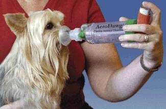
As an alternative to oral steroids, inhaled steroids can be used as is done with human asthmatics. A special facemask is used, and a spacer connects the mask to the metered dose inhaler. A couple of puffs from the inhaler doses the dog with inhaled steroids, and then it is possible to avoid the systemic side effects associated with oral steroids. Dogs typically need to use inhalers at least twice a day to control the symptoms of bronchitis. Airway dilators can also be given via inhalation in the same way as steroids.

Cough Suppressants

The cough itself is involved in the vicious cycle of irritation described above. Suppression of the cough reflex through medication may be helpful in establishing patient comfort. On the other hand, we want our patient to be able to clear the mucus from the inflamed airways if there is an excess of mucus. Common medications used are hydrocodone and butorphanol. Over-the-counter human products are often ineffective and may be combined with harmful additional medications such as decongestants or pain relievers.

Never medicate your pet without checking with your veterinarian first about what product to use and how to dose it.

Airway Dilators

Although airways technically do not constrict in chronic bronchitis, it may be helpful to relax them, allowing air to pass through bronchi that had previously been so small as to be blocked by mucus. Common medications include albuterol, terbutaline, and theophylline.  These are frequently used in combination with corticosteroids to reduce cough.

Mucolytics

An over-the-counter supplement called N-acetylcysteine may be helpful. This supplement works by breaking down sulfur bonds in the airway mucus, creating a more watery secretion. Thinner secretions may be easier to clear. The use of this supplement has not been formally studied for this use but it is an option that might be helpful.

Check with your veterinarian about what to use and how to use it.

Antibiotics?

Chronic bronchitis is not an infectious disease but all the inflammation and mucus in the lung certainly compromises the patient’s normal resistance to infection and ability to clear inhaled bacteria. Sometimes antibiotics are a helpful temporary addition to the therapies listed above.  More commonly, antibiotics are used early in the treatment course to rule out infectious causes of cough before the use of corticosteroids, which are immune suppressive.

Other Therapies

Nebulization and Coupage

It is important for the bronchitis patient to cough up inflammatory secretions as long as coughing does not become excessive and perpetuate the inflammation. It is not easy to draw the line as to when the cough is helpful and when it is excessive; only experience will tell. In patients for whom coughing is encouraged, it may be useful to moisten secretions with a vaporizer or even a nebulizer; the difference in these pieces of equipment is the size of the water droplet they generate. Inhalation of water vapor a couple of times daily will help keep secretions moist.

Coupage is a form of respiratory physical therapy that allows for moistened secretions to be moved into more central airways so they can be coughed up. See a YouTube video of how to do coupage.

Obesity Management

Pressure on the chest from body fat hampers the patient’s ability to take a deep breath. Many patients with chronic coughs do not require medication after excess weight has been lost simply because the chest can then expand properly.

Minimize Airway Irritants

It is especially important to minimize irritants circulating in the air. If possible, cigarette smoking should be limited to outdoor areas. If house painting, cleaning, or remodeling is planned, the pet should probably be boarded or stay elsewhere until any fumes have dissipated. It may be a good idea to get a good air filter for home use to minimize dust, pollens, or molds in the air.

Chronic bronchitis represents an ongoing airway change that generally cannot be cured; however, symptoms are manageable for most patients. Discuss the options for therapy with your veterinarian, expect some trial and error to be necessary, and it is likely the outcome will be a system that keeps your pet comfortable.

6037131

Chondrodystrophy in Dogs 

What is chondrodystrophy?

Chondrodystrophy is the abnormal development of cartilage. It causes the long bones of the body to grow at an abnormal rate and results in shortened legs.

What is a chondrodystrophic breed?

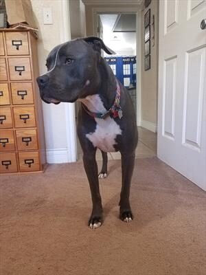
Chondrodystrophic breeds have the short leg, long body look. Classic examples include Dachshunds, Basset Hounds, Beagles, Corgis, English Bulldogs, and others. A chondrodystrophic dog can be a purebred or a mixed breed that inherited the short leg appearance.

What is a different ‘normal’?

To get the desired short legged look, certain degrees of limb deformities are selected for in these breeds. These deformities are changes in the shape or size of bones that are considered normal and expected in chondrodystrophic breeds but cause problems in other dogs. In chondrodystrophic dogs, one of the bones in the forelimb, the ulna, grows in an abnormal direction. This causes the other bone, the radius, to grow in a curved manner instead of straight. This curved bone contributes to the bow-legged appearance and is a normal finding as long as it does cause any problems for the dog.

When the legs are not as straight as they should be, it is called an angular limb deformity. This can occur in any limb. It results in toes pointing outward to the sides (duck-toed) or pointing inward towards the body of the dog (pigeon-toed). The former is common in the front legs; the latter is common in the back legs. Either way, the toes do not point forward. This is a common finding in chondrodystrophic dogs.

Another trait of chondrodystrophic breeds is misalignment of elbow bones. The bones of the elbow are meant to click into place like puzzle pieces; this does not happen in these dogs. Similar to other skeletal deformities, veterinarians don’t recommend fixing the elbow unless they cause problems for the dogs. Many dogs with this deformity move around fairly well.

Are any diseases associated with chondrodystrophy?

Many times deformities of chondrodystrophic dogs are excessive and cause enough problems walking that it’s a good idea to see a veterinarian. Some limb deformities lead to increased stress on nearby joints, resulting in joint instability, partial dislocation of bones (subluxation), pain, reduced range of motion, and lameness. The elbow joint is commonly affected but other joints are as well. Over time, these joints are more likely to develop arthritis.

Chondrodystrophic breeds are predisposed to specific diseases and conditions. One such condition is an ununited anconeal process. The anconeal process is a small projection of bone on the ulna, one of the bones in the dog’s forelimb. An ununited anconeal process means this part of the bone did not unite or fuse; this contributes to elbow instability, pain, and lameness. Chondrodystrophic dogs are also predisposed to canine intervertebral disc disease, or IVDD. This disease also causes front limb problems and lameness but is an issue within the spinal column.

Intervertebral discs are sponge-like cushions that sit between the bones in a dog’s spinal column. They provide flexibility and allow mobility of the spine during everyday movement. Chondrodystrophic breeds are associated with the premature deterioration of these discs causing disease. IVDD is a serious, expensive disease with high rates of mortality. In some cases, it can be painful enough to strongly consider euthanasia. As an owner, it is important to be aware of the risk of IVDD and other diseases of chondrodystrophic breeds; these risks should be considered before purchasing a pet.  

How do I treat my chondrodystrophic dog?

Owning and caring for a chondrodystrophic dog is more about preparation and awareness than prevention and treatment. As mentioned earlier, many limb deformities do not need to be treated. To an extent, the limb deformities are necessary and desirable; they help create short stature. Chondrodystrophic dogs live well with deformities that cause pain and lameness in non-chondrodystrophic breeds. If your dog does show signs of discomfort or lameness seek veterinary care. In some cases, these symptoms are temporary growing pains; time will tell. In other cases, they indicate a severe deformity and need to be addressed. Treating these severe deformities involves medical management, exercise restriction, and possible surgical intervention.

There is no current scientific evidence that chondrodystrophic dogs need to be restricted in jumping up and down, running or other movements to prevent IVDD. This disease is genetic and occurs regardless of activity. If you are set on owning a chondrodystrophic dog, educate yourself on the particular diseases your dog may get and consider creating a savings account and/or buying pet insurance; your dog may need expensive medical treatment at some point in their life.

What about genetic testing?

Recently, scientists have discovered mutations associated with a higher risk of IVDD in chondrodystrophic dogs. Some laboratories offer genetic testing. While this is one step closer to reducing an often fatal disease, more research is needed to determine how test results should influence owners and breeders. As an owner of a chondrodystrophic breed, tt may be more cost-effective to save for potential treatment than to spend on a genetic test. As a breeder of a chondrodystrophic breed, it is important to discuss this test with your veterinarian.

Chondrodysplasia is an abnormal finding in some breeds and there is a genetic test available for breeders seeking to eliminate the trait from their breeding program.  Breeds like the Australian Shepherd, American Cocker Spaniel, and Chesapeake Bay Retriever are among those in which the condition is considered abnormal and undesirable. 

7176337

Chagas Disease in Dogs  

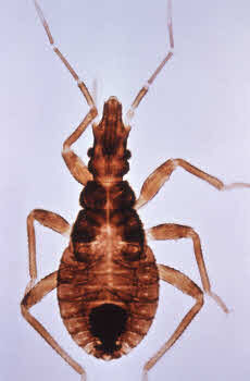
Chagas disease is an infectious disease caused by a microscopic parasite called Trypanosoma cruzi. This parasite cannot infect mammals on its own and instead is spread by an insect called the kissing bug, or, more formally, the triatomine. Kissing bugs spread the parasites through their feces. If the bug bites a dog and then defecates nearby, parasites can infect the dog through the open wound from the bite. Dogs can also be infected by eating kissing bugs, as the parasites enter the body through mucous membranes like the gums. 

Once the parasite infects an animal, if it is not treated promptly, it can reproduce and live in the body for the rest of the animal’s life. Chagas has been a serious problem in humans and dogs in South America for many years, but recently there have also been cases reported in the southern United States. 

Signs of Infection

Chagas has three stages after the animal has been infected: acute, latent, and chronic. 

Acute Phase

During the acute phase, the parasite enters the dog’s body and begins to infect macrophages, a type of white blood cell. Macrophages normally travel all through the body to fight infection and the parasite, hidden inside these cells, can move about undetected. 

When it reaches the heart, it begins to multiply and damage heart cells. The parasite can also infect many other body systems, including the nervous system, liver, kidneys, spleen, or lymph nodes. 

Most animals only have mild symptoms at this stage, such as slight fever or low energy, although dogs younger than six months are more likely to be severely affected. This stage generally lasts for several weeks and throughout this time, there are many parasites in the blood and tissues. Unfortunately, due to the lack of visible symptoms, acute phase infections of Chagas are often missed. As a patient’s immune system begins to fight the parasites, the number of parasites in the body goes down and the patient moves into the next phase of infection.

Latent Phase

During the latent phase, there are few parasites in the blood and the patient has no symptoms of illness. The immune system is generally not able to fully eliminate all the parasites in the body, but it keeps levels low enough that the patient doesn’t feel sick. A Chagas infection at this stage is difficult to identify through laboratory testing because there are so few parasites in the blood. Some dogs may remain in the latent phase for the rest of their lives and will thus continue on without any symptoms. 

Chronic Phase

Other dogs, however, will go on to the chronic phase of infection. We are not yet sure what triggers the chronic phase in some animals, but if the immune system becomes compromised through other diseases or injury, it can allow parasites to replicate again. As the immune system tries to fight against the growing number of parasites, it causes inflammation throughout the body, which can make the dog feel even sicker. Along with signs of systemic infection, like fever and lethargy, we also see symptoms of heart disease such as weakness, collapse, and, in severe cases, sudden death.

Diagnosis

Because Chagas disease is still relatively unknown in the United States, many cases are missed even after symptoms set in. Ask yourself the following questions to see if your pet is likely to have contracted Chagas:

  • Do they currently live or have they ever lived in areas where Chagas disease is common? Infections are widespread in Texas, Oklahoma, Louisiana, Georgia, and Tennessee, but have been reported all through the southern United States. 
  • Does your dog have symptoms of heart disease, such as lethargy, weakness, collapse, or labored breathing? Or do they have symptoms of systemic disease, like fever or loss of appetite?
  • Does your dog spend a lot of time outdoors, such as in a kennel, dog house, or as a working dog? Kissing bugs do not commonly live inside homes in the United States, but they can be found outside. 

If your dog fits any of the above criteria and your veterinarian suspects Chagas, they may take a blood sample to send to a laboratory for molecular tests or to look at under a microscope. They may also take X-rays or use ultrasound to look at your dog’s heart. 

Treatment

Drugs are available to fight the parasite infection and are quite effective during the acute phase. However, once the dog is in the chronic stage, it is no longer effective to only kill the parasites. Instead, the focus is on managing the symptoms of heart disease. Your veterinarian may recommend heart medications, prescription diets, or changes in exercise level.

Chagas Disease In Other Species 

Although Chagas is also a serious condition in humans, your risk of contracting it from your dog is extremely low. You cannot get Chagas by being bitten or licked by an infected dog. The main risk to humans from dogs is for those who are handling infected blood samples, such as veterinary or laboratory staff.

Chagas has been seen in both domestic cats and wild cats, such as bobcats. However, it appears to be less common in cats than it is in dogs. Research on the disease in cats is minimal, but if your cat is showing symptoms of heart disease, promptly inform your veterinarian.

Prevention

Research is being done on a canine vaccine for Chagas, but as of right now, it is not commercially available. Even if it were available, it only provides partial protection. If you live in an area where Chagas has been reported, you can take the following steps to protect your pets: 

  • Do not allow your pets to interact with or eat meat from other species that might be infected (raccoons, opossums, armadillos, rodents and skunks). Your dog can contract Chagas by ingesting kissing bugs that are on these other animals.  
  • Regularly spray outdoor kennels and spaces with pyrethrin/permethrin insecticides, which are effective against kissing bugs.
  • Before breeding female dogs, screen them for Chagas because it can be passed down to offspring. 

Note that pyrethrin insecticides are toxic to cats in high doses. Spray insecticides are 1-5% pyrethrin, while most cats have symptoms of poisoning at concentrations around 40%, so pyrethrin sprays intended to be used outside are generally safe even if you have cats. Be sure to check the concentration of pyrethrin in the product you are using, store the container where cats can’t access it and allow insecticides to dry fully before allowing cats to enter that space. Never apply a pyrethrin product directly to a cat. 

Being informed of the risk factors and symptoms of Chagas disease is the best way to ensure that your pets receive timely care. The earlier you catch the infection, the better your chances of reducing long-term complications.