4128677

Choking: First Aid

Choking is interference with breathing caused by foreign material in, or compression on, the trachea (windpipe). Thankfully, true choking is a very rare occurrence. Many pet owners will seek veterinary care because they believe their pet has something stuck in its throat, and this is rarely the case. It is far more likely that your pet has something mild and infectious such as tracheobronchitis (commonly called kennel cough) and is coughing or gagging rather than choking.

Frequently, coughing is confused with choking. Both cause the pet to forcefully exhale. With choking, the pet has difficulty inhaling. When coughing, the pet can inhale almost normally. Be careful to distinguish the two: attempting to give first aid to a pet who is merely coughing can cause injury.

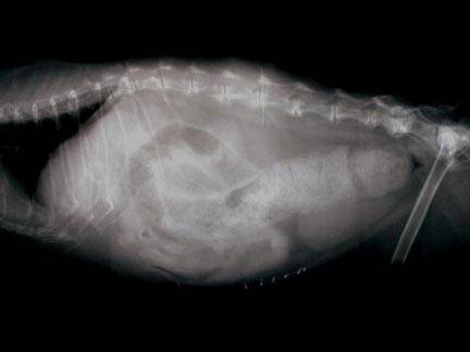
If you are in any doubt, have your pet evaluated by a veterinarian. To properly evaluate the throat, pets will require sedation and some will require evaluation with a fiber-optic endoscope or X-rays to look for foreign material. Note, however, that the throat normally contains small bones (the larynx, or voicebox), and since this area is rarely X-rayed, it may be difficult to determine if something abnormal is present.

If the Pet is Unconscious

Perform a Finger Sweep

Open your pet’s mouth and do a finger sweep by placing your finger along the inside of the mouth, sliding it down toward the center of the throat over the base of the tongue, and gently “sweeping” toward the center to remove any foreign material. Warning: there is a structure deep in the throat (Adam’s apple) that feels like a smooth bone. Do not attempt to pull it out!

Begin Rescue Breathing

Rescue breathing is described in the article on CPR. If air is not entering the lungs, slap the chest wall firmly or perform the Heimlich maneuver by putting the pet on their back, placing your hands over the abdomen near the bottom of the rib cage, and gently but firmly thrusting toward the spine. Perform a finger sweep and begin rescue breathing. Repeat until the foreign body is clear and the lungs can be inflated. Transport to the veterinarian right away.

If the Pet is Conscious

Perform a finger sweep only if it will not excite the pet. Do not perform a finger sweep if you believe your pet will bite you. Stay calm and try to keep the pet calm. If the pet is overheated, cool them with cold water applied to their extremities (ears and feet) and belly, and transport them to the nearest veterinarian or veterinary emergency clinic if there is one in your area. Running the air conditioning in the car on the way may help keep them cool.

6645376

Cardiopulmonary Resuscitation (CPR): First Aid

Cardiopulmonary resuscitation (CPR) is the treatment required to save an animal’s (or human’s) life when suffering cardiopulmonary arrest. The intent of CPR is to provide sufficient blood flow and oxygen to the brain and vital organs to support life until more advanced medical therapy can be started. Unlike what we see on television, most patients who suffer from cardiac arrest are unable to be saved, even with CPR. In a study from the University of California at Davis, researchers discovered that long-term survival among feline patients is extremely poor. In this study, only 2.3 percent of feline patients who received CPR survived to be discharged from the hospital alive. This closely parallels the findings of similar studies in human medicine.

In 2012 the American College of Veterinary Emergency and Critical Care established a committee to evaluate the scientific evidence on how to best conduct CPR in pets and generated evidence-based guidelines. The results of these guidelines are incorporated into the recommendations below.

CPR consists of two parts: rescue breathing and chest compressions.

These two techniques combine to keep the lungs supplied with oxygen and to keep blood circulating, carrying oxygen to the other parts of the body such as the brain and vital organs.

Basic CPR is CPR performed by trained bystanders at the scene of the arrest.

Advanced CPR is CPR performed by trained teams of professionals, generally in a hospital setting.

Basic CPR is the most important for pet owners, and is described in this section.

All body tissues require a steady source of oxygen. If the source is interrupted for only a few minutes, irreversible damage may be done. If cardiopulmonary arrest occurs, basic CPR must be initiated at the scene.


As long as the airway is open, compression of the chest may cause a forward flow of blood and may cause enough movement of air that less frequent breathing is recommended for single-rescuer CPR. Therefore, if only one rescuer is available to perform CPR, a ratio of 30 compressions to 2 breaths is recommended.

Basic CPR: Rescue Breathing (If Two People are Present)

Make Certain the Animal is actually Arrested and Unconscious
Talk to the pet first. Gently touch and attempt to awaken the pet. You could be seriously injured should you attempt to perform CPR on a pet who was only sleeping heavily and was startled awake.

Ensure an Open Airway


Extend the head and neck and pull the tongue forward. Look in the mouth and remove any saliva or vomitus. If it is too dark to see into the mouth, sweep your finger deep into the mouth and into the throat to remove any vomit or foreign body. Be aware of a hard, smooth, bone-like structure deep in the throat, which is likely to be the hyoid apparatus (Adam’s apple). Serious injury could result if you pull on the hyoid apparatus.

Observe for Effective Breathing


Sometimes an animal will begin to breathe spontaneously when the head is put in the position discussed above (head and neck extended, tongue pulled forward). Watch for the rise and fall of the chest while listening closely for sounds of breathing. If no breathing is evident in 10 seconds, begin rescue breathing.

Begin Rescue Breathing


Rescue breathing is performed by covering the pet’s nose with your mouth and forcefully blowing your breath into the lungs. In cats and small dogs, you must hold the corners of the mouth tightly closed while you force the air in.

In larger dogs, the dog’s tongue should be pulled forward and the mouth and lips held shut using both hands cupped around the muzzle. Force air into the lungs until you see the chest expand. Take your mouth away when the chest has fully expanded. The lungs will deflate without help. Air should be forced into the lungs until you see the chest expand.

Give 3 to 5 Full Breaths


After several breaths are given, stop for a few seconds to recheck for breathing and heart function. If the pet is still not breathing, continue rescue breathing 10 times per minute in cats or dogs. Push down on the stomach area every few seconds to help expel the air that may have blown into the stomach. If the stomach is allowed to distend with air, the pressure will make the rescue breathing efforts less effective. Try to coordinate breaths with chest compressions for 2-person CPR.

If Breathing is Shallow or Non-existent


If you find that breathing is either shallow or non-existent and the pet is still unconscious, continue the rescue breathing 10 times per minute and transport the pet to the nearest veterinary facility.

Basic CPR: Chest Compressions (If one or two people are present)

After Ensuring an Open Airway, Check for a Pulse
If no pulse is detectable, begin chest compressions.

In Small Dogs or Cats


Squeeze the chest using one or both hands around the chest. Depress the rib cage circumferentially (see illustration). Do this 100-120 times per minute.

In Large Dogs


Compress the chest wall with one or both hands, depending on the size of the dog and the size of the rescuer (whatever works best for you). If the dog is on their side, place your hand(s) on the side of the chest wall where it is widest. If they are on their back (a position best for barrel-chested dogs like bulldogs), place your hand(s) on the breastbone. Depress the rib cage 1.5 to 4 inches, depending on the dog’s size. Do this 100-120 times per minute.

Coordinate Rescue Breathing and Chest Compressions
If possible, give breaths during the compressions. If it is not possible, give two breaths after every 30 compressions.

Continue CPR until

  • You become exhausted and can’t continue. 
  • You get the animal transported to a veterinary facility and professionals can take over.
  • The pulse is palpable or heartbeats are felt and they are strong and regular.

In the vast majority of cases, artificial ventilations will continue to be required for a period of time, even though heart function has returned. This is due to the nervous system depression that occurs as a result of the arrest.

All resuscitated animals should be transported to a veterinary facility for further examination and care.

716183

Corneal Ulcers and Erosions in Dogs and Cats

One of the classic eye problems veterinarians must address is a red eye. The red eye may or may not be obviously painful but when it is, the pet can be observed squinting or even rubbing at his/her face. The conjunctiva (the pink moist tissue lining the inner surfaces of the eyelids) becomes an angry red and can even swell or puff up around the eye (a condition called chemosis). In short, it is clear when the eye suddenly hurts, and that veterinary attention is needed.

The Corneal Erosion

There are several causes of acutely red and painful eyes and one of the most common causes is a wound or scrape to the surface of the eye. The clear surface of the eye is called the cornea; because it is the outermost layer of the eye, it is prone to scrapes and tears. Common causes of corneal erosions include:

  • Rough contact with plants, thorns, or bushes
  • Scratches from another animal (note: the cat scratch wound can be especially serious as the wound quickly heals over, sealing infection within the eye.)
  • Self-trauma (rubbing or scratching at a painful ear or even at the eye due to some other eye problem can lead to an inadvertent scratch to the eye.)
  • Chemical irritation (such as getting shampoo in the eye during a bath.)
  • Foreign body injury (plant material can get stuck under an eyelid and can scrape the cornea.)

A special fluorescent stain is used to confirm if there is an ulcer or erosion. Normally, water will run smoothly off the surface of the cornea, like rain washing off a windshield. If the cornea is damaged, the stain will stick to the damaged area and look bright green under a fluorescent lamp.

Treatment

Antibiotics

A routine corneal ulcer or erosion should heal easily. Since the damaged cornea is at risk of becoming infected (or may even already be infected, as demonstrated by a purulent discharge), a topical antibiotic is needed and ideally should be used four times a day or more. Since it is a rare pet owner that can accommodate any medication administration four times a day, we usually have to make do with three times a day but to properly sterilize the eye surface, the antibiotic should be used four times a day. Either drops or ointment can be used depending on the owner’s preference.

Pain Relief

The second part of treatment is pain relief: Atropine 1% drops or ointment. The atropine acts by temporarily paralyzing the pupil’s ability to constrict (the main source of pain is spasm in the pupil). Pupillary dilation is expected when this medication is used and the pet may be reluctant to experience bright sunlight while under the influence of this medication. Because the tear duct system is connected to the nose and mouth, the patient will also taste the above medications and atropine is famous for its bitter taste. Dogs do not seem to mind this unduly but cats will drool shortly after the medication is given in an effort to get the taste out of their mouths. This is a normal reaction to the Atropine as is the dilated pupil.

A special collar, called an Elizabethan Collar, may be needed to prevent self-trauma of the eye. If you think your pet will rub the eye, it is important to have the pet wear this special collar until the erosion is healed. Be sure to request one if you think your pet needs it and if you are given one be sure the pet wears it for the entire course of treatment.

Re-Check in One Week

It is important that the eye be stained again after one week of therapy. Most ulcers will have healed in this time but some will require an additional week. If the ulcer has not healed after two weeks, it is no longer considered routine.

Some other procedures, and possibly a visit to a veterinary ophthalmologist, may be necessary. 

 If the inflammation associated with the ulcer goes deeper into the eye, the situation becomes more serious; it is very important not to skip that one-week re-check. If there is any question about the eye’s healing progress, the eye should be re-checked sooner.

It is important that the owner observe the progression of healing at home. If the eye is doing well but suddenly becomes more painful, if a discharge develops, or if the eye simply does not look right, have your vet recheck the pet sooner than the planned one-week appointment.

Some Special Ulcer Conditions

Indolent Ulcer

Some ulcers form with a small lip on their edge.  Since the ulcer is trying to heal from the bottom up, the lip interferes and creates an ulcer that seems to never get any smaller. Several techniques can be used to remedy this situation: the lip can be rubbed away, hyaluronan or PSGAG eye drops can be used to strengthen the cornea, or surgery can be performed. A technique that has gained popularity over recent years is called the grid keratectomy where a needle is used to scratch a grid of lines on the cornea. The cornea is then able to heal in, grid by grid. Poodles and boxers are notorious for these ulcers but they can occur in any breed. 

Melting Ulcers

When infection is present, the cornea will develop a yellow or tan gooey appearance because the bacteria or fungi causing the infection elaborate enzymes that actually dissolve corneal collagen fibers. The cornea softens and appears to be melting and can actually perforate. Culture and cell sampling for analysis are very helpful in determining the right antimicrobial therapy. In addition to antibiotic drops, the eye will need some sort of medication to inactivate the aforementioned collagen-dissolving enzymes. Often, this involves taking a blood sample from the patient and actually delivering the patient’s own serum as an eye drop.

Descemetocele

(Pronounced des’ĕ-met’ō-sēl) Descemet’s membrane is the thin attachment of the cornea to the fluid of the eye below. A Descemetocoele is an ulcer that has penetrated through the cornea completely except for the last thin membrane. An eye with a descemetocoele is at high risk for rupture and measures must be taken to protect the eye. Usually, surgery is needed. The brachycephalic breeds (Pekingese, pug, etc.) are predisposed to this problem due to their prominent eyes.

Again, corneal ulcers and erosions usually heal routinely but stay alert for any changes that could indicate a more serious turn of events. Be sure self-trauma (rubbing the eye) is prevented and be sure to go to a recheck at the proper time. If anything seems not to be proceeding properly, be sure to contact your veterinarian.

4128675

Cherry Eye in Dogs and Cats

Prolapse of the Tear Gland of the Third Eyelid

Unlike humans (who only have eyelid, technically called the nictitans or nictitating membrane, arises from the inner corner of the eye and covers the eye diagonally as shown. The eye is lubricated by tear film, which consists of water, oil, and mucus. The oil comes from glands lining the outer eyelids, the mucus comes from glands in the conjunctiva (the pink part inside the eyelids), and the water comes from tear (or lacrimal) glands.

Each eye has two tear glands: one just above the eye and one located in the third eyelid. The gland in the third eyelid is believed to produce a full 30 percent of the total tear film water, so it is important to maintain the function of this gland.

The tear gland of the third eyelid is held in place by tissue fibers but some individuals have weaker fibers than they should so the gland protrudes. This protrusion is called a cherry eye.  In the smaller breeds – especially Boston terriers, cocker spaniels, bulldogs, and beagles – the gland of the third eyelid is not strongly held in place for genetic reasons. The gland prolapses (drops down) out to where the owner notices it as a reddened mass. Out of its normal position, the gland does not circulate blood properly, may swell, and may not produce tears normally. 

Treatment: Replacing the Gland in its Proper Location

By far the best treatment for cherry eye is replacing the gland back into its proper location. There are two techniques for doing this. The traditional tucking method (also called tacking) is probably the most commonly performed. Here, a single stitch is permanently placed, drawing the gland back where it belongs. Complications are uncommon but be aware of the following possibilities:

  • If the stitch unties, the surface of the eye could become scratched by the suture. If this occurs, the eye will become suddenly painful and the suture thread may be visible. The suture can be removed and the problem solved.
  • The tuck may not be anchored well enough to hold permanently. In fact, this surgery is notorious for this type of failure, and frequently a second or even third tuck is needed. If more than a couple of tucks have led to failure, it may be better to try the imbrication technique as described below. Some cases are repaired using both tuck and imbrication together.
  • Sometimes cherry eye is accompanied by other eyelid problems that make the repair more difficult or less likely to succeed. In these cases, again, if the simple surgery is not adequate, ask your veterinarian if a referral to a veterinary ophthalmologist for the second surgery to maximize the chances of a permanent resolution is in the best interest of you and your pet.

In a newer surgical technique called imbrication, or pocketing, a wedge of tissue is removed from directly over the actual gland. This technique is more challenging as it is not easy to determine how much tissue to remove. Tiny stitches that will eventually dissolve are used to close the gap so that the tightening of the incision margins pushes the gland back in place. 

Complications may include:

  • Inflammation or swelling as the stitches dissolve.
  • Inadequate tightening of the tissue gap may lead to recurrence of the cherry eye.
  • Failure of the stitches to hold and associated discomfort. Loose stitches could injure the eye depending on the type of suture used.

Sometimes both surgical techniques are used in the same eye to achieve a good replacement. Harmful complications from cherry eye surgery are unusual but recurrence of the cherry eye can happen. If it recurs, it is important to let your veterinarian know so that a second surgery, either with your veterinarian or an ophthalmologist, can be planned.

Expect some postoperative swelling after cherry eye repair but this should resolve and the eye should be comfortable and normal in appearance after about a week. If the eye appears suddenly painful or unusual in appearance, have it rechecked as soon as possible.

Treatment: Removing the Gland

Historically, the prolapsed gland was treated like a small tumor; it was simply removed. This was before the full significance of the gland was realized.

If the third eyelid’s tear gland is removed, it cannot be put back in place. If the other tear gland (the one above the eye) cannot supply adequate tears, not an uncommon phenomenon in older small breed dogs, then the eye becomes dry and uncomfortable. A thick yellow discharge results and the eye develops a blinding pigment covering for protection. This condition is called simply dry eye or more scientifically keratoconjunctivitis sicca and daily medical treatment is required to keep the eye both comfortable and visual. Not only is dry eye uncomfortable, but its treatment is often frustrating and time-consuming and there is expense involved. If left untreated, the eye can become blind. We would like the dog to maintain the greatest amount of tear-producing tissue possible, thus removing the gland for cosmetic reasons is not an acceptable treatment method.

4435548

Cataracts in Dogs and Cats

The normal lens of the eye is a focusing device. It is completely clear and is suspended in position by tissue fibers (called zonules) just inside the pupil. The pupil opens and closes to control the light entering the eye so as to project an image onto the retina in the back of the eye, the way a projector projects an image onto a movie screen. The lens focuses the projected image in a process called accommodation. The focusing power of the dog’s lens is at least three times weaker than that of a human lens, while a cat’s lens is at best half the power of a human’s. 

(Dogs and cats have a sense of smell at least 1,000 times more accurate than ours and this is their primary means of perceiving the world.)

Anatomy First

Despite its clarity, the lens is in fact made of tissue fibers. As the animal ages, the lens cannot change its size and grow larger; instead, it becomes more compact with fibers. The older lens, being denser, appears cloudy. This condition is called nuclear sclerosis and is responsible for the cloudy-eyed appearance of older dogs. The lenses with nuclear sclerosis may look cloudy but they are still clear and the dog can still see through them; these are not cataracts.

The fibers making up the lens come together in the center of the lens, forming a “Y” shape that is sometimes visible when one looks into the eye. These Y-shaped lines are often called the sutures of the lens.

The lens is enclosed in a capsule that, if disrupted, allows the immune system to see the lens proteins for the first time, recognize them as foreign, and attack. The resulting inflammation (a form of uveitis) is painful and can be damaging to the eye. A cataract can result from this inflammation or from any of the numerous other reasons listed below.

A cataract is an opacity in the lens.

A Note on Diabetes Mellitus in Dogs

Cataracts can be congenital (born with it), age-related; of genetic origin (the most common cause); caused by trauma; dietary deficiency (some kitten milk replacement formulas have been implicated); electric shock; or toxins. The patient with a cataract is not able to see through the opacity. If the entire lens is involved, the eye will be blind.

Many things can cause the lens to develop a cataract. One cause is diabetes mellitus. In this condition, the blood sugar soars as does the sugar level of the eye fluids. The fluid of the eye’s anterior chamber is the fluid that normally nurtures the lens but there is only so much glucose that the lens is able to consume. The excess sugar is absorbed by the lens and transformed into sorbitol. Sorbitol within the lens unfortunately draws water into the lens, causing an irreversible cataract in each eye. Cataracts are virtually unavoidable in diabetic dogs no matter how good the insulin regulation is; diabetic cats have alternative sugar metabolism in the eye and do not get cataracts from diabetes.

What Else Could It Be?

Many owners cannot tell which portion of the eye looks cloudy. Cloudiness on the cornea, as caused by other eye diseases, can be mistaken for a cataract by an inexperienced owner. Also, in dogs, the lens will become cloudy with age as more and more fibers are laid down, as described above. Nuclear sclerosis, as described, can mimic the appearance of a cataract, yet the eye with this condition can see and is not diseased. It is a good idea to have your veterinarian examine your pet if you think there is a cataract, as you could be mistaken.

Why is it Bad to Have a Cataract?

The area of the lens involved by the cataract amounts to a spot that the patient cannot see through. If the cataract involves too much of the lens, the animal may be blind in that eye and there could be cataracts in both eyes, which means the pet could be rendered completely blind.

A cataract can luxate, which means that it can slip from the tissue strands that hold it in place. The cataractous lens can thus end up floating around in the eye, where it can cause damage. If it settles to block the eye’s natural fluid drainage, glaucoma (a buildup in eye pressure) can result, leading to pain and permanent blindness. A cataract can also cause glaucoma when it absorbs fluid and swells so as to partially obstruct fluid drainage.

Cataracts can begin to dissolve after they have been there long enough. While this sounds like it could be a good thing, in fact, it is a highly inflammatory process. The deep inflammation in the eye creates a condition called uveitis, which is in itself painful and can lead to glaucoma. If there is any sign of this type of inflammation in the eye, it must be controlled before any cataract surgery.

A small cataract that does not restrict vision is probably not significant. A more complete cataract may warrant treatment. Cataracts have different behavior depending on their origin. If a cataract is a type that can be expected to progress rapidly (such as the hereditary cataracts of young cocker spaniels) it may be beneficial to pursue treatment (i.e. surgical removal) when the cataract is smaller and softer, as surgery will be easier.

What Treatment is Available?

Cataract treatment generally involves surgical removal or physical dissolution of the cataract under anesthesia. This is invasive and expensive and is not considered unless it can restore vision or resolve pain. Pets with one normal eye and the other with a cataract can still see with their good eye and may not need surgery depending on circumstances.

Determining if a Dog is a Candidate for Cataract Removal

Obviously, the patient must be in good general health to undergo surgery; diabetic dogs must be well-regulated before cataract surgery. Also, it should be obvious that for a patient to be a good candidate for surgery, the patient must have a temperament conducive to getting eye drops at home. 

Pre-anesthetic lab work can be done with the patient’s regular veterinarian. Some ophthalmologists prefer that patients have their teeth cleaned before surgery to minimize infection sources in the eye.

A complete examination of the eye should be performed. If your veterinarian is not comfortable treating cataracts or does not have the appropriate equipment, your pet may be referred to a veterinary ophthalmologist.

It is impossible to see the retina through a cataract; an electroretinogram test can determine if the eye has a functional retina and can benefit from cataract surgery. Ultrasound of the eye can be used to look for retinal detachments. If the eye has a blinded retina, there is no point in subjecting the patient to surgery. Inflammation in the eye will require treatment before surgery. Sometimes, other eye drops are prescribed for a period before surgery depending on the veterinarian’s preference.

Cataract Removal: Phacoemulsification and Surgical Removal

Historically, removing the cataract meant surgically cutting into the eye and physically removing the lens. This is still done for older patients whose lenses are compact. For younger patients in whom the lens is soft, a technique called phacoemulsification is preferred.

This technique has become the most common method of removing cataracts in dogs. Here, the lens is broken apart by sound waves and removed with an instrument similar to a small vacuum cleaner.

In either case, the eye must be paralyzed during surgery to prevent eye movement or even blinking at a critical moment. Nerve blocks can be used to paralyze the eye, or specific medications can be used to paralyze the entire patient (in which case a mechanical ventilator is used to breathe for the patient during surgery).  After the lens is removed, an artificial lens is implanted. (Without the prosthesis, the dog’s vision will be approximately 20/800, and objects will appear to be reversed, as in a mirror.)

After surgery, the pet must wear an Elizabethan collar for a good three weeks, and eye drops to reduce inflammation will be needed for several months. A harness may be recommended for walks instead of a collar to reduce pressure on the head and eye from pulling. There will be a schedule of recheck appointments.

Complications

Some degree of uveitis (deep inflammation) is unavoidable. This can cause a pupil constriction reaction that can increase the risk of scarring within the eye. Eye drops to keep the pupil dilated are usually effective in preventing this but not always. Inflammation in the eye will resolve over weeks to months after surgery. The success rate is higher for cataract surgery if there is minimal inflammation in the eye prior to surgery, thus pre-operative anti-inflammatory eye drops are frequently prescribed.

Another complication involves the development of opacities on the remaining lens capsule. In humans, laser surgery is used to remove the lens capsule, but in dogs, the capsule is too thick for this. Some ophthalmologists prefer to remove the capsule as a preventive measure. The portion of the capsule that is involved in this reaction is present in young dogs but not in adult dogs.

Bleeding after surgery can be an enormous complication and can easily be caused by excess barking or activity after surgery. Small bleeds are of little consequence, but a large bleed could ruin vision.

Glaucoma can develop at any time after cataract surgery. This complication is not only blinding but painful as well. The risk of this complication has been decreased by placing a prosthetic lens (a formerly uncommon but now fairly standard procedure) but dogs who start off with hypermature (dissolving) cataracts or have an unusually long surgery time tend to have an increased risk for this complication.

Overall, a 95 percent vision rate is described immediately after cataract surgery with 80 percent having long-term vision success.

Before embarking on the adventure of cataract surgery, be sure to obtain a clear explanation from your veterinarian or ophthalmologist of exactly what the home care will involve.

What if the Cataract Goes Untreated?

A cataract by itself does not necessarily require treatment. If there is no associated inflammation or glaucoma and the only problem is blindness, it is perfectly reasonable to have a blind pet. Blind animals have good life quality and do well though it is important not to move furniture around or leave any hazardous clutter in the home. Some dogs, however, become anxious or even aggressive when they lose their vision. Restoring vision for the pet is weighed against risk and expense and is a decision for each owner to make individually. Many cataracts will progress to a hypermature state where they will begin to dissolve as described and anti-inflammatory eye drops are needed as mentioned. 

Can Eye Drops Dissolve Cataracts?

Products containing N-acetylcarnosine have been marketed to dissolve cataracts and have led to a great deal of false hope. N-acetylcarnosine is an antioxidant eye drop that may have beneficial effects on the eye but they do not include any sort of dissolution of a mature cataract. For smaller cataracts, it may be possible to dilate the pupil so that the pet can see around the cataract but there is some controversy about doing so as these medications have other effects on the eye.

5392483

Cushing’s Syndrome: Classifying the Type

Adrenal or Pituitary Cushing’s? Why do we Care?

Once a pet has been confirmed as having Cushing’s syndrome, the next step is to determine which form of Cushing’s syndrome the pet has since treatment and prognosis are different for each form. As described, there are two forms, commonly abbreviated PDH and AT. PDH stands for pituitary dependent hyperadrenocorticism and accounts for 85 percent of canine and feline Cushing’s patients. In PDH, there is a pituitary tumor, often extremely small, causing over stimulation of the adrenal glands. AT simply stands for adrenal tumor. Adrenal tumors can be benign or malignant. Testing to determine the type or form of Cushing’s disease is called discriminatory testing. This type of testing is done AFTER the diagnosis of Cushing’s disease has already been made. Here are some issues and concerns where the type of Cushing’s disease the pet has becomes an important matter.

  • If there is an adrenal tumor, there is a 50 percent chance that it is malignant. It is helpful to know if the pet has a condition with this kind of a malignancy rate, especially if surgery is being considered.
  • If the patient is known to have an adrenal tumor, this knowledge will direct the doctor on how and where to look for tumor spread to determine if the patient has cancer or not. On the other hand, if the patient is known to have a pituitary tumor, it becomes more relevant to image the brain to look for evidence of pressure on the brain.
  • Medication protocols may be very different for adrenal tumors versus pituitary tumors.
  • Surgery protocols are completely different for adrenal tumors versus pituitary tumors.

Approximately 85 percent of Cushing’s patients will have pituitary dependent disease while approximately 15 percent will have adrenal tumors. This statistic appears to be valid in dogs as well as cats.

Dogs

What Tests will Tell us What Type of Cushing’s Syndrome the Dog Has?

The Low Dose Dexamathasone Suppression Test
If you are lucky, the same test used to determine if a pet has Cushing’s disease can also classify the Cushing’s disease so that no further tests are needed. To get the information needed, a 4-hour sample is drawn in addition to the pre-dexamethasone sample and the 8-hour sample. If the cortisol level suppresses to a level below 50% of baseline at 4 hours, only to escape to a level high enough to indicate a positive result, only a pituitary tumor is possible. 

While pituitary tumors do not always show this kind of classic suppression at four hours, adrenal tumors never do. If suppression is seen in the 4-hour sample, further testing is not needed, though this is the only pattern that is distinctive enough to obviate further testing. Approximately, 60 percent of patients with pituitary tumors can be identified with the low dose dexamethasone suppression test.

The High Dose Dexamethasone Suppression Test


This test is similar to the low dose dexamethasone suppression test except that a higher dose of dexamethasone is used and the patient has already been diagnosed with Cushing’s. The idea behind is that a patient with a pituitary tumor will show suppression in circulating cortisol when exposed to the high dose of dexamethasone; suppression does not occur with the low dose. If there is an adrenal tumor, suppression does not occur. Unfortunately, the theory does not hold well enough to make this a reliable test; a full 20 to 30 percent of pituitary tumors will fail to suppress even with the high dose of dexamethasone. Furthermore, this test cannot identify patients with adrenal tumors; it is able to confirm about 70 to 80 percent of patients with pituitary disease. The rest will need further testing.

Endogenous ACTH Level


Many veterinarians feel this is the most accurate method of classifying Cushing’s syndrome, but the problem is that the test is technically challenging to run. Serum from the patient must be frozen when transported to the laboratory and must not thaw. The hormone ACTH is fragile and may not survive the trip to the lab. The idea with this test is that a patient with a pituitary tumor will have high ACTH levels, as this is what the tumor is secreting. A patient with low or no measurable ACTH levels has an adrenal tumor as the pituitary is trying its best not to stimulate the over-active adrenal tumor.

Imaging


Fundamental to discerning pituitary dependent from adrenal tumor is the size and shape of the adrenal glands. With pituitary dependent disease, both adrenal glands are being over stimulated so they should both be symmetrical in size and shape and most likely enlarged. With an adrenal tumor, one gland will be severely atrophied and possibly undetectable, while the other is enlarged and possibly misshapen. This difference makes imaging of the adrenal glands an accurate way to distinguish types of Cushing’s syndrome. Ultrasound, MRI, or CT scan can be used, however, ultrasound is the most readily available and least costly. In one study, ultrasound was able to correctly identify adrenal tumors in 86 percent of cases. Sedation is generally not needed for abdominal ultrasound, whereas CT and MRI require general anesthesia and usually needs referral to a specialty clinic.

An added benefit of imaging includes looking for evidence of malignancy should an adrenal tumor be identified. Malignant tumors tend to invade the vena cava, the largest vein in the body. Ultrasound has been found to be extremely accurate in evaluating for this complication. Imaging also allows for taking measurements of the adrenal glands. Glands that measure greater than 4 cm in length are highly suspicious of being malignant.

It is important to note that ultrasound can never be used to make the initial diagnosis of Cushing’s syndrome as there is too much variability in the size of normal adrenal glands. When it comes to gaining information, imaging is best used on patients already known to have Cushing’s syndrome for whom classification is needed.

Is it Reasonable to Play the Odds and not do Further Testing?


Since 85 percent of canine patients with Cushing’s have a pituitary tumor, it is not unreasonable to play the odds and treat for a pituitary tumor. The risks in doing so is that if the patient doesn’t readily respond to medication, it will be impossible to know if the dose simply needs adjustment or if the patient has an adrenal tumor and a completely different treatment is needed. A great deal of time and effort could be wasted by pursuing the wrong therapy.

Cats

For a cat with Cushing’s, the methods that can be reliably used to distinguish pituitary from adrenal disease are: high dose dexamethasone suppression testing; endogenous ACTH; and imaging.

High Dose Dexamethasone Suppression Test
The feline high dose dexamethasone suppression test is the same as the canine version except that even higher doses must be used. If the cortisol levels suppress in response to injected dexamethasone, then only a pituitary tumor is possible. If the cortisol levels do not suppress, however, then no conclusion can be drawn as 50 percent of feline pituitary tumors will not suppress. This can be a frustrating situation but since the test is non-invasive, it is worth a try to confirm a pituitary tumor in this way.

3955475

Cushing’s Syndrome (Hyperadrenocorticism): Description

This condition represents a classical excess in cortisone-type hormone circulation in the body; it’s a relatively common hormone imbalance. Both cats and dogs can be affected (though it is primarily a dog’s disease) and the onset is insidious.

Cushing’s syndrome is the resulting set of symptoms observed when the body is exposed to excess cortisone (or related hormones) over a long period of time. Cortisol (called hydrocortisone when used as a medication) is a hormone produced by the adrenal glands that are located atop the kidneys. Cortisol is stored in the adrenal glands and is released in times of stress where it helps our bodies prepare for a fight or flight situation. It adjusts the metabolism to expect physical exertion by mobilizing fat and sugar stores and retaining sodium and water. It puts us in a state of break down so that our stored resources can be used quickly. However, if the body is exposed to this hormone most of the time instead of during short stressful periods only, the state of break down becomes debilitating.

There are several mechanisms that can lead to Cushing’s syndrome and, as they are treated differently, it is important to determine which one is at work in a given animal.In the normal body, the pituitary gland, located at the base of the brain, can detect when cortisol levels in the blood are declining. In response, the pituitary secretes a stimulating substance, called ACTH, which causes the adrenal gland to release more cortisol.  When the pituitary gland detects that cortisol levels are again appropriate, it stops its stimulatory message.You can think of the pituitary gland as a thermostat for cortisol, though the pituitary gland regulates many other hormones as well. This raising and lowering of cortisol blood level is regulated throughout the day via ACTH secretion and occurs rapidly.

Pituitary-Dependent Cushing’s Syndrome

This form of Cushing’s disease accounts for 85% of dogs with Cushing’s syndrome. Basically, the pituitary gland grows a small tumor. The tumor is generally too small to cause any trouble due to its size and it is generally a benign tumor. This tumor, however, over-produces ACTH, thus leading to over-stimulation and enlargement of both adrenal glands and an over-production of cortisone. Occasionally (10% of pituitary-dependent Cushing’s dogs), these benign pituitary tumors are large enough to compress the brain. In these cases, neurological signs may be observed; these cases are unusual but very hard to treat (see section on macrotumors.)

Adrenal-Dependent Cushing’s Syndrome

In 15% of dogs with Cushing’s syndrome, an adrenal tumor is directly over-producing cortisone. The tumor is often large enough to see with radiographs or ultrasound and may be malignant. There is very little or no production of ACTH from the pituitary gland and as a result the opposite adrenal gland is usually atrophied/small.

Overuse of Cortisone-Type Hormones

Cortisone derivatives may be the most widely utilized drugs in veterinary medicine. A single corticosteroid medication can be used to treat a multitude of completely different diseases simply by using different dosing schedules. Steroids are commonly used to relieve inflammation particularly in skin allergies, arthritis pain, lung disease and more. Relief is usually rapid and many owners find themselves requesting cortisone shots or pills over and over again. In time, Cushing’s syndrome can result, not from any inherent disease in the pet’s system but from the effects of the hormones given over the long term.

The pituitary gland perceives the high steroid levels yielded by the medication and does not send stimulation to the adrenal glands. In time, the adrenal glands atrophy and are not able to release cortisone on their own should they be required to do so. This effect lasts as long as a period of three months after the cortisone medication has been discontinued. To allow the adrenal to gradually recover, cortisone pills are usually prescribed in a decreasing dose, rather than a sudden stoppage; an owner should never discontinue the pills suddenly.

Commonly prescribed cortisone derivatives include: Vetalog,

Azium, prednisone/prednisolone, dexamethasone, Depo-Medrol, triamcinolone, methylprednisolone, Medrol™ and others. These medications have important parts to play in medicine but they must be respected and not used indiscriminately, nor discontinued suddenly after prolonged use.

4128771

Cushing’s Disease in Dogs (Hyperadrenocorticism): What Is It?

Cushing’s disease, also called Cushing’s syndrome or hyperadrenocorticism, results from an overproduction of the hormone cortisol. Cortisol is produced by the adrenal glands, located next to the kidneys, and adrenal glands are stimulated to produce cortisol by the pituitary gland located at the base of the brain. There are two types of Cushing’s disease. In adrenal-dependent Cushing’s disease, an adrenal gland overproduces cortisol because of an adrenal tumor. In pituitary-dependent Cushing’s disease, the adrenal glands overproduce cortisol because they are overstimulated by a pituitary tumor.

Signs associated with Cushing’s disease may include increased thirst and urination, a voracious appetite, an enlarged and sagging belly, hair loss, lethargy/weakness, panting, and sometimes, recurrent infections.

Diagnosis

Cushing’s disease can be difficult to diagnose, especially in early cases. Tests that may be needed to diagnose Cushing’s disease include:

  • Complete blood count (CBC), chemistry panel and urinalysis: These blood and urine tests evaluate the general health of your pet and can help your veterinarian determine if other health problems should be suspected.
  • Adrenocorticotrophic hormone (ACTH) stimulation test: This test involves a blood sample, an injection of a hormone (ACTH), an hour of waiting, and then another blood sample. This test is used to diagnose Cushing’s disease in a pet.
  • Low-dose dexamethasone suppression test: This test takes longer to perform than the ACTH stimulation test and takes place over 8 hours. It involves a blood sample, an injection of a steroid, a few hours of waiting, another blood sample at 3 hours, more waiting, and then another blood sample at 8 hours. This test is used to diagnose Cushing’s disease. Sometimes this test can also determine where the problem is in the pet’s body (i.e., pituitary or adrenal).
  • High-dose dexamethasone suppression test (HDDST): This test is similar to the LDDST except that it uses a higher dose of dexamethasone. It is used to determine where the problem is in the pet’s body (i.e. pituitary or adrenal).
  • Endogenous adrenocorticotrophic (ACTH) assay, or ACTH level: This test involves a single blood sample. It is used to determine where the problem is in the pet’s body (i.e. pituitary or adrenal).
  • Radiographs, abdominal ultrasound, or CT scan: Radiographs can help us see changes in other organs that could indicate Cushing’s. An abdominal ultrasound can help us see the adrenal glands. Sometimes, a CT scan can be performed to help us evaluate the pituitary gland at the base of the brain.

Treatment

Treatment consists of medication to control the cortisol overproduction by the adrenal gland or surgery to remove the abnormal adrenal gland. Choosing the best treatment for a pet with Cushing’s disease depends upon where the problem is in your pet’s body (i.e. pituitary or adrenal gland).

4128613

Cataracts in Diabetic Dogs

Most diabetic dogs will develop cataracts and go blind. This FAQ is designed to assist the owners of diabetic dogs in knowing what to expect and to make decisions regarding cataract surgery.

What Is a Cataract?

A cataract is an opacity in the lens of the eye. The entire lens may be involved or just a part of it. The patient will not be able to see through the opacity.

Why Do Diabetic Dogs Get Cataracts?

The lens of the eye is round, hard, and normally as clear as glass. Looking at the lens, it is hard to believe it is a piece of living tissue. The lens is suspended by fibers that can adjust its position so that one can focus vision. The lens is encased in a capsule and depends on the fluids of the eye for nutrients. The lens does not receive a direct blood supply.

Normally, the lens absorbs glucose from the eye fluids, using most of this for its own energy needs. Some of the excess is converted to another sugar called sorbitol. When there is excess sugar in the eye fluids, as in diabetes mellitus, there is excess sorbitol produced. Sorbitol pulls water into the lens, which in turn disrupts lens clarity and causes the cataract. Fructose is also produced from the excess glucose and also contributes to this water absorption.

Cataracts do not necessarily imply poor diabetic control. Even well-controlled dogs can still get cataracts.

How Long Does It Take To Go Blind?

Generally, the cataract has matured, and the dog is blind in a matter of weeks.

Until recently, blindness in a diabetic dog was basically a foregone conclusion, but there is a new product called Kinostat® that has changed that outlook. To review, the lens absorbs glucose from the fluids of the eye and uses this glucose as nutrition. Any extra glucose that is absorbed into the lens is converted to sorbitol by an enzyme called aldose reductase. Sorbitol pulls water into the lens to prevent the lens from becoming dehydrated. This is all well and good, but in the diabetic state, there is lots of excess glucose, and the excess glucose gets converted to excess sorbitol, which, in turn, pulls so much water into the lens that clarity and function are disrupted, and a cataract is formed. Kinostat is an aldose reductase inhibitor that curtails sorbitol production. Early use of Kinostat may significantly delay or even completely prevent the development of cataracts.

Kinostat is preventive only and will not reverse cataract formation that has already occurred. Kinostat is unfortunately not yet commercially available, though has been reportedly near coming to market for some time.

What Does It Mean To Say That a Cataract Is Mature?

A cataract’s maturity is determined by how much visual impairment the pet is believed to have. Since we cannot ask a dog to read an eye chart, we must determine this by visually inspecting the eye. A light is used to look into the eye and view the colorful area at the back of the eye called the tapetum.

(This is the area that flashes or appears colored in certain lighting.) When less than 10 percent of the tapetum is obstructed, the cataract is very young and does not significantly change vision. When 10-50 percent of the tapetum is obstructed, this cataract is called early immature. When 51-99 percent is obstructed the cataract is late immature. The mature cataract obstructs the entire tapetum. Ideally, a cataract is removed in the early immature stage for the lowest surgical complication rate.

When a cataract is hypermature, it starts to liquefy and dissolve. This can actually lead to restoring vision, which sounds like a positive turn of events; unfortunately, the dissolution process is quite inflammatory. The resulting inflammation can be painful and can damage the eye even more.

All cataracts do not progress all the way to hypermature and may stay static or progress at changing rates; however, diabetic cataracts are notorious for reaching hypermaturity and creating inflammation. 

What Is Uveitis?

Uveitis is inflammation of the eye’s uveal tract, which consists of the eye’s vascular tissues. In this situation, uveitis is the inflammation that results when the hypermature cataract begins to liquefy. Uveitis is painful and tends to cause the eye to become reddened and the pupil to constrict. When a pet has uveitis before surgery, success (pain-free vision) occurs in only 50 percent of cases 6 months after surgery as opposed to 95 percent of cases for whom there was no uveitis pre-operatively.

Can My Dog’s Vision Be Restored?

Maybe. It depends on the health of the eye, excluding the cataract. If the only thing wrong with the eye is the cataract and the patient’s diabetes mellitus is well-regulated, surgery can remove the cataract and place an artificial lens. If the eye is deeply inflamed or is otherwise damaged, the eye may be blind even without a cataract. A veterinary ophthalmologist can evaluate the eye to determine the feasibility of cataract surgery.

It is important to note that there are numerous eye drops advertised that claim to dissolve cataracts. None of these products actually work and wasting time with them may actually allow uveitis to set in and make for a much worse prognosis for vision.

Is it Cruel To Keep a Dog Blind?

Not at all. Dogs do not depend on vision the way humans do. A blind dog can get along very well as long as the furniture isn’t moved and the dog is properly supervised.

For tips on helping a blind dog adapt:

Blind Dog Support

There are many medical conditions that render a dog blind and as long as the condition is not painful, the dog can live a normal life as a successful and happy pet.

Cataract Surgery: What Is The First Step?

The first step is a consultation with your regular veterinarian. Your dog’s diabetes must be well-regulated before surgery is considered. If pre-operative lab tests show nothing to preclude anesthesia, your veterinarian may refer you to a veterinary ophthalmologist, as clinics do not usually have the specialized equipment necessary. A regular veterinarian is not qualified to perform cataract surgery.

What Happens At The Ophthalmologist’s?

It is necessary to determine if the eye is going to be visual after cataract surgery. There is, after all, no point in performing this surgery if the eye is going to be blind anyway. The most important test is called an ERG (an electroretinogram). This test checks the retina for electrical activity, which indicates the eye should be able to see after the cataract is removed.

In addition to the ERG, the ophthalmologist will check for uveitis. It should be treated before surgery to minimize the inevitable inflammation after surgery.

What Kind of Surgical Procedures Are Done?

There are two types of surgery: lens extraction and phacoemulsification. With lens extraction, the incision tends to be larger, the post-operative inflammation is greater, and the potential for leaving bits of the lens behind is also greater.

With phacoemulsification, an ultrasonic instrument is used to liquefy the lens, and a sort of vacuum cleaner sucks the lens away. This procedure is more difficult if the patient is older, and the lens is thus harder in consistency. This method is preferred for diabetic patients.

After either surgery, an artificial lens is usually placed for optimal post-operative vision.

What Kind of After Care Is Needed?

The patient will need to wear an Elizabethan collar after surgery to protect the eye. Cortisone eye drops are needed for probably several weeks. Oral anti-inflammatories will be needed for weeks to months after. Drops to keep the pupil dilated will also be used.

What Kind of Complications Are Possible?

Complications to consider are:

  • Long-term uveitis (probably of most concern for diabetic patients)
  • Opacification of the lens capsule (usually correctable with a laser)
  • Corneal clouding (can be managed with 5 percent saline eye drops 4 to 6 times daily)
  • Bleeding in the eye
  • Glaucoma
  • Retinal detachment (particularly if the cataract is hypermature)

Should Surgery Be Done On Both Eyes?

It is important to remember the old saying that the one-eyed man is king among the blind. A dog need only have one cataract removed to have vision restored. Doing both eyes is an option to discuss with the ophthalmologist, as some dogs need all the vision they can get.

Cataract surgery requires committed patient care both in the hospital and at home. Your veterinarian will be able to answer your questions or direct you to other appropriate resources. 

4128893

Constipation and Megacolon in Dogs and Cats

Simple Constipation

An occasional episode of constipation is not cause for alarm. When a pet is constipated, stools seem unusually hard and there is unproductive straining. Hard stools might be found in unusual locations. Veterinary assistance may or may not be needed. Here is what you need to know if you think your pet is constipated.

  • One of the purposes of the colon (large intestine) is to store stool. Many pet owners become alarmed if their pet has not passed a stool in a few days, especially in a post-anesthetic situation. In fact, the colon can easily store several weeks’ worth of stool so if only a few days have passed it may be worthwhile to wait a little longer. If your pet seems to be uncomfortable or straining unproductively, then it is probably time for intervention.
  • Why do pets get constipated? Straining unproductively can be a symptom of either constipation or large intestinal diarrhea. In either case, small amounts of mucous, gooey, or even blood-tinged stool can be passed and there is a lot of pushing involved. Difficulty urinating can also appear as straining. The point is that if all you have noticed is straining, it may not be constipation. Straining to urinate is often an emergency situation so if there is any question about the pet’s ability to urinate, see the veterinarian right away.
  • It may be tempting to buy a commercially prepared enema at the drug store and attempt to relieve the pet’s problem at home. Some commercially prepared products are toxic to pets so it is important that human constipation products, be they enemas or laxatives, not be used in pets without specific veterinary instruction.
  • In fact, the colon can easily store several weeks worth of stool so if only a few days have passed it may be worthwhile to wait a little longer. If your pet seems to be uncomfortable or straining unproductively, then it is probably time for intervention and you should call your veterinarian.

Simple constipation can be caused by any number of reasons. Some animals excessively groom themselves (especially if they are itchy) and find themselves passing stools containing large amounts of hair. This is not an uncommon cause of constipation and often treatment for the excessive grooming is helpful.

Some animals, especially dogs, get in the habit of eating gravel, stones, dirt, bones, or plants. This does not usually indicate a dietary deficiency as many owners suspect, though we do not have a good explanation for this unusual dietary behavior. The stools produced can be sharp or painful to pass, often leading to straining and discomfort.

Some medications can have constipation as a side effect (sucralfate).

An important potential cause of constipation to check for is an electrolyte imbalance, especially in an older pet. This may be the only noticeable sign of an important metabolic problem such as insufficient kidney function. If constipation has been a recurring problem, then this kind of lab work database becomes especially important.

An internal obstruction may be causing the problem. For example, animals hit by cars often suffer a fractured pelvis. These usually heal without surgery but can heal such that the pelvic canal through which stool must pass is narrowed. Constipation may not result for years after the initial trauma. An old fracture is generally obvious with a radiograph of the abdomen.

Alternatively, an enlarged prostate gland is a common feature of older male dogs. The gland sits just below the colon and can press on the colon serving to narrow it. Neutering usually solves this problem, though sometimes the problem is more serious, such as a prostate tumor. An enlarged prostate is often palpable rectally though the size of the prostate is better assessed with a radiograph.

Treatment for Simple Constipation

Simply isolated episodes of constipation are easily treated with a DSS, soap and water, or K-Y jelly-based enema. It is important to appreciate that pets do not take kindly to enemas and this kind of procedure should not be attempted at home. It invites bites and scratches, especially if the patient is uncomfortable, to begin with, plus it is a messy undertaking. Enemas are best left to experienced professional staff.

A short course of medication may be prescribed. This might be stool softeners such as laxatone, lactulose, polyethylene glycol 3350 (Miralax®), or DSS or it might be a medication to increase the normal motility (contractile strength) of the large intestine such as cisapride or bisacodyl (Dulcolax®).

An old-fashioned remedy has been mineral oil taken orally (by mouth). It is best to avoid this temptation because mineral oil is a petroleum oil without flavor and it is easily inhaled into the respiratory tract accidentally when it gets in the mouth. Since it is a mineral-based compound, it cannot ever be removed by the body and the immune system will forever attempt to wall it off with inflammatory granulomas.

For a single episode of constipation, a diet change may or may not be recommended. There are two approaches that are commonly employed in this regard. The first is the addition of fiber to the diet. Fiber is not absorbed by the patient’s intestinal tract and passes to the colon where it contributes to the stool volume. The result is a larger, more bulky stool which, when passed, provides stronger sensory stimulation to the colon than a regular stool. This increased stimulation may result in better colon motility. This type of diet change is achieved most easily by switching to one of the prescription high-fiber diets formulated for this exact use; most manufacturers of therapeutic pet foods include such diets in their offerings. If this is not acceptable to the pet, fiber may be added to the regular diet in the form of:

  • Canned pumpkin
  • Bran cereal
  • Metamucil, Konsyl, Siblin or similar product

Your veterinarian can instruct you as to how much you should add.

The second theory of dietary management is that the colon would perform better with a smaller stool. In this case, a low-residue, high-digestibility diet is used. With such a diet, a greater amount of nutrients is absorbed by the patient and there is less undigested material passing to the colon to contribute to the fecal mass. One of the problems with constipation is that stool becomes dry when water is absorbed by the colon, making the stool harder to pass. The aforementioned high-fiber diets tend to absorb water and potentially make constipation worse whereas low-residue diets help preserve fecal water and create a softer stool.

Recurring Constipation

In recurring constipation, the same treatment methods as listed above are employed but on a more long-term basis. Enemas may have to be used more frequently and medications/diet changes may represent permanent management methods. The lab work database and the abdominal radiographs become especially important.

Some additional comments regarding the long-term use of the above methods:

  • Lactulose is an especially thick and overly sweet liquid and many animals find its taste objectionable (though cats are reportedly unable to taste the sweetness component). The objectionable taste frequently leads to drooling out the medicine into the fur of the chin and ruff. This may become a grooming or cosmetic problem.
  • Bisacodyl works by stimulating the pelvic nerves to increase the motility of the colon. It has been suggested that this kind of nerve stimulation should not be done indefinitely; thus this medication is typically recommended in a finite course.
  • Polyethylene glycol 3350 (Miralax powder) acts by pulling water from the body into the intestinal tract during stool formation, leading to a wetter, more pliant stool. The product is typically given twice daily in food and is generally well tolerated by pets though some trial and error is often needed to find an acceptable dose.
  • Cisapride has a broad dose range depending on individual response. Often a more conservative dose is selected to start. If this does not appear to be effective, there is a good chance that there is room to increase the dose. Do not change a drug dose on your own! If you think the cisapride dose you have been using is ineffective, notify your veterinarian and ask if a higher dose might be a good idea.
  • While neutering usually resolves most enlarged prostate problems, an old pelvic fracture may or may not necessarily be helped by surgically repairing the bones. If the above medical management is not effective, the subtotal colectomy (see below) may become needed.

Constipation vs Obstipation

When constipation becomes a more permanent and continuous problem, it is more correct to use the term obstipation. Here, patients are unable to effectively or completely empty the colon on their own (70% of affected cats are male, 30% female). The obstipated colon is dilated and packed with an enormous, rock-hard burden of feces. The patient is usually quite uncomfortable, with more frequent unproductive straining, lethargy, appetite loss, and even vomiting entering the picture.

Small hard bits of the stool is often found around the house as well as in the litter box. Sometimes liquid fecal secretions are passed around the hard fecal mass, leading the owner to incorrectly think the pet has diarrhea.

Usually, the only way to relieve this is through a more complete de-obstipation process, which frequently necessitates general anesthesia. The patient is hydrated, usually using fluids given under the skin and some enemas are given while the patient is awake. This helps moisten the hard fecal mass and sometimes helps with fecal evacuation. After this, the patient is anesthetized and the fecal mass is milked from the colon by hand. If the colon is severely backed up, often a single procedure is incompletely effective as some of the stool that is higher up may not be accessible at the time of the procedure.

There is no way to predict the frequency with which this procedure must be performed in a given individual; you must simply judge the patient’s discomfort to determine this.

At some point, repeated use of anesthesia may represent an undesirable expense or risk. At this point, home enemas may be reconsidered. As previously mentioned, this is a messy procedure that pets do not appreciate in the least. Your vet will need to show you the equipment and supplies as there are many different products available. The procedure will need to be done in an area that can be mopped or hosed off afterward (an outdoor area, for example). Often the patient will require a bath afterwards. For many pet owners, this is simply not something they want to do but for some people, this can be a valuable management procedure that can save a great deal of veterinary expense.

High-fiber diets are not appropriate after a patient has progressed from simple constipation to obstipation.

Subtotal Colectomy – A Permanent Solution for Cats

For cats, a permanent resolution of this problem can be achieved by surgically removing the diseased colon though this procedure is not nearly as effective in dogs. This generally eliminates the need for any stool softeners, pills, enemas etc. and the patient can resume a low-maintenance lifestyle. The constipation is replaced by a looser consistency stool and, though sometimes this firms up into a more normal consistency stool after a couple of months, it is important for an owner expect this change to be permanent. Patients appear much more comfortable with this new arrangement and most owners are so satisfied with results as to wish they had pursued surgical treatment sooner.

Still, it is important to realize that subtotal colectomy is a major surgery and there are special problems to be concerned about:

  • The colon’s bacterial population is enormous (some ten times higher than the bacterial population of the small intestine). This introduces special concern for any leaking from the intestinal incision. An infection could easily progress to peritonitis and become a lethal complication. This is by no means a common problem but it is important to report any post-operative lethargy or appetite issues immediately, especially if the patient had appeared to be stabilizing and the change is sudden. Leaking after intestinal surgery generally occurs around the third day after surgery if it is going to occur. Fever is a sign of infection but it is best not to attempt to take a rectal temperature at home given the proximity of the colon incision. Your cat will probably be going home on antibiotics.
  • Specific diets are often recommended during the recovery period, usually something highly digestible to minimize the amount of stool produced. The 20% of cats who have persistent loose stool problems generally must stay on this diet permanently.
  • It is not unusual for a patient to refuse food for several days after surgery. The cat must not be allowed to go without food for more than five days without some kind of nutritional support. This may require some form of assistance, such as force-feeding through a special feeding tube. 
  • Scarring of the surgery site (stricture) may lead to narrowing of the bowel and recurrence of the obstipation. If this occurs a second surgery would be necessary to remove the narrowed area.

Most cats do not experience complications with this surgery beyond the initial loose stool mentioned. Results are described as good to excellent.

In Summary: 

Some causes of constipation:

  • Side effects of some medications
  • Excessive grooming causes feces to contain hair
  • Eating gravel, rocks, bones, plants
  • An electrolyte imbalance, especially in an older pet
  • An internal obstruction, perhaps from being hit by a car even years ago

Simple constipation can be caused by any number of reasons. The colon can easily store several weeks’ worth of stool so if only a few days have passed, waiting a bit is reasonable if the pet is not straining without going.

Treatment of one episode:

  • Can be treated with veterinary enema, and possibly stool softeners or laxatives recommended by your veterinarian
  • Add fiber to the diet
  • Use a low-residue, high-digestibility diet

Some commercially prepared enemas for people are toxic to pets; do not give one without discussing it with your veterinarian. Enemas for pets whose colons are filled and cannot pass it are painful and first need sedation.
Recurring constipation uses the same treatments but on a long-term, possibly permanent, basis.

Some medical options for treating recurring constipation:

  • Lactulose, although it is a thick sweet liquid, and many animals find its taste objectionable
  • Dulcolax stimulates the pelvic nerves to increase movement in the colon
  • MiraLAX powder pulls water from the body into the intestinal tract, leading to a wetter, more pliant stool.
  • Cisapride provides improved movement of contents from the stomach to the colon.

Obstipation occurs when constipation becomes an ongoing problem, and they are unable to empty their colon. 

  • The obstipated colon is extended and packed with an enormous, rock-hard burden of feces.
  • High-fiber diets are not appropriate after a patient has progressed from simple constipation to obstipation.
  • Usually, the only way to relieve obstipation is through a more complete de-obstipation process, which frequently necessitates general anesthesia. The patient is hydrated and might be given an enema while awake because it moistens the hard fecal mass. After this, the patient is anesthetized, and the fecal mass is milked from the colon by hand. Often a single procedure doesn’t finish the job as some of the stool that is higher up may not be accessible at the time of the procedure.
  • At some point, repeated use of anesthesia may become too expensive or risky. At this point, home enemas may be reconsidered for obstipated pets.

Megacolon is obstipation in which the colon’s muscles have stretched so much that the colon becomes three or four times larger than normal and stays stretched that way. The colon muscles are unable to push feces into the rectum, so a lot of feces remain in the enlarged colon. The combination of the enlarged colon and obstipation is called megacolon.

For cats, a permanent solution called subtotal colectomy is surgically removing the stretched-out colon, though it is not nearly as effective in dogs. This procedure generally eliminates the need for any stool softeners, pills, enemas, etc. and the patient can resume a low-maintenance lifestyle. The subtotal colectomy is a major surgery with specific concerns:

  • The colon’s bacterial population is enormous, and the incision can leak. Infection can progress to peritonitis and become a lethal complication.
  • Specific recovery diets may need to be used permanently.
  • Cats may refuse food for several days afterward but must not be allowed to go without food for more than 5 days without nutritional support.
  • Scarring may lead to a narrowed bowel, causing obstipation again. A second surgery would be necessary to remove that narrowed area.
  • Results are usually described as good to excellent.