7169438

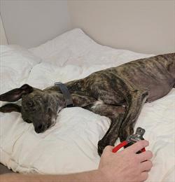
Conditioning Your Dog to Nail Trims

Teaching your dog to be calm and comfortable with the nail trimming process is necessary for your pet’s well-being. Your dog needs to hold still as their feet and nails are manipulated. The procedure can be painful if the nails are trimmed too short or if your dog struggles during physical restraint. Untrimmed toenails can break or tear, causing pain or infection. Trimmed nails help to contribute to healthy body movement.

Lay a Positive Foundation in Puppyhood

Food treats distract puppies and help keep them still without restraint. Lickable treats such as squeezable cheese, canned food, or peanut butter spread on a spatula or table are helpful. Trim one or two nails daily so your puppy enjoys short training sessions. If they become stressed, stop and try again later with higher-value treats. Routine trimming is not an emergency and forcing your dog’s compliance may cause long-term resistance to foot and toe handling.

Change the Emotion to Change the Behavior

Classical conditioning can be used to change your dog’s emotional state from neutral or negative to positive by rewarding each step with a tasty treat.

  • Touch your dog’s shoulder. Treat.
  • Touch your dog’s elbow. Treat.
  • Touch your dog’s paw. Treat.
  • Put pressure on the nail. Treat.
  • Place your nail trimmers nearby. Treat.
  • Sound of the snip nearby. Treat.
  • Place nail trimmers near the toenail. Treat.
  • Trim the nail. Treat.

Ideally, your puppy will learn to expect a yummy treat at the sight of the trimmers.

The process should proceed at your dog’s pace and they should remain relaxed. It may take several sessions to change their emotional response. Each nail trim step should occur right before the treat is delivered and the treat is given regardless of your dog’s reaction. They do not need to offer a paw or other behavior, to earn the treat. Rushing the training may cause your dog to become fearful, and then they may associate treats with a negative experience. They will then avoid the treats and nail trims.

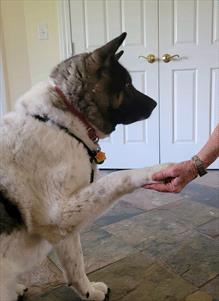
Observe your dog’s body language for subtle indications of stress or fear. When working with a partner during the conditioning process, one person can carry out each step while the other delivers the treat. Clear communication and proper timing are important. If your dog is given the treat too soon, they will not connect the handling to the treat. Your dog should be aware of each step, then rewarded before continuing. Operant conditioning gives your dog an opportunity to offer their cooperation. They can be taught to sit, lift a front paw, place the paw in your hand and hold it still. No restraint is necessary as the dog elects to participate. Positive reinforcement training methods should be used. If punishment methods such as pushing, grabbing, pinching, yelling, collar corrections, or shock are used, then fear and anxiety will be associated with nail trimming.

Your veterinarian may suggest both classical and operant conditioning for home practice and clinic nail trim visits. A referral to a positive reinforcement trainer or board-certified veterinary behaviorist may be made. If your dog needs a nail trim before being fully trained, sedation can be used to prevent further stress. Daily supplements and medications may be prescribed to lessen a dog’s anxiety before nail trims.

Nails should be kept short for your dog’s health and wellness. Classical and operant conditioning are techniques to ensure successful nail trims. With patience and low stress handling, nail trims can become routine for every dog.

6634930

Coat Clipping: Other Options to Keep your Dog Cool

When temperatures rise, it can be a challenge to keep pets cool while still being able to enjoy the outdoors with our four-legged friends. One response is to shave dogs, but here are some factors to consider before reaching for the clippers.

Does coat clipping help dogs keep cool?

Dogs have different skin and hair than people. While wearing a sweater during summer might sound uncomfortable to us, in some breeds a thick and wooly hair coat actually prevents dogs from overheating by acting as a layer of cooling insulation. Dogs also don’t have sweat glands all over their body (just on their paws), so shaving won’t help your dog release more heat through sweating.

Additionally, many other factors besides hair coat affect how well dogs cope with heat. Dogs that are overweight (obese) can overheat easily. Because panting is an important way for your dog to cool off, dogs with short snouts (brachycephalic) can be more heat intolerant than other dogs.

What are the drawbacks of coat clipping?

Many dogs have their coats clipped without any problems. However, consider the following before clipping your dog’s hair coat:

  • Clipping can injure the skin: Especially if the clippers overheat or the cut is too close to the skin, clipping can lead to burns, cuts, and scratches that become infected.
  • Less hair can mean more sunburn risk: A dog’s hair coat helps protect the skin from sunburn. Without that hair, consider minimizing UV exposure by using sunscreen.
  • Hair may regrow slowly: Many factors affect hair regrowth. If your dog has a health condition or medication that affects hair growth, the hair coat may be slow to regrow. In some dog breeds, clipping may lead to hair that grows back with a different texture or color.

If you decide to clip your dog’s hair coat, consider taking your dog to a professional groomer or your veterinarian to make sure the hair is cut properly.

What else can I do besides clipping my dog’s hair coat to help them with the heat?

Grooming your pet on a regular basis helps remove excess undercoat. Grooming will prevent and remove hair mats, which decrease air circulation around hairs. You can also help your pets cope with heat by providing shade and clean water while outdoors. During the summer months, avoid overexerting your dog and schedule runs and other activities to when the temperatures are cooler.

7440375

Clean Houses are Possible, Even With Pets!

Pets are messy. Shedding fur gets everywhere, mud tracks into the house, litter gets kicked out of the box, and water splashes out of the bowl. Plain old dirt conspires to make your pet’s living space -meaning yours – a mess. If you have multiple pets that shed, the dust bunnies can seem to be the size of a Flemish Giant rabbit. Since you can’t have pets without some mess, the main solution is to keep up with housecleaning.

The most important thing, though, is to clean up messes immediately. The longer any bodily fluid stays on the floor, the harder it will be to clean up. If accidents aren’t cleaned up right away, they will encourage your pets to use that spot for elimination, which is exactly what you do not want.

That said, set up your place with pets in mind.

Shedding is the bane of many pet owners. It can be tiresome to clean up all that hair so often, but there’s an easy way to lessen the sheer volume of loose fur: brushing your pets takes out the middle man, so to speak, as you grab the hair in a contained way rather than vacuuming and sweeping. Also, it’s a bonding experience for you and your pet, and allows you to check for lumps and bumps. However, most of us don’t do it often enough.

A good vacuum cleaner is a necessity for those with carpeting, and using one with a high-efficiency particulate air (HEPA) filter can help. Several vacuums are available that are designed with pets in mind and will likely have better success with fur-filled dust bunnies than those not designed for pets. After sweeping, sweep again with an electrostatic wipe (or start with an electrostatic mop) for non-carpeted floors. For deep cleaning, it’s hard to go wrong with a steamer mop as they do not use any chemicals and therefore cannot harm your pets or you.

Pets increase the volume of indoor dust, so dusting is usually needed a bit more often than in homes without pets. Microfiber mitts make the job easy.

If your pet has a favorite napping place, leaving a sheet or towel there allows you to scoop up the dirt and hair, shake the sheet or towel outside, and toss in the wash. That’s easier than picking up fur from all over the house. If your cat enjoys napping in front of the heat register, leave a bed or towel there with a placemat or rubber sheet underneath it just in case of an accident.

During rain or snow season, mats and towels are your best friends. Outside the door the dogs come in, place a washable mat. The rubber-backed ones wash easily, but if placed in the dryer too often the plastic backing will shred, so it’s best to let those air dry. Car mats work too. If the door height will allow it, also place one just inside the same door. Put a bowl of water (not cold) and a towel or microfiber mitt by the door and have your dog sit on the mat so you can wipe paws with a towel before the dog enters. (The microfiber mitt is also good for getting dirt off your dog’s dry paws.) You can either spend some time training your dog to sit on the mat or spend far more time during his lifetime cleaning up paw prints.

If you are shopping for new carpeting, opt for all-synthetic fibers. Unlike natural fibers, synthetic ones don’t absorb everything. Natural fibers will hold stains and odors. The deeper the carpeting, the harder it is to clean, plus the deeper rugs provide a perfect environment for fleas. Think about colors; a solid cream will show more dirt than even a slight pattern. Some people get carpeting that matches the color of their pets.

When looking at furniture, go for upholstery with a smooth surface, such as top-grain semi-aniline leathers, microfiber or microsuede, as those fabrics will resist claw marks so any couch-scratching cat will not be encouraged. Avoid upholstery with a lot of texture, and don’t use wicker. Materials with open or loose weave is just asking your dog and cat to engage in the battle of boredom with your furniture.

Keeping on top of brushing and cleaning up messes immediately goes a long ways toward keeping a house clean whether you clean it regularly or when you can’t stand it anymore.

7440377

Capturing Desirable Behaviors

You can improve your dog’s behavior in just a few minutes daily. By focusing on the desirable behaviors your dog does naturally rather than correcting the undesirable ones, you are building a stronger relationship without force or stress. Set your dog up for success by marking for easy behaviors, not unknown complicated ones.

Choose one or two behaviors each week that you want your dog to do consistently. For busy dogs, reinforce for lying quietly on their bed. To reduce barking, reward for silence as your dog watches someone walk by your house. To stop begging at the dinner table, treat your dog for choosing to lie on a mat several feet away.

Following this simple game captures the “good” behavior while ignoring the “bad”.  By not prompting your dog to do a specific behavior, such as sit or down, you create a thinking dog, and as the days go by, they will start to offer only reinforced behaviors.

  1. Count out the same number of dry kibble from your dog’s daily food intake. This could be 10 or 20 pieces for a small dog or 50 or more pieces for a large breed. You can withhold half from their breakfast portion and the other half from dinner or start out with the full amount to use throughout the day. High-value, low-calorie treats may be used in place of some kibble.
  2. Either carry the food in your pocket or have it easily accessible in central locations. 
  3. Throughout the day, when your dog offers the behavior you want, encourage them by marking that moment with a clicker or a verbal word such as “yes”. 
  4. Give them a piece of kibble from your budgeted quantity and go about your business. By doing this, you are capturing good behavior like a snapshot. This reinforcement increases the likelihood they will repeat that behavior. 
  5. Spend a week working on one or two behaviors, then switch to a different one the following week. Over time, build up to more complicated ones such as rewarding your dog for bringing you a toy instead of jumping on you when you walk through the door.

After a few days, your dog will offer the desired behaviors faster than the undesired ones. You should find your dog to be calmer and your relationship stronger as you go through your kibble supply more quickly each day.  

8156711

Cannabis Toxicity in Pets

An Overview

Cannabis generally refers to the products of the Cannabis sativa plant, commonly known as marijuana. The Cannabis sativa plant uniquely has over 100 different chemicals called phytocannabinoids. (Phyto means plant.) The most well-known of these is delta 9-tetrahydrocannabinol, referred to as THC.

THC has psychoactive effects on the brain, making it popular for recreational use. Psychoactive substances affect the mind or behavior and work on the nervous system, altering mood and feeling and making marijuana popular for recreational use. However, because THC affects many other body systems, it also has many medical uses. In fact, marijuana has been safely used medically for centuries.

In the United States, the Drug Enforcement Agency (DEA) has classified THC as a Schedule 1 drug. Schedule 1 drugs have no currently accepted medical use and a high potential for abuse. Despite this classification, almost all states have legalized marijuana for either recreational or medical use.

Another cannabinoid found in the Cannabis sativa plant, called cannabidiol (CBD), is also popular, but because it does not have psychoactive effects, its popularity is primarily because of medical benefits.

Marijuana contains at least 1% THC, although this percentage depends on what part of the plant is used and how it is processed. Products made from marijuana seeds contain little or no THC, but products made from the plant’s buds or flowers can contain up to 10% THC. In recent years, selective plant breeding has produced levels of THC up to 50%.            

The United States government has legalized a different Cannabis sativa plant called industrial hemp. Hemp can contain no more than 0.3% THC, but it can contain larger amounts of a second phytocannabinoid, cannabidiol or CBD. Because it contains little THC, the DEA does not control hemp or its products, and they are not considered illegal by the federal government. Because it is not illegal, many dietary supplements containing CBD are now being marketed for medical use in animals.

How It Works

The phytocannabinoids THC and CBD in Cannabis sativa work through the body’s natural endocannabinoid system (ECS). (Endo means self.) The chemicals in the ECS interact with the cannabinoid receptors found throughout the body, helping to regulate physiologic functions, and may be involved in different diseases. Using drugs to influence this system may help in the treatment of disease.

The medical effects of THC and CBD differ because they interact differently with the endocannabinoid system. 

In human medicine, CDB has been used to treat anxiety, improve appetite, relieve nausea, control certain types of seizures, and assist in sleep disorders. In animals, pain associated with osteoarthritis and seizures associated with epilepsy are common uses of CBD.

CBD and Supplements

CBD is not psychoactive like THC but may have medical benefits.

CBD is not a scheduled drug by the DEA and is an added ingredient in dietary supplements sold online, at dispensaries, and available over the counter. THC and CBD are also available by prescription, and your veterinarian may prescribe one or the other for treating your pet, most commonly for pain management. 

The difference between supplements and prescription drugs is that unapproved products like supplements are not regulated, nor are they assessed for consistency. Supplement labels may inaccurately list the amount of CBD in the product. More importantly, supplements may contain chemicals such as synthetic cannabinoids that are not listed on the label but may be dangerous to your pet.

In general, marijuana is safe. People are generally exposed to higher amounts of THC than pets because people more commonly smoke, rather than ingest, marijuana. THC that is ingested (for example, marijuana cookies) is removed mainly by the liver before it can get into the blood. THC that is inhaled bypasses the liver, and most of it enters the blood.

Toxicity in THC vs CBD

Because of its psychogenic effects, products containing THC are more likely to cause clinical signs that look like toxicity in dogs or cats. However, in contrast to marijuana or THC, products containing CBD have not been associated with significant toxicity or side effects in dogs. Toxicity or side effects to cannabis most likely reflect the effects of THC rather than CBD but are rarely lethal.

In cases in which side effects were reported with a supplement containing CBD, the supplement also contained THC. Even then, the side effects involved the gastrointestinal tract (vomiting, diarrhea) and were not severe.

An exception is those products that contain synthetic cannabinoids or if other drugs have been unknowingly added. These products can be life-threatening. This is another reason that it is important to bring the actual product to your veterinarian.

Different cannabinoids have varied effects. Each cannabinoid may reach a different area of the body. Responses to CBD among animals may reflect the differences in individual endocannabinoid systems.

Keep any medicinal or recreational products out of the reach of pets and children.

Signs of Marijuana Toxicity in Pets

The legalization of marijuana has led to increased reports of toxicity in dogs and cats. Most pet exposure to marijuana occurs after eating homemade baked goods containing the dried marijuana plant (about 70%), followed by eating the plant itself (about 10%).

For marijuana toxicity to occur, the pet must eat a large amount of marijuana. The amount of marijuana plant necessary to cause death in a dog or cat has not been determined. However, the least amount that has ever been associated with signs of toxicity is 3 to 9 g per kg of animal weight (about 7 to 20 g per lb.). For comparison, the average marijuana cigarette generally contains only about 0.5 to 1 gram of plant material. 

A problem with dogs ingesting marijuana in baked goods such as brownies or cookies is that they often contain chocolate, which is also toxic to dogs. Baked products may also contain concentrated forms of marijuana butter or oil. Another possible reason for marijuana toxicity in dogs is that marijuana may contain unsafe, illegal substances such as other drugs or synthetic cannabinoids. In cases where death has been reported in dogs, the marijuana most commonly was in baked goods. Chocolate may contain the sugar substitute xylitol, a substance that is toxic to dogs. 

After ingestion, the time it takes for clinical signs to occur and how long it takes for the pet to become normal again depends, in part, on how much marijuana and what else might have been ingested. Clinical signs can appear as early as 30 minutes after exposure but may take up to two hours to occur. After ingestion, pets may become normal again in as little as a couple of hours, but more often, it takes 24 to 72 hours for pets to return to normal.

A common clinical sign of marijuana toxicity is loss of urination control. Because this sign does not occur with other toxins, it helps with the diagnosis.

Other common clinical signs of toxicity include:

  •  lethargy or depression;
  •  difficulty walking or standing; 
  •  increased sensitivity to motion, sound, or touch (easily startled); and
  •  salivating (drooling).

Rarely, the pet may become aggressive. With the most severe toxicity, seizures may occur, or the pet may become comatose. Pets that already have illness may be more likely to become ill. Clinical signs in these pets may depend on what other illnesses they have.

Compared to humans, a dog has many cannabinoid receptors around the brain that help with balance. When dogs ingest a large amount of THC, they may become uncoordinated and unable to stand. This THC side effect in dogs is not likely to occur with CBD or in other species.

Diagnosing marijuana toxicity can be difficult. It is important that your veterinarian be given any information that might help with identifying the cause of the clinical signs. Bringing the actual product your pet was exposed to with you to the clinic can help. There is no risk to you as the pet owner because your veterinarian is not required to report toxicity to local law enforcement. 

In humans, urine tests are used to detect chemicals metabolized in urine from THC. However, animals do not make these metabolites, so these types of urine tests are not helpful.

Treatment for Toxicity

Diagnosis and treatment of marijuana toxicity depend on a well-informed veterinarian.

There is no specific antidote for marijuana toxicity. Treatments are given help to control the clinical signs. This might include fluids or drugs to control vomiting and, for severe toxicity, antiseizure medications.  

Usually, the patient is sent home after a physical examination. The pet can be kept in a confined space and monitored until the effects wear off. However, more intense in-hospital observation and support are needed if the patient has lost consciousness.

If less than 30 minutes have passed since the Cannabis has been eaten, it may be possible to induce vomiting. However, the pet may inhale stomach contents, particularly if it is acting sedated. This option may be necessary if the pet ingested a life-threatening toxin.

In more severe cases of toxicity, your veterinarian may give activated charcoal orally. This is a liquid that helps to bind ingested toxins. The toxin passes along with the charcoal when the pet defecates. However, this should not be given to patients that are sedated.

Cannabinoids might interact with other medications. For example, they may cause other medications to leave the body more slowly. It is important to notify your veterinarian if your pet is receiving cannabis or any supplement. 

The ASPCA National Animal Poison Control is available 24 hours a day at 888-426-4435. Expect an initial consultation fee of around $100.00 and additional follow-up is at no charge. You will be assigned a case number your veterinarian can use to communicate with a toxicology specialist before beginning treatment.

4127263

Chronic Kidney Disease in Dogs and Cats: Where to Begin

The terms chronic kidney disease, chronic renal failure, renal insufficiency, chronic kidney failure, and kidney insufficiency all mean the same thing. For simplicity’s sake in terms of this article, the term kidney failure will be used to reflect all these terms or conditions.

The kidney is an amazing organ. It is designed to do a lot more than just make urine and get rid of toxins. It also manages hydration, blood pressure, red blood cell production, pH balance in the blood, calcium, and phosphorus balance, and more. Kidneys are made up of millions of filtration units called “nephrons” and we are born with vastly more nephrons than we need to keep healthy. Over time and age, however, nephrons get damaged through the wear and tear of living and they die off. We only need enough nephrons to fill up about 1/3 of one kidney to manage normal kidney function and we start with two full kidneys packed with working nephrons at first but there may come a time when we just don’t have enough anymore.

When we don’t have enough working kidney tissue to maintain normal function, we are said to have “renal insufficiency,” with “renal” being the medical term for the kidney. Kidneys with insufficient function need help to get their work done and a long-term diseased kidney, regardless of its function or lack thereof, has “chronic kidney disease.” Chronic kidney disease patients are divided up by what stage of insufficiency they are contending with and what symptoms they have. 

 Treatment is focused on preventing progression of the insufficient function in earlier stages and on maintaining good life quality in later stages.

In the past, we used the term “chronic kidney failure” instead of “renal insufficiency” but this wording sounded far too dramatic and evoked images of ongoing sickness, expensive hospitalization, and doom when management could be as simple as diet change depending on the stage at which the disease is discovered. Early detection is paramount so let us review what the kidneys do and where they might need some help.

What our Kidneys Do/What Insufficient Kidneys Cannot Do

First, it is important to know what normal kidneys do. Kidney function goes far beyond simply making urine. Kidneys are made of millions of processing units called nephrons. These tiny little units are responsible for separating the chemicals you want to keep in your body from the chemicals you need to dispose of. The chemicals to remove are dissolved in water and make up the fluid we all know as urine. So here is what your kidneys do for you and for your pet, what they become unable to do in renal insufficiency, and some of the parameters your veterinarian will want to track.

Water Conservation

Hydration of the body depends not only on water consumed but on water removed. In times of dehydration, the kidney must respond by conserving water. This means that all the materials that the body needs to get rid of still need to be removed, but the kidney needs to do so using the smallest amount of water possible. 

Similarly, if you drink too much water, the kidney needs to efficiently remove it to prevent dilution of the bloodstream. A pet with insufficient kidney function will not be able to make concentrated urine and will need to drink extra water to process the body’s waste chemicals. For this reason, excessive water consumption is an important early warning sign and should always be investigated.

When we analyze a urine sample, one of the most important parameters is the specific gravity. It is a measure of how concentrated a urine sample is. Water has a specific gravity of 1.000. A dilute urine sample has a specific gravity of less than 1.020 (often less than 1.010). A concentrated urine sample would have a specific gravity over 1.030 or 1.040. A failing kidney, by definition, cannot make concentrated urine and the patient must drink excessively to get enough water to get rid of the day’s toxic load.

Toxin Removal

The kidneys remove our metabolic wastes for us. If there is inadequate circulation going through the kidneys or if there are not enough functioning nephrons to handle the waste load, toxins will build up. When the toxins build up and exceed the normal range, a condition called azotemia occurs. If the toxins build up to a level where the patient actually feels sick, they have a condition called uremia. If we can keep our azotemic patients below the uremia level, they will feel pretty normal and have good life quality.

The most important marker of uremia is called creatinine. Creatinine is a by-product of muscle breakdown and is always in small amounts in the bloodstream. The kidney removes it continuously unless there is a kidney function problem. A newer parameter called SDMA (symmetrical dimethylarginine) becomes abnormal much earlier than creatinine does and is becoming more commonly tracked. We are able to stage a patient’s kidney disease based on creatinine blood level and SDMA level (see the staging section below). Another marker is the BUN, which stands for blood urea nitrogen. This parameter is similar to creatinine but is influenced by dietary protein as well as kidney function. These three markers are central to determining the severity of a kidney problem.

Calcium/Phosphorus Balance

The balance between calcium and phosphorus in the blood is really important. Too much of one or the other will lead to crystals forming in body tissues and bones to the extent that they may actually become rubbery. The kidney plays an important role in this balance and when kidney function is lost, phosphorus levels begin to rise. Therapy for insufficient kidney function requires monitoring phosphorus levels and the use of diet and medications to keep phosphorus levels in a reasonable range.

Sodium/Potassium Balance

The kidney plays a major role in controlling electrolyte balance as well. In particular, conserving potassium is an important aspect. Insufficient kidneys lose their ability to conserve potassium and potassium levels begin to drop, leading to weakness. Potassium supplements are commonly needed in the treatment of kidney failure.

Blood Pressure Regulation

Blood pressure sensors in the kidney help regulate blood pressure in the body. When these are damaged, hypertension (high blood pressure) can result and can damage the kidney even more. Blood pressure is commonly measured in kidney failure patients.

Protein Conservation

There are a lot of important proteins circulating in the bloodstream and it is crucial that they are not lost in the urine. The nephron possesses a filtration system that keeps the protein in while removing harmful wastes. If this filtration system is damaged (glomerular disease) then a much more severe form of kidney failure results. Screening for this damage is an important aspect of staging kidney failure and a test called a urine protein: creatinine ratio is often included in the testing profile to assess this condition.

Red Blood Cell Production

The kidney produces a hormone called erythropoietin. This hormone tells the bone marrow to make more red blood cells. In the absence of this hormone, non-responsive anemia occurs and can get so bad that a transfusion is necessary. Erythropoietin can be given by injection to treat this problem but there are some potential pitfalls in doing this as will be discussed in a later article. Hematocrit and packed cell volume (PCV) are measures of red blood cell volume commonly monitored in kidney patients.

 pH Balance

Metabolic processes require a narrow pH range for efficiency. The kidney also regulates this balance and if it cannot, intervention, usually in the form of fluid therapy, is necessary.

That’s a lot of tasks for a pair of kidneys to handle! With chronic kidney disease/renal insufficiency, chances are at least one of these tasks is not moving along smoothly. There may or may not be any symptoms when the disease is discovered but the goal is to slow the progression to the point where symptoms develop. If there already are symptoms, the goal becomes controlling the symptoms and maintaining good life quality. 

Once kidney disease has been discovered, the next step is staging and evaluating any other aspects of the above function list that may not have been covered. Typically animals need special food and possibly medications depending on symptoms. Very advanced cases will likely need hospitalization, intravenous fluids, and possibly assisted feeding to get to where they can resume a more normal life at home. As mentioned, early screening is especially important as it is much easier to slow the progression of disease than it is to reverse it.

STAGING KIDNEY DISEASE

Kidney disease is either discovered via wellness screening tests of apparently healthy animals or during the course of the evaluation of patient illness. As mentioned, what we do for the patient will depend on the stage of the disease. The International Renal Interest Society (IRIS) has determined a staging system based on the creatinine level and SDMA level found on a blood test (see Toxin Removal section above). Patients in Stage 1 or 2 rarely have any symptoms related to their disease and those in Stage 3 or 4 usually have symptoms to control.

The chart below stages dogs and cats according to their Creatinine and SDMA levels. Once the patient has been staged then further testing for “sub-staging” begins.

Table 1: Creatinine Values for Each IRIS Stage listed in US, International, and SDMA Units

 UnitsStage I(pre-failure)Stage II(mild failure)Stage III(moderate failure)Stage IV(severe failure)
Dogmg/dl
μmol/l 
SDMA μg/dl
<1.4
<125
<18 mcg/dl
<1.4-2.8
<125-250
<18-35 mcg/dl
<2.9-5.0
<251-440
<36-54 mcg/dl
>5.0
>440
>54 mcg/dl
Catmg/dl
μmol/l 
SDMA μg/dl
<1.6
<140
<18 mcg/dl
<1.6-2.8
<140-250
<18-25 mcg/dl
<2.9-5.0
<251-440
<26-38 mcg/dl
>5.0
>440
>38 mcg/dl

Sub-staging of kidney disease involves screening for urine protein loss and measuring blood pressure. A urine protein: creatinine ratio is performed on a urine sample and the protein amount is classified as either non-proteinuric, borderline proteinuric, or proteinuric. Blood pressure is checked and the patient is classified as normotensive (normal), borderline hypertensive, hypertensive, or severely hypertensive. The ultimate classification for the patient will reflect all of these things (“Stage 3, non-proteinuria, normotensive” would be an example). Further testing and monitoring are determined based on what the actual parameters are. For more details on these substages, see the articles on glomerular disease, and hypertension.

Stage I is mostly for pets that are known to have kidney disease based on abnormal ultrasound or their slightly elevated SDMA. We check their blood pressure and urinary protein levels, and screen for urinary tract infections. If abnormalities are found, they are usually checked quarterly for any sign of progression.

Stage 2 is where diet change to a renal diet is typically recommended with the idea being that the earlier this is started, the more likely the pet will accept it. The same type of screening is performed as with Stage I, only now we need to start watching the phosphorus levels so they don’t rise above 4.6 mg/dl. Also, potassium levels may begin to drop especially in cats. Both of these factors are helped by using a renal supportive diet but if that proves inadequate, medications may become needed.

In Stage 3, the patient is likely to be showing some symptoms such as reduced appetite or occasional nausea. In addition to the tests of Stages I and 2, it is in Stage 3 that we start paying attention to red blood cell numbers and blood pH. This is a good time to consider the use of calcitriol (active vitamin D supplementation) if one is going to do so.

In Stage 4, the patient is almost certainly showing symptoms and it becomes important to be sure the patient is eating enough calories to maintain a healthy weight. Similar concerns exist as for the first three stages but a phosphorus level of 4.6 mg/dl is probably unrealistic at this point and a better goal is 6.0 mg/dl. Fluid support at home is a good idea. This may mean adding water to the food, giving fluids under the skin (subcutaneous fluids), or even a stint of hospitalization and IV fluids to drive the numbers down.

Many patients are not diagnosed with kidney disease until they are far past a creatinine of 5.0 mg/dl. It is not unusual for sick patients to come to the vet and have creatinine values of 10 mg/dl or higher. At this point, more aggressive treatment will be needed to stabilize the patient and the goal is going to be improving the dysfunction to a level where the patient has acceptable life quality and comfort at home. Transitioning to a new lifestyle will take some time. (For example, appetite and comfort must be restored but changing to a renal support diet may be something for further down the treatment path when more stability has been achieved).

A Note on Pyelonephritis

Pyelonephritis is another name for kidney inflammation and usually means kidney infection. Patients get kidney infections when a bladder infection goes unnoticed or incompletely treated long enough and the bacteria go up the ureters into the kidney where they set up shop and cause damage and pain. This form of kidney failure has the most potential to reverse or partially reverse, so it is important to culture the urine for it early. Urinalysis is frequently unable to detect infection in this situation as the patient drinks so much water that the visible markers of infection are diluted out and cannot be found. The infection also will produce a significant amount of urinary protein so the patient will be sub-staged incorrectly if an infection is not ruled out.

A Special Note on Ureteral Stones

The ureter is the tube that connects the kidney to the bladder and urine should constantly be flowing through it. Often in age (plus with calcium/phosphorus imbalance), mineralization will occur in the kidney. This may or may not be a problem depending on how big and how plentiful the mineral deposits are but sometimes a mineral deposit will break off and get lodged in a ureter. This can obstruct the ureter completely and the backlog can destroy the kidney. Or it can partially obstruct the ureter, causing obstruction and then the relief of obstruction as it rolls down the ureter and (hopefully) into the bladder where it will not cause trouble. A spike in creatinine can be a clue that this has happened and if one acts quickly intervention may be possible. For this reason, some kind of imaging of the kidneys is recommended early in the course of kidney disease and periodically after.

The bottom line is that there is a wide range of treatments when a patient has insufficient kidney function and a lot of monitoring is needed to keep on top of all the aspects. Each patient will have a different stage and a different constellation of issues to contend with. Some will need only a diet change, and some will need ongoing fluid therapy at home or hospitalization. The prognosis depends on all the factors involved. Your veterinarian will let you know what your particular pet needs and will be watching many different lab tests to keep control of your pet’s condition.

 In Summary:

  •    From a practical standpoint, the terms chronic kidney disease, chronic renal failure, renal insufficiency, chronic kidney failure, and kidney insufficiency all mean the same thing.
  •    The kidney’s job is more than just to make urine and get rid of toxins. Additionally, the kidney manages hydration, blood pressure, red blood cell production, pH balance in the blood, calcium and phosphorus balance, and more.
  •    Over time, kidney cells can become damaged. When there is not enough working kidney tissue to maintain normal function, it is called “renal failure”. A long-term diseased kidney, regardless of its function or lack thereof, has “chronic kidney disease.”
  •    Hydration of the body depends not only on water consumed but on water removed. In times of dehydration, to keep this balance, the kidney will remove the toxins using the smallest amount of water needed.
  •    If over-hydrated, the kidney must remove excess water to prevent dilution of the bloodstream.
  •    A failing kidney cannot make concentrated urine, and the patient must drink excessively to get enough water to get rid of the day’s toxic load. Excess water consumption by your pet could be an early warning sign of kidney disease.
  •    If there is inadequate circulation going through the kidneys or if there are not enough functioning kidney cells to handle the waste load, toxins will build up.
  •    If concerned about possible kidney disease and toxin levels, your veterinarian will order a urinalysis and blood work for your pet. Markers in the urine and blood will show the presence and severity of a kidney problem.
  •    Levels of kidney disease are labeled by “stages,” i.e., I (pre-failure), II (mild failure), III (moderate failure), IV. (severe failure). How far the disease has progressed will determine treatment.
  •    Treatments can be combinations of fluid therapy, diet changes, medications, and possibly vitamin supplementation. On-going examinations and laboratory tests will be a part of the treatment as your pet is monitored for changes to their condition.
  •    Prognosis depends on the stage of the disease and factors involved.
4128893

Cryptorchidism (Retained Testicles) in Dogs and Cats

Cryptorchidism is a condition in which a male’s testicles have not descended (dropped) into the scrotum. At birth, a male puppy’s or kitten’s testicles are located near the inguinal ring. 

By 8 weeks of age, testes are palpable in the puppy’s scrotum. However, scrotal and suprascrotal testes can be difficult to palpate if the testes are small or the puppy is obese. Testes may also freely move between the scrotum and inguinal area in young puppies.

By 8 to 16 weeks, they’re palpable in the kitten’s scrotum. Some retained testicles can be palpated. However, testicles retained in the abdomen are typically not palpable on physical exam.

In the fetus, a structure called the gubernaculum connects the testicle (located next to the kidney during development) to the scrotum. If this structure fails to develop properly, the testicle will not end up in the scrotum but will end up in the abdomen, the inguinal canal, etc.

Cryptorchidism can be unilateral (only occurring on one side) or bilateral (occurring on both sides). Unilateral cryptorchidism usually involves the right testicle. Bilaterally cryptorchid animals are usually sterile because the higher body temperature inside the abdomen is enough to prevent sperm production. (The animals will, however, still exhibit male behaviors.)

Cryptorchidism is a fairly common defect in dogs. Dog breeds most likely to be affected include Yorkshire terrier, Pomeranian, French poodle, Siberian husky, miniature schnauzer, Shetland sheepdog, Chihuahua, German shepherd, dachshund, and brachycephalic breeds. Still, it can happen in any breed or mixed breed.

Since cryptorchidism is considered to have a genetic basis, animals with this condition should not be used for breeding.

Dogs with cryptorchid testicles are prone to testicular torsion (twisting) and testicular cancer, so these dogs should be neutered to prevent problems later.

Cryptorchidism in cats is uncommon. The most common breed associated with cryptorchidism is the Persian. Congenital abnormalities that have been known to occur simultaneously with cryptorchidism are patellar luxation, shortened tail, kinked tail, tetralogy of Fallot, tarsal deformity, microphthalmia, and upper eyelid agenesis. Unlike dogs, it may be possible to visually differentiate between a castrated cat and one with retained testicles, because cryptorchid cats have barbs on the penis.

Laboratory Diagnosis

A testosterone assay can be used to differentiate between a castrated male and a cryptorchid male.

Treatment

Surgical removal is the only treatment for cryptorchidism. Even if the animal is a unilateral cryptorchid, both testicles should still be surgically removed. (The cryptorchid testicle should be removed to prevent testicular torsion and testicular cancer, and the normal testicle should be removed to prevent cryptorchid offspring.) This surgery is more complicated than the usual neuter surgery because the cryptorchid testicle can be difficult to locate. Depending on the case, some pets will be able to go home on the day of the surgery, and some may have to stay in the hospital overnight. A 2-week recuperation (reduced activity) is advised because this surgery usually involves opening the abdomen, and the surgical site has to have time to heal before the pet resumes normal activities.

Many males will need to wear a protective Elizabethan collar during recuperation to prevent them from licking or chewing at the incision. Owners should check the incision regularly for redness and swelling, which could indicate a post-operative infection or self-trauma. If non-dissolvable skin sutures are used, they will need to be removed by your veterinarian about 10 to 14 days after surgery.

Prognosis

Cryptorchid animals that have had both testicles removed, and have no other defects, will generally live a normal lifespan for the breed.

5425457

Calcium Phosphorus Balance in Dogs and Cats

In renal insufficiency, phosphorus is not anyone’s friend. The same phosphorus that has so many helpful roles in the body (from transferring biochemical energy to combining with calcium to form bone), turns against us in a condition called renal secondary hyperparathyroidism.

The short version is that the failing kidney is no longer good at getting rid of excess phosphorus and phosphorus levels in the blood begin to rise. The rise in phosphorus upsets the delicate balance between calcium and phosphorus and activates a regulatory hormonal cascade that attempts to re-establish control. Without healthy kidney tissue to play its role in this balance, the body is fighting a losing battle. Calcium is mobilized from bone to balance the phosphorus but in the end this only serves to demineralize and weaken the bones and cause calcium phosphate deposits to form in soft tissues. These mineral deposits are inflammatory and damaging.

The hormones that play roles in the regulation of calcium and phosphorus are parathyroid hormone and calcitriol (which most of us know as Vitamin D). The story co-stars a substance called Fibroblast Growth Factor 23. These three materials interact to maintain a normal and effective blood calcium level without allowing phosphorus levels to run away.

We’ll review the story of how this works.

Keeping Calcium Under Control

While our discussion of renal disease largely revolves around phosphorus, the importance of calcium cannot be underestimated. The movement of calcium ions is what allows our muscle fibers to contract, not just in our arms and legs but also in our hearts, and involuntary intestinal and blood vessel muscles as well. Calcium combined with phosphorus makes up bone; in fact, bone can be considered a storage depot for calcium when we need some in a pinch. The blood level of calcium is tightly regulated by hormones within a narrow range as too much calcium is as dangerous as too little.

There are four tiny parathyroid glands around the thyroid gland in the throat area. These glands produce a biochemical called parathyroid hormone (often abbreviated PTH). PTH basically raises calcium and activates vitamin D, which further raises calcium. It sounds like calcium would just continue to rise out of control but there is a safeguard. When active vitamin D levels get to a certain point, they shut off PTH production. Calcium levels then drop down and between these two hormones, blood calcium is regulated within a healthy range.

Keeping Phosphorus Under Control

When it comes to phosphorus, PTH and calcitriol no longer work together. PTH signals the kidney to dump phosphorus while calcitriol signals the kidney to save it. In the normal body, the coordination of these hormones works out to keep calcium and phosphorus levels in balance, no matter what is happening with the blood calcium.

All this goes along swimmingly until the kidney is too damaged to respond to its hormonal signals or properly activate calcitriol.

Parathyroid hormone causes the kidney to dump phosphorus while vitamin D causes the kidney to save phosphorus.

What Happens in Kidney Failure?

In early kidney failure, the kidney becomes unable to get rid of phosphorus normally, and as a result phosphorus levels begin to rise. This activates a substance called Fibroblast Growth Factor 23 (affectionately known as FGF-23) from the bones. FGF-23 encourages the kidney to dump more phosphorus and dampen the activation of vitamin D (remember that vitamin D tells the kidney to save phosphorus). As part of its mission to dampen vitamin D, FGF-23 also instructs the parathyroid glands to cut back on PTH. All this vitamin D suppression is all well and good and helps keep the phosphorus levels reasonable, but pretty soon FGF-23 has dampened both PTH and vitamin D so well that calcium levels begin to drop.

And this is where calcium-phosphorus Hell breaks loose.

At this point, kidney failure is no longer early. Dropping calcium levels supersede any FGF-23-mediated suppression of PTH and the parathyroid glands crank up PTH production to bring calcium levels back up. To meet the body’s demand for circulating calcium, the bones liberate their structural calcium to save the day, but unfortunately doing this also liberates more phosphorus. This leads to an even higher phosphorus level. Circulating phosphorus binds the circulating calcium creating crystals that deposit in the body’s soft tissues and generate inflammation. Bones are weakened and replaced with fibrous tissue. Calcium is depositing uselessly all over the place and the parathyroid glands must crank even harder to keep calcium levels livable. A metabolic disaster has occurred at this point.

Making matters worse are other effects of excess parathyroid levels. In high amounts, nerves cannot conduct electrical impulses properly. Patients become dazed and poorly responsive.

The goal is to keep the phosphorus level from getting out of control in the first place. If this is not possible, the goal is to get the phosphorus level back under control and keep it there. 

Treatment

How do we Control Phosphorus Levels?

The goal phosphorus level defined by the International Renal Interest Society is between 2.5 and 4.6 mg/dl for earlier renal insufficiency stages and to keep phosphorus levels under 6 mg/dl for Stage Four (more advanced) patients.

Fluid Therapy

Often simply giving fluids under the skin at home provides enough extra circulation through the kidneys for the extra phosphorus to be excreted normally. Further treatment may not be needed. Hydrating a damaged kidney effectively maximizes the kidney’s remaining function, and this may be enough to control phosphorus.

Therapeutic Diet

It is important to remember that ultimately phosphorus balance is a matter of balancing phosphorus entering the body with phosphorus leaving the body. If the kidney is no longer effective at removing phosphorus, we can perhaps work on limiting the amount of phosphorus entering the body. The first step in doing this is with a therapeutic renal diet. These diets are designed to give the kidney less work to do, and that includes limiting phosphorus entering the GI tract. After one to two months on a phosphorus-restricted diet, blood tests will indicate whether or not additional phosphorus treatment is needed.

Phosphate Binders

These products are given with food to bind phosphorus in the food. The complex of phosphorus and phosphate binder cannot be absorbed into the body with the result being reduction of incoming phosphorus. If the pet will not eat food containing the binder, the binder can be given just before or just after the meal but the binder must be in the GI tract at the same time as the food or it will not work. There is no point in giving a binder to an animal that is not eating. Giving the binder before the meal can induce enough stress so that the pet will not eat so it is better to give the binder after the meal if the binder is not accepted mixed in the food.

There are a number of phosphate binders available and which one is selected will depend in part on the concurrent calcium situation. This means that the ionized calcium level (not the total calcium level) must be measured. Some binders also supplement calcium while others drop or have no influence on calcium. Some patients require more than one binder to get their phosphorus value low enough.

Calcitriol


You might think that calcitriol would not be helpful in this situation since it leads the kidney to retain phosphorus. The good news is that when small enough doses are given, calcitriol can still act as the off switch for parathyroid hormone without causing the kidney to retain phosphorus. The amounts needed for this beneficial effect are so small (they are measured in nanograms) that a compounding pharmacy is needed to custom-make the product at the proper dose.

  • Calcitriol cannot be used in patients with elevated blood calcium levels.
  • Calcitriol cannot be used in patients with phosphorus levels that are already abnormal. This is a preventive measure more than a treatment.

Renal secondary hyperparathyroidism and its associated high blood phosphate levels are one of the most common uremic toxin issues in treating kidney disease. Dedication is required to get phosphate binders into the pet along with food when the pet’s appetite is weak. Phosphate binders come in powders, liquids, capsules, compounded flavored chews, and other formats. Consider what approach is likely to work best for your pet but, remember, the binder only works with food.

4128653

Cervical (Neck) Disk Disease in Dogs and Cats

Cervical (neck) disk disease is an intervertebral disk pressing on the spinal cord, causing pressure and pain. It’s essentially the same condition as intervertebral disk disease, except it occurs in the neck instead of in the back.

Intervertebral disks are located between the vertebrae (bones of the spine). Each disk has two parts, a fibrous outer layer and the jelly-like interior. When disk herniation occurs, the interior either protrudes (bulges) or extrudes (ruptures) into the vertebral canal, where the spinal cord resides. The onset of herniations can be either acute or chronic. Generally speaking, when this type of herniation happens in the neck, disk disease is the most likely cause, although it can be due to trauma (falling off furniture or a deck, getting hit by a car, collar trauma, etc.). Small chondrodystrophic breeds – those with a long back such as dachshunds, for example – are the breeds most typically affected, although other breeds can be affected. It’s more common in middle-aged pets.

When the spinal cord is compressed by cervical disk material, the dog or cat can experience signs ranging from mild (neck pain) to severe (complete paralysis of all four legs and with no pain perception).

Usually, the neck signs appear suddenly. The neck muscles may tense up or shiver, the pet’s nose may point toward the ground, the pet’s back may arch, and there may be muscle tremors in the neck and shoulder. The pet is usually reluctant to move his head from side to side or to lift his chin. Sometimes nerve control of urinary and bowel functions will be affected.

Other signs may include clumsiness and paralysis. Severe damage to the spine will have severe effects and a worse prognosis than mild damage. Chronic spinal cord damage also has a worse prognosis.

An acute onset is an absolute emergency, and the pet should be seen immediately by a veterinarian.

Diagnosis

After obtaining your pet’s medical history, your veterinarian will perform a general physical examination and then a neurological examination. The veterinarian will see how much pain perception your pet has; inability to feel any pain is not good. Radiographs are usually taken, although they may not be definitive. If possible, a myelogram (where dye is injected into the spinal canal), MRI, or a CT scan will be run.

Treatment

Treatment options depend on how severe the problem is. If the situation is mild, it may be treated with just medication, typically anti-inflammatories or glucocorticoids, and crate rest. If the problem resolves on therapy, but recurs when treatment is finished, surgery may be required.

More serious cases may require surgery, although surgery is not an instant cure. The surgeon will remove the extruded disk material to take pressure off the spinal cord. Surgery does nothing to the cord itself. It takes time for the spinal cord to heal, but the good news is that the neck portion of the spinal cord is more tolerant of pressure from the extruded disk material than the back portion is.

Strict crate rest is necessary for either medical or surgical management. If your pet is not confined to strict crate rest, the herniation could recur and possibly be worse than the original episode. After surgery, your pet will probably require a month of crate rest. Another month of restricted activity may then be necessary. During that month, the use of steps should be avoided, and there should be no heavy exercise. If you customarily use neck collars or choke collars on your dog, those should not be used for another three months; body halters would be less likely to cause trauma to your dog’s neck.

Prognosis

Your pet’s prognosis depends, of course, on his particular case. Generally speaking, if he could walk at all before surgery, his post-surgical prognosis is excellent. If he couldn’t walk, but still had some deep pain sensation, the post-surgical prognosis is good to excellent. If he lost deep pain sensation completely, the prognosis is guarded.

4128682

Cryptosporidium is a Particularly Challenging Type of Coccidia for Pets

What are Coccidia and Why they are Bad

Coccidia are a common parasite of young puppies and kittens. They reproduce inside intestinal cells, ultimately killing the cells within which they divide and producing nasty diarrhea when too many intestinal cells have been killed. As the coccidia increase in number, so does the number of intestinal cells being killed. Potentially (for very small or immune-compromised pets), so many intestinal cells can be killed that the host animal dies from its severe bloody diarrhea. 

Survival is a matter of the host’s immune system versus the dividing coccidia. There are drugs that can kill certain types of coccidia, but for Cryptosporidium, drugs just reduce the number of organisms and may not be able to clear the infection fully. (More on this later).

Coccidia infection is the scourge of the dairy industry, as it is one of the most common causes of calf death. This infection is similarly a nightmare for small puppies and kittens.

Generally speaking, carnivores like dogs and cats get infected with Cystoisospora species, while livestock (goats, sheep, calves, and even rabbits) get Eimeria species, and these species of coccidia do not cross over.

Cryptosporidium vs. “Garden Variety” Coccidia

The easiest way to understand Cryptosporidium species and how they cause disease is to think of them as another kind of coccidia.  Cryptosporidium have some uniquely unpleasant features, however, that are not shared by routine species of coccidia.

  • Cryptosporidium oocysts (the eggs that can be seen on a routine fecal test) are so small that they are difficult to detect under the microscope with normal testing methods. More routine coccidia oocysts are about ten times larger and thus less likely to go undetected.
  • Routine intestinal coccidia is not a human threat. Cryptosporidium is another story, and human threat depends on the species of Cryptosporidium involved and the immune status of the person in question. Cryptosporidium from dogs and cats do not readily infect humans, with the exception of immunosuppressed individuals. For these people, the infection can actually be life-threatening. (Cryptosporidium from livestock more readily infects humans, causing severe diarrhea that sometimes results in hospitalization even in humans who are not immune-suppressed).
  • Drugs that would work on routine species of coccidia do not have any effect on Cryptosporidium.
  • In calves, only ten oocysts are needed to establish a significant infection (i.e., that is how many the calf has to swallow). This is a particularly small number. We do not know how small the number is for other species but the point is that this is a virulent organism and a small exposure can lead to a very pronounced disease in an individual under stress or with immune compromise due to immaturity, concurrent disease, or small size.
  • Possibly, the most ominous difference between routine coccidia and Cryptosporidium is that Cryptosporidium can yield a self-perpetuating infection, whereas routine coccidia will run its course.

How Infection Occurs

Out in the world, Cryptosporidium oocysts (the infective stage shown below) are very tough. They resist bleach and most other normal cleansers. Only prolonged exposure to ammonia or extreme temperatures can kill them. This photograph depicts the oocyst releasing sporozoites after it has been swallowed by its new host (as described below). The sporozoites are the stage that kills intestinal cells.

The life cycle of this organism is rather complicated, and it is not necessary to understand all the stages. The short version is that an oocyst (sort of like an egg) is passed in the feces of an infected animal. This oocyst is swallowed by another animal by licking the dirt off its fur, drinking contaminated water, or some such activity. The oocyst releases sporozoites (sort of like a spore) into the intestinal tract of the new host. 

The sporozoite infects an intestinal cell and divides. The spores divide into other stages with other names, which in turn infect more cells.

All this cell division occurs asexually for a while until, eventually, the Cryptosporidium begins a sexual phase: instead of making more copies of itself by simple cell division, it produces male and female cells. Fertilization occurs, yielding oocysts like the egg that started it all.

There are actually two types of oocysts: one that is thick-walled and ready to be passed in feces to face the external world, and one that is thin-walled and just infects the host over again from the beginning.

This is an important and bad thing so we will say it again: the thin-walled oocyst infects the host over again from the beginning. No contaminated water is needed. No dirty fur is necessary. This is now a self-perpetuating infection.

If there is any good news in this, it is that most hosts have healthy immune systems and are able to coexist with low numbers of Cryptosporidium without diarrhea. As will be seen, it is unlikely that medications will eradicate Cryptosporidium; the goal in therapy is to eliminate diarrhea rather than clear the organism completely.

How Rare is this Parasite?

The prevalence of Cryptosporidium oocyst shedding in dogs has varied from 2% to up to 15 -20% in stray dog populations. Fecal specimens from 200 stray dogs impounded at the San Bernardino City and County animal shelters were screened for Cryptosporidium oocysts, and 2% of dogs were found to be oocyst positive. A similar survey of 206 cats revealed oocyst shedding in 5.4%. Most infections are subclinical, meaning that the host animal is not sick.

Humans tend to get their own species of it (Cryptosporidium hominis), while cats and dogs each have their own Cryptosporidium. Pet ownership has not been found to be a significant risk for humans with cryptosporidiosis (i.e., most infected humans get infected from other humans or from livestock).

This is generally good news except for the immunosuppressed owner, who might adopt an infected pet without knowing it. Remember, in the immunosuppressed individual, cryptosporidiosis can be serious, so a screening test for a new pet is a good idea in this situation.

Screening for Infection

A routine fecal flotation test, as is recommended once or twice a year for most pets, is likely to miss CryptosporidiumThere are two main reasons: first, Cryptosporidium is extremely small, and second, oocysts are only intermittently shed. At this time more specialized testing such as PCR testing for Cryptosporidium DNA or ELISA tests for Cryptosporidium proteins are the best way to go, but these tests were developed to detect the Cryptosporidium species that infect humans so there may be some issues with test sensitivity. Screening routinely for Cryptosporidium is probably not warranted for healthy animals unless they are going to be spending time with immune-compromised individuals.

Treatment

The bad news is that treatment is difficult. Nothing can really be described as highly effective. A medication called paromomycin has effective but is highly toxic to the kidneys. A medication called nitazoxanide has been effective but causes nausea and diarrhea. Azithromycin and tylosin also have activity against Cryptosporidium and are sometimes used. Fortunately, most infected animals have healthy immune systems and, even though medication may not be fully effective, the patient’s own immune system is usually able to control, if not fully curtail, the infection. Many animals appear to carry this organism without symptoms.

The U.S. Public Health Service and the Infectious Diseases Society of America both recommend that HIV-infected individuals should not bring into their homes:

  • Animals with diarrhea
  • Stray dogs or cats
  • Dogs or cats under the age of six months.

See more details on pet-related guidelines for HIV-positive individuals.

The Centers for Disease Control and Prevention (CDC) has information on preventing infections from pets.

CDC tip: Protect yourself against getting Crytosporidium from animals. Simply wash your hands with running water and soap after any contact with animals and animal feces (stool).