Actinic (Solar) Dermatitis
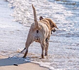
Most of us know that sun exposure can be damaging to the skin. We get wrinkles, sunburn, and even sun- induced skin cancers. Dogs have similar issues.
Pets like to sunbathe just like people do. Many like to lie happily on their backs and soak up the damaging UVB light. UVB light is pretty unpleasant stuff. For one thing, UVB light induces the inflammatory response of skin cells, damages the skin’s immune cells, and induces mutations in skin cells of DNA. At first, the skin becomes pimply and red, but then plaques of thick skin develop and spread. Skin cancer can form, and not all forms of skin cancer stay confined to the skin.
Dogs at Risk
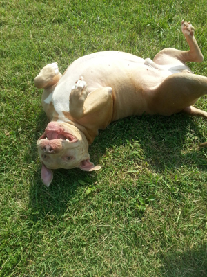
Most of the time, a good fur coat is all the protection a dog needs to avoid damage from the sun. After all, protection from the environment is what fur is all about. There are two problems, however: the tummy (which is sparsely furred in most dogs), and short white fur (which sunlight readily penetrates). This puts the pit bull terrier at special risk but any dog with similar hair length (greyhound, pointer, Dalmatian etc.) can have a problem with too much sun exposure, on the tummy or otherwise.
What Does it Look Like?
The white fur becomes patchy, and the skin below becomes red and irritated. In contrast, if there are adjacent patches of dark fur, the coat is lush and normal with no patchy hair loss, and the skin underneath is not inflamed. Passing your fingers across the fur from white to colored will yield a dramatic difference in skin texture as the colored fur is protective against the sun.
Infection (usually a pimply red rash) is common in many skin conditions, including this one, so sometimes a course of antibiotics can make the underlying sun-induced situation more clear. Biopsy of sun-induced skin damage is very characteristic and should be able to settle any questions.

Courtesy Morguefile
Treatment
As long as skin cancer has not actually cropped up, treatment focuses on inflammation and reducing sun exposure.
Antibiotics
Most actinic dermatitis cases have a hefty secondary Staphylococcus infection and need a couple of weeks of antibiotics.
Topical Steroids
If the skin has a lot of inflammation and discomfort, a week or so of topical steroids can relieve this. Be sure to use a product made for animals as human products are not made with the expectation that they will be licked.
Vitamin A
Protocols using daily Vitamin A have been shown to reduce the development of skin cancer and lessen damage from the sun. Your veterinarian can tell you what to get.
Pentoxifylline
This medication can improve circulation to the skin, thus assisting with perfusion and healing.
Reduce Sun Exposure!
If possible, keep the pet indoors during the hours of 11 a.m.—2 p.m.
- Beware of outdoor areas featuring large spaces of white concrete. This type of flooring is highly reflective of sunlight and will expose a tummy to sunlight even without sunbathing.
- Use sunscreen on the pet’s skin but be careful of human products as they have irritating scents and the potential for toxicity (zinc oxide can be toxic if licked). For dogs, Epi-Pet® can be purchased at many outlets. Dermoscent SunFree lotion is made for both dogs and cats.
- Sunsuits and even a T-shirt are better than nothing. Leg holes can be cut out so that the tummy and back are well covered. This will, unfortunately, leave the legs vulnerable. Surfing dogs have used canine sunsuits for years, protecting the whole body. Unfortunately, most of the companies that make these are in Australia, but some American companies sell appropriate protective dog clothing.
Pre-Cancer and Cancer
If sun-induced dermatitis does not respond to treatment or you find growths or “blood blisters,” a biopsy is probably in order. The biopsy will identify sun-induced tumors as well as skin in a precancerous state. Any tumors should be removed as soon as possible. Any skin in a precancerous state needs attention.
Tumors
The sun can induce squamous cell carcinoma or hemangiosarcoma in the skin. Surgery is often curative, especially if performed early, but the problem is that multiple areas of tumor can arise, and in an area like the belly, there is only so much skin that can be removed. Your veterinarian will guide you, but many dogs must come in monthly as new skin tumors appear. Small tumors can be removed with a local anesthetic. Your veterinarian will determine your best protocol. You may wish to discuss with your veterinarian whether a referral to an oncologist is in your and your pet’s best interests.
Precancer
Remove growths wherever possible and implement some sort of prevention for the rest of the skin, which is undoubtedly in a precancerous state. Obviously, the treatments listed above should be used, but some extra steps may also help.
Non-Steroidal Anti-Inflammatories
These not only help with pain but can also inhibit sun-induced cancers. Never use a human product in your pet without specific instructions from your veterinarian, as many human NSAIDs are toxic to animals.
Imiquimod
This is a topical ointment with anti-tumor effects. It can be expensive for a larger dog, but the ointment can cover a large expanse of sun-exposed tummy. This medication should not be licked by your dog, so special wraps may be needed if it is used.
If you have a light-coated, short-haired dog, be careful of sun exposure because the fur is likely not enough protection alone. Any “blood blisters” on the skin, especially the tummy, should be seen by the veterinarian. Time in the sun should be fun for everyone, including the family dog.