Cats, like children, often resist what is best for them. The two most frequent comments that I hear from people when trying to convince them to feed their cats a healthier diet are “my cat won’t eat canned food” and “but my cat really likes his dry food.” Children really like potato chips and ice cream, but that certainly does not mean those food items constitute optimal nutrition.
The transition process often involves much more than just plunking down a new food item. Time, patience and tricks are often required.
One reason that cats like dry food so much is because the pet food companies do not play fair when manufacturing this sub-optimal food source. They coat the kibble with extremely enticing animal digest sprays that are very pleasing to a cat – making a poor quality diet very desirable to the target animal.
In addition to the aforementioned coating of dry food with animal digests, another issue is one of a crunchy texture, which is very different from canned food. Cats are very resistant to such a drastic change in the texture of their food.
If you are convinced that getting your cat off of dry food is the way to go, read on for some tips on how to accomplish this.
The key is to do it slowly and with patience and incorporate various tricks for the stubborn cats. The most important issue is actually making the change, not how fast you accomplish it.
I must say that my cats tested every ounce of patience I had over a 3+ month period of time during their transition from dry to canned food. They had been on dry food their entire lives and did not recognize canned food as food. My cats ranged in age from 2 years to 10 years at the time of the transition.
The single biggest mistake I see people make time and again is to say that their cat “won’t touch” the new food and then panic and fill up the bowl with dry food. In many cases, it is simply not that easy to get cats off of dry food.
There are two categories of cats – those that will eat canned food and those that will be extremely resistant to eating anything other than dry food. If your cat falls into the first category, lucky you. These cats will take to it with the attitude of “finally – an appropriate diet for my species.” In this case, if your cat has been on all dry food, or only receives canned food as an occasional ‘treat,’ start by feeding canned food in increasing amounts. Gradually decrease the dry, taking about a week to fully switch the cat over to 100 percent canned food.
Some cats may experience softer stools during the transition. I do not worry if this happens and tend to ‘ride it out.’ If diarrhea results from the diet change you will either need to experiment with different canned foods or slow the transition down and do it over a period of several weeks.
Note that in over 40 years spent in this profession, I have never met a cat that needed dry food to stay healthy but some need to be transitioned more slowly than others.
The average cat should eat about 180 – 220 calories per day which will be found in 5-6 ounces of the average canned food.
However, note that high protein/low fat/low carb foods like Weruva Paw Lickin’ Chicken and some Tiki Cat varieties are very low in calories (see the Cat Food Composition chart, far right column) so you will need to feed much more than 5-6 ounces which can get quite expensive.
The necessary daily caloric intake should be split between 3 to 4 meals/day (or just free-fed if they are not overweight).
When determining how much you should be feeding your cat once transitioned to canned food, keep it simple. Too fat? Feed less. Too thin? Feed more.
Now….for the stubborn cats……
If you are unlucky like I was, and your cat does not recognize the fact that he is a carnivore and would live a healthier life if eating canned food, (or a homemade diet) then you will have some work to do. Some cats that have been on dry food for their entire life will be quite resistant to the diet change and may take several weeks or longer to make the transition to a healthier diet.
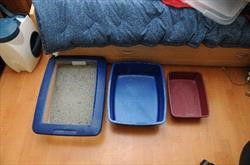
For ‘resistant-to-change’ cats, you will need to use the normal sensation of hunger to help with the transition. For this reason, it is very important to stop free-feeding dry food. This is the first, and very critical, step. You need to establish set mealtimes. They are not going to try anything new if their bowl of junk food is in front of them 24/7.
Cats do not need food available at all times. It really is okay for them to experience a hunger pain! That said, it was very hard for me to listen to my cats begging for food even though I was strong in my conviction that I was heading them in the best direction for optimal health. It truly was a stressful time for me and them. Actually, I think it was harder on me!
This is where many people fail and just give in and fill up the dry food bowl. There were a few times when I had to call my ‘sponsor’ and was instructed to “just leave the house if you can’t take looking into those eyes!” I left the house. Those pitiful little cries of “I have not had food for two WHOLE hours!” were hard to take. But, lo and behold, they were just fine when I returned. Not one cat had died from hunger.
On the other hand, do not attempt to withhold food for long periods of time (greater than 24 hours) with the hope that your cat will choose the new food. You need to ‘convince’ them that a high quality canned food really is good for them, rather than to try starving them into it – which does not work anyway. Allowing a cat to go without food – especially an overweight cat – for a long period of time (greater than 48 hours) can be quite dangerous and may result in hepatic lipidosis (fatty liver disease).
Hepatic lipidosis can also develop when a cat consumes 50 percent or less of his daily caloric requirements over a period of many days. The definition of “many” varies from cat-to-cat. For this reason it is important to understand that you need to have some idea of the calories from canned food combined with the calories from dry food that your cat is consuming on a daily basis while you are implementing the transition to canned food.
I have never seen a cat develop hepatic lipidosis when consuming at least 15 calories per pound per day. This number is figured on lean body weight, not fat weight.
If your cat weighs 18 pounds but really should weigh 12 pounds, please make sure that he is consuming about 180 calories per day. (12 pounds lean body mass X 15 calories/pound/day = about 180 calories/day)
In reality, the cat in the above example would probably be completely safe at only 150 calories per day.
If you have a small female cat that should only weigh 9 pounds, please make sure that she is consuming at least 135 calories per day.
Canned foods never list the calorie content on the can but many dry foods do list this information on the bag. A rough guideline for the calorie content of most canned foods that are 78 percent moisture is about 30 calories per ounce, but can range from 20 to 40 calories/ounce as shown by the chart linked above.
Most cats will lose some weight during the transition to canned food. Given that a very high percentage of cats are overweight to begin with, this is a favorable result of the diet change – as long as they do not lose too much weight too fast. A cat should never lose more than one to two percent of body weight per week.
I highly suggest that all cat caregivers weigh their cats periodically, especially if they are over 10 years of age. This will help ensure a safe transition to a healthier diet and, in general, weight loss is often the first sign of ill health for any reason. I make it a point to weigh my cats at least once each month, especially since they are now over 10 years of age.
Here is a scale that is reasonably priced: Salter Baby and Toddler scale. It weighs to the nearest half ounce and has a ‘hold’ button on it that helps obtain an accurate weight even for a cat that is moving around a bit.
Here is another scale that may be even better because its base is as long as the scale. Red Cross Baby Scale. This is important for cats that are trained to walk onto it otherwise, scales like the Salter one linked above may tip. This would scare the cat and harm the scale.
All of my cats lost weight during the three months that it took to switch them to canned but none of them became too thin. They slimmed down to a nice lean body weight – losing fat while maintaining their muscle mass. They also became much more active.
If your cat is overweight, please see the feline obesity article.
Resign yourself to the fact that you will be very frustrated at times and you will be wasting canned food as they turn up their nose at it. Also, you may want to immediately switch your cat to a dry food that has fewer calories from carbohydrates than most dry foods. Talk to your veterinarian about specific brands.
The low-carb dry foods are very high in fat and therefore are very calorie dense. These foods must be portion-controlled, otherwise your cat may end up gaining weight. Let’s presume that a certain dry food has 612 calories per cup. One quarter of a cup contains 153 calories so be very careful to pay attention to how much of these high calorie dry foods you feed.
The caloric needs of an average cat can range between 150 to 250 calories/day depending on their lean body weight and activity level.
The low-carb dry foods are also very high in phosphorus. This is especially detrimental for cats with compromised kidney function.
And, of course, these low-carb dry foods are water-depleted – just like all dry foods – putting your cat at risk for serious urinary tract problems. They are also cooked at high temperatures in order to dry them out.
I do not recommend these dry foods for long-term feeding for all of the reasons stated above. Please use them only as transition diets.
Be sure to stay away from any “light” varieties since those types of foods are very high in carbohydrates.
Here are some various tricks for the stubborn ones.
Keep in mind that different tricks work on different cats:
- If your cat has been eating dry food on a free-choice basis, take up the food and establish a schedule of 2 to 3 times per day feedings. I really do prefer just twice-daily feedings when trying to transition them. A normal, healthy hunger response after 12 hours goes a long way to convince them to try something new.
- If you want to take the transition very slowly, you can feed the amount that your cat normally consumes in a 24 hour period – split up into two feedings to get him used to meal feeding. Many people, however, are unsure as to how much their free-fed cat really eats so I would start off by figuring out the calories that your cat needs to maintain his weight if he does not need to lose any weight.
- Leave the dry food down for 20 minutes, and then remove any uneaten portion. Repeat in 8 to 12 hours depending on if you are feeding two or three times per day. During the first few days of transitioning to a set schedule, you can offer canned food during the dry food meals, or in-between meals. The stubborn ones, however, will not touch it. Do not despair – all cats will eventually eat canned food if their caregiver is determined, methodical, and patient enough. Once your cat is on a schedule, you will notice that he is more enthusiastic about food during his proper mealtimes and will be much more inclined to try something new.
- Again, most cats only need 150 to 250 calories/day. The dry food bag should tell you how many calories are in a cup of food but if it does not, you can call the company.
- Once the cat has transitioned to canned food, I prefer to either free-feed them (if they are not too fat) or to put out a meal 3 to 4 times per day. Small cats in the wild eat 8 to 10 small meals per day. I do not worry about leaving canned food out for up to 12 hours at a time. Keep in mind that a lion is not going to eat his entire prey immediately.
- Once you have established scheduled mealtimes, you will most likely need to start feeding a bit less at each mealtime in order to get the normal sensation of hunger to work in your favor. Again, we are trying to use the normal sensation of hunger to help us out. We are not trying to starve the cat into the diet change.
- Once your cat is on a schedule of meal-feeding instead of free-feeding, try feeding a meal of canned food only. If he will not eat it – and the very stubborn ones won’t, try not to get frustrated – and do not put down dry food. Try some of the other tips listed below. If he still will not eat the canned food, let him get a bit hungrier. Offer the canned again in a couple of hours, or just leave it out. Some cats will be more apt to try something new if they keep walking by it and seeing/smelling it. Try a different brand/flavor or a different ‘trick.’ Once it has been about 18 hours since he has eaten anything, give him just a small amount (1/4 of a cup) of his dry food – keeping track of his daily caloric intake.
- Remember to be patient.
- Exercising your cat with a tassel toy before feeding can also help stimulate his appetite.
- Cats’ noses are much more sensitive than ours are. They can smell the dry food in the cupboards. I suggest either putting it in the refrigerator or putting it in a tightly sealed container. If they can smell it, they will hold out for it. Some people recommend getting it out of your house completely, but this is not possible when you are dealing with a very stubborn cat that needs a bit of time and patience to make the transition happen.
- The following worked for my cats: Sprinkle a very small amount of tuna – or any other favorite treat (some cats do not like fish and would prefer cooked chicken) – on top of the canned food and then once they are eating this, start pressing it into the top of the new food. (The “light” tuna is better than the fancy white tuna because it has a stronger smell. Or, Trader Joe’s makes a Cat Tuna that is very stinky.) Be careful to decrease the amount of fish as soon as possible. Health problems can occur with a predominantly fish-based diet. Plus, you do not want to create a situation where your cat will only eat very fishy foods.
- Make sure that any refrigerated canned food is warmed up a bit. Cats prefer their food at ‘mouse body temperature.
- Try offering some cooked (or raw – whole meats, rinsed well or partially baked) chicken or meat baby food. One of the goals is to get your cat used to eating food that does not crunch. He needs to get used to a different texture. Also, chicken is a great source of protein to point him in the proper direction toward a high protein, low carbohydrate diet. If he eats the chicken, he may head right into eating canned food. Then again….he may not.
- Try sprinkling some parmesan cheese on the canned food. Most cats love parmesan cheese and this trick has been very successful for me.
- Try a product called FortiFlora, feline version. Most cats LOVE FortiFlora and this has recently become my favorite trick. This is a probiotic made by Purina but you are not going to use it for its probiotic properties. You are just going to use it as a flavor enhancer. The base ingredient in FortiFlora is animal digest – the very substance that makes dry food so very enticing to cats. The directions say to use one package per day – and you can use this much if you want to – but this amount is not usually necessary. You may only need about 1/4 of a package, or much less, with part mixed into the food and part sprinkled on top of the food just as you would use salt and pepper on your own food.
- FortiFlora can be purchased online but an easier product to find is Temptations treats. I trap a lot of feral cats for spaying/neutering purposes and this is one of the best baits that I can use. These tasty treats can be found at most pet stores. Put a few in a plastic baggie and crush them with a hammer. Use the crushed treats as described for the FortiFlora above.
- There are numerous freeze dried meat treats on the market that you can also sprinkle on top of the canned food. Halo’s Liv A Littles is a popular choice.
- Speaking of texture, a common question is “can I just soak the dry food in water?” I hedge more than just a bit at this question. Dry food often has a very high bacterial content. Mold is also often found in dry food. Both organisms flourish in moist environments. There have been many deaths of dogs and cats secondary to eating mold mycotoxins, vomitoxins and aflatoxins that often contaminate the grains found in dry food. If you want to try the trick of wetting down the dry food to alter the texture, please leave it out for only 20-30 minutes then discard it.
- Try dipping some dry food pieces in the juice from the canned food. Some cats may refuse to eat it if the dry food even touches the canned food. But if he will eat it with a bit of canned juice on it, try the ‘chip and dip’ trick. Scoop up a tiny bit of canned food onto the piece of dry food. Put them on a separate plate from his small portion of dry food. Some cats will eat their small portion of dry and then go investigate the dry food with a tiny bit of canned on it.
- Going one step further, try adding a few small pieces of the canned food to the small portion of dry food. Your cat may pick around the canned food but will get used to the smell – and texture – even if he does not eat any pieces of the new food.
- Crush some dry food and sprinkle it on the top of the canned food.
- If you do not think it will upset your cat, try gently rubbing a bit of canned food or juice on the cat’s gums This may get him interested in the taste and texture of the new food – but do it gently. You do not want to make this a stressful situation and create a food aversion. (This trick is commonly used to get just-weaned kittens used to eating canned food.
- If you do not think it will upset your cat, use your finger to put a tiny bit of canned food or juice on his paw for him to lick off. This has not worked for me in the two cats I have tried it on, but it is another idea. Make sure you do it without stressing your cat. Again, you do not want to create a food aversion.
- If you have a multiple cat household, some cats like to eat alone in a less stressful environment, so you may need to take these cats into a separate, quiet room to think about the error of their ways – their carbohydrate/dry food addiction. Once in a quiet setting, away from the other cats, two of my cats would eat canned food/tuna ‘meatballs’ by hand. Not from a bowl, mind you, but only from my hand. I’m not sure who was being trained. They did eventually start eating from a bowl after a few hand feedings.
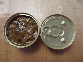
- Try various brands and flavors of canned foods. Try Friskies, 9-Lives, Fancy Feast, etc. Many cats prefer the foods that are all by-products and turn their noses up when offered the by-product-free diets like Wellness, etc. You can worry about feeding a a different canned food later if you want to and you can always mix different types of food together. The initial goal is just to get your cat used to eating canned food and not dry kibble. And remember what I said above. I would much rather see a cat eating a canned food like Friskies, 9-Lives, or Fancy Feast rather than any dry food.
- Syringe-feeding is also another option but has to be done with finesse and patience so as to avoid a food aversion. If you choose to syringe-feed, your goal is not to feed him a full meal. Sometimes just syringing a 1-2 cc’s can ‘jump-start’ your cat into eating the canned food – maybe not the first time but it will at least get him to taste the new food and experience a foreign texture. The best way to syringe-feed is to kneel on the floor with your cat between your legs so he is facing the same way as you are. Then, using a small (1cc/TB) syringe, slip it in the side of his mouth and give about 1/2 cc at a time. He may spit it out but you are just trying to get him used to the taste and texture, not stress him.
- Few canned foods will make it through the tip of a syringe but human meat baby food works well for this trick. You can also water it down a bit if you need to.
- If you want to use canned cat food instead of baby food, you will need to cut the end off of the syringe so that the opening is as big as the barrel. Make sure that the tip is smooth. If you do not want to cut the tip of the syringe off, you will need to puree a pate (versus chunks) type of food. I puree Wellness for this. I run it through the blender with a small amount of water (about 3-4 tablespoons/5.5 ounce can). Then I strain it to remove anything big enough to clog the small tip of the syringe. Wellness is also a balanced diet, unlike human baby food.
- Even though human baby food is not a balanced diet for long-term use, it is a great tool that can be used to help transition a cat to a texture that he is not used to.
- I did have to take drastic measures for a foster cat named Molly. She was dangerously obese (20 lbs – double what she should have weighed) and would not eat canned food even after two weeks of syringe-feeding her. She needed to go in for a dental so while she was under general anesthesia, I put in a feeding tube. This took the stress off of both of us. After two weeks of feeding her via the tube she started licking the canned food from my fingers then suddenly decided it was time to eat it. She then started to finally lose weight. Before the 7-pound weight loss, she could barely walk, could not clean herself, and was quite possibly headed for diabetes.
- Don’t give up. One of my barn cats ate dry food for the first 12 years of her life. She would never touch the canned food that the other cats ate. Then, one day, she found her ‘inner carnivore’ and started eating canned food out of the blue! I was shocked. That was 4 years ago and she has been on a 100 percent canned food diet since she made the switch.
These are just a few tricks that you can try. Different tricks work on different cats. The key is to be patient. Remember, it took me three months to get my cats on 100 percent canned food. Most cats, however, will not take this long.