7341109

Horner’s Syndrome in Cats and Dogs

A syndrome is a collection of symptoms that have significance when they go together. It is important to realize that having a syndrome is not the same as having a diagnosis. A syndrome, however, often has a limited number of causes such that recognizing a specific syndrome brings one substantially closer to a diagnosis.

What is Horner’s Syndrome?

Horner’s syndrome consists of five signs:

  • Constricted pupil
  • Elevated third eyelid
  • Retraction of the eyeball into the head
  • Slight drooping of the eyelid
  • Increased pink color and warmth of the ear and nose on the affected side (very hard to detect in small animals)

These signs appear on the side of the face (and eye) with damaged sympathetic nerves.

What is the Sympathetic Nervous System?

Our bodies have numerous functions that are controlled by our nervous systems, yet we are completely unaware of them. Our heart and respiratory rates, the amount of sweat and other secretions we produce, circulation to different body areas, pupil dilation (enlargement), and constriction (shrinking) are all regulated by our nervous systems automatically and without our knowledge or control. The part of our nervous system dedicated to these automatic systems is called the autonomic nervous system.

Sympathetic vs Parasympathetic

The autonomic nervous system is divided into the sympathetic nervous system and the parasympathetic nervous system. The parasympathetic system maintains a status quo, a normal business-as-usual state; the sympathetic system prepares the body for a fight-or-flight situation.

For example, the sympathetic nervous system kicks in with anxiety or fear leading to increased sweating, pupil dilation, increased heart and respiration rates, and increased blood flow to the muscles. The body is preparing to defend its life either by running or fighting. When danger passes, the parasympathetic nervous system kicks in to return everything to normal.  Both systems coexist to provide balance in a healthy body.

In the eye, the sympathetic nerve fibers dilate the pupil, widen the eyelids, drop the third eyelid, and keep the eye in a forward position in the socket. The parasympathetic nerves constrict the pupil, raise the third eyelid, and retract the eye for protection. Both systems are working at the same time, one system slightly dominating the other depending on what is happening.

When the sympathetic nerves controlling one of the eyes are damaged, only the parasympathetic nerves work and Horner’s syndrome is created.

How can the Sympathetic Damage Occur?

The nerve carrying the tiny nerve fibers that provide sympathetic control to the eye has a long path, and damage may occur anywhere along it. Also, some types of injuries are more likely to occur in certain areas along the path. 

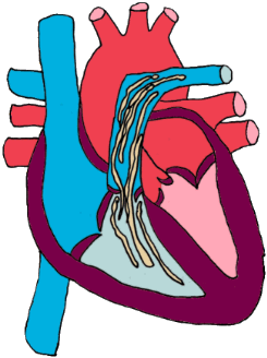
This path begins in the brain’s hypothalamus. A group of nerves exit the hypothalamus and travel down the brainstem, continuing down the spinal cord, down the length of the neck, and just into the chest. This segment is called the central segment and is shown in blue in the illustration to the right. This pathway extends from the brain and brain stem down to the level of the second thoracic vertebra, all within the spinal cord.

From here, the nerves form right and left bundles and exit the spinal cord to make a U-Turn and travel back towards the eyes. The bundles are called the right and left sympathetic trunks (or the “pre-ganglionic segments”) and they extend from the top of the chest back to the area of the middle ear. They are shown in red in the illustration.

From there, the nerves connect to the last segment of nerves (the postganglionic segments – one on the right and one on the left), as shown in yellow. This segment starts just below the ear and travels to the eye.

The damage can occur in the neck or spinal cord area, the ear area, or the eye area. Damage can occur in the form of trauma, tumor involvement, infarction (abnormal blood clot), middle ear infection, or diseases of the eye itself. Each segment of the nerve pathway is vulnerable to different types of damage, so knowing which segment is involved gives us a good idea of what caused the damage.

Locating the Damage

As mentioned, localizing which area of the sympathetic nervous system is affected goes a long way in determining the nature of the damage, as different areas of the system are prone to different types of injury. Eye drops can be used to stimulate different areas of the above pathway and determine which area is damaged. Damage is described as being first order, second order, or third order. Most lesions turn out to be third order.

First Order Lesions (involving the blue segment)

Diseases that hit nerve fibers in the brain, brainstem, or spinal cord include tumors of the brain, vascular accidents (such as stroke) in the nerve tissue, fibrocartilaginous embolism in the spinal cord where disk material sprays into the spinal cord, or even a herniated intervertebral disk in the area of the neck. Horner’s syndrome stemming from any injury such as one of these might prompt a search for other neurologic issues. Advanced imaging such as an MRI might be a good idea.

Second Order Lesions (involving the preganglionic red segment)

Diseases that strike the sympathetic trunk include foreleg injuries especially if the foreleg is pulled and the nerves that exit the spinal cord in the armpit area become over-stretched. Sometimes a mass in the chest, such as a tumor or fungal granuloma, will damage the sympathetic trunk. Neck trauma such as pulling very hard on a leash could be severe enough to cause a second order lesion. If there is no obvious history to suggest injury, it might be a good idea to radiograph the chest to see if there are masses in the lung that might be involved in a second order lesion.

Third Order Lesions (involving the postganglionic yellow segment) 


These are the most common causes of Horner’s syndrome because ear infections are so common for small animals. Inflammation in the middle ear can easily lead to Horner’s syndrome. Third order lesions are associated with vestibular disease, the imbalance and dizziness of the middle ear infection, in many cases. When Horner’s syndrome localizes as third order, the ears should be thoroughly investigated as the source.

Treatment

It is not necessary to treat Horner’s syndrome. The syndrome is not painful and does not interfere with vision. The significance of the syndrome is that it indicates nerve damage that must be recognized. If you wish to treat the syndrome for cosmetic reasons, phenylephrine eye drops can be prescribed to relieve clinical signs. The most important thing is to determine what caused the Horner’s syndrome. Horner’s syndrome itself probably does not need treatment, but its underlying cause very well might.

4128558

Heartworm Treatment for Dogs

It has been said that the treatment of heartworm infection is something of an art. There are several strategies that can be used depending on the dog’s medical condition, including the option of not treating at all. The important concept to realize is that, while there are off label alternative heartworm-killing protocols, the FDA-approved method of killing adult heartworms involves very harsh arsenic-based drugs and killing large adult heartworms is neither simple nor safe. Let’s review some of the dangers and options in clearing the body of this parasite.

Patient Evaluation

Prior to therapy, the heartworm patient is assessed and rated for risk into one of four categories. Important factors include: how many worms are thought to be present based upon the tests performed, the size of the dog, the age of the dog, concurrent health factors, severity of the heart disease, and the degree to which exercise can be restricted in the recovery period. Some hospitals use computerized formulas to categorize heartworm infected patients. The categories into which patients are grouped are as follows:

Class I: Lowest Risk. These dogs are typically without symptoms and their infection is a surprise discovery during a routine annual health examination when a positive test comes up. Other blood tests are normal and radiographs show mild changes if any change at all.

  • Class II: Moderately Affected. Healthy dogs with minimal signs as above, occasional coughing, fatigue only with exercise but with radiographs that show definite evidence of heart disease. Lab testing shows mild anemia, urine dipsticks show some protein present but not severe urinary protein loss.

Class III: Severely Affected. Dog is suffering from weight loss, cough, difficulty breathing, and blatant damage to the vasculature is apparent on radiographs; lab work reveals a more severe anemia and marked urinary protein loss. The damage to the lung blood vessels from the worms creates extra resistance for the heart to pump against, and often, episodes of collapse occur with exercise. 

If the damage is severe enough, the heart can actually fail trying to pump through all the clogged-up blood vessels. Class III dogs are expected to die without treatment but are, unfortunately, sick enough that treatment itself is not without risk.

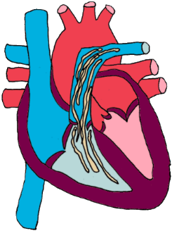
  • Class IV: Caval Syndrome. Dog is collapsing in shock with dark brown urine evident. Heartworms are visible by ultrasound in the AV valve of the right side of the heart, with very abnormal bloodwork. These dogs are dying and can only be saved by the physical removal of adult heartworms via an incision through the jugular vein. If such a dog can be saved from this crisis, further treatment cannot be contemplated until the dog is stable enough to fit into one of the other categories above. In this video (we warn you this is not for the faint of heart), view the physical removal of adult heartworms from the jugular vein of a dog with caval syndrome.

After knowing what class the patient fits in, treatment can be determined. The dog has three groups of heartworms in their body: microfilariae (basically newborn worms whose parents are living inside the dog in question), newly arrived immature worms (relatively fresh from the mosquito bite and living in the skin tissues), and adult worms themselves.  

  • Microfilariae are the first stage larvae (basically the youngest stage). These are the children of the adult worms living in the heart and blood vessels. The mother worm gives live birth (no eggs involved) and the babies swim freely in the host’s bloodstream. They must be killed so that they cannot be picked up by mosquitos and transmitted to new dogs..

The newly arrived heartworm larvae, freshly delivered from mosquito bites in the last 3 months. These are third and fourth stage larvae living in the skin. For practical purposes, we will include the L5 larvae in this group. The L5’s are old enough to leave the skin tissues and enter the circulation but they are not really mature enough to be called adults. This group of young worms (L3’s and L4’s in the skin and L5’s in the circulation) represent the first 5-7 months of infection. We need to kill them before they reach maturity and begin to wreak havoc on the cardiovascular system of the host.

The newly arrived heartworm larvae, freshly delivered from mosquito bites in the last 3 months. These are third and fourth stage larvae living in the skin. For practical purposes, we will include the L5 larvae in this group. The L5’s are old enough to leave the skin tissues and enter the circulation but they are not really mature enough to be called adults. This group of young worms (L3’s and L4’s in the skin and L5’s in the circulation) represent the first 5-7 months of infection. We need to kill them before they reach maturity and begin to wreak havoc on the cardiovascular system of the host.

Stabilization First

Expect strict exercise restriction to be needed after adulticide therapy but moderately affected dogs will need restriction from the first. If the dog has heart failure, this will need to be controlled as well. Some dogs will need anti-inflammatory doses of steroids to control the inflammation that stems from the presence of worms. This is often done concurrently with the second step as described below.

Killing the Microfilaria and Migrating Worms 

The next step in treatment is clearing the migrating immature worms. Heartworm treatment is a project that lasts at least a couple of months. We do not want immature worms to mature in that time frame. Since our goal is to have fewer adult worms when the time comes to kill the adult worms, it behooves us to kill the younger worms right off the bat.

The tissue-based L3 and L4 larvae are killed by monthly macrocyclic lactone-based heartworm preventive products (avermectins and milbemycin). Killing the L3 and L4 larvae prevents heartworm infection. Some of these products can also kill circulating microfilariae (which need to pass through a mosquito before becoming infective to other animals).  The only FDA-approved product for killing microfilariae after adulticide therapy is Advantage Multi®, which contains moxidectin. Other products can be used for this purpose also, but it should be noted that an anaphylactoid reaction can occur if a large number of microfilariae are killed rapidly with products such as high-dose ivermectin (off-label) or high-dose milbemycin (e.g., Interceptor). This problem has not been noted with Advantage Multi. While low-dose ivermectin products (e.g., Heartgard) will gradually eliminate microfilariae in most dogs, the package insert discourages it for this use.

Killing Wolbachia

Wolbachia is a genus of rickettsial organisms (sort of like bacteria, but not exactly) that live inside heartworms of all developmental stages, but their numbers begin to expand once the young worms reach their infectious stage. Wolbachia organisms seem to be protective or beneficial to the heartworms that harbor them, aiding in metabolism and general worm health. Without Wolbachia colonization, many female worms are not able to reproduce, and third-stage larvae are not able to infect new hosts. 

Wolbachia organisms possess inflammatory surface proteins that are released when adult worms die or are killed in treatment, and these proteins greatly increase the chances of severe circulatory reactions in the canine host.

It has been found that killing Wolbachia with an antibiotic such as doxycycline is helpful in treating the heartworm infection as the Wolbachia can be removed from their heartworm hosts, thereby weakening the worm and minimizing the release of inflammatory proteins when the worm finally dies. To minimize reactions, the American Heartworm Society recommends using doxycycline along with heartworm preventives for a good month as soon as the diagnosis is made and prior to the use of the arsenic compounds needed to kill the adults.

Killing the Adult Worms

he only product currently available for treating adult heartworms is melarsomine dihydrochloride (Immiticide® or Diroban®. If you go by the manufacturer’s recommendations, treatment can be done in two or three doses, depending on the class of heartworm infection. Most universities, however, opt to treat all patients with the three-dose protocol as it creates a more gradual kill of the adult worms (which is safer in terms of embolism and shock).

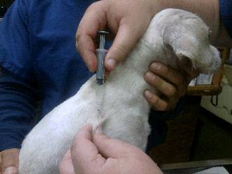
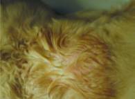
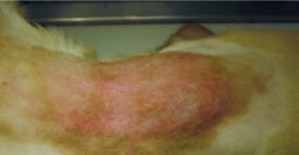
The patient receives an intramuscular injection deep in the lower back muscles. This is a painful injection with a painful substance, and it is common for the patient to be quite sore at home afterward. Pain medication may be needed. Be careful around the injection site, as the pet may bite. The site may form an abscess that requires warm compresses. Approximately 30% of dogs experience some injection site reaction. These generally resolve in 1-4 weeks. 

In the two-dose protocol, the dog returns for a second injection the next day on the opposite side of the lower back. In the three-dose protocol, the dog comes back one month later for two doses 24 hours apart (the first dose is an introductory treatment to kill some of the more sensitive worms.) Keep in mind that too many worms dying at once creates circulatory shock. The benefit of the three-dose protocol (sometimes called the split dose protocol) is that the first injection serves to kill off any older or weaker worms without killing off the stronger, younger ones. When the two consecutive doses occur one month later, there will be fewer worms dying at once.

After treatment, the patient must be strictly confined for one month following the final treatment. No walks, no running around. The dog must live an indoor life. The reason for this is that embolism, to some degree, is inevitable, and it is important to minimize embolism-related problems. Exercise increases heart rate and oxygen demand, and we need the heart to rest during this recovery period.

Watch for:

  • Coughing
  • Fever
  • Nose bleeds

If any of these occur, report them to the vet as soon as possible. The most critical time period is seven to 10 days following a melarsomine treatment, but these signs can occur anytime in the following month.

The “Slow Kill Method” with Ivermectin Only

Melarsomine treatment is expensive and often out of reach for rescue groups, shelters, and many individuals. If the dog is stable (Class I), one option is to simply leave the dog on an ivermectin-based preventive. This option has led to a great deal of misconception about the ability of ivermectin to kill adult heartworms.

Let us lay the rumors to rest now:

  1. Ivermectin does not kill adult heartworms.
  2. Ivermectin does shorten the lifespan of adult heartworms.
  3. Ivermectin does sterilize adult heartworms.
  4. Ivermectin does kill microfilaria (keeping the dog from being a source of contagion)
  5. Ivermectin does kill L3 and L4 larvae (preventing new infections).

This means that if you opt to treat a heartworm-positive dog with only an ivermectin heartworm preventive, you can expect the dog to remain heartworm-positive for as long as two years and the heartworm disease will be progressing during that time. This is not good for the dog but certainly beats getting no treatment of any kind.

The “Slow Kill Method” with Mox-Dox 

Adding doxycycline to ivermectin has changed the way slow kill works. As the “slow kill” protocol evolved, topical moxidectin has been substituted for ivermectin, and a month of twice daily doxycycline is used to kill the Wolbachia bacteria that are so protective of the living heartworms. The result seems to be a much faster slow kill with microfilaria clearing three weeks or so (addressing the resistance in microfilaria issue of slow kill). The adult worms die over 3-4 months, which is similar to the 3-dose protocol (1-3 months on preventive, one dose of melarsemine with two doses following a month after that).

Exercise restriction is still needed, and relatively high doses of moxidectin are also needed. (The topical product is essential; long-acting injectables wane too quickly). Heartworm antigen testing is performed nine months after starting the protocol and continued monthly until negative. If the dog is still heartworm antigen positive after 15 months, new decisions should be made (do mox-dox again, use melarsomine, etc.) The American Heartworm Society views this protocol as a salvage procedure, meaning that it could be considered for a dog for whom melarsomine is not an option. Research and controversy continue with regard to this method.

4128256

Hookworms in Cats and Dogs

Hookworms (Ancylostoma caninum, Ancylostoma braziliense, Uncinaria stenocephala) are one of the classical groups of internal parasites of puppies, the others being roundworms, tapeworms, and coccidia.  Hookworm infection has several features that are of interest to the caretakers of dogs.

  • Hookworms (particularly Ancylostoma caninum) suck blood.
  • Hookworms can be transmitted to unborn pups.
  • Hookworms can infect humans.

Before elaborating on these aspects of hookworm infection, it is important to understand the life cycle of the hookworm, encompassing how infection happens, how the parasite lives, etc.

Life Cycle of the Hookworm

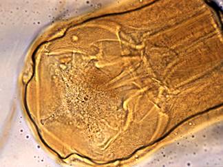
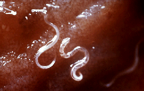
The adult hookworm lives in the small intestine of its host where it hangs on to the intestinal wall using its 6 sharp teeth. This means that, like other parasitic worms, they are bathed in intestinal contents but while other worms share the host’s food by absorbing it directly through their skin, hookworms feed by drinking their host’s blood. The adult worm lives and mates within the host’s intestine and ultimately, the female worm produces eggs. Hookworm eggs are released into the intestinal contents and passed into the world mixed in with the host’s stool.

The egg hatches in the environment and develops from a first-stage larva (the hatchling) to a second-stage larva and finally, a third-stage larva, which is ready to infect a new host.

The larva can infect its new host in several ways. One way is to penetrate the host’s skin directly through the feet or belly or whatever part of the skin is touching the ground. Another way for the larva to gain entry to the new host is to be in soil that is licked and swallowed by the host as it cleans itself.

The pet can be infected by contaminated dirt or by eating an infected animal. This could be a prey animal, such as a rodent, or could be an insect such as a cockroach.

Once the larvae are inside the host, they make their way to the intestine where some worms simply stay and mature into adulthood. Other individuals are bolder, tunneling out of the intestine and migrating to the lung tissue. In the lung, the larvae develop into fourth-stage larvae, and when they are ready they break out of the lung, climb up the trachea (windpipe), get coughed into the throat, and swallowed. Once back in the intestine, these well-traveled worms will complete their maturation to adulthood, rejoining any friends they had that never left the intestine on a migration.

Not all the worms that begin this treacherous migration complete it. As they emerge from one tissue to move on to the next, some fall into a state of arrest where they go dormant and encyst. These larvae remain inactive, periodically emerging and continuing their migration. Only hookworms in the intestinal tract are vulnerable to deworming medications; those in various stages of migration are protected.

Summary: Adult worms live by sucking blood from the intestine. Their eggs are passed by the host into the environment where a new host picks them up. The developing larvae may migrate widely through the new host’s body before settling down to complete their maturation.

It is worth repeating that the host is not always a pet. Other vertebrates, such as rodents and birds, can pick up hookworm larvae from the soil. If the pet eats an infected rodent or bird, the pet will become infected just the same as if the infection came directly from the soil.

Now let’s return to the points we want to emphasize.

Hookworms Suck Blood

Hookworm infection can be looked at as a natural check in the canine population as it is frequently lethal to young puppies. A young puppy is growing, and growth includes making enough new blood to serve not only its current oxygen needs but what is required for growth as well. Growing requires a tremendous red blood cell production from the puppy’s bone marrow, yet in the hookworm-infected puppy, this process is being sabotaged by numerous tiny vampires within. The puppy may effectively bleed to death.

Infected puppies are commonly pale, weak, and have long-standing deficiencies. They may or may not have diarrhea.

Treatment involves deworming with one of several products: mebendazole (Telmintic®), milbemycin (Interceptor®), moxidectin (Advantage Multi®, Proheart6®, Proheart12®), fenbendazole (Panacur®), pyrantel pamoate (Nemex®, Drontal®, or Strongid T®) and others. Some of these products are not absorbed into the host’s body from the GI tract and can only kill worms in the GI tract. These products are typically given every 2-4 weeks to cover worms returning from their migration. We currently do not have a deworming strategy effective against the encysted larvae in other areas of the host’s body.

Simply killing the worms will not be sufficient to save the life of a severely affected puppy. Like any other blood loss, a blood transfusion may be needed to keep the puppy alive until it can replace its own lost red blood cells. An iron supplement is frequently needed as well.

Hookworms are Transmitted to Unborn Pups

Infection of a very young puppy can occur in two ways not addressed above.

Typically an infected mother dog will have encysted larvae all around her body. Throughout the adult dog’s life, some larvae will awaken, break out of their cysts, and complete their migration to the GI tract.

Pregnancy hormones, unfortunately, serve as little wake-up calls to encysted hookworm larvae only this time, the little worms migrate to the unborn puppies and to the mammary gland.

 This means that most puppies will become infected by drinking their mother’s contaminated milk. If this is not enough to infect the entire litter, others will become infected from the soil of their own nest which will quickly become contaminated with the stool of their infected litter mates.

It is clear why puppies are at a special risk over adult dogs when it comes to hookworms. Ask your veterinarian about this method if you are contemplating breeding a female dog. 

Can We Prevent Transmission from the Mother?

The answer is yes but daily deworming is required through the second half of pregnancy and into the nursing period. Regular single deworming will not be effective in protecting the litter. A specific protocol using fenbendazole (Panacur®) has been found to be effective in preventing both roundworm and hookworm infections in unborn puppies.

Female dogs using Proheart6 or Proheart12 (injectable moxidectin) for heartworm prevention are believed to pass fewer hookworm larvae onto their pups.

Hookworms Can Infect Human Beings

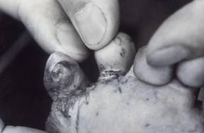
Contaminated soil is an important hookworm source when it comes to a human disease called cutaneous larva migrans. Running barefoot through the park or beach may seem pleasant but if the soil has been contaminated with canine fecal matter, the eager infective larvae may be waiting to penetrate your skin.

Hookworm infection in the skin is intensely itchy but usually treatable. Local restrictions on bringing dogs to beaches and the strict clean-up laws reflect concern for hookworm (and roundworm) infection in people.

Humans can also become infected by eating improperly washed vegetables that may harbor contaminated soil. Humans have been found with actual hookworm intestinal infections. This would be a challenging diagnosis as it is not usually expected but the good news is that it is treated fairly easily when it is discovered.

For additional information, see the Centers for Disease Control and Protection’s (CDC) hookworm fact sheet.  

Decontaminating the Environment

Many people are concerned about how to decontaminate the backyard or property that has housed an infected dog. The good news is that unlike roundworms, which are extremely hardy in the environment, hookworm eggs deplete their energy reserves in a few months and die. Further, hookworm eggs do not survive freezing temperatures. Boric acid can be raked into the soil to kill hookworm eggs, but this will kill grass and vegetation as well.

Preventing Future Infections

The Companion Animal Parasite Council recommends that dogs be dewormed monthly starting at age 8 weeks. In most areas of the U.S., heartworm products are needed monthly to protect against heartworm disease and most (but not all) heartworm preventives will also prevent hookworm infection. Alternatively, tablets or liquids can be given separately. The same products listed above as treatments are also preventives.

Explore products for heartworm prevention that also work against other parasitic worms.

Resistant Hookworms

The influx of retired racing greyhounds into the pet population has brought with it resistant populations of hookworms. It was reported in 2020 that most, if not all, racing greyhounds are coming off the track infected with hookworm infections that do not respond to the usual medications. As these dogs mingle with other canine pets, resistant hookworm populations have spread.

When a hookworm infection seems stubborn, a special test can be performed. This test is called a Fecal Egg Count Reduction Test which compares egg counts in fecal samples before and 10-14 days or so after treatment (with the product that is believed not to have worked). Actually counting eggs before and after treatment gives a better idea of whether the hookworm infection is truly resistant.

A special laboratory may be needed to run this type of testing as it is more commonly done in livestock where resistant parasites have been a long-standing farming issue. The goal of therapy becomes not clearing the infection, but clearing the clinical signs and maintaining as low an egg count as possible. Testing is done using pyrantel, fenbendazole, and milbemycin oxime in the hope of finding an effective product. An egg count reduction of greater than 95% is considered effective. An egg count reduction of less than 75% is considered ineffective and a combination of multiple dewormers will be needed monthly.

Of all the dewormers to which hookworms are generally considered vulnerable, moxidectin is probably the newest, and as long as the dog has not been previously treated with this product, there is a good chance that a combination of dewormers will be effective as long as moxidectin is included. That said, moxidectin-resistant hookworms most certainly exist.

Keep in mind that hookworm larvae encyst throughout an infected dog’s body and “leak” into the intestine periodically as they mature. There is no way to clear these encysted dormant hookworms and they cannot be addressed until they make it back to the intestinal tract. There is no way to distinguish an old infection stemming from migrating worms from an infection with resistant hookworms. Of course, a resistant hookworm infection may lead to the migration of resistant worms into the intestine theoretically for a dog’s entire life.

Feline Hookworms

There are two species of hookworms in cats: Ancylostoma tubaeforme and Ancylostoma braziliense, the former being the most aggressive bloodsucker. The story is pretty much the same for cats as for dogs with a few exceptions:

  1. Kittens cannot be infected before birth nor can they be infected by nursing. Cats are generally infected by larvae invading the skin or by eating an infected prey animal, although infection through the skin from the ground can also occur. 
  2. Both dogs and cats can be infected by eating a vertebrate host, such as a rodent, but it is important not to forget the cockroach. A scuttling bug can be a tempting toy for a cat in particular and if eaten, the cockroach can transmit hookworm larvae it is carrying. A cockroach can also infect dogs.
  3. The Companion Animal Parasite Control Council recommends deworming kittens beginning at age 2 weeks with pyrantel pamoate and continuing deworming every 2 weeks through age 8 weeks. After that, a monthly deworming is recommended (easily combined with a flea product). The indoor-only lifestyle will greatly lessen the need for this level of parasite protection, however.
  4. There are numerous products approved for the treatment of feline hookworm infection: ivermectin, milbemycin oxime, emodepside (the active ingredient in Profender®), selamectin, and moxidectin.
  5. See a chart of heartworm preventives that also protect against hookworms.

Hookworms are significant parasites in both dogs and cats and constitute a human hazard as well. Very young pets are at the highest risk for blood loss so it is important to deworm regularly. If you have further questions or concerns about hookworms, remember your veterinarian is always there to see that you get the answers you need.

6709916

Oral Squamous Cell Carcinoma in Cats

Squamous cell carcinoma is not only the most common oral malignancy in cats, it has one of the poorest outcomes. In most cases, the goal is to minimize infection and pain until the tumor has advanced to a state where comfortable eating and/or breathing is not possible. The tumor is treatable only if detected early in its course.

Prevention and Risk Factors

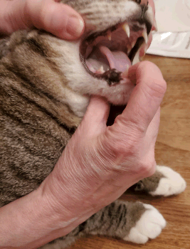
While sun exposure is an important risk factor for squamous cell carcinoma on the ears and nose, the owner’s cigarette smoking habits turn out to be one of the most important risk factors for oral squamous cell carcinoma. The risk goes up four times when the owner smokes 1 to 19 cigarettes daily. Cats who have at one

Photo of cat with open mouth showing a tongue squanoma

time lived with a smoker have an increased risk over cats that have always lived in non-smoking homes.

The use of flea collars was associated with a five-fold increase in the development of oral squamous cell carcinoma, although the study that found this correlation was not able to differentiate between different types of flea collars. Interestingly, regular flea shampoo use was associated with a 90% reduction in risk (perhaps a surface toxin is washed away by frequent bathing).

Regular feeding of canned food, especially tuna fish, was associated with increased risk for the development of oral squamous cell carcinoma. In humans, poor oral hygiene has been associated with an increased risk for oral squamous cell carcinoma, so it is speculated to be the basis of the canned food correlation in cats as well.

The average age at diagnosis is 12.5 years. This tumor usually occurs in older cats. Get comfortable looking in your cat’s mouth.

Where to Look

The squamous cell carcinoma is a destructive tumor. Underlying bone is commonly eaten away. Findings of bone destruction on a radiograph may also be helpful in establishing diagnosis.

Computed tomography (CT or CAT scan) is needed to determine the extent of the tumor. If surgery is considered, knowing the tumor margins is crucial and radiographs simply cannot accurately show how far the tumor extends.

Treatment

Sadly, options are limited for treating oral squamous cell carcinoma; fewer than 10% of cats survive one year from the time of diagnosis. Cats are euthanized when they become too disfigured to eat or breathe. Still, the situation is not without options and some situations are more favorable than others. 

Photo of a cat's tongue being held by two hands

Your primary veterinarian or veterinary oncologist will recommend treatment options specifically for your pet’s condition.

Surgery (Mandibulectomy)

When it comes to oral squamous cell carcinoma, the best-case scenario is a tumor in the front of the lower jaw, because this area can be surgically removed.

If the tumor is located on the lower jaw, it may be possible to remove the part of the lower jaw that contains the tumor. This surgery is called mandibulectomy and is preceded by a CT scan (where available) to determine the margins of the tumor.

  • Median survival times of five to seven months were obtained with this technique, depending on the study. The goal is to remove 1 cm (approx. 1/2 inch) around the tumor in all directions. 
  • After mandibulectomy, a cat can be expected to experience at least one of the following problems: drooling, difficulty grooming, poor appetite, and/or trouble swallowing in the first month after surgery. For most cats (76%), at least one of these problems is permanent, although most owners (83%) felt their cats had good life quality anyway and would choose to do the procedure again in similar circumstances.
  • If a cat survives to the one-year mark, there is a good chance of survival to the two-year mark as well. (The same survival percentages are found at one year and at two years.)
  • The recurrence rate after mandibulectomy is approximately 38%.

If the tumor is located in the upper jaw, surgery is probably not an option (see below).

Mandibulectomy Combined with Radiotherapy


The longest survival times (14 months) have been yielded by a combination of surgery and radiotherapy. The study reporting this rather long survival time only reviewed seven affected cats, a relatively small number upon which to draw generalizations. These cats, however, all required feeding tubes for a median of 15 days and had drooling issues and required regular cleaning (usually daily) on the chest and feet long term.

Radiotherapy with or without Chemotherapy


Radiotherapy can be definitive (intended to cure) or palliative (intended to slow disease and improve life quality but not cure). If surgery can reduce the tumor size to what is called microscopic disease, meaning no tumor is visible to the naked eye, radiation after healing provides the longest survival times. If the tumor is too large to remove to this state, definitive radiation is not going to be possible, but palliative radiation is still an option if it is aggressively done (twice daily treatments for 9-10 days). In this situation, median survivals of 5-6 months were obtained. Similarly, another study showed promising results: a group of 11 cats with oral squamous cell carcinoma treated with a combination of radiation and the chemotherapy agent mitoxantrone yielded complete remission in 8 cats with a median duration of 170 days. It should be noted radiotherapy is generally a very expensive treatment and, when the mouth is involved, can create enough inflammation that a feeding tube is necessary to support the patient.

Chemotherapy alone has shown such poor efficacy that it is not worth the attempt for this tumor.

Non-steroidal anti-inflammatory Drugs (NSAIDs)

It has recently been found that certain tumors, particularly carcinomas, have receptors to bind the enzyme cyclooxygenase (lovingly known as COX). This enzyme is involved in the production of biochemicals called prostaglandins that are involved in the generation of inflammation. Cyclooxygenase inhibitors such as piroxicam and even meloxicam, two drugs more commonly used against arthritis pain, may have anti-cancer properties beyond simply controlling inflammation and pain associated with tumors. Often, these drugs are prescribed for cats with oral squamous cell carcinoma but be aware that cats can be sensitive to their side effects. Often, treatment of the tumor justifies this risk but still, you should be aware of potential complications and discuss the timing of monitoring blood tests with your veterinarian.

The bottom line is that the oral squamous cell carcinoma is a very bad tumor for a cat, and as yet, research has not yielded a reliably good therapy. Because the tumor does not spread until late in its course, removing the tumor when it is small is the best hope for a worthwhile survival time. The lower jaw tumors are best suited to this kind of therapy; other forms, such as tumors under the tongue or in the upper jaw, are nearly impossible to address meaningfully, though aggressive radiotherapy is emerging as an option. It is important to keep a realistic attitude about what to expect.

Information in this article was largely obtained from the following review article: Marretta, J.J., Garrett, L.D., Marretta, S.M. Feline oral squamous cell carcinoma: an overview. Veterinary Medicine, June 2007; p392-406.


Other Types of Oral Squamous Cell Carcinoma

As mentioned, it is difficult to achieve meaningful treatment for a squamous cell carcinoma in the upper jaw and less than 10% of patients are still alive one year after diagnosis. In fact, most cats have stopped eating and require euthanasia within one month or so. Even with palliative radiotherapy only 1 to 4 months of survival are reported. That said, a new therapy is emerging in the form of aggressive radiotherapy. This protocol involves twice daily radiation of the tumor area for 7 days. With this protocol, after therapy 56% of feline patients had no discernible tumor for an average of 13 months. A feeding tube is placed during the time of therapy and kept throughout the recovery time until the cat is eating well and the radiation site is healed. This kind of therapy is relatively expensive but for a 50:50 shot at a long remission it may well be worthwhile.

4128692

Hard to Regulate Diabetic Dogs

The goal in long-term management of diabetes mellitus is the alleviation of unpleasant clinical signs (constant thirst, weight loss, etc.) and the prevention of dangerous secondary conditions (infections, ketoacidosis, etc.). To accomplish this, blood sugar levels should be regulated between 250 mg/dl (13.88 mmol/L) and 80 mg/dl (4.44 mmol/L). This is not as good as the body’s natural regulation, but it is a fair and achievable goal in most cases.

Some patients just seem completely unaffected by even high doses of insulin, and it is important to have a step-by-step plan to rule out causes of insulin resistance so that regulation can be achieved.

Insulin resistance is defined as:

Persistent high blood sugar levels throughout the day when three units of insulin per four pounds of patient body weight are used.

or

A unit or more of insulin per pound of patient body weight (or more) is needed to achieve regulation.

Typically, glucose curves show levels that are consistently too high with virtually no drop in sugar level when insulin activity is peaking. So what is going on here? The possibilities may seem surprising.

Step One: Rule out Owner-Related Factors

Owner error is actually the most common cause of what is thought to be insulin resistance.

This may seem basic, but it is important not to skip the basics. Confirm that the amount of insulin being drawn into the insulin syringe is correct, that the injection technique is correct, and that the patient is actually receiving the injection. Rule out any snacking or changes in the patient’s feeding schedule. Be sure the bottle of insulin is not expired and that it has been properly stored. Ideally, the veterinary staff will observe insulin administration and will check the bottle while the pet is being checked in the hospital.

Review insulin storage and handling.

Step Two: Determine for sure that the Patient is Insulin Resistant

A glucose curve is needed to distinguish the following three phenomena.

Somogyi Overswing

In the case of Somogyi overswing, the insulin dose is too high and drives the blood sugar low for part of the day. When the blood sugar is low, other hormones such as cortisone or adrenalin are released to raise blood sugar. These hormones can have a prolonged effect (many hours), thus creating hyperglycemia (high blood sugar). If the patient’s high blood sugar has been caused by a Somogyi overswing, a lower dose should be used and a new curve performed in a week or two. One might think a flat glucose curve (i.e. one not showing hypoglycemia followed by hyperglycemia) would rule out Somogyi overswing but, in fact, a Somogyi hyperglycemia can persist for days and create a flat curve. Often a good approach is to cut the insulin dose back and see what the curve looks like.

Rapid Insulin Metabolism

In this case, the insulin simply is not lasting long enough to create sustained normal blood sugar levels. If the curve shows that the insulin effect is wearing off too soon, twice a day administration of the insulin may solve this problem or a longer-acting insulin may be needed. Longer acting insulins tend to have poorer absorption into the body from an injection site. This may also necessitate change to a different insulin type. Often it is necessary to change to a human insulin from a canine insulin.

True Insulin Resistance


Here no significant drop in blood sugar level (levels stay greater than 300 mg/dl[16.65 mmol/L]) is seen in response to the insulin dose used. Usually there is a history of prior increases in insulin dose, all met with minimal response.

Step Three: Spay

Hormone fluctuations will easily account for insulin resistance as progesterone is one of many hormones that raise blood sugar levels (by stimulating growth hormone secretion). Canine unspayed female diabetics need to be spayed if regulation is to be achieved. (Feline patients do not have a similar relationship with naturally occurring progesterones and do not require spaying; however, progesterone-type medications can produce diabetes in cats. Diabetic patients of either species should not take progesterone-related medications.)

Step Four: Rule out Infection

Diabetic animals are at risk for developing bladder infections because they have so much sugar in their urine. Stress of any kind will contribute to high blood sugar and infection would lead to stress. A urine culture should be done to rule out bladder infection, plus the teeth and skin should be inspected for infection in these areas. If infection per se is not found, the patient should be screened for other chronic illnesses that might constitute a stress. A basic blood panel would be a logical starting point.

If infection or other stress is allowed to go unchecked, ketoacidosis, an especially life-threatening complication of diabetes mellitus, can develop.

To avoid infection issues, it is a good idea to plan for diabetic patients to have their teeth cleaned annually and to have the urine periodically cultured whether there are symptoms of a problem or not.

Step Five: Control Obesity

Insulin response is typically blunted in obese patients. If obesity is an issue, it should be addressed. A formal weight loss program using measured amounts of a prescription diet and regular weigh-ins is necessary for success. See general information about weight loss for pets. If these steps do not reveal a relatively simple explanation for the poor insulin response, then it is time to seek more complicated causes. This generally means an additional hormone imbalance.

Hyperadrenocorticism (Cushing’s Disease)

This condition is relatively common in dogs but less so in cats. This condition involves an excess in cortisone-type hormones, either from over-production within the body or over-treatment with medication. Cortisone – more accurately referred to as cortisol – is secreted naturally in response to a fight or flight situation and prepares the body for exercise by mobilizing sugar stores. If sugar is mobilized into the blood stream in the absence of a fight or flight situation, diabetes mellitus can result. If the excess cortisone situation is resolved, it is possible that the diabetes will also resolve.

About 10% of dogs with Cushing’s disease are also diabetic. About 80% of cats with Cushing’s disease are also diabetic. Testing for Cushing’s disease cannot proceed until some degree of diabetic control has been achieved and the patient is not ketoacidotic.

Excess Circulating Lipids

High levels of circulating triglycerides (fats) interfere with binding insulin to the cells it needs to act upon. While most dogs with excess circulating lipids are not diabetic, when a diabetic patient is insulin resistant it may be helpful to attempt to reduce the circulating fats. Excess lipids in the blood usually occur because of some other hormone imbalance (Cushing’s disease, hypothyroidism in dogs etc.) but they can simply happen spontaneously in the miniature schnauzer.

Circulating fats are reduced by treating the hormone imbalance that caused them if there is one. If there is no underlying disease, circulating lipids can be reduced with diet and omega 3 fatty acid supplementation.

Acromegaly

Acromegaly results from an over-secretion of the pituitary hormone known as growth hormone. This hormone normally is responsible for one’s growth from infancy to adulthood. When adulthood is achieved, its secretion dramatically slows, bone growth plates close, and growth essentially stops. If for some reason, this hormone begins secreting again, growth resumes but not generally in normal proportions as the limb bones have closed their growth areas.

One of the effects of growth hormone is causing the body tissues to become resistant to insulin by interfering with tissue insulin receptors. Animals with acromegaly are frequently diabetic.

The prognosis and treatment for acromegaly is different between dogs and cats. Dogs generally develop acromegaly due to excess progesterone secretion, as would occur from an ovarian cyst. Canine patients are thus usually older, unspayed females and spaying may be curative depending on the remaining ability of the pancreas to secrete insulin.

Antibodies against Insulin

When a patient is treated with insulin from another species, the immune system recognizes the introduction of the foreign protein and generates antibodies. It was because of this phenomenon that most commercial insulin available is genetically engineered human insulin so that the world’s human diabetics no longer need to worry about making insulin antibodies.

But where does this leave dogs and cats? It leaves them making antibodies against human insulin, that’s where. There is no feline insulin product available; the closest thing is beef insulin. Dogs are luckier as their insulin is identical to pig insulin and porcine insulin is available as Vetsulin® (Caninsulin® in Canada). If a human insulin is used, some degree of antibody production is a forgone conclusion.

You would think this would pose a big problem but in fact insulin antibodies are not always bad. Most of the time the antibodies simply interfere with removal of the insulin, leading to a longer-acting insulin than would be achieved with the same type of insulin made from the native species. For example, Humulin N® (human insulin) will last longer in the dog than Canine Insulin N (if there were such a product). This may be desirable depending on the patient; changing the species of origin of the insulin is one way to get the insulin to last a bit longer.

To become insulin resistant from antibodies, a body must lose 70% or more of the insulin injection to antibody binding. This is very unusual but possible and should not be forgotten as a possible cause of insulin resistance. Blood tests to measure insulin antibodies are available in some areas. Insulin can be switched to a species of origin more closely related to the species desired.

If a Cause Cannot be Found

If a cause cannot be found or if treatment for that cause is not practical or possible, the good news is that multiple high doses of insulin can generally overcome the resistance. Sometimes combinations of short and long-acting insulins are used together to achieve reasonable regulation.

info-box-img-6

Ovarian Remnant Syndrome in Dogs and Cats

Whether the patient is a dog or a cat, ovarian remnant syndrome is suspected when a spayed female pet appears to be coming into heat, something that should be impossible after spaying. A spayed female has had both her ovaries removed and should not cycle and should not display behaviors typical of heat. If she is displaying signs of an active heat cycle, a search for an estrogen source is required. In particular, we need to rule out the presence of a piece of ovarian tissue left inside. If a piece of ovary (or similarly active tissue) is indeed inside, the only treatment is surgically removing it, and since this is an invasive solution, we want to be as certain as possible that there is no other likely explanation.

The Girl Dog in Heat

A female dog comes into heat every 6 to 8 months on average. During this time, the most prominent feature is the vaginal discharge which begins bloody and switches to a more golden straw color. The girl dog in heat will attract males and will most likely flirt playfully with them. This period of hormonal activity lasts about 3 weeks and culminates in a false pregnancy (or potentially a real one if she has been bred). The false pregnancy period lasts roughly a couple of months.

Drawing of a dog

The Girl Cat in Heat

A female cat in heat does not have an apparent vaginal discharge. For her, the signs of heat are more behavioral. She becomes restless and loudly vocal. She may urinate in the house in an attempt to notify local tomcats of her state. She is often unusually affectionate and has a tendency to stick her rear in the air and even tread her feet when she is petted over her back.

A female cat cycles seasonally, when the days are longer, and tends to stop cycling when the days begin to get shorter. She is in heat for five days or so and then out of heat for a couple of weeks, then back in, then back out over and over until she is either bred or the seasons change. If she is bred, she will ovulate and go into a false pregnancy or a real one if the breeding was successful. In any case, it is the behavior that is the clue that her hormones are active, especially since the occasional female cat will not follow the usual rules about the seasons.

Signs of heat (also referred to as estrus) imply estrogen in the pet’s system. The first step is to confirm that there genuinely are estrogens in her system.

Testing for Hormones

Vaginal Smear

You might expect a blood test to be necessary to look for estrogen or progesterone, but in fact, the best test is a vaginal smear test, at least for dogs. Using a cotton swab, the veterinarian harvests some cells from the vaginal wall and looks at them under the microscope. If the female is in heat at the time of the test, the cells will have a characteristic appearance called cornification. This means there are estrogens in her body.

Blood Testing

Testing for estradiol, one of the estrogen hormones, can be done but is not as accurate as the vaginal smear test. If the spayed female has high estrogen levels during the time she appears to be in heat, this would be considered a positive test. The problem is that low levels can still be present in the bloodstream even if there is an ovarian remnant, so low levels are hard to interpret.

Another approach is to give her a shot of GnRH (gonadotropin releasing hormone) while she appears NOT to be in heat. If she responds to the injection by producing progesterone two weeks later (detectable with a blood test), this would be consistent with an ovarian remnant.

The vaginal smear costs a fraction of the amount of blood testing and can generally be done in a matter of minutes in the doctor’s office, so these types of blood tests are rarely performed.

Witness LH and SpayChek® Testing

There is an in-house test kit for luteinizing hormone (LH) that can determine if a female cat or dog has been spayed or not, provided she is not experiencing a heat at the time of the test. This test could be performed to assist in diagnosis but would have to be done during a time without symptoms of heat. A low LH level is compatible with the presence of estrogen and presumably ovarian tissue.

The newer SpayChek® test for anti-mullerian hormone is reportedly more reliable but requires that at least two to three months have passed since the original spay surgery for accuracy. It is best performed in conjunction with a progesterone level for the best accuracy.

Your veterinarian may suggest more than one type of test to be sure there is little room for ambiguity. After it is clear that estrogens are definitely present, the next question to address is, “Where did they come from?”

Where Did they Come from?

Estrogen Creams for Human Use

Topical estrogen-containing creams have numerous human uses. These creams invariably end up on the user’s hands or arms, where a loving pet can lick them. A female pet exposed to a hormone crème in this way can easily manifest signs of heat but will not show a hormone cycle in any predictable way. Generally, people are aware that hormone crèmes are in use at home and can be more careful about avoiding exposure to pets. Creams may be applied using gloves that can be discarded away from the pet, hands can be washed carefully, etc.

Did Something go Wrong with the Original Surgery?

One would think that if there is a remnant of ovarian tissue in the spayed pet’s abdomen, the surgeon must have left it behind. This is not necessarily so. It appears that some females possess accessory ovarian tissue separate from the main ovary, and this tissue becomes active only after the main ovaries are removed. Some cats actually grow invisible ovarian tissue down the length of their ovarian ligament, which is cut during the spay surgery but not fully removed. Furthermore, if an ovary contacts the abdominal wall, cells appear able to adhere and vascularize effectively, creating a new, albeit small, ovary. In many cases, the

Gloved hands removing feline ovary

original spay surgery was performed months or even years before. These secondary bits of ovary are growing in this time and do not show themselves until they have achieved sufficient hormone-producing power. Once it has been determined that a pet has an ovarian remnant, surgery should be performed to locate and remove it.

It is worth mentioning that adrenal tumors can produce hormones. These can be difficult, if not impossible, to remove, but exploratory surgery should at least identify them over an ovarian remnant. In the situation of this type of tumor, signs of heat are constant; they do not cycle as with ovarian tissue.

Treatment

The ovarian tissue will be most prominent or visible just following the active heat period. This is the time after ovulation when the ovary is normally ripe with large progesterone-secreting structures called corpora lutea, and this is the best time for surgical exploration. If errant ovarian tissue is found, it can be removed generally without difficulty. It is not unheard of, however, for the ovarian remnant or remnants to be impossible to find. If that is the case with your pet, Discuss with your veterinarian whether referral to a surgical specialist would be best for you and your pet; however, a surgery specialist may be necessary to find the remnants. Biopsy of the removed tissue should settle the question as to whether the hormone source was removed intact or not.

Why Do We Need to do Anything at all?

The problem with all this is not limited to the inconvenience of hormone-related behaviors and discharges. The actual problem is that hormones can cause harm. Long-term exposure to female hormones can promote mammary cancer. If a small piece of uterus persists after spaying, the influence of female hormones can promote chronic infection (“stump pyometra”). While neither of these outcomes is definite, their repercussions are serious, and it is best to take these hormones out of play.

What if it isn’t Heat?

Female dogs can have a bloody vaginal discharge for reasons other than heat. It is possible the owner is seeing bloody urine, for example, and the medical workup will need to shift to address the different causes of this problem. Alternatively, a transmissible venereal tumor, urinary tract infection, or vaginal injury might lead to bleeding.

As for a female cat, she may simply have behavioral issues to address.


7963100

Hypertrophic Osteodystrophy (HOD) in Dogs

Hypertrophic osteodystrophy is a developmental, auto-inflammatory disease of the bones that is usually first seen between 7 weeks and 8 months of age.  In general, most cases will appear for the first time when the puppy is between 3 – 5 months of age. Relapses may occur until about 20 months of age.

HOD is most often seen in fast-growing puppies of large and giant breeds.

Some medium-sized breeds, such as the Australian cattle dog and pit bull may also develop HOD. The Weimaraner, in particular, appears to be predisposed to HOD.

Predisposed breeds include:

  • Australian kelpie
  • Boxer
  • Bullmastiff
  • Doberman pinscher
  • German shepherd dog
  • Giant breed dogs
  • Great Dane
  • Irish setter
  • Irish wolfhound
  • Kuvasz
  • Labrador retriever
  • Large breed dogs
  • Rottweiler
  • Saint Bernard
  • Standard poodle
  • Weimaraner

In HOD, blood flow decreases to a part of the bone next to the joint. This interrupts bone formation. The interruption means that bones don’t harden appropriately, nor do they grow as strong as those of a healthy puppy.

HOD can be very painful. Sometimes HOD is straightforward and responds quickly to treatment, but it can also be tricky to diagnose and treat.

HOD is somewhat similar to panosteitis (pano) in that it affects the growing leg bones of large- or giant-breed puppies. However, pano usually affects only one leg at a time, and is thought of as “growing pains.” HOD can affect more than one leg at the same time and is more painful than panosteitis. In addition, unlike pano, HOD can permanently damage the growth plates.

HOD doesn’t appear to be inherited, and at this time no one knows how puppies get it, although auto-immune diseases and dietary excesses can trigger it. However, because it is more common in some breeds if the puppy is being considered for breeding purposes, your veterinarian may recommend a thorough review of the pup’s relatives, to see how many of them have had HOD. If the puppy comes from a bloodline that has few cases, or the cases were mild and self-limiting, that may be better for breeding purposes than bloodlines that have had many cases, or have severe cases. (Every pup, of every breed, being purchased for breeding purposes should always have an extensive review of the relatives, to avoid known breed-related issues.)

Clinical Signs

Clinical signs depend on how mild or severe the HOD is. For mild cases, the puppy usually has a slight limp and appears to have pain in the affected bone. Puppies with more severe cases may have a decreased appetite and subsequent weight loss, fever, and depression. Unfortunately, these non-specific signs can happen with many diseases.

More specific signs of HOD include swollen, warm, and painful leg bones. The puppy may refuse to bear weight. If multiple limbs are affected, the puppy may prefer to remain lying down, be reluctant to get up, and be unwilling to walk.

And not just the legs may be involved. Some cases have swollen muzzles, excessive salivation, and pain when the dog tries to open his mouth; these cases may involve the jaw. Also, radiographs may show changes in the jaw, spine, ribs, shoulder, and eye socket bones.

Systemic signs can include:

  1. Fever ranging up to 106⁰F (a dog’s normal body temperature is 101-102.5⁰F)
  2. Lack of appetite
  3. Discharge from the eyes or nose
  4. Bumps on the skin, some with pus
  5. Diarrhea that can be bloody
  6. Inflamed vulva or vagina
  7. Increased respiratory sounds

At the very least, HOD dogs will have a fever, lameness, and typical HOD lesions showing on the radiographs. The more signs the puppy has, the more severe the case. (In severe cases, with multiple systems and organs affected, more aggressive treatment to suppress the immune system will be necessary.)

A complicating factor to remember is that, for the most part, HOD patients are very young puppies that do not have bodily reserves and can decline rapidly with fever and lack of appetite.

HOD can be a painful condition and typical treatment to control even significant pain may not work.

HOD generally comes in episodes that last a few weeks. Often the first episode lasts a week and it can be followed by a full and spontaneous remission.

Diagnosis

Diagnosis is made via physical exam (seeing obviously swollen soft tissue over the leg bones), radiographs, blood tests, etc.

Treatment

Your veterinarian will want to control the fever, lethargy, and bone pain. Nonsteroidal anti-inflammatory drugs (NSAIDs) are often used for this. However, if the puppy develops gastrointestinal signs, the NSAIDs may have to be discontinued.

Some puppies may need immunosuppressive doses of corticosteroids, usually prednisone. Some dogs may need to take them for a year, in which case the drug must be tapered off slowly, rather than simply stopped. Your veterinarian will recommend tapering only once the dog is free of pain and walks normally.

In some cases, narcotics may be necessary to control severe pain. These may have to be given by intravenous injection.

Some breed-specific studies have shown better responses to corticosteroids than to NSAIDs. In one study of 53 Weimaraners, 50 percent did not respond to NSAIDs. In another study of six related Weimaraners, NSAIDs had little effect and the dogs had to be switched to corticosteroids. A similar experience has been reported in affected Irish setters and Australian kelpies.

Your veterinarian may need to prescribe antibiotics for pneumonia or to prevent secondary infections resulting from immune systems that have been depressed by the corticosteroids. Antacids, such as famotidine, may be given while the dog is taking the corticosteroids. Some veterinarians may recommend probiotics when the GI tract is involved. Pain medications may be needed.

Other care may include fluid therapy, analgesics (e.g. tramadol, opiates), and rest or restricted activity. Even when the puppy starts feeling better, the veterinarian may still want exercise to be restricted until the bones have structurally recovered.

Vaccines should be avoided during an active HOD episode.

Monitoring and Prognosis

Mild cases of HOD may resolve on their own or with only supportive care. Watch for a possible return of any clinical signs once the dog has stopped/tapered the NSAIDs or prednisone because recurrences are common until the leg bone has finished growing. Dogs with affected litter mates are more likely to relapse. Complete recovery is expected once the leg is finished growing, but relapses (episodes of fever and malaise) have been reported in adult dogs. Most relapses in adult dogs respond to NSAIDs or corticosteroids.

Permanent limb deformities are rare. However, if they occur, surgery may help. Your veterinarian will use radiographs to determine if surgery is indicated.

Prevention

While it’s difficult to prevent a disease when you don’t know what causes it, some common sense can boost the chances that a puppy won’t get it. Large- or giant-breed puppies should not be given any mineral or vitamin supplements that aim to increase their growth rate and adult size. Slow and consistent growth is ideal for the puppy’s health because rapid growth can cause skeletal abnormalities. Puppies need a completely balanced diet. Puppies should be discouraged from doing jumping exercises until their growth plates have closed, which is at about a year of age. (Check with your veterinarian for an appropriate estimate of growth plate closure based on your puppy’s breed and growth.) Avoid housing puppies on hard surfaces, such as concrete. If floors are not carpeted, rugs may help so the puppies don’t slip as much.  

Prognosis

Most cases of HOD are resolved with or without medical care. Unfortunately, sometimes HOD is so severe, painful, and uncontrolled by treatment that owners are forced to discuss the pet’s quality of life issues, resulting in euthanasia.

4128244

Hot Spots (Pyotraumatic Dermatitis) in Dogs and Cats

What are Hot Spots?

When the weather gets warm, we start seeing more dogs with hot spots. These wounds are weepy, wet, red, and sometimes bloody when they are fresh, and they are dry and scabby when they are resolving. They can cover large expanses of the pet’s skin or they can appear as smaller solitary lesions (sometimes in multiple locations). Hot spots generally have very clear margins defining them and separating them from normal skin.

They are caused by over-zealous self-licking, chewing, scratching, and rubbing (the pet creates them him/herself), and they can arise especially quickly (10 minutes of chewing can create a big one). The good news is that they almost always look worse than they are. The infection is quite superficial and often will resolve with topical treatment alone. The bad news is that about 30 percent of the pets who come to my hospital for hot spots actually have other kinds of skin diseases, such as deeper skin infections, bite wounds or other trauma, or even immune-mediated ulceration. If you aren’t entirely sure about what a hot spot looks like, it is probably best to have the veterinarian look at it.

The dog causes the hot spot by self-licking, chewing, scratching, and rubbing. What causes the pet to self-chew and lick is another story.

The pet may have an allergy, may have come in contact with an irritating substance, may have irritation from a grooming clipper, or may have some pain in the area from the underlying tissues. In many cases, the pet simply has fleas and is allergic to their bites. Anything that makes the dog itch will make the dog lick and chew, and if the licking and chewing is obsessive enough, a hot spot will soon follow.

First Aid

Treating a hot spot may or may not be a do-it-yourself project. Smaller hot spots can be treated at home with topical products made for this use. The important thing is to be aware that these areas are tender and so the pet may bite if you use something on the area that stings. Also, be careful about using human topical products as these may be toxic to pets when licked. Zinc oxide, for example, can be toxic when licked and is common in many human skin ointments.

Initial treatment usually involves removing the surrounding hair so that the hot spot can be disinfected. Once the superficial infection is properly cleansed, topical products can be used to relieve the associated inflammation. The lesion dries and scabs while it is healing. If the pet is really itchy or there are multiple hot spots, pills or injectable medication become necessary. Hot spots just under the ear/on the facial cheek, for example, are notorious for covering up a deeper skin infection below and often require more extensive treatment, especially in Golden Retrievers. More extensive treatments might include oral corticosteroids and/or oral antibiotics in addition to topical antiseptics and anti-inflammatories.

Good flea control is important for any itchy pet and is the foundation of itch prevention for most dogs and cats. Always be sure you have flea control secured in the approach to managing itchy skin in pets.

6721539

Hydrometra, Mucometra, and Pyometra in Dogs and Cats

Uterine disease in dogs and cats is often influenced by the hormone progesterone, which prepares a female for pregnancy and also helps the mother maintain a pregnancy. However, sometimes things don’t go as planned. One such event is when the uterus, or one of its horns, becomes filled one of several different types of fluid, and the effects can be relatively simple or substantial enough for an emergency hysterectomy to prevent death.

After a pet is no longer in heat, high levels of the hormone progesterone remain for nearly two months, and that high level of progesterone causes the uterine lining to thicken so as to be ready for pregnancy. Progesterone also suppresses the uterus’s immune function. If there is no pregnancy after several cycles, the uterine lining keeps getting thicker until eventually some cysts form inside the lining, a syndrome called cystic endometrial hyperplasia. Those cysts in the thickened lining leak fluid into the uterus.

The first stage of that fluid buildup is hydrometra. Unfortunately, two of the three stages do not have any significant clinical signs, so the pet owner is unaware that there is a medical problem.

A fluid-filled uterus has three stages called hydrometra, mucometra, or pyometra:

  • Hydrometra – watery fluid
  • Mucometra – fluid with mucous
  • Pyometra – pus (white blood cells and bacteria)

In hydrometra and mucometra, the fluid is sterile and there is no infection. Only pyometra is an infection, which occurs when bacteria crawl up the vagina through the cervix and into the uterus, causing a risk of developing endotoxemia (toxins in the bloodstream) and sepsis (in which the body’s response to an infection damages its own tissues) that are generally introduced by uterine disease. In pyometra, the uterus sometimes ruptures, which releases large amounts of pus and dead tissue into the abdomen.

Discovering hydrometra and mucometra is important: the conditions lead to decreased fertility and likely increase the risk of developing more severe uterine disease.

These conditions are more common in countries or areas where it is not typical practice to spay or neuter healthy dogs. They are not uncommon in sexually intact animals, especially in older females.

Radiography (X-rays) may detect uterine enlargement, but an ultrasound can tell if there is fluid in the uterus and may also indicate the type of fluid as well as rule out pregnancy. A dog with pyometra may be  quite sick, have a bloated belly, vaginal discharge, a poor appetite, and may be vomiting or drinking far too much water. Blood tests can detect infection and other underlying organ problems.

Treatment

Medical management sometimes involves using a drug called dinoprost (Lutalyse) to clean out the uterine contents; it is used to induce labor and the same contractions as occur in labor help eject the excess fluid. Sometimes vaginal infusion of warm saline can help empty the uterus.

While any stage of the first two conditions can be treated medically, they will recur. They will not resolve on their own and are a risk. Spaying is the only permanent fix.

Pyometra generally occurs in middle-aged to older female dogs in the six weeks following heat. The uterus fills with pus, bacteria, dying tissue, and toxins; at some point the uterus dies, releasing all of that dangerous material into the abdomen. It is a life-threatening emergency. A dog or cat with pyometra must be surgically spayed immediately (after stabilization) or she will die.

Oddly enough, the easiest one to fix is pyometra: treatment is a single surgery, despite being a bit more complex than a regular spay, and some antibiotics. Given that it’s otherwise fatal, it’s a good thing that the pet will have signs so that you know something is going on. Hydrometra and mucometra can be slow to respond to the dinoprost that lowers progesterone – sometimes taking a few months – and thus take much longer to treat than a surgical fix.

Pyometra is one of the main reasons for preventive spays. Removing the uterus is the only way to prevent hydrometra, mucometra, and pyometra.

6039086

Hepatitis in Dogs

What is Hepatitis?

Hepatitis is inflammation of the liver. It is a syndrome – a group of clinical signs or symptoms – rather than a specific disease, and it has many causes. Your dog can have immediate inflammation in their liver called acute hepatitis or long-term inflammation called chronic hepatitis. Chronic hepatitis (CH), also called chronic active hepatitis, can lead to scar tissue formation and cirrhosis in the liver. Cirrhosis is extensive, end-stage scar tissue. Some dogs with acute hepatitis progress to the chronic form.

Infectious Canine Hepatitis

One disease that causes chronic hepatitis is infectious canine hepatitis (ICH). ICH is caused by a virus but not the same as the Hepatitis A, B or C viruses that cause disease in humans; you and your family won’t get sick if your dog is infected. Similarly, The hepatitis viruses that cause human hepatitis do not affect dogs. ICH is spread through animal feces, urine, saliva and objects. Since most dogs are vaccinated as puppies against ICH, it is considered a rare infection. If you have a young puppy or an unvaccinated dog in your household, they can get ill with this virus. However, outbreaks sometimes occur, mostly in dogs less than 1-year-old or unvaccinated dogs. An infected dog may show signs of depression, fever, vomiting, diarrhea, and abdominal pain. Treatment involves supportive care such as fluids and medications to boost liver function; the latter may include SAMe, milk thistle, ursodiol, and vitamin E.

Causes 

Aside from ICH, there are many other causes of hepatitis. Causes of acute hepatitis include toxins, drugs, viruses, bacteria (particularly leptospirosis), and fungi. On the other hand, chronic hepatitis is caused by bacteria, viruses, excess copper in the liver, drugs, and immune system issues. If a cause cannot be determined, this is referred to as idiopathic hepatitis and is the most common cause. Idiopathic means no cause can be found.

Age and Breed Predispositions

Any dog can have an acute episode of hepatitis. Chronic hepatitis, on the other hand, is most common in middle-aged to older dogs; most dogs diagnosed with chronic hepatitis are 4-10 years old.

Some breeds are suspected to have a genetic predisposition to CH, making them more likely to develop it. These include the Bedlington Terrier, Doberman pinscher, West Highland white terrier, Dalmatian, English springer spaniel, American and English cocker spaniels, Jack Russell terrier, and standard poodle, Great Dane, Labrador retriever, and others. CH is also more common in females than males.

Signs

Dogs with acute hepatitis often have more severe signs than those with CH. Symptoms may include anorexia (lack of appetite), vomiting, diarrhea, dehydration, depression, fever, and jaundice (yellow discoloration of the eyes, gums, and skin), enlarged abdomen and weight loss. How serious your dog’s symptoms depend on how much liver damage they have and this may vary from mild to severe. In severe cases, there are signs of bleeding and bruising. Bleeding can occur inside the body or through any opening of the body, such as the nose and mouth.

Dogs with chronic hepatitis often appear normal early on. It is often surprising how normal the dog appears, even with significant progressing disease. This is why it is a good idea to pay attention to elevations in the liver enzymes (particularly ALT), as there is a better chance of slowing down the disease the sooner it is detected and appropriate management begins. As the disease gets worse, common signs are poor appetite, vomiting, diarrhea, depression, increased thirst and urination, weakness, jaundice, and weight loss. A small number of dogs have severe signs such as bleeding, incoordination, and behavior changes. Similar to acute hepatitis, bleeding can occur through any opening in the body.

Diagnosis

Similar to many illnesses, a thorough history and physical examination from your veterinarian are crucial to making a diagnosis. Drug history and potential exposure to toxins are particularly important as these can cause significant liver damage. Some dogs with hepatitis have increased levels of liver enzymes that can be seen on lab work. These increases do not have to be large to be important; they may indicate liver damage but can also be caused by many different diseases.

Elevated liver values are a common abnormality, but they are often non-specific and can be confusing to pet owners. Not all cases of elevated liver enzymes indicate specific liver disease. The liver performs many functions and, in some cases, the liver enzymes may elevate due to problems elsewhere in the body, or simply due to age. It is a good idea to look into these sooner rather than later, especially if your dog is one of the breeds predisposed to hepatitis. They may be the only abnormality a dog with chronic hepatitis has at that time.

To add to the confusion, some dogs with hepatitis can have normal liver enzymes. This normalcy does not rule out chronic hepatitis: CH can be so severe in some dogs that there is little functioning liver tissue left to release enzymes, making test results look ‘normal.’ If hepatitis is suspected, your veterinarian may recommend a serum bile acids test specifically to evaluate liver function. Another diagnostic test is an abdominal ultrasound. Abnormalities on these tests may strongly suggest hepatitis, but often do not pinpoint a cause. A liver biopsy is the most accurate way to make a diagnosis.

Treatment

Treating a dog with acute hepatitis involves supportive therapy such as fluids, medications and antioxidants.

Treating chronic hepatitis has several goals: treat the underlying cause, reduce inflammation and scarring/cirrhosis, provide supportive care, and treat complications. This support may involve changing current medications, use of antibiotics, and medication meant to reduce excess copper. Steroids, and/or other immunosuppressive drugs, may be used to reduce inflammation if infection is not suspected. Supportive care works to boost liver function, while treating complications depends on the specific problems.

Your dog may benefit from specific dietary changes depending on their individual condition and the cause of their chronic hepatitis. Dogs whose chronic hepatitis is associated with excess copper in the liver should be fed diets reduced in copper. They can also have a zinc gluconate supplement added to their food which acts to reduce copper levels as well. Regardless of cause, dogs with chronic hepatitis may benefit from diets with restricted protein levels. Restricted protein diets are not always necessary however and are only considered if your dog has evidence of protein intolerance. Your veterinarian will work with you to determine if your dog needs to be on a protein restricted diet. Another important dietary factor to consider for a dog with CH is how tasty and nutritious the food is. Dogs with CH often do not want to or are reluctant to eat. It’s important your pet’s food is appetizing to them and has high levels of carbohydrates and moderate levels of fat to provide them with their necessary calories. There is evidence that fiber may benefit dogs with liver disease and is another factor to consider increasing in your dog’s diet.

Prognosis

Your pet’s prognosis depends on the underlying cause of their hepatitis. In general, acute hepatitis has a better prognosis than the chronic form. If the liver is not too damaged, most dogs recover. Some, however, progress to chronic hepatitis.

According to one study, the average survival time with chronic hepatitis was two to three years, although individual results depend on the condition and response to treatment. Identification of the liver disease (via biopsies) is the best way to ensure the proper treatment. The prognosis is much worse if scarring and/or cirrhosis is extensive or blood clotting test results are abnormal. To ensure the health of your pet, it is important to not only treat hepatitis as soon as it is diagnosed, but also to look into abnormal test results such as elevated liver enzymes; these may be early signs of liver issues.